A Portable Microfluidic
Paper-Based Device for Performing ELISA
The Harvard community has made this
article openly available.
Please share
how
this access benefits you. Your story matters
Citation Chen, Austin. 2016. A Portable Microfluidic Paper-Based Device for Performing ELISA. Bachelor's thesis, Harvard College.
Citable link http://nrs.harvard.edu/urn-3:HUL.InstRepos:38934425 Terms of Use This article was downloaded from Harvard University’s DASH
repository, and is made available under the terms and conditions applicable to Other Posted Material, as set forth at http://
nrs.harvard.edu/urn-3:HUL.InstRepos:dash.current.terms-of-use#LAA;This article was downloaded from Harvard University’s DASH repository, and is made available under the terms and
3
Table of Contents
Abstract ... 4
Introduction ... 5
Rapid Test Devices ... 6
Syphilis ... 7
Enzyme-Linked Immunosorbent Assay (ELISA) ... 8
Experimental Design ... 9
Overview ... 10
Sliding Strip ... 12
Sensing Area ... 13
Choice of Analyte ... 13
Detection Method ... 14
Complex Microfluidic Paths ... 14
Blood Test ... 15
Stored Reagents ... 15
Procedure ... 16
Results and Discussion ... 19
Results ... 20
Challenges ... 22
Future Work ... 23
Conclusion ... 24
4
Abstract
5
Chapter 1:
6 Rapid Test Devices
In low-resource settings, many of the typical assays used for the diagnosis of various diseases (i.e., Western blot, fluorescent antibody absorption, particle agglutination, and enzyme immunoassays) cannot be performed because they require materials and conditions found in well-equipped clinical laboratories as well as skilled personnel.1 To provide analytical tools for
the diagnosis of diseases in resource-limited settings, rapid test devices—portable devices that incorporate some of these techniques at a smaller scale—have been created for use in low-resource environments.2 Rapid test devices are typically thought of as a “lab-on-a-chip” where
the miniature device is performing all of the complicated functions that are typically performed in a lab. The allure of this technology is its potential for simplifying complex assays. Many of the rapid test devices require a few simple steps to perform and lead to a result that is easy to understand; a classic example of this is the pregnancy test where urine is the only reagent the user has to provide. The advantages of rapid test devices stem from their miniature size and portability; the devices use small volumes of fluids, have fast turnaround times, and are useable in most environments.3 These devices are typically designed to fulfill the ASSURED
(Affordable, Sensitive, Specific, User-friendly, Rapid and robust, Equipment free, and Deliverable to end-users) standards of the World Health Organization for diagnostic devices to be used in developing countries.
Currently, however, one of the limitations of rapid test devices is that they are unable to perform multi-step assays. The majority of rapid test devices are lateral flow devices using one-step assays—“one-step” is defined here as an assay only requiring the addition of one reagent— such as particle agglutination as their method of detection.4–6 While there are many advantages to
a lateral flow assay, the one-step nature of the assay has some shortcomings including not being able to enhance the signal and not being able to include wash steps. These shortcomings can be addressed in a multi-step assay.7 Thus, this project hopes to make a more sensitive and versatile
7 Syphilis
According to the World Health Organization, there are 12 million new cases of syphilis each year.8 Syphilis is caused by the coil-shaped spirochaete Treponema pallidum (Tp). The
disease causes rashes and lesions that facilitate the spread of the bacteria through venereal contact. Depending on the level of disease activity, syphilis infection is divided into five stages: incubating, primary, secondary, latent (early latent and late latent), and tertiary syphilis.9 Various
techniques are more effective in diagnosing the disease for its various stages, but the current gold standard method regardless of the stage of the infection is the Treponema pallidum particle agglutination (TP-PA) assay. The assay sensitizes colored gelatin particles with Tp antigen and mixes it with a serum sample of the patient. If the test is positive, the gelatin particles will agglutinate because of the attraction to the antibodies in the serum and clumps of color will be seen.1
Since a plethora of rapid test devices exist for syphilis, we decided to use syphilis as the target that we would detect, as there is plenty of literature to compare our results. Many of the rapid tests for syphilis that exist on the market are lateral flow immunoassay tests. In these rapid tests, a liquid sample flows through a membrane and flows across areas where biomolecules have been pre-attached. Typically, there is one test line and one control line; the test line will hold anti-analyte antibodies while the control line will hold anti-immunoglobulin G (IgG) antibodies. The spot where the sample is added will have anti-analyte antibodies conjugated with a nanoparticle made of either gold or latex. When the sample is added, these conjugated antibodies will bind to the target analyte from the sample, and as this antibody/analyte combination flows through the nitrocellulose membrane, the analyte will be bound to the pre-attached anti-analyte antibodies in the test line, resulting in a color from the collected nanoparticles. The same will occur for the control line, except that the anti-analyte antibodies conjugated with the nanoparticles will be bound by the pre-attached antibodies at the control line regardless of the analyte existing in the sample, leading to color.7 As stated before, the
shortcomings of this method come from its lack of an amplification step in its method of detection.10 Previous research has shown that enzyme-linked immunosorbent assays (ELISAs)
are more sensitive and specific than TP-PA and lateral flow immunoassays, especially with individuals co-infected with HIV.1 Therefore, we aimed to incorporate the multi-step ELISA into
8 Enzyme-Linked Immunosorbent Assay (ELISA)
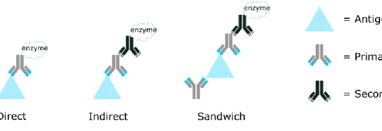
[image:7.612.113.501.348.481.2]Typically performed in a wet-lab setting, the ELISA is a high-sensitivity test that uses antibodies in conjunction with enzymes to identify a substance. As with all immunoassays, the ELISA relies on the interaction between antibodies and their antigens to detect a particular target molecule. There are three main types of ELISAs: direct, indirect, and sandwich. The various types of ELISAs describe the method by which the enzyme-linked antibodies will bind their antigen. In the direct ELISA, the enzyme is attached to the primary antibody whose antigen will be the target molecule. For the indirect ELISA, the enzyme is attached to the secondary antibody that is specific to the primary antibody that will bind to the target molecule. The sandwich ELISA is similar to the indirect ELISA, except the antigen is first sandwiched between two primary antibodies—called a capture antibody and a detection antibody—and then the secondary antibody conjugated to the enzyme is added for visualization. Figure 1 shows the various types of ELISA and how they function.
Figure 1: Various types of ELISA
9
Chapter 2:
10 Overview
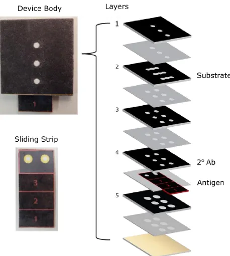
[image:9.612.89.541.268.378.2]The device consists of multiple layers of wax-patterned chromatography paper with double-sided tape to transform the two-dimensional paper sheets into a three-dimensional device with complex microfluidic paths. The key feature of the device is a sliding strip that moves within the device to various positions, allowing for the device to incorporate assays with multiple steps. The strip is made from chromatography paper, nitrocellulose, and cellulose acetate. The key component of the strip is the wax-patterned piece of nitrocellulose used as the reaction matrix for immunoassay reactions to occur.
Figure 2: Three positions that the sliding strip can occupy.
The various layers of the device are patterned with wax, which creates a hydrophobic barrier in the paper and results in channels where the wax was not laid. The fluid flows along the channels of the hydrophilic paper by capillary action.11 The device consists of five layers. Each
11
[image:10.612.80.538.93.604.2]
12 Sliding Strip
The sliding strip is crucial to the design of the device, and is one of its truly novel components. The sliding strip essentially introduces the idea of a mobile sensing area. In previous devices, the sensing area is stationary, and therefore all channels will cross paths at some point within the device en route to the sensing area or after passing the sensing area. Without the strip, multiple reagents, and therefore multiple steps, are unable to exist within a rapid test device because the sensing area would interact with multiple reagents simultaneously. A lack of control in when the reagents reached the sensing area of the device prevents the success of multiple step assays. With the incorporation of the sliding strip, the device is effectively partitioned into multiple sections; in the case of our device, there are 3 partitions, one for each step of the ELISA. This partitioning grants the user control over when a reagent should be used in the assay taking place within the device.
The significance of partitioning the device is that it allows multiple environments to exist within a single device. Partitioning will be defined as the various positions that the sliding strip can occupy. For example, if the strip is at position 1, as shown in Figure 2, what is stored in this pathway can be completely different from what is stored in the pathways for the strip at position 2 or position 3. Each time the strip slides to a new position, the sensing area is connected to a new channel, and therefore a new environment. In the case of the sliding strip device designed for detecting syphilis, the secondary antibody conjugated with the enzyme is stored in position 2 while the substrate that will react with the enzyme is stored in position 3, as shown in Figure 4.
13
Figure 4: From left to right: sample layer, splitting layer (yellow is the NBT/BCIP substrate), 6-hole layer, 6-6-hole layer (red is the secondary antibody), sliding strip (green is the antigen, Tp17), larger
6-hole layer. The antigen used in this figure is specific to the device used for syphilis.
Sensing Area
The sensing area on the strip is made from nitrocellulose. The ideal material used for a sensing area has consistency throughout the surface pore size and high binding capacity of proteins.12 Nitrocellulose has a high binding capacity for macromolecules and is commonly used
in molecular biology for various assays that require the adsorption of macromolecules onto a membrane. Over the last two decades, nitrocellulose has been used frequently as the membrane for the sensing area in lateral flow assays.12 While it is not well understood how nitrocellulose
binds macromolecules, it is hypothesized that both electrostatic and hydrophobic interactions play a role, with hydrophobic interactions playing a more dominant role.13 Nitrocellulose’s
ability to bind macromolecules without extra modifications to the membrane make it compatible with a variety of assays, and therefore make it a desirable substance to be used to bind the antigen in our device.
Choice of Analyte
Treponema pallidum (Tp) is the spirochaete bacterium that causes syphilis, as well as other diseases such as bejel, pinta, and yaws that are caused from treponemal infection. The outer membrane of the Tp bacterium contains several outer membrane proteins such as Tp15, Tp17,
Tp45, and Tp47. These four membrane proteins are considered important candidates for the serological diagnosis of syphilis because of their strong immunogenicity and presence throughout all stages (primary, secondary, tertiary, and latent) of syphilis infection.14,15 For our
14 Detection Method
The device employs an indirect ELISA as its method of detection. The Tp17 antigen is bound on one side of the sensing area of the sliding strip. If a patient is positive for syphilis, their body’s antibodies will bind to the Tp17 on the sensing area, and these antibodies will act as the primary antibody in the ELISA. Next, the previously stored, anti-human IgG secondary antibodies conjugated with enzyme will flow onto the sensing area and will bind to the primary antibodies. Finally, the stored substrate will flow to the sensing area and will interact with the secondary antibodies and the enzymes, completing the assay.
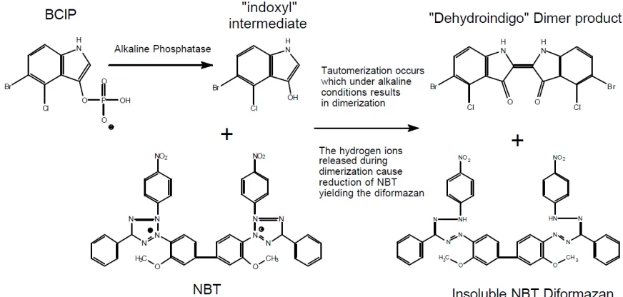
[image:13.612.82.518.393.601.2]The enzyme used in the assay is alkaline phosphatase (ALP), and it is used in conjunction with the substrate nitro blue tetrazolium chloride/5-Bromo-4-chloro-3-indolylphosphate (NBT/BCIP). When ALP comes into contact with NBT/BCIP, ALP cleaves the phosphate group from the BCIP and creates an intermediate that reacts with the NBT. The reaction results in the tautomerization of the blue dye intermediate, and the reduction of the NBT which is a solid, dark blue precipitate as shown in Figure 5.
Figure 5: NBT/BCIP Reaction16
Complex Microfluidic Paths
15
lateral flows. A device with vertical flow capabilities allows for more complex assays to be incorporated into a small, rapid test device.17 An example of this is the combination of the
vertical and horizontal flows working in tandem in the second layer of the device where the fluid flows from layer 1 to layer 2 and dissolves the NBT/BCIP substrate before continuing to flow horizontally towards the inlets of layer 3. The complex flow helps to create a homogenous solution containing the substrate as the chromatography paper channels double as a filter for the larger substrate particulates that were not completely dissolved in the initial flow from layer 1 to layer 2.
Furthermore, having both vertical and horizontal flow allows the device to remain compact despite having pathways that are more complex. With the ability to build vertically, branching and splitting of flow lets a device have powerful multiplexing capabilities while remaining a reasonable size.17
Blood Test
The ultimate goal of the device is to be able to place a drop of blood onto the inlet of the device and allow the device to process the blood and analyze its contents. Currently, we are using human serum based VIROTROL® Syphilis Total and VIROCLEAR® as our positive
control and negative control, respectively. Both products are produced by Bio-Rad Laboratories and are used as quality assurance reagents for immunoassays. VIROTROL® Syphilis Total
contains IgG and IgM antibodies to the Tp bacterium as well as non-treponemal antibodies, making it an ideal mimic for the serum from a patient who has been infected by syphilis. VIROCLEAR® is guaranteed to be non-reactive for Tp IgG and IgM antibodies as well as
non-treponemal antibodies; it is commonly used as a non-reactive quality control for assays for syphilis.
Stored Reagents
16
diameter of 1.0 mm. Once the powder is in place, the first layer with tape is placed on top of the device to seal the substrate within the device.
The secondary antibody conjugated with ALP is Anti-Human IgG (Fc specific) − Alkaline Phosphatase antibody produced in goat. The secondary antibody is diluted in 1x PBS and StabilCoat® Immunoassay Stabilizer until the concentration of the secondary antibody is 100 µg/mL and the solution consists of 10% StabilCoat® Immunoassay Stabilizer. 2 µL of this solution is transferred to the middle two channels of the fourth layer of the device, as shown in Figure 4. The layer is then placed in a vacuum desiccator for 3 hours. After 3 hours, the third layer with tape attached is placed on top of the fourth layer to seal the secondary antibody into the device.
To store the antigen, in this case the Tp17 outer membrane protein, 2 µL of Tp17 with concentration of 2.8 mg/mL is added to the testing region of the nitrocellulose sensing area. The nitrocellulose is then placed in an incubator at 20 degrees Celsius for 15 minutes to allow the antigen to dry. After 15 minutes, the nitrocellulose is removed from the incubator, placed in a solution of 3% bovine serum albumin (BSA), and placed on a shaker for one hour in order to block the sensing area. After one hour, the nitrocellulose is removed from the solution and washed three times with molecular grade water. The nitrocellulose is once again placed in the incubator at 20 degrees Celsius for 15 minutes until the sensing area has dried. The nitrocellulose is then placed on the sliding strip.
Procedure
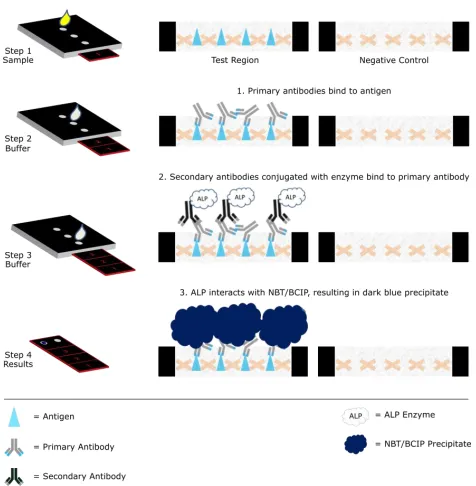
The device is run in three steps – one step for each location the strip occupies, as seen in Figure 6. In the first step, 20 µL of the sample (VIROTROL® or VIROCLEAR®) is added to the first inlet. After the sample has flowed through the device, 100 µL of 1x phosphate buffered saline (PBS) solution with 0.05% Tween 20 is added to the same inlet as the sample. The PBS not only ensures that the sample has passed through the entirety of the device, but also acts as a washing step to remove any unbound molecules from the sensing area. At this point in the procedure, if the sample is positive for the target disease, the primary antibody from the sample should be bound to the antigen on the sensing area.
17
the first storage zone and the PBS causes the stored secondary antibodies to flow through the device. At the end of this step, the secondary antibody should be bound to the primary antibody, and therefore bound to the sensing area.
After all of the fluid has wicked through step 2, the strip is moved to the third position, where 150 µL of Tris buffer of pH 9.5 is added to the third inlet. The Tris buffer dissolves the stored solid substrate and wicks it through the device. The Tris/substrate solution comes into contact with the enzyme attached to the secondary antibody, and the reaction between the ALP and NBT/BCIP occurs, leaving a blue-purple precipitate on the sensing area.
18
19
Chapter 3:
20 Results
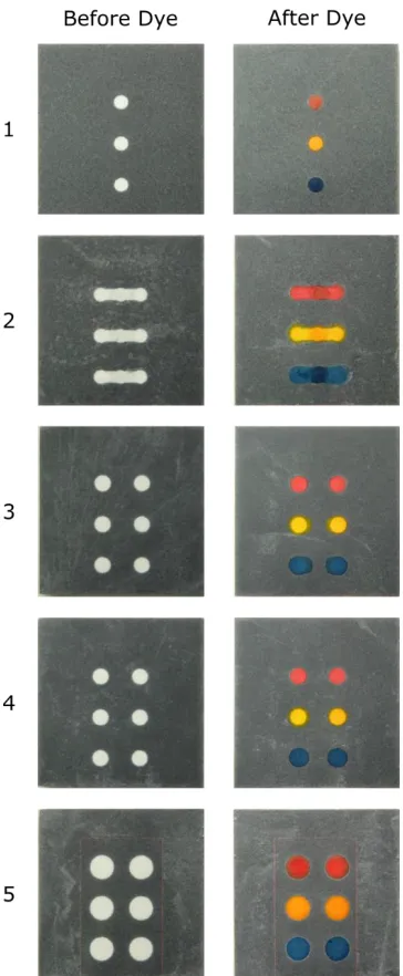
[image:19.612.215.397.222.663.2]Figure 7 shows the flow of a colored dye through the various layers of the device. The dye spreads through the entire hydrophilic channel and is effectively blocked by the hydrophobic wax. The dyes do not interact with each other, showing that each step is isolated. Due to this, the reagents stored in the device are isolated from each other as well. Buffers that are used to run the device only interact with the partitions that they are designed to interact with. It takes ~2 minutes for the dye to flow through the device.
21
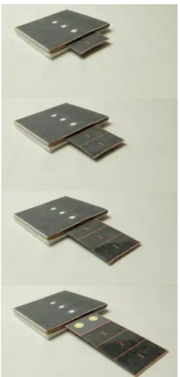
[image:20.612.179.435.93.628.2]Each location the strip moves to is a different step in the ELISA, as shown in Figure 8.
22
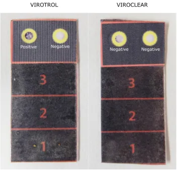
[image:21.612.131.482.162.501.2]The result presented in Figure 9 shows the contrast between the positive signal and the negative signal within a device, in the left hole and right hole, respectively, and the comparison between devices run using VIROTROL® and VIROCLEAR®, on the left strip and the right strip, respectively.
Figure 9: Positive result using VIROTROL® diluted by 100 times on the left strip. Negative result using VIROCLEAR® diluted by 100 times on the right strip.
Challenges
23
purpose of rapid test devices is to save lives by making the process of diagnosing patients simpler, and therefore, they need to have high sensitivity and specificity.
Another challenge of the device was the longevity of the reagents in storage. Although we used StabilCoat® Immunoassay Stabilizer to try and maintain the life of the secondary antibodies and the enzymes, good results—where the positive and negative signals were clearly differentiated—were more likely if the devices were run on the same day that they were
assembled. Devices that were left overnight after assembly and run the following day had a higher incidence of unsatisfactory results where the difference between the positive and negative signals were not as apparent. This may have been a result of protein degradation of the enzyme and antibodies from uncontrolled temperature and exposure to the environment. As well, the NBT/BCIP may not have been viable after being stored overnight; we saw that if left in the open for a couple of hours, the NBT/BCIP would become a darker shade of yellow and would begin to clump. While we stored all of the unused devices in a desiccator and found that leftover substrate stored in a desiccator remained light yellow and powdery, the results, nonetheless showed a higher rate of failure.
Future Work
Since the antigen spotted onto the nitrocellulose is the only disease-specific part of the assay, by exchanging the syphilis antigen for the antigen of another disease, in theory, the sliding strip device would be able to detect other diseases as well. Changing the antigen would most likely require an adjustment to the protocol for running the device, and different amounts of reagents would be necessary as well in order to account for a change in the properties of the device. While a change in protocol and amounts of reagents stored would be necessary, the potential for what the device can detect is large.
24
device: water and the sample. Having a device that can be run using water would greatly simplify the procedure and would be more user-friendly, especially in resource-poor locations.
While the implication of being able to detect syphilis in a paper-based diagnostic device using an enzyme immunoassay may not be significant in terms of revolutionizing the way that syphilis is diagnosed, the incorporation of a multistep assay into a portable device is significant. Being able to perform multistep assays involving enzymes within a portable device opens the doors for the detection of various analytes without the use of lab equipment. Theoretically, if the reagents and their amounts are altered, any disease that can be detected by an antibody can be detected using this device.
Conclusion
25
References
(1) Ratnam, S. The Laboratory Diagnosis of Syphilis. Canadian Journal of Infectious Diseases and Medical Microbiology.2005, 16 (1).
(2) Yager, P.; Edwards, T.; Fu, E.; Helton, K.; Nelson, K.; Tam, M. R.; Weigl, B. H.
Microfluidic Diagnostic Technologies for Global Public Health. Nature.2006, 442 (7101), 412–418.
(3) Chin, C. D.; Linder, V.; Sia, S. K. Commercialization of Microfluidic Point-of-Care Diagnostic Devices. Lab on a Chip.2012, 12 (12), 2118.
(4) Branson, B. Rapid Tests for HIV Antibody. AIDS Reviews.2000, 2.
(5) Strep A Rapid Test Device. CLIA Waived 2004.
(6) Clarity MONO Mononucleosis Rapid Test Device. Diagnostic Test Group 2007.
(7) Posthuma-Trumpie, G. A.; Korf, J.; van Amerongen, A. Lateral Flow (immuno)assay: Its Strengths, Weaknesses, Opportunities and Threats. A Literature Survey. Analytical and Bioanalytical Chemistry.2009, 393 (2), 569–582.
(8) Meredith, S.; World Health Organization. The Global Elimination of Congenital Syphilis: Rationale and Strategy for Action; World Health Organization: Geneva, 2007.
(9) Fantry, L. E.; Tramont, E. C. Treponema Pallidum (Syphilis) http://www.antimicrobe.org/b242.asp (accessed Mar 30, 2016).
26
(11) Carrilho, E.; Martinez, A. W.; Whitesides, G. M. Understanding Wax Printing: A Simple Micropatterning Process for Paper-Based Microfluidics. Analytical Chemistry.2009, 81
(16), 7091–7095.
(12) Yetisen, A. K.; Akram, M. S.; Lowe, C. R. Paper-Based Microfluidic Point-of-Care Diagnostic Devices. Lab on a Chip.2013, 13 (12), 2210.
(13) Nitrocellulose (Molecular Biology) http://what-when-how.com/molecular-biology/nitrocellulose-molecular-biology/ (accessed Mar 30, 2016).
(14) Ran, S.; Lai, D.; Ren, R.; Lian, S.; Zhang, H. Treponema Pallidum-Specific Antibody Expression for the Diagnosis of Different Stages of Syphilis. China Medical Journal.2013,
126 (2), 206–210.
(15) Syphilis Reagents for Commercial Assay Development. Meridian Life Science, Inc.
(16) Colorimetric Alkaline Phosphatase and Peroxidase Substrate Detection Systems http://www.sigmaaldrich.com/technical-documents/articles/biofiles/colorimetric-alkaline.html (accessed Mar 31, 2016).