Adrenergic receptor genotype
influences heart failure severity
and β-blocker response in children
with dilated cardiomyopathy
The Harvard community has made this
article openly available. Please share how
this access benefits you. Your story matters
Citation
Reddy, Sushma, Alan Fung, Cedric Manlhiot, Elif Seda Selamet
Tierney, Wendy K. Chung, Elizabeth Blume, Beth D. Kaufman,
Elizabeth Goldmuntz, Steven Colan, and Seema Mital. 2015.
“Adrenergic receptor genotype influences heart failure severity
and β-blocker response in children with dilated cardiomyopathy.”
Pediatric Research 77 (2): 363-369. doi:10.1038/pr.2014.183. http://
dx.doi.org/10.1038/pr.2014.183.
Published Version
doi:10.1038/pr.2014.183
Citable link
http://nrs.harvard.edu/urn-3:HUL.InstRepos:14065446
Terms of Use
This article was downloaded from Harvard University’s DASH
repository, and is made available under the terms and conditions
applicable to Other Posted Material, as set forth at
http://
Articles
Translational Investigation
nature publishing group
Background: Adrenergic receptor (ADR) genotypes are
associated with heart failure (HF) and β-blocker response in adults. We assessed the influence of ADR genotypes in children with dilated cardiomyopathy (DCM).
Methods: Ninety-one children with advanced DCM and 44
with stable DCM were genotyped for three ADR genotypes associated with HF risk in adults: α2cdel322-325, β1Arg389, and β2Arg16. Data were analyzed by genotype and β-blocker use. Mean age at enrollment was 8.5 y.
results: One-year event-free survival was 51% in advanced and 80% in stable DCM. High-risk genotypes were associated with higher left ventricular (LV) filling pressures, higher systemic and pulmonary vascular resistance, greater decline in LV ejec-tion fracejec-tion (P < 0.05), and a higher frequency of mechanical circulatory support while awaiting transplant (P = 0.05). While
β-blockers did not reduce HF severity in the overall cohort, in the subset with multiple high-risk genotypes, those receiving
β-blockers showed better preservation of cardiac function and hemodynamics compared with those not receiving β-blockers (interaction P < 0.05).
conclusion: Our study identifies genetic risk markers that may help in the identification of patients at risk for developing decompensated HF and who may benefit from early institution of β-blocker therapy before progression to decompensated HF.
d
ysregulation of the adrenergic system can contribute to the development and progression of heart failure (HF) through altered signaling of pre- and postsynaptic α- and β-adrenergic receptors (ADRs). Presynaptic α2C ADRs inhibitnorepinephrine (NE) release, cardiac β1 ADRs are targets for
NE-induced chronotropy and inotropy, and β2 ADRs mediate
vascular smooth muscle relaxation (1–4). Activation of ADRs initially promotes maintenance of cardiac output but ultimately acts to accelerate HF progression (5). This forms the basis for the use of sympathetic antagonists such as β-blockers in the
management of HF with reduced ejection fraction (HFREF) (6). Unlike in adults, β-blockers have not shown symptomatic or survival benefit in pediatric HFREF in the only prospective, placebo-controlled clinical trial to date (7). Lack of effective therapies contributes to poor outcomes with ~50% of children with dilated cardiomyopathy (DCM) either dying or requiring a heart transplant within 5 y of diagnosis (8).
Single-nucleotide polymorphisms (SNPs) in the ADR genes have been implicated in the progression of HFREF in adults. These polymorphisms include α2Cdel322-325 (associated with increased NE release) (5,9–11), β1Arg389 (associated with increased receptor sensitivity to NE) (12), and β2Arg16 (associ-ated with receptor downregulation and impaired vasorelaxation) (13–16). Furthermore, homozygosity for both α2Cdel322-325 and β1Arg389 genotypes is associated with higher risk of HF in African Americans compared with Caucasians (17,18). Overall, the presence of genetic variations that increase cardiac sympa-thetic tone and/or peripheral vasoconstriction may increase the risk of HF progression by increasing cardiac work, myocardial oxygen consumption, and myocyte loss (4,19–25). The asso-ciation of ADR polymorphisms with response to β-blockers is however conflicting (4,13,26–29). While the response to meto-prolol and carvedilol, the most commonly used β-blockers in adult HFREF patients, was independent of ADR genotype, survival benefit with bucindolol was limited to the subset of patients carrying the β1Arg389 and α2Cdel322-325 genotype via downregulation of NE release (30–32).
Despite the role of these genetic variations in adult HFREF, their role in HF progression and response to β-blocker ther-apy in children is not known. Given widespread empiric use of
β-blockers despite negative results of the pediatric carvedilol HF trial (7), knowledge of genetic markers that predict β-blocker response is particularly important since it would allow target-ing of β-blockers only to potential responders and avoid futile therapy in nonresponders. However, this requires a pediatric-focused study since adult ADR genotype associations may not
Received 17 February 2014; accepted 15 August 2014; advance online publication 10 December 2014. doi:10.1038/pr.2014.183
1Department of Pediatrics, Stanford University, Stanford, California; 2Department of Pediatrics, Hospital for Sick Children, University of Toronto, Toronto, Ontario, Canada; 3Departments of Pediatrics and Medicine, Columbia University, New York, New York; 4Department of Pediatrics, Children’s Hospital Boston, Harvard University, Boston,
Massachusetts; 5Department of Pediatrics, Children’s Hospital of Philadelphia, University of Pennsylvania, Philadelphia, Pennsylvania. Correspondence: Seema Mital
(seema.mital@sickkids.ca)
adrenergic receptor genotype influences heart failure
severity and
β
-blocker response in children with dilated
cardiomyopathy
Sushma Reddy1, Alan Fung2, Cedric Manlhiot2, Elif Seda Selamet Tierney1, Wendy K. Chung3, Elizabeth Blume4,
Beth D. Kaufman1, Elizabeth Goldmuntz5, Steven Colan4 and Seema Mital2
Pediatr Res
363
369 2015
Pediatric Research 10.1038/pr.2014.183
10December2014
77
2
17February2014
15August2014
Copyright © 2015 International Pediatric Research Foundation, Inc.
Adrenergic genotypes in heart failure Reddy et al.
Translational Investigation Articles
Articles
Reddy et al.
be reproducible in children since there are differences in ADR receptor subtype density, calcium transients, contractile force, and relaxation velocity (33). The objective of this study was to determine if variants in ADRA2c (α2Cdel322-325), ADRB1
(β1Arg389), and ADRB2 (β2Arg16) genes increase HF severity and influence β-blocker response in children with DCM.
RESULTS
Allele Frequencies
The frequency of α2cdel322-325, β1Arg389, and β2Arg16 geno-types was similar in the 91 advanced HF patients (21, 48, and 48%, respectively) and 44 stable HF patients (22, 42, and 35%, respectively) and was comparable to previous studies in healthy adults (26,34,35). All genotypes were in Hardy–Weinberg equilibrium. Individual genotype assays did not yield results in five patients in the advanced HF cohort.
Clinical Characteristics
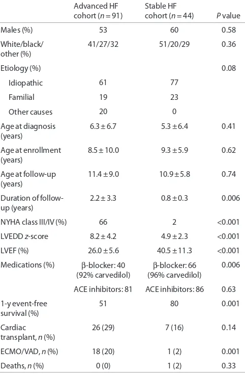
Clinical characteristics at enrollment are shown in Table 1. Demographic characteristics were similar in the two cohorts. There was no difference in age, gender, and etiology of HF at
presentation among patients in the different genotype groups with 61% having sporadic idiopathic DCM, 19% familial, and 20% other causes including burnt out viral myocarditis (n = 8), neuromuscular disorder (n = 2), maternal lupus (n = 1), anthracycline cardiomyopathy (n = 3), and metabolic disease (n = 4). In the advanced HF group, the LV end-diastolic volume z score was 5.2 ± 2.6, and the shortening fraction was 13.1 ± 5.8%. Detailed echocardiographic data were available in the stable HF group as part of the ventricular volume variabil-ity (VVV) study (Table 2). Baseline clinical and echocardio-graphic characteristics of patients in this genetic substudy were comparable to the parent VVV patient cohort from which this subset was derived (data not shown).
Genotype Association With Hemodynamics, Outcomes, and Response to β-Blocker Therapy (Advanced HF)
During follow-up, 57 of the 91 patients in the advanced HF cohort underwent cardiac catheterization to assess HF sever-ity. Median time from diagnosis to cardiac catheterization was 3 mo reflecting a cohort presenting in advanced HF. Average hemodynamic measurements at cardiac catheterization were central venous pressure, 7 ± 4 mm Hg; pulmonary capillary wedge pressure, 19 ± 8 mm Hg; indexed pulmonary vascular resistance, 3.7 ± 2.5 mm Hg/l/min/m2, indexed systemic
vas-cular resistance, 24 ± 10 mm Hg/l/min/m2, and cardiac index,
3.1 ± 1.5 l/min/m2. There was a significant association between
[image:3.594.46.287.343.710.2]ADR genotypes and hemodynamic severity of HF manifested by higher pulmonary capillary wedge pressure, indexed pul-monary vascular resistance, and indexed systemic vascular resistance in patients with a higher number of ADR high-risk genotypes. There was an additive effect of multiple high-risk genotypes with a linear increase in central venous pressure, pulmonary capillary wedge pressure, indexed pulmonary vas-cular resistance, and indexed systemic vasvas-cular resistance with increasing number of high-risk genotypes (Figure 1). Since factors like age, use of general anesthesia, or oxygen supple-mentation during catheterization can independently influence cardiac hemodynamics, we analyzed but found no influence of these factors on the association between ADR genotype and hemodynamic severity of HF. Forty percent of patients were
table 1. Clinical characteristics at enrollment
Advanced HF
cohort (n = 91) Stable HF cohort (n = 44) P value
Males (%) 53 60 0.58
White/black/ other (%)
41/27/32 51/20/29 0.36
Etiology (%) 0.08
Idiopathic 61 77
Familial 19 23
Other causes 20 0
Age at diagnosis
(years) 6.3 ± 6.7 5.3 ± 6.4 0.41 Age at enrollment
(years) 8.5 ± 10.0 9.3 ± 5.9 0.62 Age at follow-up
(years) 11.4 ± 9.0 10.9 ± 5.8 0.74 Duration of
follow-up (years) 2.2 ± 3.3 0.8 ± 0.3
0.006
NYHA class III/IV (%) 66 2 <0.001 LVEDD z-score 8.2 ± 4.2 4.9 ± 2.3 <0.001 LVEF (%) 26.0 ± 5.6 40.5 ± 11.3 <0.001 Medications (%) β-blocker: 40
(92% carvedilol) β
-blocker: 66 (96% carvedilol)
0.006
ACE inhibitors: 81 ACE inhibitors: 86 0.63 1-y event-free
survival (%) 51 80 0.001
Cardiac
transplant, n (%) 26 (29) 7 (16) 0.14 ECMO/VAD, n (%) 18 (20) 1 (2) 0.001 Deaths, n (%) 0 (0) 1 (2) 0.33
[image:3.594.306.550.581.727.2]ACe, angiotensin-converting enzyme; eCMO, extracorporeal membrane oxygenation; HF, heart failure; LVeDD, left ventricular end-diastolic diameter; LVeF, left ventricular ejection fraction; NYHA, New York Heart Association; VAD, ventricular assist device.
table 2. Echocardiographic characteristics at enrollment in the
stable HF cohort (n = 44)
Left ventricular end-diastolic volume z 4.4 ± 4.1 Left ventricular end-systolic volume z 4.5 ± 2.1 Ejection fraction (%) 40.5 ± 11.3 Ejection fraction z −4.5 ± 2.2 Left ventricular mass z 2 ± 1.8 Left ventricular mass/volume z −1.3 ± 1 Mitral: early deceleration time (msec) 151.3 ± 75.2 Mitral: peak early velocity (E) (cm/s) 99 ± 22 Mitral: peak early/late velocity (E/A) 2 ± 0.7 Average E/e′ (septal + lateral wall) 12.1 ± 5.9
Adrenergic genotypes in heart failure
Articles
receiving β-blockers at enrollment (92% carvedilol; mean dose: 0.57 ± 0.53 mg/kg/d). The subset of patients with the high-risk genotype receiving β-blockers showed an improvement
in their hemodynamics compared with those not receiving
β-blockers (genotype group × β-blocker use, interaction P
[image:4.594.43.290.112.321.2]values ≤ 0.05; Figure 2). There was no significant difference in freedom from transplantation between the two genotype groups (hazard ratio: 0.88; 95% confidence interval: 0.52–1.46;
Figure 3a). There was, however, a lower freedom from
ven-tricular assist device/extracorporeal membrane oxygenation in listed patients with ≥2 ADR high-risk genotypes awaiting transplantation (hazard ratio: 2.57; 95% confidence interval: 1.05–7.23; Figure 3b).
Genotype Association With Change in LV Systolic and Diastolic Function Stratified by β-Blocker Therapy (Stable HFREF)
To assess the influence of ADR genotypes in earlier stages of HF, we enrolled 44 patients with stable HF. Sixty-six percent were receiving β-blockers at enrollment (96% carvedilol; mean dose: 0.44 ± 0.28 mg/kg/d). There was no association between ADR genotypes and New York Heart Association/Ross HF severity score. Regression analyses were performed to assess association of ADR genotypes with change in ventricular sys-tolic and diassys-tolic function on serial echocardiograms in 35 stable HF patients in whom serial echocardiographic data were available. Figure 4 shows the change in echocardiographic measurements during follow-up for each additional ADR risk genotype stratified by β-blocker use. There was a greater decline in LV ejection fraction for every additional ADR risk genotype, but this association was not seen in patients receiv-ing β-blockers (genotype group × β-blocker use, interaction P
[image:4.594.171.423.454.645.2]= 0.003 vs. those not receiving β-blockers). There was a greater increase in LV end-systolic volume z-score for every additional ADR risk genotype, and this association was seen independent
Figure 1. Cumulative effect of multiple adrenergic risk genotypes on
cardiac hemodynamics (advanced HF cohort). The squares represent the point estimates from regression models, and the solid bars represent the 95% confidence interval around that estimate (n = 57). A higher number of adrenergic high-risk genotypes were associated with (a) higher CVP (P = 0.02), (b) higher PCWP (P = 0.06), (c) higher PVRI (P = 0.004), and (d) higher SVRI (P = 0.005). The gray zones represent normal hemodynamic ranges, i.e., CVP, 1–5 mm Hg; PCWP, 4–12 mm Hg; PVRI, 0.25–1.6 mm Hg/l/min/m2; and SVRI, 9–20 mm Hg/l/min/m2. ADR, adrenergic receptor; CVP, central venous pressure; PCWP, pulmonary capillary wedge pressure; PVRI, indexed pulmonary vascular resistance; SVRI, indexed systemic vascular resistance.
12 24
20
16
12
8
4
0 10
CVP (mm Hg
)
PCWP (mm Hg
)
8
6
4
2
0
0 1 2
Number of high-risk ADR genes 3
10 32
28
24
20
16
12
8
PVRI (mm Hg/l/min/m
2)
PVRI (mm Hg/l/min/m
2) 8
6
4
2
0
0 1 2
Number of high-risk ADR genes
3 0 1 2
Number of high-risk ADR genes 3
0 1 2
Number of high-risk ADR genes 3
a b
c d
Figure 2. Interaction between adrenergic genotype and hemodynamic response to β-blockers (advanced HF cohort). The markers represent the point
estimates (red: not on β-blockers, n = 27; black: on β-blockers, n = 28) from regression models, and the solid bars represent the 95% confidence interval around that estimate. Patients not receiving β-blockers showed (a) higher CVP (P = 0.02), (b) higher PCWP (P = 0.004), (c) higher PVRI (P = 0.0003), and (d) higher SVRI (P = 0.009) for every additional high-risk genotype. This increase in filling pressures and resistances was either blunted (CVP) or absent (PCWP, PVRI, and SVRI) in patients with high-risk genotypes receiving β-blockers. There was a significant interaction between ADR genotype and
β-blocker therapy for PCWP (interaction P = 0.05), PVRI (interaction P = 0.03), and SVRI (interaction P = 0.02). Red, no β-blocker, black; β-blocker. The gray zones represent normal hemodynamic ranges, i.e., CVP, 1–5 mm Hg; PCWP, 4–12 mm Hg; PVRI, 0.25–1.6 mm Hg/l/min/m2; and SVRI, 9–20 mm Hg/l/min/m2. ADR, adrenergic receptor; CVP, central venous pressure; HR, high-risk genotype numbers; PCWP, pulmonary capillary wedge pressure; PVRI, indexed pulmonary vascular resistance; SVRI, indexed systemic vascular resistance.
16 32
28 24 20 16 12 8 4 0 14
12 10
CVP (mm Hg) PCWP (mm Hg
)
32 36
28 24 20 16 12 8
SVRI (mm Hg/l/min/m
2) 8
6 4 2 0
HR-0 HR-1 HR-2 HR-3
14 12 10
PVRI (mm Hg/l/min/m
2)
8 6 4 2 0
HR-0 HR-1 HR-2 HR-3 HR-0 HR-1 HR-2 HR-3
HR-0 HR-1 HR-2 HR-3
a b
Articles
Reddy et al.
of β-blocker use (interaction P = 0.12). There was a greater decline in peak mitral E wave velocity and in early diastolic tricuspid annular velocity for every additional ADR risk genotype, but this association was not seen in patients receiv-ing β-blockers (interaction P < 0.001 vs. those not receiving
β-blockers). Interaction of individual ADR genotypes with
β-blockers was not assessed due to the small sample size. Overall, these results suggest that patients with a higher num-ber of ADR high-risk genotypes show more rapid progression of systolic and diastolic dysfunction but that this progression may be prevented by β-blocker therapy with an improvement or stabilization in LV size and function.
DISCUSSION
DCM is the most common form of cardiomyopathy and the leading indication for transplantation in adults and children. Almost 50% of children with DCM either die or require a heart transplant within 5 y of diagnosis (3,8). Carvedilol is widely used but has failed to show clinical benefit in one of the largest clinical trials of pediatric HF to date, although the trial did not evaluate response by genotype (7). Our study identifies genetic
risk markers that may help in the identification of patients at risk for developing decompensated HF who may benefit from early institution of β-blocker therapy before progression to decompensated HF.
[image:5.594.309.542.59.263.2]An important finding of our study is the association of high-risk ADR genotypes with hemodynamic severity of HF in patients with advanced DCM. Those with a higher number of ADR high-risk genotypes had higher left- and right-sided filling pressures and higher systemic and pulmonary vascu-lar resistance, faster progression of LV systolic and diastolic dysfunction, as well as a higher incidence of acute decom-pensation requiring mechanical circulatory support during follow-up. The combined physiologic effects of these geno-types are most likely related to an increase in the release and responsiveness to NE as well as a decrease in the sensitivity of β-ADRs which can contribute to impaired myocardial con-tractility, impaired peripheral vasodilation, and progression of cardiac dysfunction (5,18). Our results suggest that genotyping for ADR polymorphisms may help in the early identification of patients at risk for decompensated HF who may benefit from closer monitoring and timely listing for transplantation before hemodynamic decompensation.
Figure 3. Adrenergic genotype and event-free survival (advanced HF
cohort). (a) Time to transplantation during follow-up was not different between patients with <2 (black, n = 48) vs. ≥2 (red, n = 43) ADR high-risk genotypes (hazard ratio: 0.88; 95% confidence interval: 0.52–1.46). (b) Patients listed for transplantation with ≥2 ADR high-risk genotypes (n = 28) showed lower freedom from mechanical circulatory support (VAD/ECMO) as a bridge to transplantation compared with those with <2 risk genotypes (n = 33) despite similar waiting times (hazard ratio: 2.57; 95% confidence interval: 1.05–7.23). Number of patients remaining at each time point are shown in black for patients with <2 high-risk genotypes and in red for patients with ≥2 high-risk genotypes. ADR, adrenergic receptor; ECMO, extracorporeal membrane oxygenation; VAD, ventricular assist device.
100
80
60
40
Percent free from transplantatio
n
20
0
0 1
48 N left:
43 24 17
14 9
8 8
6 8
6 8
100
80
60
40
Percent still listed an
d
free from ECMO/VAD 20 0
0 30 60
Days since listing
90 120
33 N left:
28 11
9
5 2
2 1
2 0
2 3
Years since presentation
4 5
a
b
Figure 4. Adrenergic genotype and change in ventricular systolic and
dia-stolic dysfunction (stable HF cohort). Regression analyses were performed to assess association of ADR genotypes with change in ventricular systolic and diastolic function on serial echocardiograms in 35 stable HF patients. Graphs show parameter estimate of change in echocardiographic measure-ments for each additional ADR risk genotype stratified by β-blocker use. Red, on no β-blockers; black, on β-blockers. There was a greater decline in LV ejection fraction for every additional ADR risk genotype, but this association was not seen in patients receiving β-blockers (interaction
P = 0.003 vs. those not receiving β-blockers). There was a greater increase in LV end-systolic volume z-score for every additional ADR risk genotype, but this did not reach statistical significance between those receiving vs. those not receiving β-blockers (interaction P = 0.12). There was a greater decline in peak mitral E wave velocity for every additional ADR risk genotype, but this association was not seen in patients receiving β-blockers (interaction
P < 0.001 vs. those not receiving β-blockers). There was a greater decline in early diastolic tricuspid annular velocity with each additional ADR risk genotype, but this association was not seen in patients receiving β-blockers (interaction P = 0.001 vs. those not receiving β-blockers) (n = 7 for 0, n = 22 for 1, n = 22 for 2, and n = 4 for 3 risk genotypes).
−10 −5 0 5 10 Ejectionfraction (%)
End systolic volume z-score
Peak early mitral velocity (cm/s)
Peak early diastolic tricuspid annular velocity (cm/s)
−1.0 −0.5 0.0 0.5 1.0
−0.4
−3 −2 −1 0
Change per high-risk genotype
1 2 3
[image:5.594.71.263.62.348.2]Adrenergic genotypes in heart failure
Articles
We further evaluated the pharmacogenetic interactions between the ADR genotypes and response to β-blocker ther-apy. Although our numbers are small, we found results simi-lar to the carvedilol trial—patients receiving β-blockers did not show improvement in HF severity in the overall cohort. However, when stratified by ADR risk genotypes, β-blocker therapy was associated with better hemodynamic adaptation in the high-risk genotype groups. This suggests that the benefit of β-blocker therapy in pediatric DCM may be seen primar-ily in those in whom disease progression is mediated by dys-regulated ADR signaling likely due to variant ADR genotypes. This is consistent with the findings by Liggett et al. (31) who showed that the β-blocker, bucindolol, a sympatholytic agent, was effective in improving HF survival only in adults with the
β1Arg389 genotype, not in those with the β1Gly389 genotype. In light of the observable benefit of β-blocker therapy in advanced HF patients with high-risk genotypes, we analyzed if
β-blocker therapy would show a similar benefit in patients with earlier stages of DCM. We found that patients with high-risk genotypes showed a decline in ejection fraction and diastolic function during follow-up but that this decline was attenuated, and even improved, in high-risk genotype patients receiving
β-blockers. These findings suggest that β-blocker therapy in the subset of stable HF patients with high-risk genotypes may not only prevent decline in function but may also lead to recovery of ventricular function and reverse remodeling. This exciting find-ing highlights the importance of early institution of β-blocker therapy in this high-risk subset to prevent disease progression. This finding is particularly important in light of the results of the only randomized trial with β-blockers in children with HF that failed to reproduce the benefits seen in adult HF (7). Our findings suggest that while not all pediatric DCM patients ben-efit from β-blocker therapy, the subgroup of patients with high-risk ADR genotypes may show benefit. Although genotype did not influence carvedilol response in adult studies, children may be more catecholamine dependent compared to adults, and therefore, the impact of receptor genotype may be greater than that in adults. Given the considerable phenotypic heterogene-ity in sporadic as well as inherited DCM where considerable intrafamilial variability exists, knowledge of disease modifiers may enable better risk prediction not only in sporadic but also in familial cases and, more importantly, facilitate risk modifi-cation strategies by targeting therapies to potential responders.
Limitations
Since patients were not always enrolled at the time of initial diagnosis of DCM, this may introduce a survivor bias in our cohort. The study was not powered to analyze the effect of underlying genetic mutation type on HF severity and progres-sion. The study was also limited by the relatively short mean follow-up of 0.8 y in the stable HF cohort; therefore, longer-term outcomes were not assessed. Although results from our study show a difference in rates of acute decompensation requiring mechanical circulatory support between the two genotype risk groups, these results require further validation in a larger patient cohort.
Conclusions
The characterization of the ADR genotype may aid in risk stratifying patients with DCM who are more likely to prog-ress to decompensated HF. The preferential responsiveness of patients with high-risk genotypes to β-blocker therapy high-lights the importance of early institution of β-blocker therapy in genetically susceptible patients while avoiding futile therapy in those unlikely to respond. Prospective, randomized, geno-type-guided trials of β-blocker therapy in this high-risk subset are needed to validate these observations and develop pharma-cogenetic-guided therapies in this vulnerable cohort.
METHODS
Study Population
The study included patients with advanced DCM from a single US institution and patients with stable DCM from a multi-institutional North American cohort. The advanced DCM group consisted of 91 DCM patients aged <21 y, who were on medical therapy with symp-tomatic HF and undergoing evaluation for transplantation at the Columbia University Medical Center (2002–2005). The stable DCM group included 44 DCM patients with stable HFREF enrolled in a multicenter Pediatric Heart Network observational study of DCM, the VVV study (accrual 2005–2007; Clinical Trials Registration: #NCT00123071). Detailed design of the Pediatric Heart Network study has been previously published (36). The inclusion criteria for the VVV study from which this stable HF cohort was derived were pediatric DCM patients, disease duration >2 mo, on outpatient medi-cal therapy, no hemodynamic instability, and not being assessed or listed for transplant (due to the intent to assess longitudinal natural history). Patients were enrolled across eight North American centers and followed with serial echocardiography for 18 mo. Four of eight VVV sites participated in a genetic substudy and provided DNA sam-ples from enrolled patients—Columbia University Medical Center, Children’s Hospital Boston, Children’s Hospital of Philadelphia, and Hospital for Sick Children, Toronto. In both cohorts, children with active myocarditis, hypertrophic or restrictive cardiomyopa-thy, or congenital heart disease were excluded. Informed consent was obtained from the parents and assent from older subjects. The study was approved by the local Institutional Review Boards of all participating sites—Columbia University Medical Center, Children’s Hospital Boston, Children’s Hospital of Philadelphia, and Hospital for Sick Children, Toronto.
Clinical Data
Articles
Reddy et al.
mortality compared with clinical data and LV ejection fraction alone in adult studies with only limited tissue Doppler data available in chil-dren with DCM (37). The interpreters of the echocardiograms were blinded to the genotype data (36).
Candidate Genes and SNP Selection
Three candidate ADR genes (ADRA2c, ADRB1, and ADRB2) were selected based on their role in modulating sympathetic tone and asso-ciation with adult HF (5,9,12,14,16). Genotypes previously associated with HF in adults were defined as high-risk genotypes for the pur-pose of this study (α2Cdel322-325-ADRA2c, β1Arg389-ADRB1, and β2Arg16-ADRB2; Table 3) and evaluated for association with
echo-cardiographic evidence of disease progression, hemodynamic param-eters, and clinical outcomes (4,5,15,34,38).
Genotyping
Genomic DNA was isolated from whole blood (Gentra Systems, Minneapolis, MN). ADRA2c SNP was genotyped by end-labeling primers (Table 4) with γ-32P-ATP (PerkinElmer Life Science, Boston,
MA) using T4 polynucleotide kinase (Promega, Madison, WI) and using PCR to amplify the genomic fragment. The PCR products were resolved on an 8% nondenaturing polyacrylamide gel to resolve the size difference. ADRB1 and ADRB2 SNPs were genotyped individu-ally using pyrosequencing (PSQ96; Biotage, Westborough, MA) with a vacuum system with streptavidin sepharose beads (Amersham Biosciences AB, Uppsala, Sweden). PCR reactions consisted of 5 pmol of each of the appropriate forward and reverse primers (Table 4), 1 U AccuPrime GC-Rich DNA polymerase (Invitrogen, Carlsbad, CA), 1× buffer A, and 30 ng of genomic DNA in a 25 ml reaction volume for 30 cycles at an annealing temperature of 62 °C for ADRA2c and ADRB2 and touch-down from 65 to 50 °C for ADRB1. Each assay was performed in duplicate to ensure accuracy.
Statistical Analysis
Hardy–Weinberg equilibrium was determined using Pearson χ2
analy-sis of actual and predicted genotypes. Presence of a single risk allele was defined as a high-risk genotype. Data are reported as means with SDs, median with minimum and maximum and frequencies as appro-priate. The combined effect of multiple high-risk genotypes on echo-cardiographic and hemodynamic variables was assessed in a univariate regression model using maximum likelihood estimates and appropri-ate mathematical transformations to normalize the distribution of the response variable. Regression models adjusted for repeated measures through a compound symmetry covariance structure were used to determine the effect of ADR genotypes on serial echocardiographic
measurements and on hemodynamic measures. The associations between high-risk genotype and outcomes were also tested in multi-variable models including an interaction multi-variable between genotype and β-blocker use at enrollment. Age and time since diagnosis were also included in the model. HF progression was defined as worsening hemodynamics and/or echocardiographic progression of LV dilation and dysfunction. β-Blocker responsiveness was defined as an improve-ment or stabilization in LV size and function. Event-free survival, i.e., freedom from heart transplantation and freedom from extracorpo-real membrane oxygenation/ventricular assist device while listed for transplantation was modeled using Kaplan–Meier nonparametric esti-mates, and distributions of time to event were compared using the log-rank test. Follow-up time was censored at death or transplant when analyzing time to extracorporeal membrane oxygenation/ventricular assist device initiation. All statistical analyses were performed using SAS Statistical Software v9.1 (The SAS Institute, Cary, NC).
ACKNOWLEDGMENTS
The authors thank Claudia Fontes, BA, and Alisa Nakamine, BA, for help with sample processing for genotyping; Patricia Lanzano and Liyong Deng for genotyping of the primary cohort; Tara Paton for genotyping the second-ary cohort; and Shing Lee for help with statistical analysis. The authors also thank the SickKids Labatt Family Heart Centre Biobank Registry for access to patient DNA. This work was presented, in part, at the Annual Scientific Ses-sions of the American Heart Association, November 2011.
STATEMENT OF FINANCIAL SUPPORT
This work was supported by the US National Institutes of Health (NIH; Bethesda, MD, USA; grant number: RR00645); Children’s Cardiomyopathy Foundation, Tenafly, NJ, USA; and the Syde Hurdus Foundation, New York, NY, USA. This work was also supported by U01 grants from the National Heart, Lung, and Blood Institute (HL068279, HL068285, HL068290, and HL068288), NIH.
Disclosure: None.
REFERENCES
1. Majewski H. Modulation of noradrenaline release through activation of presynaptic beta-adrenoreceptors. J Auton Pharmacol 1983;3:47–60. 2. Brede M, Wiesmann F, Jahns R, et al. Feedback inhibition of catecholamine
release by two different alpha2-adrenoceptor subtypes prevents progres-sion of heart failure. Circulation 2002;106:2491–6.
[image:7.594.46.557.76.136.2]3. Hein L, Kobilka BK. Adrenergic receptor signal transduction and regula-tion. Neuropharmacology 1995;34:357–66.
table 3. Candidate genes and polymorphisms
Gene Nucleotide substitution Genomic context SNP ID Cytogenetic locus
α2C adrenergic receptor (ADRA2C) 964–975 nucleotide deletion del322-325 rs6846820 4p16
β1 adrenergic receptor (ADRB1) 1165G>C Gly389Arg rs1801253 10q24-26
β2 adrenergic receptor (ADRB2) Nucleotide 46G>A Gly16Arg rs1042713 5q31-32
sNP, single-nucleotide polymorphism.
table 4. Primers used in pyrosequencing and real-time PCR
Gene Primers for pyrosequencing Primers for real-time PCR
ADRA2C del F3 AGCCCGACGAGAGCAGCGCA TCTTCTGGATCGGCTACTGC
ADRA2C del R GTCAGCGCCCCCGACTGAGC AGGATGTGCTTGAAGGATGG
ADRB1 nonA389A F ATCTACTGCCGCAGCCCCGACT AGGGGAACGAGGAGATCTGT
ADRB1 nonA389A R Biotin-GCGTGGCCCCGACGACAT TCCCTTCCCAAACTTTCCTT
ADRB1 nonA389A Sq CGCAAGGCCTTCCA —
ADRB2 G16A F GAACGGCAGCGCCTTCTTGCT TCTTCTGGATCGGCTACTGC
ADRB2 G16A R Biotin-ACCTCGTCCCTTTCCTGCGTGA GTCTTGAGGGCTTTGTGCTC
ADRB2 G16A Sq TTGCTGGCACCCAAT —
[image:7.594.42.560.172.301.2]Adrenergic genotypes in heart failure
Articles
4. Kaye DM, Smirk B, Finch S, Williams C, Esler MD. Interaction between cardiac sympathetic drive and heart rate in heart failure: modulation by adrenergic receptor genotype. J Am Coll Cardiol 2004;44:2008–15. 5. Small KM, Wagoner LE, Levin AM, Kardia SL, Liggett SB. Synergistic
polymorphisms of beta1- and alpha2C-adrenergic receptors and the risk of congestive heart failure. N Engl J Med 2002;347:1135–42.
6. Packer M, Bristow MR, Cohn JN, et al. The effect of carvedilol on mor-bidity and mortality in patients with chronic heart failure. U.S. Carvedilol Heart Failure Study Group. N Engl J Med 1996;334:1349–55.
7. Shaddy RE, Boucek MM, Hsu DT, et al.; Pediatric Carvedilol Study Group. Carvedilol for children and adolescents with heart failure: a randomized controlled trial. JAMA 2007;298:1171–9.
8. Daubeney PE, Nugent AW, Chondros P, et al.; National Australian Child-hood Cardiomyopathy Study. Clinical features and outcomes of childChild-hood dilated cardiomyopathy: results from a national population-based study. Circulation 2006;114:2671–8.
9. Hein L, Altman JD, Kobilka BK. Two functionally distinct alpha2-adrenergic receptors regulate sympathetic neurotransmission. Nature 1999;402:181–4.
10. Small KM, Forbes SL, Rahman FF, Bridges KM, Liggett SB. A four amino acid deletion polymorphism in the third intracellular loop of the human alpha 2C-adrenergic receptor confers impaired coupling to multiple effec-tors. J Biol Chem 2000;275:23059–64.
11. Minatoguchi S, Ito H, Ishimura K, et al. Modulation of noradrenaline release through presynaptic alpha 2-adrenoceptors in congestive heart fail-ure. Am Heart J 1995;130(3 Pt 1):516–21.
12. Sandilands AJ, O’Shaughnessy KM, Brown MJ. Greater inotropic and cyclic AMP responses evoked by noradrenaline through Arg389 beta 1-adrenoceptors versus Gly389 beta 1-adrenoceptors in isolated human atrial myocardium. Br J Pharmacol 2003;138:386–92.
13. de Groote P, Lamblin N, Helbecque N, et al. The impact of beta-adrenore-ceptor gene polymorphisms on survival in patients with congestive heart failure. Eur J Heart Fail 2005;7:966–73.
14. La Rosée K, Huntgeburth M, Rosenkranz S, Böhm M, Schnabel P. The Arg-389Gly beta1-adrenoceptor gene polymorphism determines contractile response to catecholamines. Pharmacogenetics 2004;14:711–6.
15. Small KM, McGraw DW, Liggett SB. Pharmacology and physiology of human adrenergic receptor polymorphisms. Annu Rev Pharmacol Toxicol 2003;43:381–411.
16. Dishy V, Sofowora GG, Xie HG, et al. The effect of common polymor-phisms of the beta2-adrenergic receptor on agonist-mediated vascular desensitization. N Engl J Med 2001;345:1030–5.
17. Liggett SB. Polymorphisms of the beta2-adrenergic receptor. N Engl J Med 2002;346:536–8.
18. Metra M, Zani C, Covolo L, et al. Role of beta1- and alpha2c-adrenergic receptor polymorphisms and their combination in heart failure: a case-control study. Eur J Heart Fail 2006;8:131–5.
19. Kaye DM, Lefkovits J, Jennings GL, Bergin P, Broughton A, Esler MD. Adverse consequences of high sympathetic nervous activity in the failing human heart. J Am Coll Cardiol 1995;26:1257–63.
20. Unverferth DV, Magorien RD, Moeschberger ML, Baker PB, Fetters JK, Leier CV. Factors influencing the one-year mortality of dilated cardiomy-opathy. Am J Cardiol 1984;54:147–52.
21. Campana C, Gavazzi A, Berzuini C, et al. Predictors of prognosis in patients awaiting heart transplantation. J Heart Lung Transplant 1993;12:756–65. 22. Morley D, Brozena SC. Assessing risk by hemodynamic profile in patients
awaiting cardiac transplantation. Am J Cardiol 1994;73:379–83. 23. Stevenson LW, Tillisch JH, Hamilton M, et al. Importance of
hemody-namic response to therapy in predicting survival with ejection fraction less than or equal to 20% secondary to ischemic or nonischemic dilated cardio-myopathy. Am J Cardiol 1990;66:1348–54.
24. Nohria A, Tsang SW, Fang JC, et al. Clinical assessment identifies hemody-namic profiles that predict outcomes in patients admitted with heart fail-ure. J Am Coll Cardiol 2003;41:1797–804.
25. Cohn JN, Levine TB, Olivari MT, et al. Plasma norepinephrine as a guide to prognosis in patients with chronic congestive heart failure. N Engl J Med 1984;311:819–23.
26. Liggett SB. beta(2)-adrenergic receptor pharmacogenetics. Am J Respir Crit Care Med 2000;161(3 Pt 2):S197–201.
27. Johnson JA, Terra SG. Beta-adrenergic receptor polymorphisms: car-diovascular disease associations and pharmacogenetics. Pharm Res 2002;19:1779–87.
28. Mialet Perez J, Rathz DA, Petrashevskaya NN, et al. Beta 1-adrenergic receptor polymorphisms confer differential function and predisposition to heart failure. Nat Med 2003;9:1300–5.
29. Lobmeyer MT, Gong Y, Terra SG, et al. Synergistic polymorphisms of beta1 and alpha2C-adrenergic receptors and the influence on left ventricular ejection fraction response to beta-blocker therapy in heart failure. Phar-macogenet Genomics 2007;17:277–82.
30. Sehnert AJ, Daniels SE, Elashoff M, et al. Lack of association between adrenergic receptor genotypes and survival in heart failure patients treated with carvedilol or metoprolol. J Am Coll Cardiol 2008;52:644–51. 31. Liggett SB, Mialet-Perez J, Thaneemit-Chen S, et al. A polymorphism
within a conserved beta(1)-adrenergic receptor motif alters cardiac func-tion and beta-blocker response in human heart failure. Proc Natl Acad Sci USA 2006;103:11288–93.
32. Bristow MR, Murphy GA, Krause-Steinrauf H, et al. An alpha2C-adren-ergic receptor polymorphism alters the norepinephrine-lowering effects and therapeutic response of the beta-blocker bucindolol in chronic heart failure. Circ Heart Fail 2010;3:21–8.
33. Molenaar P, Bartel S, Cochrane A, et al. Both beta(2)- and beta(1)-adren-ergic receptors mediate hastened relaxation and phosphorylation of phos-pholamban and troponin I in ventricular myocardium of Fallot infants, consistent with selective coupling of beta(2)-adrenergic receptors to G(s)-protein. Circulation 2000;102:1814–21.
34. Leineweber K, Büscher R, Bruck H, Brodde OE. Beta-adrenoceptor poly-morphisms. Naunyn Schmiedebergs Arch Pharmacol 2004;369:1–22. 35. Moore JD, Mason DA, Green SA, Hsu J, Liggett SB. Racial differences in
the frequencies of cardiac beta(1)-adrenergic receptor polymorphisms: analysis of c145A>G and c1165G>C. Hum Mutat 1999;14:271.
36. Colan SD, Shirali G, Margossian R, et al. The ventricular volume variabil-ity study of the Pediatric Heart Network: study design and impact of beat averaging and variable type on the reproducibility of echocardiographic measurements in children with chronic dilated cardiomyopathy. J Am Soc Echocardiogr 2012;25:842–54.e6.
37. Wang M, Yip GW, Wang AY, et al. Peak early diastolic mitral annulus veloc-ity by tissue Doppler imaging adds independent and incremental prognos-tic value. J Am Coll Cardiol 2003;41:820–6.
38. Grocott HP, White WD, Morris RW, et al.; Perioperative Genetics and Safety Outcomes Study (PEGASUS) Investigative Team. Genetic polymor-phisms and the risk of stroke after cardiac surgery. Stroke 2005;36:1854–8.