DISSERTATION ON
A STUDY ON THE CLINICAL AND ETIOLOGICAL PROFILE OF
PATIENTS WITH PANCYTOPENIA
Submitted in partial fulfilment of Requirements for
M.D.DEGREE EXAMINATION
BRANCH-I GENERAL MEDICINE
INSTITUTE OF INTERNAL MEDICINE
MADRAS MEDICAL COLLEGE
CHENNAI-3.
THE TAMILNADU DR.M.G.R MEDICAL UNIVERSITY
CHENNAI
CERTIFICATE
This is to certify that the dissertation entitled “A STUDY ON THE CLINICAL AND ETIOLOGICAL PROFILE OF PATIENTS WITH
PANCYTOPENIA” is a bonafide work done by DR.M.MYVIZHI SELVI, post graduate student, Institute of Internal Medicine, Madras Medical College, Chennai-3 in partial fulfillment of the University Rules and Regulations for the award of MD Branch – I General Medicine, under our guidance and supervision, during the Academic period from January 2010 to August 2010.
Prof. C.RAJENDIRAN, M.D., Prof.A.RADHAKRISHNAN, M.D.,
Director & Professor, Professor of Medicine,
Institute of Internal Medicine, Institute of Internal Medicine, MMC & GGH, MMC & GGH,
Chennai – 3. Chennai – 3.
Prof.J.MOHANASUNDARAM, M.D.,DNB.,Ph.D.,
Dean,
Madras Medical College, Government General Hospital,
DECLARATION
I solemnly declare that the dissertation entitled “A STUDY ON THE CLINICAL AND ETIOLOGICAL PROFILE OF PATIENTS
WITH PANCYTOPENIA ” is done by me at Madras Medical College,
Chennai-3 during Jan 2010 to August 2010 under the guidance and supervision of Prof.A.RADHAKRISHNAN, M.D., to be submitted to The Tamilnadu
Dr. M.G.R Medical University towards the partial fulfillment of requirements for the award of M.D DEGREE IN GENERAL MEDICINE BRANCH-I.
Date: Dr. MYVIZHI SELVI.M,
Place: Chennai Postgraduate Student,
ACKNOWLEDGEMENT
I thank Prof. J.MOHANASUNDARAM, M.D., DNB, Ph.D.,
Dean, Madras Medical College, for having permitted me to conduct the study and use the hospital resources in the study.
I express my heartfelt gratitude to Prof.C.RAJENDIRAN, M.D.,
Director, Institute of Internal Medicine for his inspiration, advice, and guidance in making this work complete.
I express my deep gratitude to my chief
Prof. A.RADHAKRISHNAN, M.D., Professor of Medicine, Institute of Internal Medicine for his comments, corrections and guidance to complete the study.
I am extremely thankful to Assistant Professors of Medicine
Dr. HARIDOSS SRIPRIYA VASUDEVAN, M.D., Dr. R. KALPANA,
M.D., Dr. JAYAKUMAR, M.D.,for their co-operation and guidance.
I thank Dr.T.USHA, M.D., D.M., Associate professor, Department of Clinical hematology for her guidance and cooperation in the study.
ABBREVIATION
s
FOL - Folate
MDS - Myelo Dysplastic Syndrome
PCV - Packed cell volume
MCV - Mean Corpuscular Volume
MCHC - Mean Corpuscular Hemoglobin Concentration
MCH - Mean Corpuscular Hemoglobin
ESR - Erythrocyte Sedimentation Rate
RPI - Reticulocyte Production Index
HIV - Human immuno deficiency virus
HBV - Hepatitis B Virus
HCV - Hepatitis C Virus
HBsAg - Hepatitis B surface antigen
TSH - Thyroid stimulating hormone
CLD - Chronic liver disease
SLE - Systemic lupus erythematosis
CTD - Connective tissue disease
CONTENTS
S.NO. PARTICULARS PAGE
1. INTRODUCTION 01
2. AIM OF THE STUDY 04
3. REVIEW OF LITERATURE 05
4. MATERIALS AND METHODS 40
5. OBSERVATIONS & RESULTS 45
6. DISCUSSION 65
7. LIMITATIONS 74
8. CONCLUSION 75 9. BIBLIOGRAPHY
10. ANNEXURES I - PROFORMA
II - MASTER CHART
INTRODUCTION
Cytopenia is a reduction in the number in any of the three types of
peripheral blood cell. A reduction in all the three types of cellular
components is termed pancytopenia and this involves anemia, leucopenia,
and thrombocytopenia.
Initially, mild impairment in marrow function may go undetected
and pancytopenia may become apparent only during times of stress or
increased demand (e.g., bleeding or infection). As severity increases, the
peripheral blood count decreases even in the steady state. Peripheral
pancytopenia may be a manifestation of wide variety of diseases which
can primarily or secondarily affect the bone marrow.
The frequency of underlying pathology causing pancytopenia
varies considerably depending upon various factors including geographic
distribution and genetic disturbances. The severity of pancytopenia and
the underlying pathology determine the management and prognosis of the
patients with pancytopenia. The basic investigations in a suspected case
of pancytopenia include Complete Blood Count with peripheral blood
The presenting symptoms are usually attributable to anemia or thrombocytopenia. Red blood corpuscles survive much longer than platelets or neutrophils.Hence anaemia develops slowly (unless there is significant bleeding) and the typical symptoms of tiredness, fatigue, puffiness of face, edema, lassitude, and effort intolerance may not be striking in the initial phase.
The platelet count is first to be affected. Mucocutaneous bleeding is typical of thrombocytopenia with petechial haemorrhages in skin and mucous membranes (commonest being epistaxis, haematuria, GI bleeding, menorrhagia, and only rarely intracranial bleeding). The presence of spontaneous bleeding with platelet count <20 x 109/l indicates severe marrow failure.
Pancytopenia can be due to decrease in hematopoietic cell production in the bone marrow e.g. by infections, toxins, malignant cell infiltration or suppression or can have normocellular or even hypercellular marrow, without any abnormal cells, e.g. ineffective hematopoiesis and dysplasia, maturation arrest of all cell lines and peripheral sequestration of blood cells. Bone marrow biopsy plays a significant role in understanding the etiology of pancytopenia. Hence Bone marrow examination is indicated in all cases of pancytopenia.
Pancytopenia is an important clinico haematological entity encountered in our day-to- day clinical practice. There are varying trends in its pattern, treatment modalities, and outcome. There are very few data about the clinical presentation, and the etiology of pancytopenia in Indian patients. The aim of this study to evaluate the clinical presentation and etiological spectrum of pancytopenia on the basis of peripheral smear and bone marrow examination.
AIMS
&
AIMS AND OBJECTIVES
REVIEW
OF
REVIEW OF LITERATURE
Pancytopenia is defined as a decrease in all cellular elements of the circulating blood, including erythrocytes, leukocytes, and platelets. The degree of decrease in any one of these elements may vary at any time in the course of the illness. Some disorders that cause pancytopenia may initially come to attention as single-cell cytopenia. Anemia is defined as hemoglobin level less than 13g/dl in men and 12g/dl in women. Thrombocytopenia is a platelet count less than 150,000 per microliter
and leucopenia is a white blood cell count less than 4,000 per microliter. Although leucopenia may be caused by lymphopenia or neutropenia, the most common finding is neutropenia with an absolute neutrophil count less than 1,500 per microliter.
PRESENTATION
Patients with granulocyte counts less than 500 per microliter may have overwhelming sepsis. A patient with a platelet count less than 20,000 per microliter may have life-threatening, spontaneous hemorrhage. Acquired or congenital qualitative platelet dysfunction may cause increased hemorrhage. Patients may have simultaneous severe bleeding and infection. Symptoms associated with anemia often are more insidious because of the usual gradual onset of anemia. In addition to the findings of the underlying disease, findings at physical examination often are pallor, fever, necrotic abscess, and spontaneous petechiae and ecchymoses.
Severe pancytopenia is a medical emergency. Because this condition is life threatening, immediate supportive care, rapid diagnosis, and definitive and preventive treatment should be instituted. Pancytopenia is a symptom complex of an underlying disorder in which there is an absolute or relative decrease in effective hematopoietic production of peripheral blood cell elements.
PATHOPHYSIOLOGY
pancytopenia may be caused by other pathologic states. A decrease in normal hematopoiesis may be caused by replacement of the bone marrow cavity, the site of adult hematopoiesis, through a pathologic process that may be hematopoietic in origin or be caused by invasion by extrahematopoietic cells. Reactive myelofibrosis may be present. This process is called myelophthisis. Ineffective hematopoiesis results in progenitors that produce normal to increased numbers of cellular elements that are incapable of normal maturation and succumb to intramedullary destruction. This situation is manifested in severe folate or vitamin B12 deficiency or myelodysplasia. Pancytopenia may be caused by increased peripheral destruction of cellular elements with a hypercellular marrow incapable of adequate compensatory production. Some disorders, such as HIV infection, that cause pancytopenia may do so by more than one mechanism.
DIFFERENTIAL DIAGNOSIS OF PANCYTOPENIA
Pancytopenia with Hypocellular Bone Marrow
Acquired aplastic anemia
Constitutional aplastic anemia (Fanconi’s anemia, dyskeratosis
Some of the myelodysplasias
Rare aleukemic leukemia (AML)
Some cases of acute lymphoid leukemia
Some cases of lymphomas of bone marrow
Pancytopenia with Cellular Bone Marrow
Primary bone marrow diseases/disorders
Myelodysplasia
Paroxysmal nocturnal hemoglobinuria
Myelofibrosis
Some aleukemic leukemia
Myelophthisis
Bone marrow lymphoma
Hairy cell leukemia
Systemic lupus erythematosus
Hypersplenism
Vitamin B12, folate deficiency
Overwhelming infection
Alcohol
Brucellosis
Sarcoidosis
Tuberculosis
Leishmaniasis
Hypocellular Bone Marrow +/- Cytopenia
Q fever
Legionnaires’ disease
Anorexia nervosa, starvation
HISTORY AND PHYSICAL EXAMINATION
The history and physical examination often lead to the diagnosis of pancytopenia. Because of the diversity of manifestations and causes of pancytopenia, a detailed and careful history and physical examination are required. Anemia may cause increasing fatigue and dyspnea on exertion, but because of the gradual onset of symptoms, it may be insidious and not detected earlier. Among elderly patients, increasing signs and symptoms of congestive heart failure may be evident. Despite severe anemia, the blood volume may be increased. Leukopenia and neutropenia may cause unexplained high fevers, shaking chills, and repeated, persistent infections. Thrombocytopenia may come to attention as increasing ecchymoses and petechiae, particularly in the lower extremities. Patients with severe thrombocytopenia may report bleeding from their gums or oral mucosa or visual changes caused by retinal hemorrhage. Patients may have signs and symptoms of life-threatening intracranial hemorrhage. Women of childbearing age may have severe menorrhagia.
surgical procedures is important. That a patient has undergone gastrectomy or ileal resection suggests the possibility of vitamin B12 deficiency. A careful drug history, including the months before the onset of symptoms, is important in ascertaining whether the patient has drug-induced aplastic anemia. Administration of chemotherapy or high-dose radiation therapy to the bone marrow often causes expected pancytopenia of finite duration. A history of collagen vascular disease or rheumatoid arthritis suggests aplastic anemia or a lymphoproliferative disorder. The patient should be asked specifically about the presence of dark urine at the initial morning void, which suggests the presence of paroxysmal nocturnal hemoglobinuria.
The history should include an adequate dietary evaluation that includes alcohol ingestion. The possibility of HIV infection should be entertained, and the history should include potential risk factors. To determine the possibility of a genetic disorder such as lipid storage disease or congenital aplastic anemia, a complete family history should be obtained. Although primarily diagnosed in childhood, these disorders may occur in young adulthood or later. A travel history should be established, because infectious diseases may cause pancytopenia.
the absence of adequate neutrophils, the findings of pneumonia may be subtle.
The physical examination should be an attempt to determine the cause of the pancytopenia. In many disorders, there may be no further physical findings. However, splenomegaly may indicate that pancytopenia has been caused by hypersplenism or a myelophthisic state with extramedullary splenic hematopoiesis. With massive splenomegaly, the spleen extends into the lower abdomen, and the examination should begin in the lower abdomen and carefully proceed cephalad. Massive splenomegaly occurs in Gaucher’s disease, myelofibrosis, hairy cell leukemia, and lymphoma. Splenomegaly usually does not occur with aplastic anemia, acute lymphocytic leukemia, ineffective hematopoiesis, and myelodysplastic syndromes.
LABORATORY STUDIES AND DIAGNOSTIC TESTS
Laboratory studies are the most definitive aspect of the evaluation for pancytopenia. An immediate and careful examination of a good-quality peripheral blood smear is mandatory. The complete blood cell count and differential count may indicate that some cell lines are more severely affected than others. Cell lines should be individually examined.
Erythroid macrocytosis may indicate ineffective hematopoiesis, as in vitamin B12 or folate deficiency, or the defect may occur as leukoerythroblastosis in myelophthisic disorders. The latter condition may be associated with poikilocytosis, nucleated RBCs, and teardrop-shaped cells. Nucleated RBCs are findings of stress erythropoiesis. Although aplastic anemia is classified as normochromic normocytic anemia, macrocytosis indicative of terminal stress erythropoiesis may be present. Polychromatophilia is indicative of erythroid production.
absence of myeloid cells causes relative lymphocytosis. A leukoerythroblastic smear has a few immature myeloid cells in addition to the RBC changes. Pelger–Huet anomaly and hypogranulation of the myeloid cells suggest an underlying myelodysplastic syndrome. Hypersegmentation of polymorphonuclear leukocytes is associated with megaloblastic anemia. An almost normal differential count and percentage of myeloid cells may indicate peripheral destruction caused by hypersplenism. Decreased numbers of small platelets indicate decreased platelet production, and the presence of giant platelets is associated with a myelophthisic process or compensatory thrombopoiesis.
Most important, examination of the peripheral blood must include a reticulocyte count with a supravital stain for numeric assessment of erythroid production. For more accurate measurement of erythropoiesis, the reticulocyte count should be corrected for an abnormal hemoglobin level.
malignant disease of the hematopoietic system, lipid storage disorders, megaloblastic anemias, and myelodysplastic syndromes. Metastatic cancer cells sometimes are found in the aspirate, although these findings are more frequent in core biopsy. Prussian blue stains may demonstrate the presence of the ringed sideroblasts indicative of the myelodysplastic syndromes. Examination of the aspirate should include flow cytometry, which may aid in the identification of clonal abnormal cell populations. Cytogenetic abnormalities are found in the congenital bone marrow failure syndromes, such as Fanconi’s aplastic anemia, and in some types of leukemia and myelodysplastic syndrome.
lymphoma may be demonstrated in the marrow. Bone marrow aspiration and biopsy often establish the diagnosis among patients with an enlarged spleen, particularly if the splenomegaly is caused by an infiltrative disorder or extramedullary hematopoiesis.
Careful evaluation of the peripheral blood and bone marrow often establish the cause of the pancytopenia. The need for further laboratory testing is dictated by the results of these examinations.Below are discussed some of the common causes of pancytopenia.
MEGALOBLASTIC ANEMIA
The most common cause of cobalamin deficiency is pernicious anemia (PA), a condition in which the portion of gastric mucosa that contains the parietal cells is destroyed through an autoimmune mechanism. Cobalamin deficiency leads not only to megaloblastic anemia but also to a demyelinating disease that manifests itself as peripheral neuropathy, spastic paralysis with ataxia (so-called combined system disease of the spinal cord), dementia, psychosis, or a combination of the foregoing. "Subtle" cobalamin deficiency, manifested as neurologic symptoms without anemia, appears to be relatively widespread among the elderly.
Definition:
maturity of the cytoplasm as it acquires hemoglobin contrasts with the immature-looking nucleus, a feature termed nuclear-cytoplasmic asynchrony. Megaloblastic granulocyte precursors are larger than normal.
They show nuclear-cytoplasmic asynchrony, with cytoplasm that looks less mature than the cytoplasm of their normal counterparts. A characteristic cell is the giant metamyelocyte, which has a large horseshoe-shaped nucleus, sometimes irregularly shaped, containing ragged chromatin. Megaloblastic megakaryocytes may be abnormally large, with deficient granulation of the cytoplasm. In severe megaloblastosis, the nuclteus may show unattached lobes.
Causes of Megaloblastic Anemias
Folate deficiency
Decreased intake
Poor nutrition Old age, poverty, alcoholism
Hyperalimentation Hemodialysis Premature infants Spinal cord injury Children on synthetic diets
Goat's milk anemia
Impaired absorption
Nontropical sprue Tropical sprue Other disease of the small intestine
Increased requirements
Pregnancy
Increased cell turnover Chronic hemolytic anemia
Acute megaloblastic anemia
Nitrous oxide exposure Severe illness with Extensive transfusion Dialysis
Total parenteral nutrition
Exposure to weak folate antagonists (e.g., trimethoprim or low-dose methotrexate)
Drugs
Dihydrofolate reductase inhibitors Antimetabolites
Inhibitors of deoxynucleotide synthesis Anticonvulsants Oral contraceptives Others Inborn errors Cobalamin deficiency Imerslund-Gräsbeck disease
Congenital deficiency of intrinsic factor Transcobalamin II deficiency
Exfoliative dermatitis Cobalamin deficiency Impaired absorption Gastric causes Pernicious anemia Gastrectomy Zollinger-Ellison syndrome Intestinal causes
Ileal resection or disease Blind loop syndrome Fish tapeworm
Pancreatic insufficiency
Decreased intake
Vegeterians
Congenital folate malabsorption Dihydrofolate reductase deficiency
N5-methyl FH4 homocysteine– methyltransferase deficiency Errors of cobalamin metabolism "Cobalamin mutant" syndromes with homocystinuria
Other errors
Hereditary orotic aciduria Lesch-Nyhan syndrome
Thiamine-responsive megaloblastic anemia
Unexplained
Congenital dyserythropoietic anemia Refractory megaloblastic anemia Erythroleukemia
Laboratory Features:
Peripheral smear:
reticulocyte count. The more severe the anemia, the more pronounced the morphologic changes in the red cells. When the hematocrit is less than 20 percent, erythroblasts with megaloblastic nuclei, including an occasional promegaloblast, may appear in the blood. The anemia is macrocytic (mean corpuscular volume [MCV] = 100–150 fl or more), although coexisting iron deficiency, thalassemia trait, or inflammation can prevent macrocytosis. Slight macrocytosis often is the earliest sign of megaloblastic anemia.
Bone Marrow in megaloblastic anemia:
Aspirated marrow is cellular and shows striking megaloblastic changes, especially in the erythroid series. The high apoptosis rate of erythroid precursor cells in the marrow creates more globin lysis and, thus, jaundice. Sideroblasts are increased in number and contain increased numbers of iron granules. The ratio of myeloid to erythroid precursors falls to 1:1 or lower, and granulocyte reserves may be decreased. In severe cases, promegaloblasts containing an unusually large number of mitotic figures are plentiful.
Transfusion occasionally is required when the hematocrit is less than 15 percent or the patient is debilitated, infected, or in heart failure. In such instances, packed cells should be given slowly to avoid pulmonary edema. Infections can impair the response to cobalamin and must be treated vigorously.
HYPERSPLENISM
Definition:
The designation hypersplenism refers to exaggeration of the spleen's normal filtration and phagocytic functions. The disorder can occur primarily by enlargement of the spleen from vascular congestion, histiophagocytic hyperplasia, cellular infiltration, or secondarily by the inability of physically abnormal red cells, such as sickle cells, or antibody-coated cells, such as in immune thrombocytopenia purpura, to navigate the circulation or avoid engulfment by the mononuclear phagocyte population of the normal spleen. Hypersplenism usually is associated with the triad of splenomegaly, blood cytopenias, and compensatory marrow hyperplasia; it is characteristically corrected by splenectomy.
Pathophysiology:
The increased size of the filtering bed is more pronounced when the splenomegaly is caused by congestion (as in portal hypertension) than when it is caused by cellular infiltration (as in leukemias, thalassemias, or amyloidosis). Nevertheless, even in space-occupying disorders such as Gaucher disease and myelofibrosis, splenomegaly may be associated with severe hypersplenic sequestration of normal cells.
Splenomegaly increases the vascular surface area and thereby the marginated neutrophil pool. Platelets are especially likely to be sequestered in an enlarged spleen, and up to 90 percent of the total number of platelets in blood may be found in massively enlarged spleens. However, sequestered white cells and platelets survive in the spleen and may be available when increased demand requires neutrophils or platelets, although their release may be slow.
Varying amounts of erythrophagocytosis are present, reflecting the normal culling of senescent red cells. Erythrophagocytosis increases as a result of hemolytic anemia and viral infections, and in alloimmunized transfusion recipients. Macrophages within the sinusoids contain red cell fragments. When the process is pronounced, the littoral cells become cuboidal and stand out on the basement membrane ("hobnails"). Sickle cell disease and red cell membrane disorders such as hereditary spherocytosis lead to sequestration of the poorly deformed red cells in the cords but little extrasinusoidal erythrophagocytosis, in contrast to immune hemolytic anemia where macrophage erythrophagocytosis is prominent.
Splenomegaly with Appropriate
Hypersplenism
Hereditary hemolytic anemias
Splenomegaly with
Inappropriate Hypersplenism
Congestion (Banti syndrome) Hereditary spherocytosis Cirrhosis of the liver
Hereditary elliptocytosis Portal vein thrombosis Thalassemia Splenic vein obstruction Sickle cell anemia (infants) Budd-Chiari syndrome
Congestive heart failure Autoimmune cytopenias
Essential neutropenia Leukemias, chronic and acute Acquired hemolytic anemia Lymphomas
Polycythemia vera
Infections and inflammations Agnogenic myeloid metaplasia Infectious mononucleosis Gaucher disease
Subacute bacterial endocarditis Niemann-Pick disease Miliary tuberculosis Glycogen storage disease Rheumatoid arthritis (Felty
syndrome)
Amyloidosis
Lupus erythematosus, Sarcoidosis Brucellosis, Leishmaniasis
Schistosomiasis,Malaria
Laboratory Features:
of a compensatory increase in neutrophil or platelet production is more difficult to identify morphologically. Tests such as epinephrine mobilization have been used to distinguish sequestration from ineffective cellular production. Epinephrine releases neutrophils and platelets from the spleen, but the test may be difficult to interpret since epinephrine also releases the cells from marginal pools. The pathophysiology usually can be inferred from the associated clinical findings and other tests including the marrow examination.
Pancytopenia is a common finding in patients with hepatic cirrhosis and portal hypertension. However, why some patients with cirrhosis develop marked cytopenias and others do not is not clear. About one third of patients with cirrhosis develop severe hypersplenism. Decompensated liver disease and history of alcohol consumption are independent risk factors for hypersplenism. The presence of hypersplenism in patients with chronic liver disease increases the probability of variceal bleeding.
MYELOPHTHISIC ANEMIA
replacement by nonhematologic tumors and nonhematopoietic tissue. The two major features of idiopathic myelofibrosis are extramedullary hematopoiesis (in spleen, liver, and other organs) and bone marrow fibrosis. Minimal to moderate involvement usually does not cause symptoms or hematologic changes. However, such infiltration is clinically significant because in patients with an established diagnosis of cancer, it indicates metastatic dissemination of the tumor and usually an incurable disorder. Extensive infiltration may lead to pancytopenia. The condition accompanied by teardrop-shaped red cells, prematurely released nucleated red cells, and immature myeloid cells is referred to as leukoerythroblastic reaction. In myelofibrotic disorders of both idiopathic and secondary origin, the malignant clone releases fibroblastic growth factors. The resultant fibrosis restricts the available bone marrow space and disrupts bone marrow architecture. The disruption may cause cytopenias with production of deformed red cells, especially poikilocytes and teardrop-shaped cells, and premature release of erythroblasts, myelocytes, and giant platelets.
and nucleated red cells is particularly suggestive of marrow infiltration. The combination of nucleated red cells and immature myeloid precursors constitutes the leukoerythroblastic picture that is characteristic of marrow infiltration. Marrow biopsy is the most reliable procedure used to diagnose marrow-infiltrative disease and should be performed in all patients with suspected metastatic carcinoma or hematologic features of myelophthisic anemia. The inability to aspirate marrow (dry tap) leads to a high degree of suspicion of marrow replacement.
APLASTIC ANEMIA
Acquired aplastic anemia
Idiopathic
Secondary
Chemicals
Benzene
Insecticides
Glue
Solvents
Drugs
Cytotoxic agents
Antibiotics
Nonsteroidal anti-inflammatory drugs
Anticonvulsive agents
Gold salts
Epstein-Barr virus
Non-A, non-B, non-C hepatitis viral agent
Human immunodeficiency virus
Immune and rheumatologic diseases
Graft-versus-host disease
Rheumatoid arthritis
Systemic lupus erythematosus
Paroxysmal nocturnal hemoglobinuria
Pregnancy
Inherited aplastic anemia
Fanconi’s anemia
Dyskeratosis congenita
Pathophysiology:
The pancytopenia in aplastic anemia reflects failure of the hematopoietic process manifested as a severe decrease in the numbers of all hematopoietic progenitor cells. Two mechanisms have been suggested for bone marrow failure. The first mechanism is direct hematopoietic injury by chemicals (eg, benzene), drugs, or radiation to both proliferating and quiescent hematopoietic cells. The second mechanism, supported by clinical observations and laboratory studies, is immune-mediated suppression of marrow cells
Clinical presentation:
The signs and symptoms of patients presenting with aplastic anemia are typically related to the decrease or absence of peripheral blood cellular components. The clinical presentation ranges from insidious to dramatic.
Hepatosplenomegaly, lymphadenopathy, or bone pain is less common in patients with aplastic anemia.
Diagnostic Evaluation:
response is low or absent despite the anemia. Bone marrow aspiration and biopsy must be performed to rule out other possible causes for pancytopenia, such as MDS or leukemia. In normal bone marrow, 40% to 60% of the marrow space is typically occupied with hematopoietic cells depending on the age of the person; by contrast, the bone marrow in patients with aplastic anemia typically contains very few hematopoietic cells and consists primarily of fatty space and stromal cells.
MYELODYSPLASTIC SYNDROMES
The MDS are a group of clonal hematopoietic stem cell disorders that are characterized by abnormal bone marrow differentiation and maturation, which leads to bone marrow failure with peripheral cytopenias, dysfunctional blood elements, and probability of leukemic conversion. Most cases occur between the ages of 50 and 90 years. Most cases are sporadic.
stippling. The marrow usually contains increased erythroid precursors with dysmorphic features, including nuclear distortions and scanty, poorly hemoglobinized cytoplasm. Ringed sideroblasts are almost a constant feature. Neutrophils have anomalies, including bilobed nuclei and hypogranulated cytoplasm, in association with increased marrow granulocyte precursors. Giant and microcytic platelets, often with abnormal granulation, in the blood are associated with megakaryocytic hyperplasia and atypical lobulation of the nucleus and decreased size of megakaryocytes in the marrow. The bone marrow in MDS is typically hypercellular or normocellular, although hypocellularity may also be detected. It is important to distinguish hypocellular MDS from aplastic anemia because the diagnosis dictates clinical management and prognosis. A critical feature that identifies hypocellular MDS is an associated clonal cytogenetic abnormality (such as deletions in chromosome arms 5q and 7q).
increased, cytopenias are more severe, and the disease has high morbidity and mortality from infection and bleeding. Each of the syndromes has a propensity to evolve into frank AML ranging from approximately 10 to 15 percent in the clonal (refractory) anemias to approximately 40 to 50 percent of patients with trilineage cytopenias and increased marrow blast cells. Mortality from infection is a high risk in patients with severe leucopenia.
Disorders of the hematopoietic system including lymphadenopathy,
anemia, leukopenia, and/or thrombocytopenia are common throughout
the course of HIV infection and may be the direct result of HIV,
manifestations of secondary infections and neoplasms, or side effects of
therapy.
Direct histologic examination and culture of lymph node or bone
marrow tissue are often diagnostic. A significant percentage of bone
marrow aspirates from patients with HIV infection have been reported to
contain lymphoid aggregates, the precise significance of which is
unknown. Initiation of HAART lead to reversal of most hematologic
CAUSES OF BONE MARROW SUPPRESSION IN PATIENTS WITH HIV INFECTION
HIV infection
Mycobacterial infections
Fungal infections
B19 parvovirus infection
Lymphoma
Medications
Zidovudine
Dapsone
Trimethoprim/sulfamethoxazole
Pyrimethamine
5-Flucytosine
Ganciclovir
In patients with SLE apart from antibodies to double strand DNA and anti-smith antibodies, antibodies that target each of the cellular blood elements are also common. Anemia is present in about 50% of patients and is multifactorial. It can be associated with a positive Coombs test or microangiopathic hemolysis or reflect chronic disease (normochromic, normocytic). Leukopenia, particularly lymphopenia, is observed, with the lymphocyte count decreasing in the setting of increased disease activity. Antibodies that bind to lymphocytes and neutrophils have been described, and an increased tendency for lymphocytes to undergo spontaneous apoptosis may contribute to lymphopenia. Idiopathic thrombocytopenic purpura can be an early manifestation of SLE, and thrombocytopenia, induced by antiplatelet antibodies, can sometimes lead to a life-threatening risk for hemorrhage.
MATERIALS
&
MATERIALS AND METHODS
Study Design:
Prospective Cross-Control Study.
Study Population:
Patients admitted to general medical wards at Government General Hospital, Chennai.
Inclusion criteria:
Patients admitted to general medical ward with
Age > 13 yrs,
Hb hemoglobin of less than 12 g per dL in women and less than 13 g per dL in men,
WBC < 4000 cells/ l,
Platelet count < 1,50,000 / l.
Exclusion criteria:
Patients with a known hematological condition
Patients on cancer chemotherapy
Patients who received blood transfusion.
Obtained.
Informed Consent:
Obtained from all patients.
Methodology:
A total of 65 patients were identified over a period of 8months (jan 2010 – aug 2010 ) according to the above criteria and were included in the study.
In all patients a detailed relevant history was taken including the dietary history, treatment history, alcohol intake,drug intake as well as radiation exposure. Meticulous clinical examination of every patient was done for pallor, fever,bleeding tendencies, jaundice, hepatomegaly, splenomegaly, sternal tenderness, and lymphadenopathy.
abdominal ultrasonography were done all patients. Peripheral smear examination and Bone marrow examination were done in all patients and wherever required, a trephine biopsy was also performed. Vit B12 levels , serum folate levels, upper and lower gastrointestinal endoscopy were done in selected patients.Also thyroid profile is done in selected patients.
Statistical Analysis:
Data analysis was done with use of SPSS, version 13. Descriptive statistics were used to calculate the frequency, mean, and standard deviation. To examine the linear trend of the proportions, trend chi-square was used and to find the test of association chi-square was computed.
Financial support:
Nil
Conflicts of interest:
S.NO Parameter Method
1. Complete blood count Automated flow cytometry
2. ESR Westergren method
3. Urea GLDH/urease
4. Creatinine Picrate method 5. Serum albumin Bromocresol green
6. Serum bilirubin Calorimetric endpoint diazo
7. Total protein Biuret method
8. Serum Vitamin B12 Electro Chemi Luminescence Immuno Assay
9. Serum Folate Electro Chemi Luminescence Immuno Assay
10. HIV Enzyme Linked Immunosorbent Assay
11. HbSAg Enzyme Linked Immunosorbent Assay
12. HCV Enzyme Linked Immunosorbent Assay
13. Serum T4 Chemi Luminescence Immuno Assay
14. Serum TSH Chemi Luminescence Immuno Assay
15. Occult blood in stools Standard guiac test
16. Serum ferritin Electro Chemi Luminescence Immuno Assay
NORMAL VALUES
Hemoglobin M: 13.3 – 16.2gms% F: 12.0 – 15.8gms% PCV M: 38.8 – 46.4 35.4 – 44.4 Total Count 3.54 – 9.06x 10³/mm3
Platelet Count 165 – 415 x 103/mm3
MCV 80 – 100 fL
MCHC 32.3 – 35.9g/dL
MCH 26.7 – 31.9 pg/cell
ESR M: 0 -15mm/hr F: 0-20mm/hr
Reticulocyte count M: 0.8 – 2.3% F: 0.8 – 2.0 % Creatinine M: 0.6 – 1.2mg/dl F: 0.5 – 0.9mg/dl
BUN 7 – 20 mgs/dl
Se.Iron 50 – 150µg/dl Se.Vitamin B12 279 – 996 pg/ml Se.Folate 5.4 – 18.0 ng/ml
Se.LDH 115-221 IU/L
Se.TSH 0.3 – 5.5 µIU/ml Se.T4 0.7 – 1.8 ng/dl Total Bilirubin 0.3 – 1.3 mgs/dl Direct Bilirubin 0.1 – 0.4 mgs/dl
SGOT 12 – 38 U/L
SGPT 7 – 41U/L
Se.Alkaline phosphatase 33 – 96 U/L Total Protein 6.7 – 8.6 gms/dl Se.Albumin 3.5 – 5.5 gms/dl
OBSERVATIONS
&
OBSERVATIONS & RESULTS
STUDY POPULATION CHARACTERISTICS:
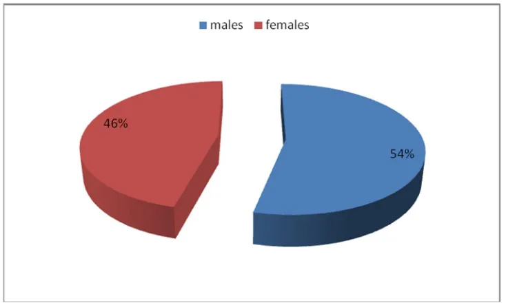
[image:57.595.88.474.252.423.2]A total no of 65 patients were found to have pancytopenia between Jan 2010 and Aug 2010.
TABLE:1 SEX DISTRIBUTION
NO. OF
PATIENTS PERCENTAGE p - value
Males 35 53.85%
0.385
Females 30 46.15%
Total 65 100.00%
[image:57.595.99.466.528.748.2]Among 65 patients included in our study, 35 (53.85%) patients were male, and 30 (46.15%) patients were female, though it is not statistically significant with p-value of 0.385.
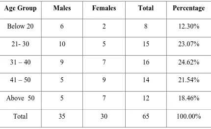
TABLE:2 AGE AND SEX DISTRIBUTION OF STUDY POPULATION
Age Group Males Females Total Percentage
Below 20 6 2 8 12.30%
21- 30 10 5 15 23.07%
31 – 40 9 7 16 24.62%
41 – 50 5 9 14 21.54%
Above 50 5 7 12 18.46%
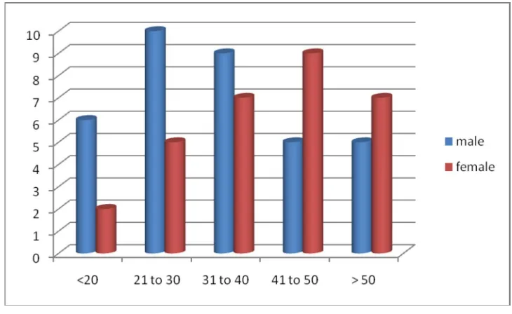
FIGURE: 2 DISTRIBUTION OF PATIENTS BASED ON AGE
Male preponderance is seen in patients < 30 yrs of age while female preponderance is seen in age group above 40 yrs of age though this finding is statistically insignificant (p – value of 0.545)
The age of the patients ranged between 14 yrs and 60 yrs with maximum no of patients seen between 31 – 40 yrs of age with a slightly male preponderance.
CLINICAL PRESENTATION:
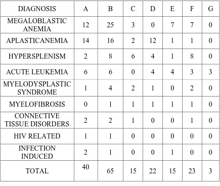
TABLE: 3 CLINICAL FEATURES OF PATIENTS ACCORDING TO
VARIOUS ETIOLOGIES
DIAGNOSIS A B C D E F G
MEGALOBLASTIC
ANEMIA 12 25 3 0 7 7 0
APLASTICANEMIA 14 16 2 12 1 1 0
HYPERSPLENISM 2 8 6 4 1 8 0
ACUTE LEUKEMIA 6 6 0 4 4 3 3
MYELODYSPLASTIC
SYNDROME 1 4 2 1 0 2 0
MYELOFIBROSIS 0 1 1 1 1 1 0
CONNECTIVE
TISSUE DISORDERS 2 2 1 0 0 1 0
HIV RELATED 1 1 0 0 0 0 0
INFECTION
INDUCED 2 1 0 0 1 0 0
TOTAL 40 65 15 22 15 23 3
A-FEVER B-PALLOR C-ICTERUS D-BLEEDING
E-HEPATOMEGALY F-SPLENOMEGALY
Pallor was present in almost all cases. Fever was the second most common presentation in our study as(40 out of 65 patients) 61.54% of patients had fever during the course of illness which is stastically insignificant with a p value of 0.063.
FIG: 3 VARIOUS CLINICAL FEATURES OF PATIENTS WITH
33.84% of patients (22) had bleeding manifestations, and are found
to have either aplastic anemia or acute leukemias which is stastically
significant(p vaue 0.009)
Icterus was present in 23.07% of the patients(15).Most of them had
chronic liver disease or megaloblastic anemia which is stastically
significant(p vaue < 0.001,significant at 5% level).
Liver was palpable in 15 (23.07%) of patients and was
predominantly seen in patients with megaloblastic anemia and leukemia
which is stastically significant(p vaue < 0.001,significant at 5% level).
Splenomegaly was found in 23 patients (35.38%); found in all
cases of hypersplenism, and in few cases of megaloblastic anemia, MDS,
and myelofibrosis which is stastically significant(p vaue < 0.018).
Lymphadenopathy was found only in 3 (4.62%) cases and seen in
patients with acute leukemia which is stastically significant(p vaue <
PERIPHERAL SMEAR FINDINGS:
[image:63.595.93.524.292.660.2]A detailed peripheral smear examination was done in all patients.
TABLE: 4 PERIPHERAL SMEAR FINDINGS ACCORDING TO
VARIOUS DIAGNOSIS
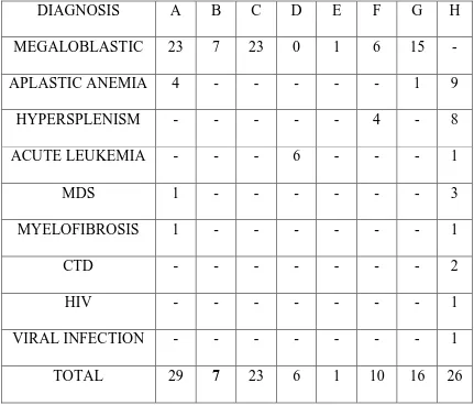
DIAGNOSIS A B C D E F G H
MEGALOBLASTIC 23 7 23 0 1 6 15
-APLASTIC ANEMIA 4 - - - 1 9
HYPERSPLENISM - - - 4 - 8
ACUTE LEUKEMIA - - - 6 - - - 1
MDS 1 - - - 3
MYELOFIBROSIS 1 - - - 1
CTD - - - 2
HIV - - - 1
VIRAL INFECTION - - - 1
A - Anisopoikilocytosis B - Erythroblasts
C - Hypersegmented neutrophils D - Immature WBC
E – Lymphocytosis. F – Reticulocytosis G- Macrocytes
[image:64.595.103.504.446.727.2]H-Microcytic,hypochromic RBCs
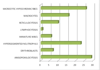
FIG:4 PERIPHERAL SMEAR FINDINGS IN PATIENTS WITH
Anisopoikilocytosis and Hypersegmented neutrophils were the predominant finding (92%) in cases of megaloblastic anemia; other findings being erythroblasts, lymphocytosis, and reticulocytosis.
Anisopoikilocytosis was also found in few cases of aplastic anemia. Peripheral blood WBC blasts were found in 4 cases of acute leukemia and in a case of MDS.
Atypical lymphocytes were found in two cases of viral infection.
VARIOUS ETIOLOGIES OF PANCYTOPENIA:
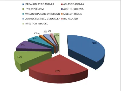
FIGURE: 5 DISTRIBUTION OF VARIOUS ETIOLOGIES OF
PANCYTOPENIA
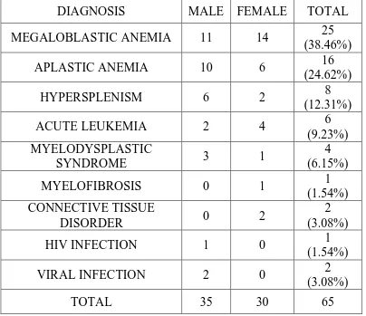
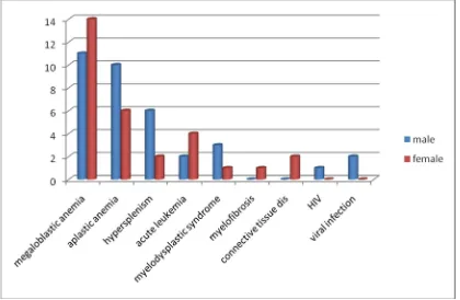
TABLE: 5 DISTRIBUTION OF CAUSES OF PANCYTOPENIA
ACCORDING TO SEX
DIAGNOSIS MALE FEMALE TOTAL MEGALOBLASTIC ANEMIA 11 14 25
(38.46%)
APLASTIC ANEMIA 10 6 16
(24.62%)
HYPERSPLENISM 6 2 8
(12.31%)
ACUTE LEUKEMIA 2 4 6
(9.23%) MYELODYSPLASTIC
SYNDROME 3 1
4 (6.15%)
MYELOFIBROSIS 0 1 1
(1.54%) CONNECTIVE TISSUE
DISORDER 0 2
2 (3.08%)
HIV INFECTION 1 0 1
(1.54%)
VIRAL INFECTION 2 0 2
(3.08%)
FIGURE: 6 DISTRIBUTION OF CAUSES OF PANCYTOPENIA
ACCORDING TO SEX
Megaloblastic anemia was more common in females (14) compared to males (11) in our study. While Aplastic anemia is common in males (10) than females (6); both of which are statistically significant.
MEGALOBLASTIC ANEMIA:
Mean age of patients with megaloblastic anemia is 39.32 yrs.
[image:69.595.100.513.329.575.2]84 % of patients had macrocytosis; with a maximum of 127.3femtolitres; 16% of patients had normocytic anemia.
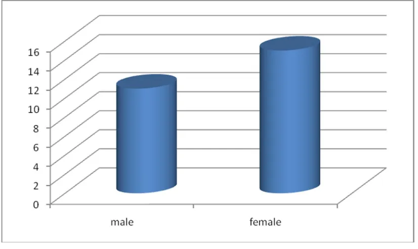
FIGURE 7: DISTRIBUTION OF MEGALOBLASTIC ANEMIA
ACCORDING TO SEX
56% of patients (14) with megaloblastic anemia are females, and the remaining 44% are males (11).
FIGURE 8: DISTRIBUTION OF PATIENTS ACCORDING TO
MCV
FIGURE 9: PERIPHERAL SMEAR FINDINGS IN PATIENTS
[image:70.595.101.510.465.701.2]Anisopoikilocytosis (92%), and hypersegmented neutrophils (92%) are the most common findings in peripheral smear of patients with megaloblastic anemia.
FIGURE 10: CLINICAL FEATURES OF PATIENTS WITH
MEGALOBLASTIC ANEMIA
Pallor was present in all cases. The other features were fever (48%), icterus (12%), hepatomegaly (28%), and splenomegaly (28%).
features of malabsorption and another one was a pure vegetarian.Among 14 female patients with Megaloblastic anemia 4 were diagnosed to have hypothyroidism,2 patients had pernicious anemia.3 female patients were pure vegeterians.another 2 patients had history of consumption of anti epileptic drugs.
APLASTIC ANEMIA:
24.62% of patients had aplastic anemia. Of which 10(62.5%) were male and 6(37.5%) were female.Mean age of patients with aplastic anemia is 33.43 yrs.Average Hb% of these patients is 4.80 gms%.
56.25% of patients had thrombocytopenia < 10,000 cells/cumm.
FIGURE 11: DISTRIBUTION OF APLASTIC ANEMIA
[image:72.595.101.513.461.755.2]68.75% of patients with aplastic anemia had MCV<100 femtolitres
[image:73.595.164.454.221.407.2]while 31.25% had MCV>100 femtolitres.
FIGURE 12: DISTRIBUTION OF PATIENTS WITH APLASTIC
ANEMIA ACCORDING TO MCV
FIGURE 13: CLINICAL FEATURES OF PATIENTs WITH
[image:73.595.103.509.494.763.2]The various clinical features of patients with aplastic anemia were pallor (100%), fever (87.5%), icterus (12.5%), bleeding (75%), hepatomegaly (6.25%), and splenomegaly (6.25%).
HYPERSPLENISM
8 patients (12.31%) with pancytopenia were diagnosed to have hypersplenism of which 6 were male and 2 were female patients.
[image:74.595.108.514.469.717.2]Out of 8 patients 2(25%) had Extra Hepatic Portal Obstruction, while the remaining 6 (75%) had Cirrhosis of liver leading to hypersplenism.
ACUTE LEUKEMIA
[image:75.595.109.514.341.644.2]6 patients (9.23%) was diagnosed to have acute leukemias; 4(66.7%) of them with acute myeloid leukemia; 2(33.3%) with acute lymphoid leukemia.out of the 6 patients 2 were males and 4 were females.
FIG:15 DISTRIBUTION OF PATIENTS WITH LEUKEMIA
ACCORDING TO SEX
MYELODYSPLASTIC SYNDROME
MDS was diagnosed in 4 patients (6.15%); of which 3(75%) were
male, and 1 (25%) was female.
Mean age of these patients is 56.25yrs .Pseudo Pelger Huet
cells(Hyposegmented neutrophils) were present in two cases of MDS.
Erythroblasts were found in peripheral smear of one of these patients.
MYELOFIBROSIS
One (1.54%) female patient was diagnosed to have myelofibrosis.
VIRAL INFECTIONS
2 patients (3.08%) had features of systemic viral infection; their
peripheral smear showed atypical lymphocytes but no blasts.
1 (1.54%) male patient was positive for HIV.
COLLAGEN VASCULAR DISORDER
2 patients (3.08%) were found to have Systemic lupus
Fig: 1 peripheral smear of a patient with megaloblastic anemia showing anisopoikilocytosis with hypersegmented neutrophil.
[image:77.595.120.479.420.642.2]Fig: 3 Bone marrow examination of a patient with aplastic anemia showing hypocellular marrow replaced by fat.
[image:78.595.136.464.426.671.2]DISCUSSION
Pancytopenia is a serious hematological problem which presents with anemia, bleeding manifestations and infections. There are only a limited number of studies from India on the frequency of various causes of pancytopenia.The variation in the frequency of various entities causing pancytopenia has been attributed to the differences in methodology and stringency of diagnostic criteria, geographic area, period of observation,genetic differences and exposure to various myelotoxic agents. Our study included 65 patients and the age group of the patients range from 14 to 60 years. Of these 30 were females and 35 were males. The mean age of the patients is 37.12 years. Most male patients are under 30 years and females more than 40 years.
Incidence of Megaloblastic anemia varies from 0.8 to 32.26% in various studies. In our study the incidence of Megaloblastic anemia is 38.46%. Similar results were obtained in the studies done by Kumar et al1(22%) , Aziz et al2 (40.9%) , Savage et al 3,Iqbal et al7. However the study by Tilak et al4 showed the incidence to be 68%.
In our study, the predominant signs present in patients with megaloblastic anemia were pallor(100%) followed by fever(48%), hepatomegaly(28%), splenomegaly(28%) and icterus(12%).Similar results were shown in the studies done by Tilak et al4 ,Khunger et al5(pallor in 100% of patients with Megaloblastic anemia)and Aziz et al2(pallor in 98% of patients with megaloblastic anemia).
In our study, 92% of Megaloblastic anemia patients had hypersegmented neutrophils in their peripheral smear and 28% had circulating megaloblasts.This is in concordance with the studies done by Tilak et al4,Khunger et al5and Ishtiaq et al15.
In our study,84% of patients with megaloblastic anemia had MCV
more than 100fl.16% had MCV less than 100 fl. The maximum MCV
noted was 127.3 fl.Megaloblastic anemia should be considered on top if
MCV is greater than 100 fl.Most of the patients had increased levels of
serum iron and ferritin.Most of the precursor cells are destroyed in the
marrow due to ineffective erythropoiesis and therefore serum iron is not
utilized.Once treatment is started with Vit B12 or folate the serum iron
and ferritin levels fall.
Bone marrow aspiration showed typical megaloblasts with sieved
chromatin and asynchronic nuclear cytoplasmic ratio.Though we have
done bone marrow aspiration for all patients it can be deferred in
those cases presenting with hepatosplenomegaly and having
hypersegmented neutrophils and or circulating megaloblasts in their
peripheral smear.These patients can be put on a trial of hematinics with a
close hematologic follow up.
Megaloblastic anemia was present in all age groups.Among the 11
male patients with megaloblastic anemia in our study 7 were chronic
alcoholics and 2 patients had undergone abdominal surgeries(one patient
resection).One patient had loose stools along with loss of weight for more
than three months.Upper GI endoscopy showed erosions in the stomach
and Lower GI endoscopy revealed cryptitis with cryptic abscess
suggestive of Ulcerative colitis.One male patient was a pure vegetarian
with even no milk consumption.
In our study, there were 14 females with megaloblastic
anemia.Among them 4 patients were diagnosed to have
hypothyroidism.Most of them were above 50 years.One young female
presented with jaundice,anemia and fever.she was diagnosed to have Vit
B12 deficiency with features of hemolysis.2 female patients had atrophic
gastritis in upper GI scopy suggestive of Pernicious anemia confirmed by
histopathological examination.Of these two patients one had
arthralgias,myalgias and history suggestive of skin rashes.The other
patient had family history of similar illness. 3 female patients with
megaloblastic anemia were pure vegeterians. In two patients with
megaloblastic anemia there is a general decrease in the intake of food due
to poverty. The higher prevalence of nutritional deficiency has been cited
for the increased frequency of megaloblastic anemia.2 patients had
history of consumption of antiepileptic drugs for a longer period with no
The incidence of Aplastic anemia varies from 10% to 52.7% in patients with pancytopenia in various studies. In our study the incidence of Aplastic anemia is 24.61%.The mean age of the patients is 33.43 years.The incidence was more common in males(62.5%) as compared to females(37.5%).The most common presentation was pallor(100%) followed by fever(87.5%) and bleeding(75%).One of the patients with Aplastic anemia in our study had splenomegaly.This could be due to the repeated blood transfusions. It is the second most common cause of pancytopenia in our study as shown by Aziz et al2(31.88%) , Savage et al3, Khunger et al5(14%) .The study done by Kumar et al1 showed aplastic anemia to be the most common cause of pancytopenia with 29.5%.
In Aplastic anemia hematopoiesis fails. Blood cell counts are extremely low and the bone marrow appears empty. 31.25% of patients with Aplastic anemia had MCV more than 100 fl. 56.25% of patients had MCV less than 100 fl . The platelet count was less than 10,000 in 56.25% of patients.
to the increased exposure to the toxic chemicals and increased availability of over the counter medications.
Hypersplenism is the third most common cause (12.3%) of pancytopenia in our study. Most of them are males. The study by Aziz et al2 showed the incidence of hypersplenism in pancytopenic patients to be 6.81% .In our study, 25% of hypersplenism were due to Extra hepatic portal vein obstruction and 75% were due to chronic liver disease. One of the patients with chronic liver disease was hepatitis C positive . The bone marrow of this patient showed adequate hematopoiesis with mild to moderate megaloblastic changes. This ruled out the possibility of Hepatitis C induced myelosuppression.One of the patients was Hepatitis B positive.
Acute leukemia was found to be the fourth most common cause
(9.23%) of Pancytopenia in our study which is similar to a study
conducted by Savage et al3 who observed that the most common cause of
pancytopenia was megaloblastic anemia followed by aplastic anemia,
acute leukemia,
AIDS and hypersplenism, and another study by Kumar et al1 who
reported the causes of pancytopenia in order of frequency as Aplastic
Anemia at 29.5%, megaloblastic anemia at 22%, Aleukemic Leukemia or
lymphoma (18%) and hypersplenism at 11.4%.
Of the 6 patients with acute leukemias, 4 had acute myeloid
leukemia, and 2 had acute lymphocytic leukemia.
The incidence of connective tissue disorder as a cause of pancytopenia is 3.07%. One of the patients had autoimmune myelofibrosis with autoimmune thyroiditis.
Myelofibrosis was diagnosed in one patient(1.54%) with pancytopenia. There was an extensive marrow replacement by fibrosis. There will be increasing splenomegaly and hepatomegaly due to extramedullary hematopoiesis .The peripheral blood cells are low due to decreased production from the marrow as well as due to hyperfunctioning of the spleen.
Lymphocytes 22%,transformed lymphocytes present,increased Reticuloendothelial cells with hemophagocytosis.
LIMITATIONS OF THE STUDY
The major limitation of our study is the small number of subjects we have included in our study.
A
ntibodies to Intrinsic factor was not done in pernicious anemiacausing Megaloblastic anemia.
Trephine biopsy evaluation was not done in all patients.Therefore more definitive evaluation of cellularity and exclusion of focal lesions could not be done.
CONCLUSION
Megaloblastic anemia is the most common cause of pancytopenia in our setting.
This probably indicates the poor nutritional status of the general population in our community.
All patients with pancytopenia should be sought for megaloblastic anemia as it is a potentially treatable condition.
Physicians should have a high index of suspicion for Vitamin B12
deficiency when dealing with patients presenting with symptoms of
anemia such as pallor and weakness and/or diagnosed with
pancytopenia on further workup.
The finding of hypersegmented neutrophils in the peripheral smear will guide in the diagnosis of megaloblastic anemia.
Aplastic anemia is the second most common cause of pancytopenia in our study.
The blood counts were extremely low in patients with aplastic anemia.
Hypersplenism is an important cause of pancytopenia in our setting.
Hypersplenism should be sought as a cause of pancytopenia in patients with chronic liver disease especially alcoholics.
Myelodysplastic syndrome is a common cause of pancytopenia in the elderly population.
After the common causes have been ruled out patient should be evaluated for rare causes like HIV infection, systemic viral infection, connective tissue disorders, and myelofibrosis.
Peripheral smear and bone marrow examination would help in identifying the etiology of pancytopenia in almost all patients.
RECOMMENDATIONS
Bone marrow aspiration can be deferred in those cases presenting with hepatosplenomegaly and having hypersegmented neutrophils and or circulating megaloblasts in their peripheral smear.These patients can be put on a trial of hematinics(Vit B12/folate) with a close hematologic follow up.
BIBLIOGRAPHY
1. Kumar R, Kalra SP, Kumar H, Anand AC, Madan H.
Pancytopenia—a six year study. J Assoc Physicians India
2001;49:1078-81.
2. Aziz T, Ali L, Ansari T, Liaquat HB, Shah S, Ara J. Pancytopenia:
Megaloblastic anemia is still the commonest cause. Pak J Med Sci
2010;26(1):132-136
3. Savage DG, Allen RH, Gangaidzo IT, Levy LM, Gwanzura C, et. al. Pancytopenia in Zimbabwe. Am J Med Sci 1999 Jan;317(1):22-32.
4. Tilak V, Jain R. Pancytopenia—a clinico-hematological analysis of
77 cases. Indian J Pathol Microbiol 1999;42(4):399-404.
5. Khunger JM, Arulselvi S, Sharma U, Ranga S, Talib VH.
Pancytopenia—a clinico haematological study of 200 cases. Indian J
Pathol Microbiol 2002;45(3):375-9.
6. Tariq M, Khan N, Basri R, Amin S. Aetiology of pancytopenia. Professional Med J Jun 2010;17(2):252-256.
8. Kar M, Ghosh A. Pancytopenia. J Indian Acad Clin Med
2002;3(1):29-34.
9. Khalid G, Moosani MA, Ahmed L, Farooqi AN. Megaloblastic
anemia seen in 48 cases of pancytopenia. Ann Abbasi Shaheed Hosp
Karachi Med Dent Coll 2005;10(2):742-4.
10. Gupta P K, Saxena R, Karan AS, Choudhry VP. Red cell indices for
distinguishing macrocytosis of aplastic anemia and megaloblastic
anemia. Indian J Pathol Microbiol 2003;46(3):375-7.
11. Ziaei JE, Dastgiri S. Role of myeloperoxidase index in
differentiation of megaloblastic and aplastic anemia. Indian J Med
Sci 2004;58(8):345-8
12. García-Casal MN, Osorio C, Landaeta M, Leets I, Matus P,
Fazzino F et al. High prevalence of folic acid and vitamin B12
deficiencies in infants, children, adolescents and pregnant women in
Venezuela. Eur J Clin Nutr 2005;59(9):1064-70.
13. Savage DG, Lindenbaum J, Stabler SP, Allen RH. Sensitivit of
serum methylmalonic acid and total homocysteine determinations
for diagnosing cobalamin and folate deficiencies. Am J Med
14. Stabler SP, Allen RH. Vitamin B12 deficiency as a worldwide
problem. Annu Rev Nutr 2004;24:299-326
15. Ishtiaq O, Baqai HZ, Anwer F, Hussain N. Patterns of
Pancytopenia patients in a General Medical Ward and a proposed
diagnostic approach. J Ayub Med Coll Abottabad 2004;16(1):8-13
16. Ng SC, Kuperan P, Chan KS, Bosco J, Chan GL. Megaloblastic
Anemia- a review from University Hospital, Kuala Lumpur. Ann
Acad Med Sing 1988;17:261.
17. Modood-ul-Mannan, Anwar M, Saleem M, Wigar A, Ahmad MA.
Study of serum vitamin B12 and folate levels in patients of
megaloblastic anemia in northern Pakistan. J Pak Med Assoc
1995;45:187
18. Niazi M, Raziq F. The incidence of underlying pathology in
Pancytopenia - an experience of 89 cases. J Postgrad Med Inst
2004;18(1):76-9.
19. Varma N, Dash S. A reappraisal of underlying pathology in adult
patients presenting with pancytopenia. TropGeogr Med
20. Bakhshi S. Aplastic Anemia- Facts and statistics. eMedicine J
2001;2(6):1.
21. Young NS. Acquired Aplastic Anemia. Ann of Int Med
2002;136(7):534
22. Issaragrisil S, Chansung K, Kaufman DW, Sirijirachai J,
Thamprasit T, Young NS. Aplastic anemia in rural Thailand: its
association with grain farming and pesticide exposure. Am J Public
Health 1997;87:1551.
23. Kaufman DW, Issaragrisil S, Anderson T, Chansung K, Thamprasit
T, Sirijirachai J, et al. Use of household pesticides and the risk of
aplastic anemia in Thailand. Int J Epidemiol 1997;26:643-50.
24. Savage DG, Allen RH, Gangaidzo IT, Levy LM, Gwanzura C,
Moyo A, et al. Pancytopenia in Zimbabwe.
25. Kar M, Ghosh A. Pancytopenia. JIACM 2002;3(1):29-34.
27. International Agranulocytosis and Aplastic Anemia study group:
Incidence of Aplastic Anemia; The relevance of diagnostic criteria. Blood 1987;70:1718
28. Keisu M, Ost A. Diagnosis in patients with severe Pancytopenia
suspected of having Aplastic Anemia. Euro J Haematol 1990;45;11.
29. Iqbal W, Hassan K, Ikram N, Nur S. Aetiological breakup of 208
cases pancytopenia. J Rawal Med Coll 2001;5 (1):7-9.
30. Rahim F, Ahmad I, Islam s, Hussain M, Khattak TAK, Bano Q. Spectrum of hematological disorders in children observed in 424 consecutive bone marrow aspirations/biopsies. Pak J Med Sci 2005;21:433-6.
31. Dodhy MA, Bokhari N, Hayat A. Aetiology of Pancy topenia, A
five-year experience. Ann Pak Inst Med Sci 2005;1(2):92-5.
32. Niazi M, Raziq F. The incidence of underlying pathology in
pancytopenia. An experience of 89 cases. J Postgr Med Inst 2004;18(1):76-9.
incidence of aplastic anemia in Thailand. The Aplastic Anemia Study Group. Am J Hematol 1999;61: 164-8.
34. Young NS. Hematopoietic cell destruction by immune mechanisms
in acquired aplastic anemia. Semin Hematol 2000;37:3-14.
35. Memon S, Shaikh S, Nizamani AA. Etiological spectrum of
pancytopenia based on bone marrow examination in children. J Coll Physicians Surg Pak 2008;18(3):163-7.
36. Ng SC, Kuperan P, Chan KS, Bosco J, Chan GL Megaloblastic
Anemia- a review from University Hospital, Kuala Lumpur. Ann Acad Med Sing 1988; 17:261.
37. Chandra J, Jain V, Narayan S, Sharma S, Singh V, Kapoor AK, et al. Folate and cobalamin deficiency in megaloblastic anemia in children. Indian Pediatr 2002; 39:453-7.
38. Mannan M, Anwar M, Saleem M, Wigar A, Ahmad MA. Study of
serum vitamin B12 and folat levels in patients of megaloblastic anemia in northern Pakistan. J Pak Med Assoc 1995;45:187.
39. Rosse WF. The control of complement activation by the blood cells
40. Firkin F, Chesterman C, Pennigton D, Rush B. De Gruchy’s Clinical Hematology in Medical Practice. 5th ed. Oxford: Blackwell; 1989.
41. Rappeport JM, Bunn HF. Bone marrow failure: aplastic anemia and
other primary bone marrow disorders. In: Isselbacher KJ, Braunwald E, Wilson JD, Martin JB, Fauci AS, Kasper KL, eds. Harrison’s Priniciples of Internal Medicine. 13th ed. New York: McGraw-Hill; 1994:1754-7.
42. Bain BJ, Clark DM, Lampert IA. Bone marrow pathology. Oxford:
Blackwell; 1992
43. Bain BJ. Blood Cells: A practical guide. 2nd ed. Oxford: Blackwell;
1995.
44. Qazi RA, Masood A. Diagnostic evaluation of pancytopenia. J
Rawal Med Coll 2002;6(1):30-3.
45. Khokhar N. Bone marrow suppression in chronic hepatitis C.
Pakistan J Med Sci 2001;17(4):251-3
47. Young NS, Barrett AJ. The treatment of severe acquired aplastic
anaemia.J Am Soc of Hematol 1995; 85: 3367- 77.
48. Camitta BM, Storb R, Thomas ED. Aplastic anemia : pathogenesis,
diagnosis, treatment and prognosis. N Engl J Med 1982; 306: 645-52.
49. Gutiernez-Unena S, Molina JF, Garcia CD et al. Espinoza LR.
Pancytopenia secondary to methotrexate therapy in rheumatoid arthritis.Arthritis Rheum 1996; 39 (2): 272-6.
50. Mehta BC. Haematological emergencies. Medicine Update 1992; Ed. Mukherjee S 1992; Vol 2 : 177-8.
51. Boogaerts M, Cavalli F, Cortes-Funes H et al. Granulocyte growth
factors: achieving a consensus. Annals of Oncol1995; 6: 237-44.
52. Peschel C, Huber C, Aulitzky WE. Clinical applications of
cytokines. La Medicine En France 1994; 176-88.
53. Katz R, Chuang LC, Sutton JD. Use of granulocyte colony –
54. Fakunle YM. Tropical splenomegaly, Part 1: Tropical Africa.Clin
Haematol 1981; 10: 963-75.
55. Chapman WC, Newman M. Disorders of the spleen. In: Richard
Lee G, Foerster J, Lukens J, Paraskevas F, Greer JP, Rodgers GM Eds. Wintrobe’s Clinical Hematology, 10th edition. Philadelphia, Lippincot Williams and Wilkins. 1999; 1969-89.
56. Duckett JW. Splenectomy in treatment of secondary hypersplenism.
Ann Surg 1963; 157: 737-46.
57. Thanopoulos BD, Frimas CA, Mantagos SP, et al. Gaucher disease:
A STUDY ON THE CLINICAL AND ETIOLOGICAL PROFILE OF PATIENTS WITH PANCYTOPENIA
PROFORMA
Name: Age: Sex: OP.NO: Address: Ph.No:
Presenting Complaints:
Easy fatiguability: Fever/chills & rigor: Dyspnoea: Giddiness:
Jaundice: Abdominal distension: Lymphadenopathy:
Purpura/Skin rash: Pedal edema: Loss of Wt: Loss of Appetite: Night Sweats: Bone Pain:
Bleeding Tendency: Gum bleed/Hematamesis/ Malena/Bleeding PR Purpura/ Skin rash:
Cough/ burning micturition/loose stools: Drug intake:
Alcohol intake/smoking:
Co-morbid Illness: DM: HT: TB: Cardiac illness: any malignancy:
EXAMINATION:
Fever:
Pallor: Jaundice: Lymphadenopathy: Cyanosis: Clubbing: Pedal edema: Purpura/Petechiae/Ecchymosis:
Sternal tenderness: Others: CVS:
RS:
Abdomen: Genitals:
Per rectal / pelvic examination:
INVESTIGATIONS FOR ALL PATIENTS:
CBC
HB%: PCV: ESR: Reticulocyte Index:
TC: P: L: E: B: M: Platelets:
Occult Blood in Stools: Blood sugar:
RFT: Urea: Creatinine:
LFT: Tot.bilirubin: Direct: SGOT: SGPT: Alkaline phosphatase:
ELISA for HIV:
HBsAg: Anti-HCV: X-RAY CHEST:
ECG:
USG-Abdomen: Serum B12 level: Serum Folate:
PT: INR: aPTT: Peripheral Smear Study:
Bone Marrow Study: Upper GI scopy: Lower GI scopy Serum Electroporesis: Se.Ferritin:
Others:
