COMPARISON OF NEUTROPHIL FUNCTION IN
DIABETIC AND NON DIABETIC WITH
GENERALIZED CHRONIC PERIODONTITIS AND IN
GENERALIZED AGGRESSIVE PERIODONTITIS
PATIENTS AND TO COMPARE THIS STUDY IN
HEALTHY CONTROL GROUP
A Dissertation Submitted
in partial fulfillment of the requirements
for the degree of
MASTER OF DENTAL SURGERY
BRANCH – II
PERIODONTOLOGY
Certificate
This is to certify that Dr. SUNIL D. PENDOR, Post graduate student (2008 – 2011) in the Department of Periodontics, Tamilnadu Government Dental College and Hospital, Chennai – 600 003 has done this dissertation titled “COMPARISON OF NEUTROPHIL FUNCTION IN DIABETIC AND NON DIABETIC WITH GENERALIZED CHRONIC PERIODONTITIS AND IN GENERALIZED AGGRESSIVE PERIODONTITIS PATIENTS AND TO COMPARE THIS STUDY IN HEALYHY CONTROL GROUP.” under our direct guidance and supervision in partial fulfillment
of the regulations laid down by The Tamil Nadu Dr. M.G.R. Medical University, Chennai -600 032 for M.D.S., (Branch – II) Periodontology degree examination.
DR.K.MALATHI M.D.S.
Professor and Guide
DR.K.MALATHI M.D.S.
Professor and Head
Department of Periodontology
Tamilnadu Government Dental College and Hospital
Chennai – 600 003.
DR.K.S.G.A. NASSER
PRINCIPAL
ACKNOWLEDGMENT
I express my deep sense of gratitude to DR. K. MALATHI M.D.S., Professor and H.O.D., Department of Periodontics, Tamilnadu Government Dental College and Hospital, without her unstained
guidance, support and encouragement, this study would not have been
possible. Words cannot express my gratitude for her quiet confidence in
my ability to do the study, her willingness to help clear the stumbling
blocks along the way and her tremendous patience till the end of the
study. I am also extremely grateful to DR. S. KALAIVANI M.D.S.,
Professor, DR. MAHEASWARI RAJENDRAN M.D.S., Associate Professors, Department of Periodontics, Tamilnadu Government Dental
College and Hospital, Chennai – 600 003, for their expert guidance and
moral support during the completion of this study. I consider myself
privileged, to have studied, worked and completed my dissertation under
them in the Department.
My sincere thanks to DR. K.S.G.A. NASSER, M.D.S., Principal, Tamilnadu Government Dental College and Hospital, Chennai – 600 003,
for his kind permission and encouragement.
I am extremely grateful to DR.A.MUTHUKUMARASAMY, M.D.S., DR. M. JEEVA REKHA, M.D.S., DR.P.KAVITHA, M.D.S., Assistant professors, Department of Periodontics, Tamilnadu Government Dental
College and Hospital, Chennai – 600 003 for their valuable suggestions,
constant encouragement and timely help rendered throughout this
I thank to DR. RAMA GOPALAN, M.D., Ph.D., Professor and HOD, Department of Pathology, Institute of Basic Medical Science, Taramani, Chennai for granting me permission to conduct this study in the Department.
I am extremely grateful to DR. P. SHANTI, M.D., PH.D Professor, Department of Pathology, Institute of Basic Medical Science, Taramani, Chennai for having taken special interest in my study and teaching me the basic of neutrophil function test.
I would like to express my very special gratitude to Mrs. Amidha,
Technician, for helping me in my study.
I thank DR.R. RAVANAN Msc.,M.Phil.,Ph.D.,Associate Professor, Department of Statistics, presidency college ,Chennai,) for
helping me with the statistics in the study.
I take this opportunity to express my gratitude to my
colleagues and well wishers for their valuable help and suggestions
throughout this study.
A special mention of thanks to all my patients for their consent,
co-operation and participation in this study.
All glory and honour to THE LORD ALMIGHTY who gives me the strength to persist against all odds, whose loving kindness and mercies
endureth forever.
My heartfelt and deep gratitude to all my family members, for their
help, patience, love and prayers which have sustained me throughout
ABSTRACT
BACKGROUND
Epidemiological studies have shown that the risk for periodontitis in diabetes
mellitus patient is greater than non diabetics. Diabetes mellitus is now documented to
significantly enhance susceptibility to severe periodontitis. Increased susceptibility to
periodontitis has been associated with impaired neutrophil function in diabetes mellitus.
The objectives of this study are to compare the neutrophil function in diabetic and non
diabetic patients with generalized chronic periodontitis and in generalized aggressive
periodontitis patients and to compare this study in control group.
AIM
To assess neutrophil function i.e. Chemotaxis, Phagocytosis and Specific Granule
Release in Diabetic and Non Diabetic patients with Generalized Chronic Periodontitis
and Generalized Aggressive Periodontitis in Indian population.
Materials and Method:
60 patients were selected to participate in the study which was divided into four groups.
These patients were selected according to clinical and radiographic criteria. Blood
samples from 60 patients were collected after drawing 5ml peripheral venous blood and
then various assays were carried out to assess the neutrophil functions mentioned above.
Statistical analysis of the results was done using ANOVA followed by Tukey HSD test.
Results:
It was found that defective chemotaxis present in diabetic and in generalized
aggressive periodontitis patients. Defective phagocytosis was observed in diabetic,
generalized chronic, and in generalized aggressive periodontitis patients. And defective
specific granule release assay was observed only in diabetic patient when compared to
healthy control.
Conclusion:
This study has proved that there is association between neutrophil dysfunction
and severity of periodontal diseases. Hence, further longitudinal and clinical trials with
DECLARATION
TITLE OF DISSERTATION Comparison of neutrophil function in
diabetic and non diabetic patient with generalized chronic periodontitis and in generalized aggressive periodontitis patients and to compare this study in healthy control group.
PLACE OF STUDY Tamil Nadu Government Dental College & Hospital, Chennai-600003
DURATION OF THE COURSE 3 Years
NAME OF THE GUIDE Dr.K.Malathi
HEAD OF THE DEPARTMENT Dr.K.Malathi
I hereby declare that no part of the dissertation will be utilized for gaining financial
assistance/any promotion without obtaining prior permission of the Principal,
Tamil Nadu Government Dental College & Hospital, Chennai-600003. In addition,
I declare that no part of this work will be published either in print or in electronic
media without the guide who has been actively involved in dissertation. The author
has the right to reserve for publish of work solely with the prior permission of the
Principal, Tamil Nadu Government Dental College & Hospital, Chennai-600003.
CONTENTS
S.NO. TITLE PAGE
NO.
1. INTRODUCTION 1
2. AIMS AND OBJECTIVES 3
3. REVIEW OF LITERATURE 4
4. MATERIALS AND METHOD S 22
5. RESULTS 48
6. DISCUSSION 58
7. SUMMARY AND CONCLUSION 66
8. BIBLIOGRAPHY 69
LIST OF ABBREVIATIONS
1. AGE Advanced glycated end products
2. A.A. Aggregatibacter Actinomycetemcomitans
3. B Bacteriods
4. CAL Clinical attachment level
5. DM Diabetes mellitus
6. FMLP Formyl methionyle leucine phosphate
7. GCF Gingival crevicular fluid
8. GI Gingival index
9. GagP Generalized Aggressive Periodontitis
10. IDDM Insulin dependent diabetes mellitus
11. JP Juvenile Periodontitis
12. LJP Localized Juvenile Periodontitis
13. NBT Nitro Blue Tetrazolium
14. NADPH Nicotine amide adenine-di-nucleotide
phosphate (reduced)
15. NIDDM Non insulin dependent diabetes mellitus
16. PMN Polymorphonuclear leukocyte
17. P. gingivalis Porphyromonas gingivalis
LIST OF PHOTOGRAPHS
S. No. Title Page
No
1. Clinical status of Group I subject 36
2. Clinical status of Group II subject 36
3. Clinical status of Group III subject 37
4. Clinical status of Group IV subject 37
5. Measurement of probing depth with William’s periodontal probe 38
6. Photograph of Radiographs of group I 38
7. Photograph of Radiographs of group II 39
8. Photograph of Radiographs of group III 39
9. Photograph of Radiographs of group IV 40
10. Armamentarium used for periodontal examination and blood
sample collection
40
11. Collection of venous blood 41
12. Armamentarium for sample transportation 41
13. Armamentarium used for neutrophil function test 42
14. Microscope 42
15. Incubator 43
S. No. Title Page No
17. Photomicrograph showing neutrophils migrating toward
Chemoattractant – Low Power
44
18. Photomicrograph showing neutrophil migrating toward
Chemoattractant – High Power
44
19. Photomicrograph showing neutrophil phagocytosis of Candida 45
LIST OF TABLES
Table No.
Title Page No
I Comparison of mean plaque index between four groups 51
II Comparison of mean periodontal index between four groups 51
III Comparison of mean and standard deviation of neutrophil
chemotaxis between four groups
52
IV Multiple Comparison of mean and standard deviation of
neutrophil Chemotaxis between four groups
52
V Comparison of mean and standard deviation of neutrophil
phagocytosis between four groups
53
VI Multiple Comparison of mean and standard deviation of
neutrophil Phagocytosis between four groups
53
VII Comparison of mean and standard deviation of specific granule
release between four groups
54
VIII Multiple Comparison of mean and standard deviation of specific
granule release between four groups
LIST OF FIGURES
Figure
No. TITLE PAGE NO
I Comparison of mean plaque index between four groups 55
II Comparison of mean periodontal index between four groups 55
III
Comparison of mean and standard deviation of neutrophil
Chemotaxis between four groups 56
IV Comparison of mean and standard deviation of neutrophil
phagocytosis between four groups 56
V Comparison of mean and standard deviation of specific
INTRODUCTION
INTRODUCTION
Diabetes mellitus is a multifactorial disease. The disease is characterized by
hyperglycemia, hyperlipidemia, and associated complication.10, 18 Diabetes mellitus
represents one of the major chronic health problem facing the world today. Diabetes
mellitus develops from either defect in insulin production or an impaired utilization of
insulin. Based upon these two conditions diabetes mellitus can be divided into two main
types. Type I (IDDM) or juvenile diabetes caused by destruction of pancreatic islets cells.
Type II (NIDDM) or adult onset diabetes which results from the defect in the insulin
molecule or alter cell receptors for insulin and represents impaired insulin function rather
than the deficiency. Periodontal disease is an infectious inflammatory process of
multifactorial origin that involves the interplay between bacteria, host and environmental
factors.47 The dynamics between the host immune responses and oral bacteria is essential
to understand the pathogenesis of periodontal diseases.26
Although numerous host defense mechanisms are called into an action by the
bacterial invasion at the gingival sulcus, substantial evidence indicates that the
Polymorphonuclear Neutrophils (PMNs) are the key cellular elements of the innate
immune system, providing protection from invading bacteria.48 Neutrophils impairment
leads to increased susceptibility to Periodontitis in diabetic patient. Epidemiologic studies
have shown that the risk for Periodontitis in diabetes mellitus patient is greater than non
diabetic. Type I and II diabetes are significantly enhances susceptibility to severe
Periodontitis. In Aggressive Periodontitis there is decreased responsiveness of PMN to
source of chemotactic factor.65 The important role of PMNs play in optimal functioning
of the immune defense system, has led to speculation that a partly compromised system
could severely weaken the defense mounted against a bacterial insult and permit the
occurrence and progression of infections.23 Since there are few studies which have been
done in this context and very little data available in Indian population, the present study
was undertaken to assess the invitro neutrophils chemotaxis, phagocytosis and specific
granule release in diabetic and non diabetic patient with Generalized Chronic
AIM
The study was aimed to assess neutrophils function i.e. Chemotaxis, Phagocytosis and
Specific Granule Release in Diabetic and Non Diabetic patients with Generalized
Chronic Periodontitis and Generalized Aggressive Periodontitis in Indian population.
OBJECTIVES
1. To assess Chemotactic activity of neutrophils in Diabetic and Non Diabetic patients with Generalized Chronic Periodontitis and in Generalized Aggressive Periodontitis
(GAgP) subjects.
2. To assess Phagocytic activity of neutrophils in Diabetic and Non Diabetic patients with Generalized Chronic Periodontitis and in Generalized Aggressive Periodontitis
(GAgP) subjects.
3. To assess Specific Granule Release activity in Diabetic and Non Diabetic patients with Generalized Chronic Periodontitis and in Generalized Aggressive Periodontitis
(GAgP) subjects.
4. To compare these neutrophil functions within the above forms of Periodontitis with the control group of periodontally healthy subject.
REVIEW OF LITERATURE
DIABETES MELLITUS
Diabetes mellitus is a clinically and genetically heterogenous group of metabolic
disorders manifested by abnormally high levels of glucose in the blood. The
hyperglycemia is the result of a deficiency of insulin secretion caused by pancreatic beta
cell destruction or of resistance to the action of insulin in liver and muscle or a
combination of these. The current classification of diabetes is based upon the
pathophysiology of each form of disease.11
Type 1 diabetes is one of the most frequent chronic childhood diseases. According to
the American Diabetes Association, this form is present in the 5 -10% of patients with
diabetes which is caused due to the pancreatic beta cell destruction as a result no insulin
is produced.11
Type2 diabetes is the most common form of the disease. It has a stronger genetic
component than type 1. It occurs because of a decreased responsiveness to insulin at the
target organs. It typically begins at around middle age (40 years) and may be treated by
dietary modification, oral hypoglycemic agents or may require insulin in uncontrolled
cases. Patients with long standing diabetes frequently experience pathologic changes in
many tissues and organs and the extent of diabetic complications is related to the degree
DIAGNOSTIC CRITERIA FOR DIABETES MELLITUS 11
1. Classical symptoms includes polyuria, polyphagia and polydipsia
2. Fasting plasma glucose level ≥126mg/dl (≥7.o m mol|L) 3. Random or 2 hour post glucose loading venous plasma
Glucose level of ≥ 200mg/dl (≥11.1 m mol\L).
INFLAMMATION AND DIABETES MELLITUS 11
Inflammation is significantly pronounced in the presence of diabetes mellitus.
Diabetics have impaired immune defense mechanisms which are more susceptible to
infection and infections in diabetics are more severe when compared to non-diabetics. A
bacterial infection decreases the effectiveness of insulin receptors on target tissue cells
which reduces the ability of the body to utilize glucose. Poor metabolic control of
diabetes results with a consequent increased risk of developing diabetic complications.
DIABETES – PERIODONTITIS – INTERRELATIONSHP 11
The association between diabetes mellitus and Periodontitis has been the subject of
much research and debate, with a myriad of research reports having produced conflicting
results. Recent data suggests that Periodontitis may cause changes in systemic
physiology. The inter relationship between Periodontitis and diabetes provides an
example of systemic disease predisposing to oral infection and once that infection is
established, the oral infection exacerbates the systemic disease. Both types of diabetes are
Joseph J Zambon et al (1988) 31 demonstrated that the sub gingival micro flora and serum antibody response was examined in Periodontitis patients with NIDDM. The
predominant cultivable micro flora was determined for sub gingival plaque sampled from
two deep periodontal pockets in each of 8 adult Periodontitis patients with NIDDM. Sub
gingival plaque samples examined, demonstrated a high prevalence of black-pigmented
bacteriods and suggested that the proportion of B. gingivalis (P. gingivalis) but not B.
intermedia ( P.intermedia ) was higher in NIDDM with Periodontitis than in other groups.
Hugoson A et al (1989) 29 found thatthe prevalence and severity of periodontal disease in age and sex matched adults with long and short duration insulin dependent diabetics
and non- diabetics were compared. The study involved 82 subjects with long duration and
72 short duration diabetics and 77 non-diabetics, all aged 20-70 years. Diabetics,
irrespective of the duration of the disease, had a higher prevalence of sites with gingivitis
than non-diabetics. Overall, there were no significant differences between the groups
regarding the prevalence of tooth surfaces with probing pocket depths of 4 and 5mm.
However, on comparison between age subgroups, long duration diabetics younger than
45 years had significantly more 4 and 5 mm pockets than non diabetics.
De Pommereau V et al (1992) 20 evaluated the periodontal status of 85, 12-18 year old French adolescents with IDDM and 38 healthy controls in the same age groups. Diabetic
children had significantly more gingival inflammation than children without diabetes, in
spite of similar plaque scores.
years. The differences is evaluated. The only statistically significant difference observed
in the diabetic group was clinical attachment loss which was higher, compared to the
controls and statistically significant. They concluded that diabetes modifies the clinical
status of the periodontal tissues and increases the clinical attachment loss.
Rylander H et al (1997) 51 concluded in their studies that there were no significant correlations between the periodontal variables and the duration of diabetics or insulin
dosage.
Paul A Moore et a1 (1999) 50 shows in a group of 320 adult dentate subjects (type I DM mean age 32.1) received a periodontal examination as part of the comprehensive oral
health assessment Attachment loss was significantly greater for older patients whereas
bleeding on probing and calculus levels were relatively constant across age categories
PERIODONTITIS
Periodontitis is an inflammatory disease that results in destruction and degradation of
the soft and mineralized connective tissues that supports and houses the dentition. It is
widely accepted that initiation and progression of periodontal disease are caused by the
presence of pathogenic micro-organisms that invade the host. These microorganisms can
cause direct damage to the periodontium, and they can also cause damage indirectly by
inactivating a variety of host mediated pathways that result in connective tissue
destruction.2
Neutrophils (Polymorphonuclear leucocytes [PMNs]) are critical components of the
bacterial challenge. PMNs are the first line of defence against a microbial challenge to
the periodontium and serve a protective function through their ability to phagocytose and
kill microorganisms. However, along with protecting the periodontium from microbial
invasion, PMNs release potent lysosomal enzymes and oxygen radicals that can be
destructive to the periodontal tissues.2
The vital role that PMNs play in protecting and maintaining the periodontium is
evident in the oral manifestations present in patients with known qualitative and/or
quantitative neutrophils disorders, such as benign neutropenia, Leukocyte Adhesion
Deficiency type 1, and agranulocytosis. Although these diseases may have different
clinical presentations and aetiologies, they are all associated with varying degrees of
susceptibility to infection that manifest orally as severe Aggressive Periodontitis and the
premature loss of primary and permanent dentition.2
Till now there have been several classifications proposed and none of them stood the
test of time, because of their dependence on clinical presentation of disease alone. In
1999, the international workshop of a classification of periodontal diseases and
conditions agreed upon new classification of aggressive periodontitis.13
AGGRESSIVE PERIODONTITIS is classified into 1. Localized
PRIMARY features of AGGRESSIVE PERIODONTITIS are (Lang et al 1999)
1. Non-contributory medical history
2. Rapid attachment loss and bone destruction
3. Familial aggregation
SECONDARY features of AGGRESSIVE PERIODONTITIS are
1. Amount of microbial deposits inconsistent with the severity of periodontal
destruction.
2. Elevated proportions of Aggregatibacter Actinomycetemcomitans and
Porphyromonas Gingivalis
3. Phagocyte abnormalities
4. Hyper-responsive macrophage phenotype, including elevated production of PGE2
and IL-1ß in response to bacterial endotoxin.
5. Progression of attachment loss and bone loss may be self arresting
Features for Localized and Generalized Forms of Aggressive Periodontitis Are LOCALIZED AGGRESSIVE PERIODONTITIS 3, 13
1. Circumpubertal onset.
2. Localized first molar/ incisor presentation with interproximal attachment loss on at
least two permanent teeth one of which is a first molar and involving no more than
two other than first molars and incisors.
GENERALIZED AGGRESSIVE PERIODONTITIS 3, 13
1. Usually affecting persons under 30yrs of age, but patient may be older.
2. Generalized interproximal attachment loss affecting at least three permanent teeth
other than first molars and incisors.
3. Pronounced episodic nature of the destruction of attachment loss and alveolar bone.
4. Poor serum antibody response to infecting agents.
MICROBIAL ETIOLOGY OF AGGRESSIVE PERIODONTITIS 54 LOCALIZED AGGRESSIVE PERIODONTITIS
The most notorious relationship between specific bacterial infection and any
periodontal disease is that between Aggregatibacter Actinomycetemcomitans and
localized Aggressive Periodontitis.
1. Aggregatibacter Actinomycetemcomitans displays a number of virulent factors that
would support its capacity to induce or propagate Aggressive Periodontitis.
2. Serological data indicates that the aggressive periodontitis patients can have very high
antibody titers against Aggregatibacter Actinomycetemcomitans, indicating that they
have been infected with the bacteria. As a group, Aggressive Periodontitis patients with
such high antibody titers against Aggregatibacter Actinomycetemcomitans (and
Porphyromonas Gingivalis) have less severe and extensive disease than do those who
3. A limited number of studies of treatment of localized Aggressive Periodontitis have
shown that elimination of detectable levels of A.A. through modalities including scaling,
root planning, surgery and antibiotic treatment resulted in cessation of attachment loss as
well as diminution of antibody response.
4. A. Actinomycetemcomitans has been detected within diseased periodontal tissues in
affected sites in localized aggressive periodontitis patients. Furthermore, the invasive
properties of A. Actinomycetemcomitans can be demonstrated in invitro. Thus the data
indicates that A. Actinomycetemcomitans is an important pathogen in localized
aggressive periodontitis and likely to be responsible for a large percentage of associated
lesions.
GENERALIZED AGGRESSIVE PERIODONTITIS
The bacteriology of Generalized Aggressive Periodontitis appears to be somewhat
more complex than that of localized form. This may be due to the fact that the
populations are more heterogeneous and thus could represent subpopulation with
different aetiologies. The bacteria that are apparently the most representative of this
group include Porphyromonas gingivalis and Aggregatibacter Actinomycetemcomitans
as well as Prevotella Intermedia, Bacteriodes Forsythus, Eubacterium species,
Fusobacterium Nucleatum, Campylobacter Rectus and number of spirochetes. Each is
emphasized here because of its documented presence in the disease, induction of immune
responses in human and virulent factors.
PMNs constitute one of the body’s primary defences against invading bacteria or
that the organisms generate the neutrophils task, once they reach them, to phagocytose
and destroy the organisms. Their bactericidal functions lie in the contents of their
granules, plasma membrane and cytoplasmic contents allows them to generate in or inject
into the phagosome enclosing the organism.
The roles of chemoattractant and phagocytizable entities are very different and their
effect on the neutrophils proceeds through different receptors and receptor mediated
processes.
Neutrophils migrate to the sites of bacterial ingress or tissue damage through the
process of chemotaxis. The term chemotaxis was introduced in 1884 by Pfeiffer, who
described it as directional migration of leukocytes along a chemical gradient.23 PMN
chemotaxis can be stimulated by various N- Formylated peptides such as FMLP. These
small peptides are of importance since they are thought to be structural analogs of
bacterial products, and mimic the effect of these bacterial products on human PMN.21
Other chemoattractants include C5a, a product of the complement cascade, products of
phospholipids metabolism (leukotriene B4), and chemokines such as IL-8. The
chemotactic response is initiated by the binding of the chemoattractant to specific PMN
plasma membrane or to its cell surface receptor. This binding of chemoattaractants causes
activation of the cytoskeletal machinery which in turn causes an array of cellular
responses, including release of arachidonic acid and lysosomal enzymes and superoxide
production as well as locomotion.23
Neutrophils chemotaxis is the first step of host defense and plays an important role in
the inflammatory site.32 The Nitro Blue Tetrazolium (NBT) test measures the respiratory
burst activity in PMN’s by the reduction of NBT to formazan by the superoxide anion
generated in the burst. It is an indicator of the degree of activity in the enzyme systems
which are usually triggered by phagocytosis and which ultimately lead to bacterial
killing. NBT tests have been described for peripheral blood PMN's however, healthy
control values (unstimulated cells) always fall between 0-15% positive despite attention
to technical detail.52
Clark, Page et al (1977) 14 evaluated the neutrophils chemotaxis in 9 patients with juvenile Periodontitis, 4 with normal subjects and 5 patients with the adult form of
periodontitis as controls. Defective chemotactic responses were observed in Neutrophils
from seven of nine juvenile patients, and a reduced level of complement-derived
chemotactic activity was demonstrated in serum from four patients. These determinations
were normal in all the patients with adult Periodontitis.
Cianciola LJ, Van Dyke TE et al. (1980) 62 Studied Polymorphonuclear leukocyte (PMNL) chemotaxis of 32 patients with localized juvenile Periodontitis (LJP), 10 adult
patients with a history of LJP (post-LJP), 8 patients with Generalized juvenile
Periodontitis (GJP), and 23 adults with moderate to severe Periodontitis. Based upon
statistical analysis of chemotaxis assays, most carried out on at least two and often three
or more separate occasions, 26 of 32 LJP patients, 7 of 10 post-LJP patients, and 5 of 8
GJP patients exhibited cellular defects of chemotaxis, whereas only 2 of 23 of the
patients with adult Periodontitis exhibited depressed chemotaxis. Elevated PMN
chemotaxis was occasionally found in subjects with juvenile Periodontitis (2 of 32 with
of patients with adult Periodontitis. The results indicate that the PMNL chemotaxis defect
observed in juvenile Periodontitis is due to a cell-associated defect of long duration.
Van Dyke TE, Levine MJ et al. (1983) 63 studied the binding of the chemotactic complement fragment, C5a, to peripheral blood neutrophils of Localized Juvenile
Periodontitis (LJP) patients and normal controls which was quantitated using iodinated
human C5a and a rapid centrifugation assay. They observed a significant reduction in the
number of binding sites per cell on neutrophils from the patient group, whereas the
binding affinity remained the same as control values.
Suzuki, Neumann et al. (1983)56 examined peripheral blood neutrophils from 29 LJP, 24 GJP and 24 healthy subjects and assessed for chemotaxis, phagocytosis and spore
germination. Chemotaxis assay was performed using Boyden diffusion chamber using
10-8 M N-FMLP as chemoattractant and found that 23 of 29 LJP and 14 of 25 GJP
subjects had a neutrophils defect.
Phagocytosis assays were performed in vitro using radiolabel bacterial spores Bacillus
cereus. 18 of 29 LJP and 7 of 24 GJP had a phagocytic defect. Neutrophils induced spore
germination assessed in vitro showed that 13 of 20 LJP and 3 of 8 GJP had this defect.
Ellegaard Borregaard et al. (1984) 8Performed quantitative studies on phagocytosis on peripheral blood neutrophils isolated from 12 young and 10 adult patients with advanced
Periodontitis. Chemotaxis was significantly reduced only in the young patients whereas
no difference could be demonstrated between young and adult patients or controls in
ATP, in the rate of oxygen consumption and oxygen liberation and lysozyme during
phagocytosis.
Page RC, Altman LC et al. (1985) 48 examined cell motility of PMNs and MNs from 27 patients with rapidly progressive Periodontitis, 5 patients with juvenile Periodontitis, and
37 normal control subjects by using a micro chamber technique and the synthetic peptide
N-formyl-methionyl-leucyl-phenylalanine (FMLP) as the chemoattractant. As a group,
PMNs and MNs from patients with rapidly progressive Periodontitis manifested
significantly enhanced random migration relative to control cell suppressed directed
migration (chemotaxis) at FMLP doses of 10(-9) and 10(-8) M and enhanced directed
migration at a dose of 10(-6) M FMLP. In contrast, PMNs from patients with juvenile
periodontitis exhibited normal random migration, and directed migration was
significantly suppressed at all doses of FMLP tested. An abnormality of either PMN or
MN motility was observed in 26 of 27 patients with rapidly progressive Periodontitis.
Enhanced random migration was seen in PMNs in 63%, MNs in 39%, and both cell types
in 26% of the patients. Suppressed chemotaxis was seen in PMNs in 85%, in MNs in
74%, and in both cell types in 69% of the patients. Thus, most, if not all, of this subgroup
of patients with early onset, highly destructive Periodontitis have abnormalities in PMN
or MN motility, and these defects may differ from those seen in cells from patients with
the juvenile form of the disease.
S. Singh, Golub et al. (1985) 57developed an in vivo assay to monitor the crevicular leucocyte response to chemotactic agents, e.g., casein and N-formyl peptides. They
included a control group with little or no gingival disease (C group), gingivitis (G group),
Casein (2mg/ml) was placed into an isolated gingival crevice of each subject with a
calibrated wire loop and the time was recorded (t=0). Leucocytes were counted in
crevicular washes 15 minutes later and every 5 minutes thereafter up to t=50 minutes.
This protocol was repeated for the crevice of an adjacent tooth except that the crevicular
fluid response to the chemotactic challenge was monitored. The C,G,CP group showed a
similar pattern of response to the chemoattractant with a single “peak” of leucocytes at
approximately t=25 minutes. LJPs showed an abnormal pattern with two leukocytes
peaks, one at approximately 25 minutes and the other at 45 minutes.
Taufiq A, Offenbacher S, Van Dyke TE. (1986) 64evaluated chemotaxis, phagocytosis, specific granule release and superoxide production in a group of 23 previously unreported
LJP patients. Both granule release and superoxide production were found to be normal in
chemo tactically defective LJP patients.
Cogen, Roseman et al. (1986) 53 compared 13 LJP, 5 GJP patients and their matched controls with respect to selected leukocyte functions and indicated that there were
significant decreases in the phagocytic and chemotactic abilities of PMN in both LJP and
GJP. All JP patients displayed intrinsic cell defects in chemotaxis compared with controls
and some patients displayed multiple defects including those which were serum
associated.
Page, Beaty et al. (1987) 49 observed that both neutrophils and monocytes from patients with generalized prepubertal periodontitis do not adhere to surfaces normally, and this
Kinane DF. Cullen. (1989) 33The locomotory behaviour of peripheral blood neutrophils (PMNs) from patients with juvenile (JP) and rapidly progressive (RPP) forms of
early-onset periodontal disease was studied under agarose technique and formyl
methionyl-phenylalanine (FMLP) as the chemoattractant. PMNs from experimental patients showed
normal random, chemotactic and chemokinetic locomotory behaviour when compared
with control subjects. Further investigation of single-cell movements using time-lapse
video analysis also failed to show any significant differences in locomotory behaviour
between the PMNs of experimental and control individuals. They concluded that the
differences in technique may account for much of the variation which exists in the
literature with respect to PMN locomotion in periodontal disease. In the final analysis, it
is difficult to dispute direct observation of moving cells and using this approach, they
have been unable to confirm the presence of any PMN locomotory defect in their series
of patients with early-onset periodontal disease.
Heikki Repo, Saxen et al. (1990) 27 studied the chemotaxis of peripheral blood polymorphonuclear leukocytes (PMNs) and monocyets and the production of tumour
necrosis factor alpha by monocytes of patients with juvenile Periodontitis (JP). As a
group, the patients' PMNs showed significantly increased chemotaxis determined by
counting the number of migrating cells within a 3um-pore-size filter. Determined as
distance of migration within the filter, as chemotactic increment based on checkerboard
analysis, as leukotactic index calculated on the basis of distance of migration and cell
count at different depths within a 3-µm-pore-size filter, as distance of migration under
agarose, and as the number of PMNs migrating across a 5-µm-pore-size filter, the
Evaluation of the data on an individual basis suggested that in terms of PMN
Chemotaxis, some patients were hyper responsive and some were hypo responsive.
Chemotaxis, spontaneous migration and the rates of lipopolysaccharide-induced Tumour
Necrosis Factor Alpha production by localized aggressive monocytes were similar to
those of control cells.
Zafiropoulos, Flores et al. (1991) 71 examined the oxidative metabolism of PMNs in 19 RPP, 10 LJP, 10 AP and 39 healthy control subjects and was compared using the luminal
chemiluminiscence (CL) method. In all groups, CL was significantly higher with
autologous serum than with normal pooled serum and there was a significant linear
relationship between the two values. There was a serum induced defect in 2 patients and
1 control.
Schenkein, Best et al. (1991) 58examined chemotaxis of PMNs from 492 subjects with various periodontal diagnoses and found that there was an influence of race on the
chemotactic response of periodontally healthy individuals whether or not they were
related to patients with Early Onset Periodontitis.
Ashkenazi, White et al. (1992) 4 carried out a study to determine if an extract of A. Actinomycetemcomitans could induce changes in PMN chemotaxis and showed that
when neutrophils were preincubated with the bacterial extract, chemotaxis toward
zymosan-activated serum, FMLP and the bacterial extract was inhibited in two different
chemotaxis assays i.e. Boyden chamber and under agarose assay and suggested that A.
Actinomycetemcomitans may contribute to the pathogenesis of localized aggressive by
Biasi D, Bambara et al. (1999) 9evaluated neutrophil function in patients suffering from the generalized form of early onset periodontitis (EOP). They concluded that neutrophils
function in patients suffering from early onset periodontitis does not differ from control
subjects, suggesting that the overall defence function of these cells are normal.
Kobayashi T, Sugita N et al. (2000) 35 evaluated whether FcγR polymorphisms are
associated with generalized early onset Periodontitis in Japanese patients. Thirty eight
Japanese patients with G-EOP and 83 Japanese patients with aggressive periodontitis
were included in study. FcγR genotypes for 3 bi-allelic polymorphisms were determined
in these generalized early onset periodontitis and adult periodontitis and 104 races-
matched healthy controls by means of allele’s specific polymerase chain reaction. There
was a significant difference in the distribution of FcγRIIIb genotypes between G-EOP
patients and healthy controls. The study indicates that FcγRIIIb-NA2 alleles could be
associated with susceptibility to generalized early onset Periodontitis.
Kazuyuki Shibata, Warbinton et al. (2001 )36examined Nitric Oxide Synthatase (NOS) involvement in chemotaxis of normal neutrophils and NOS activity in neutrophils from
10 normal subjects and 10 LAgP subjects .peripheral venous blood was isolated and
membrane associated NOS and soluble NOS were extracted from cells with or without
FMLP stimulation. NOS activity was measured using the radiolabeled arginine to
L-cit-rulline conversion assay and they suggested that NOS is present in human neutrophils
and may be involved in FMLP induced chemotaxis in normal neutrophils and NOS
Loos et al. (2003) 39 evaluated the frequency of FcγRIIIb in 21 aggressive periodontitis
patients and 26 healthy controls and found that FcγRIIIb-NA1/NA1 genotype in
aggressive patients was significantly higher than in healthy control.
Chung H-Y et al. (2003) 15 done a case-control study in which FcγRIIa, FcγRIIIa, FcγRIIIb in 32 aggressive patients, 72 chronic patients and 72 race-matched healthy
controls in Japanese individuals. There was a significant over-representation of the
FcγRIIIb-NA2 allele and FcγRIIIb 232 THr in the aggressive periodontitis group as
compared with healthy control groups. No differences in FcγRIIa, FcγRIIIa
genotype/allele distribution were demonstrated between the two groups.
Nibali L et al. (2006)44 done a case-control association study to test the association of specific gene polymorphisms affecting PMN functions with aggressive periodontitis. A
blood sample was collected from subjects and genotypes p22phox (CYBA) and FcγR were
analyzed in a blind fashion. Results showed concomitant presence of C242T p22phox
NADPH oxidase T allele and FcγRIIIb-NA1 homozygosity was associated with
generalized Aggressive phenotype in Caucasians. C242T p22phox NADPH oxidase and
FcγR polymorphisms may predispose to Aggressive Periodontitis through modulation of
neutrophils superoxide production.
A M Johnstone, Koh et al. (2007) 5 examined neutrophils function of 12 non-smoking patients who had been diagnosed with refractory Aggressive Periodontitis (RAP), 10
patients with chronic Periodontitis (CP), and 13 periodontally healthy controls (HCs).
phagocytic activity in PMNs derived from patients with RAP compared to PMNs derived
from CP patients and periodontally HCs.
Shetty, Thomas et al. (2008) 45 evaluated neutrophils functions i.e. chemotaxis, superoxide production, phagocytosis and killing of Porphyromonas Gingivalis in 30
diabetics and 30 non diabetic patients with chronic generalized Periodontitis relative to
healthy and matched controls. Their analyses revealed a significant depression in the
number of diabetic PMNs migrating along an FMLP gradient, significant enhancement of
diabetic PMN superoxide production was observed and phagocytosis and killing by
diabetic PMN of P. gingivalis was also impaired significantly.
Carvalho, Mesquita et al. (2009) 16 evaluated the correlation between PMN phagocytosis and oxidative burst with the sub gingival microbiota of patients with
Generalized Aggressive Periodontitis (GAgP). They concluded that GAgP subject
presented diminished phagocytic activity of peripheral PMNs and high prevalence and
levels of classical periodontal pathogens. No differences between groups were found for
MATERIAL AND METHOD
MATERIALS AND METHOD
SOURCE OF DATA:
This study was carried out on patients selected from the Out Patient Department,
Department Of Periodontology, Tamilnadu Government Dental College and Hospital, Chennai. The study was carried out after the institutional ethical committee approval. The
clinical material for the study consisted of 60 patients of either sex ranging in age
between 13 - 60 years.
INCLUSION CRITERIA:
Number of teeth present should not be less than - 20
Number of sites involved should have ≥ 2mm clinical attachment loss with
presence of disease activity as recorded by Gingival index (LOE AND SILNESS)
Full mouth intraoral periapical radiographs were taken.
EXCLUSION CRITERIA:
History of any systemic disease for the control group.
History of any systemic disease other than diabetes for the test group
Brittle diabetics.
Pregnant women and lactating mothers
Presence of any habits like smoking and alcoholism
SUBJECT STRATIFICATION:-
60 patients in the age group varying between 13-60 years from both sexes were selected.
60 patients were divided into four groups:-
Group I – 15 Diabetic with Generalized chronic Periodontitis patients.
Group II – 15 Non Diabetic with Generalized chronic Periodontitis patients.
Group III – 15 Generalized Aggressive Periodontitis patients.
Group IV - 15 Control group of periodontally healthy patients
SELECTION OF THE PATIENTS
All the patients selected for this study were subjected to an intraoral examination under
artificial light using a mouth mirror and William’s probe. Plaque index (SILNESS AND LOE 1964) and RUSSELL’S PERIODONTAL INDEX (1956) were used for the assessment of plaque and the periodontal condition of the patient.
CRITERIA FOR SELECTION
GROUP I - DIABETIC WITH GENERALIZED CHRONIC PERIODONTITIS 3
Patients selected for this group on the basis of the following features:
1. Age 13 to 60 years
2. Fasting plasma glucose should be equal or greater than 126mg/dl.
3. Generalized alveolar bone loss with multiple horizontal and vertical osseous
GROUP II – NON DIABETIC WITH GENERALIZED CHRONIC PERIODONTITIS 3
1. Age 30 -35 years or more
2. Amount of microbial deposits is consistent with the severity of periodontal tissue
Destruction
3. Generalized severe alveolar bone loss with multiple horizontal and vertical
osseous defects.
4. Disease usually affects many teeth withno rapid progression of the disease
GROUP III – GENERALIZED AGGRESSIVE PERIODONTITIS 13
1. Include patients under 30 years of age or older,
2. Pronounced destruction of alveolar bone and generalized attachment loss
affecting three permanent teeth other than first molar and incisors.
3. Amount of microbial deposits is inconsistent with the severity of periodontal
tissue destruction.
4. Except for the presence of Periodontitis, patients are otherwise clinically healthy.
.
GROUP IV - CONTROL GROUP
Periodontally healthy subjects who had Plaque and Russell’s Periodontal Index score 0
were included in this group.
A standard proforma consisting of the following data: Name, age, sex, past medical and
PERIODONTAL INDEX (1956) and clinical attachment loss for the each patient was recorded.
PLAQUE INDEX (Pl) (SILNESS AND LOE 1964) 59
Index used for assessment of plaque. It assesses only the thickness of plaque at the
gingival area of the tooth. The surfaces examined are the four gingival areas of the tooth
i.e. the disto-facial, facial, mesio-facial and lingual surfaces. The teeth and the gingiva
were air dried and examined with mouth mirror and a dental explorer. Scoring criteria
used is as follows:
0 – The gingival area of the tooth surface is literally free of plaque. The surface is tested
by running a pointed probe across the tooth surface at the entrance of the gingival crevice
after the tooth has been perfectly dried. If no soft matters adhere to the point of probe, the
area is considered clean.
1 – No plaque can be observed in situ by the naked eye. A film of plaque adhering to the free gingival margin and adjacent area of the tooth which can be recognized only by
running the explorer/pointed probe across the tooth surface or by using a disclosing
agent.
2 – A thin to moderate accumulation of soft deposits within the gingival pocket or on the tooth and gingival margin, which can be seen with the naked eye.
3 – Abundance of soft matter within the gingival pocket and or on the tooth surface and gingival margin. The interdental area is stuffed with soft debris.
Plaque index (Pl I) for a tooth – scores from four areas of tooth
Nominal scale for patient evaluation:
PERIODONTAL INDEX (PI) (RUSSELL 1956) 59
The PI was intended to estimate deeper periodontal disease by measuring the presence or
absence of gingival inflammation and its severity, pocket formation, and masticatory
function.
All the teeth present are examined. All of the gingival tissue circumscribing each tooth is
assessed for gingival inflammation and periodontal involvement.
Calculation of the index:
PI score per person = sum of individual scores
Number of teeth present
SCORES RATING
0 Excellent
0.1-0.9 Good
1.0-1.9 Fair
PI scoring criteria :
SCORE CRITERIA X-RAY CRITERIA
0 NEGATIVE: there is neither overt
inflammation in the investing tissues
nor loss of function due to destruction
of supporting tissues.
Radiographic appearance is
essentially normal.
1 MILD GINGIVITIS: there is an
overt area of inflammation in the free
gingivae, but this area does not
circumscribe the tooth.
2 GINGIVITIS: inflammation
completely circumscribing the tooth,
but there is no apparent break in the
epithelial attachment.
4 There is early, notch like
resorption of alveolar crest
6 GINGIVITIS WITH POCKET
FORMATION: the epithelial
attachment has been broken and there
is a pocket. There is no interference
with normal masticatory function, the
tooth is firm and has not drifted.
There is horizontal bone loss
involving the entire alveolar
crest, up to half of the length of
8 ADVANCED DESTRUCTION WITH LOSS OF MASTICATORY FUNCTION: the tooth may be loose, may have drifted, may sound dull on
percussion with a metallic instrument,
may ne depressible in socket.
There is advanced bone loss,
involving more than one half of
the length of tooth root, or a
definite infrabony pocket with
widening of periodontal
ligament. There may be root
resorption or rarefaction at apex.
Clinical conditions and periodontal scores:
CLINICAL CONDITION GROUP PI SCORES STAGE OF DISEASE
Clinically normal
supportive tissues
0 – 0.2
simple gingivitis 0.3 – 0.9 Reversible
Beginning destructive
periodontal disease 0.7 – 1.9 Reversible
Established destructive
periodontal disease 1.6 – 5.0 Irreversible
COLLECTION OF BLOOD ARMAMENTARIUM
Disposable hypodermic syringes
23 gauge needle
Sterile vials
Tourniquet
Cotton swabs
Spirit
Skin preparation was done and 5 ml of venous blood was drawn from the anticubital vein
with a needle and a disposable syringe; 2.5 ml of the blood was transferred into a plain
vial (for phagocytosis) and the remaining 2.5ml into the vial containing Heparin and
transported immediately to the laboratory in a box containing ice pack.
TESTING FOR NEUTROPHIL FUNCTIONS 41, 46, 43, 60, 71
The patients selected for the present study underwent the following neutrophils function
tests
1. CHEMOTAXIS
2. PHAGOCYTOSIS
3. SPECIFIC GRANULE RELEASE ESTIMATION.
NEUTROPHIL CHEMOTAXIS ASSAY
DETAILS OF TECHNIQUE:
UNDER AGAROSE TECHNIQUE 41, 46
Migration of neutrophils in response to chemoattractant in the environment is carried out
by using viable cells suspended in a balanced salt solution (Hank’s) or Minimal Essential
Media (MEM) using agarose technique.
PREPARATION OF CELLS
Blood was drawn into sterile disposable syringe with 10 -20 unit of Heparin per ml of
blood erythrocytes were sedimented by gravity at 370c. The blood was centrifuged at
2000 RPM for 30 minute and polymorphs obtained.
EQUIPMENT 1) 37◦C Incubator
2) A means for low power magnification
REAGENTS:
1.2G/ml agarose
120 mg agarose powder was dissolved in 5ml distilled water by heating it 10-15
min and cooled to 480C for
SUPPLEMENTED
a) 100 µl of 10x MEM
b) 100 µl of heat inactivated pooled human serum c) 100 µl7.5% sodium bicarbonate
3. 10 -8 M FMLP as chemoattractant was used
STAINING REAGENT used were A) 2 to 3 ml methanol
B) 2 to 3 ml formalin
C) Wrights stain
PREPARATION OF AGAROSE PLATE
120 mg of agarose powder was dissolved in 5ml distilled water by heating it for10-15
minutes and cooled to 480c. The agarose was mixed with an equal volume of prewarmed
MEM and fetal calf serum mixture whose pH was brought to alkalinity with sodium
bicarbonate. 6 ml of the agarose medium was layered onto the rectangular glass slide and
allowed to harden for half an hour to facilitate the cutting of wells.
PREPARATION OF AAROSE WELLS
1. Series of three wells 3mm in diameter were cut and spaced 3mm apart for this
purpose and 11 gauge punches such as Pasteur pipette attached to a vacuum was
used
a. FMLPchemoattaractant
b. White blood cells
c. MEM
d. Serum
METHOD FOR CHEMOTAXIS
1. 0.01ml of FMLP chemoattaractant was added to A well
2. 0.01ml of WBC was added to B well
3. 0.01ml MEM was added to C well
4. 0.01 ml serum was added to D well
PMN’s were incubated for 370
C for 2 hours. After incubation, cells were fixed and
stained which are as follows:
a. Culture plates were flooded with 3-5 ml methanol for 30 min and removed.
b. 3 -5 ml formalin was flooded for 30 min.
c. Agarose was removed.
d. WBC’s were stained on bottom of culture plates with Wright’s stain for 15 min
and washed with distilled water.
CALCULATION:
1. Culture plates were projected under the microscope.
2. Chemotaxis was measured as linear distance (in cm) that WBC cells (from B)
have migrated from margin of well toward FMLP chemoattractant (A).
PHAGOCYTOSIS ASSAY: 43, 60
Suspension of killed spore’s Candida albicans was incubated with buffy coat preparation
of leukocytes. The number of Candida spores internalized by each phagocyte was then
counted under the microscope from smears of centrifuged deposits. Complement factors
needed for phagocytosis were provided by pooled serum and the leukocyte was kept
viable in Hank’s Balanced Salt Solution.
MATERIALS
1. Leukocytes suspended in Hank’s Balanced Salt Solution.
2. Hank’s Balanced Salt Solution.
3. Candida albicans suspension.
Candida cells suspended in Hank’s solution at a concentration of 5 x10⁶ cells/ml. The
yeasts were killed by heating at 100 0 C for 30 mins. A large batch was thus prepared and
divided into small aliquots sufficient for each test. These were stored at -200C until use.
4. Pooled Human serum
5. Phosphate Buffered Saline (PBS)
METHOD
A sets of test tubes which include 0.25ml Hank’s solution, 0.25 ml pooled human serum,
0.25ml of heat killed Candida and 0.25ml leukocytes suspension were added. The test
tubes were incubated at 370C for 30 minutes. Then they were centrifuged at 2000 RPM
for 30 minutes and the supernatant was removed with a Pasteur pipette leaving a droplet
into which the sediment was resuspended. Smears were made air dried and stained with
Candida ingestion counted. The number of Candida spores ingested per cell was also
counted. Phagocytosis was thus expressed as the number of particle internalized per 100
neutrophils.
RESULT:
Normal neutrophils may contain anything from no Candida to five or more per cell. A
normal control tested at the same time as the patient’s cells and any difference is noted.
SPECIFIC GRANULE RELEASE ASSAY 71
Specific granule release from stimulated human neutrophils was done using qualitative
test by Nitro Blue Tetrazolium (NBT) test.
MATERIALS USED:
1. Phosphate Buffered Saline (PBS) PH -7.2
2. Endotoxin (E.Coli-H sigma Chemicals, U.S.A.)
3. Heparin 150 to 200 Unit/ml of blood
The endotoxin is made up as a solution of 1mg/ml in PBS.
0.1ml of endotoxin is added to the NBT and heparinised blood mixture for stimulation of
cells.
METHOD
2ml of venous blood was collected and 200 unit of Heparin added as an anticoagulant.6
drops of working NBT solution were added to 6 drops of Heparinised blood and gently
mixed. The mixture was incubated at 370C for 30 mins. At the end of 30 minutes the
solution was gently mixed and cover-slip smears were made. The smears were dried and
stained with Wright’s stain. The stained smear was then examined under oil immersion
counted. A total of 100 neutrophils were counted and the percentage of positive
neutrophils noted. The test was mathematically expressed as the number of neutrophils
out of 100 that were positive for NBT reduction.
RESULT:
Less than 10% of normal unstimulated neutrophils contain blue granules. Stimulated cells
Photograph No. 1
Group I - Diabetic with Generalized Chronic Periodontitis
Photograph No. 2
Photograph No. 3
Group III - Generalized Aggressive Periodontitis
Photograph No. 4
Photograph No. 5
Measurements of probing depth with William’s periodontal probe
Photograph No. 6
Photograph No. 7
Photograph of Radiographs of Group II
Photograph No. 8
Photograph No. 9
Photograph of Radiographs of Group IV
Photograph No. 10
Photograph No. 11
Collection of the venous blood sample
Photograph No. 12
Photograph No. 13
Armamentarium used for neutrophil function test
Photograph No. 14
Photograph No. 15
Incubator
Photograph No. 16
Photograph No. 17
Photomicrograph showing neutrophil migrating towards chemoattractant- low power
Photograph No. 18
Photograph No. 19
Photomicrograph showing neutrophil phagocytosis of candida
Photograph No. 20
STATISTICAL ANALYSIS
The statistical package SPSS PC + (Statistical Package for Social Science,
Version 4.0.1) were used for statistical analysis.
Analysis of variance (ANOVA) followed by Tukey – HSD test was the technique used in this study to compare the mean values between control, study group A
and B. ANOVA is used to test equality of means, when more than two populations are
considered.
A set of N = Cr observations classified in one direction may be represented as
follows:
X1r X2r Xcr Mean =
X1 X2 Xc
X1 + X2 + ... Xc X =
C Then
`Between column' sum of squares: SSC =
2
j
j X
X
`Within column' sum of squares: SSE =
2j j ij i X X
Total sum of squares: SST =
2
Xj X j
Tukey’s HSD Post Hoc Test Steps
FORMULA:
M =
treatment/group mean
n = number per treatment/group
Steps
1. Calculate an analysis of variance (e.g., One-way between-subjects ANOVA).
2. Select two means and note the relevant variables (Means, Mean Square Within,
and number per condition/group)
3. Calculate Tukey's test for each mean comparison
4. Check to see if Tukey's score is statistically significant with Tukey's
probability/critical value table taking into account appropriate df within and
RESULT
RESULT
In the present study, 60 subjects were included, among them 15 Diabetic with
Generalized Chronic Periodontitis were categorized as Group I, 15 Non Diabetic
patient with Generalized Chronic Periodontitis were categorized as Group II, 15
Generalized Aggressive Periodontitis were categorized as Group III, and healthy
control as Group IV. The clinical parameters used were plaque index, periodontal
index, probing depth and clinical attachment loss.
The neutrophils function tests were performed under identical condition for the
entire sample. The individual score for each test for each sample were calculated
and finally, the mean values and standard deviation obtained for all three tests in
all four groups.
The results were subjected to statistical analysis using ANOVA followed by
Tukey HSD.
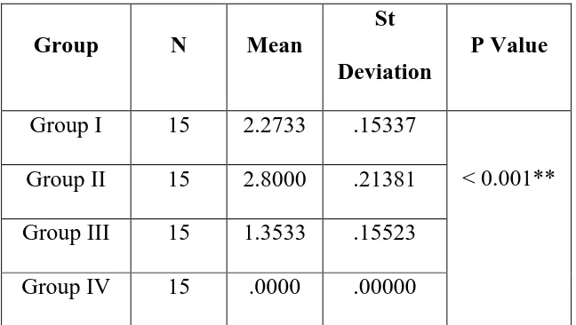
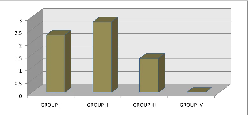
Table no.1 & Figure 1 Shows the comparison of mean and standard deviation of
plaque index. It was found that plaque index was less in Group I and in Group III
as compared to Group II with mean value of 2.273 ± 0.1533 for Group I, 2.800 ±
0.2181 for Group II, 1.353 ± 0.155 for Group III and 0 for Group IV with p value
<0.001 which is highly significant statistically.
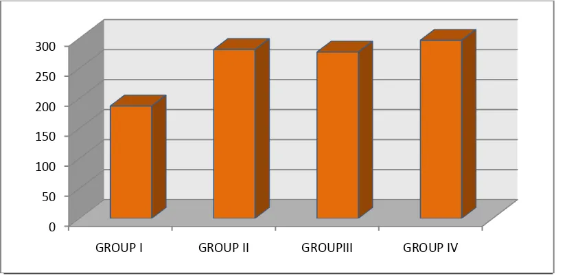
Table no.2 & Figure 2 Shows the comparison of mean and standard deviation of
Group III as compared to Group II with mean value of 4.166 ± 0.134 for Group I,
3.813 ± 0.130 for Group II, 6.126 ± 0.162 for Group III and 0 for Group IV with
p value <0.001 which is highly significant statistically.
Table no.3 & Figure 3 Shows the comparison of mean and standard deviation of
neutrophil chemotaxis between four groups. It was found that neutrophil
chemotaxis was defective in Group I and in Group III as compared to control and
Group II with mean value was 1.340 ± 0.2028 for Group I, 2.747 ± 0.130 for
Group II, 1.287 ± 0.135 , for Group III, and 2.76 ± 0.129 for Group IV, with
P<0.001 which was statistically highly significant.
Table no.4 Shows the multiple comparison of mean and standard deviation of
neutrophil chemotaxis between four groups.
Table no.5 & Figure 4 Shows the comparison of mean and standard deviation of
neutrophil phagocytosis between four groups. It was found that neutrophil
phagocytosis was defective in Group I, Group II, and in Group III as compared to
control with mean value of 187.33± 6.207, for group I, 281.40 ± 6.057,for group
II , 277.13 ± 4.373 for Group III and 296 ± 4.367 for Group IV with P>0.001
which was statistically significant.
Table no.6 Shows the multiple comparison of mean and standard deviation of
neutrophil phagocytosis between four groups.
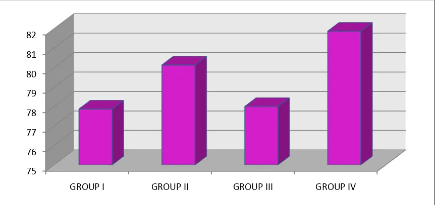
Table no.7 & Figure 5 Shows the comparison of mean and standard deviation of
was defective in Group I as compared to control with mean value 77.87 ± 2.295,
for Group I, 80.13 ± 3.021 for Group II, 78. ± 2.449 for Group III and 81.87 ±
2.326 for Group IV with P>0.001 which was statistically significant.
Table no.8 Shows the multiple comparison of mean and standard deviation of
Specific Granules Release assay among four groups.
Note
P value
0 to 0.01 - ** significant at 1% level
0.011 to 0.05 - * significant at 5% level
TABLE NO.1
COMPARISON OF MEAN & STANDARD DEVIATION OF PLAQUE INDEX BETWEEN FOUR GROUPS
Group N Mean
St
Deviation
P Value
Group I 15 2.2733 .15337
< 0.001** Group II 15 2.8000 .21381
Group III 15 1.3533 .15523
Group IV 15 .0000 .00000
TABLE NO.2
COMPARISON OF MEAN & STANDARD DEVIATION OF PERIODONTAL INDEX BETWEEN FOUR GROUPS
Group N Mean
Std.
Deviation
P Value
Group I 15 4.1667 .13452
< 0.001** Group II 15 3.8133 .13020
Group III 15 6.1267