Dissertation on
Age Estimation by the Study of Cranial Suture Closure
Submitted for
M.D., DEGREE EXAMINATION
Branch-XIV
FORENSIC MEDICINE
Madras Medical College & Govt. General Hospital
Chennai – 600 003.
THE TAMILNADU DR. M.G.R.MEDICAL UNIVERSITY
CHENNAI – 600 032.
CERTIFICATE
This is to certify that the dissertation entitled, “Age Estimation by the Study of Cranial Suture Closure” submitted by Dr. K.Gokula Ramanan, in partial fulfillment for the award of the Degree of Doctor of Medicine in Forensic Medicine by the Tamilnadu Dr.M.G.R. Medical University, Chennai is a bonafide record of the work done by him in the Institute of Forensic Medicine, Madras Medical College, during the academic year 2007– 2010.
Dean Director and Professor,
Madras medical College & Institute of Forensic Medicine, Govt. General hospital, Madras Medical College,
CONTENTS
Sl.No.
Title
Page No.
1.
INTRODUCTION
1
2.
AIM OF THE STUDY
3
3.
REVIEW OF LITERATURE
4
4.
MATERIALS AND METHODS
35
5.
RESULTS
39
6.
DISCUSSION
48
7.
CONCLUSIONS
50
BIBLIOGRAPHY
INTRODUCTION
Identification is recognition of an individual by means of various physical features and biological parameters, which are unique to each individual. There are various established parameters for identification of an individual. These are external features (such as birth marks, scar, tattoo marks ,occupational marks, malformations), personal features (such as clothes, speech, habits, handwriting), assessment of age and sex, determination of race and stature, anthropometric measurements, finger prints and foot prints, DNA finger printing.1,2 Question of identification arises in everyday medico legal practice both in civil and criminal cases.
Since the bone resists putrefaction and destruction by animals, they can lead to the reliable determination of age, sex, race, stature of the individual. Age being once of the cardinal parameter for establishing the identity, its estimation is of paramount importance and requires special attention in cases where bodies are found in decomposed, mutilated state or only fragmentary remains are discovered.
In adults mainly there are macro and microscopic methods of age estimation. The principal macroscopic changes are metamorphosis of pubic symphysis, closure of cranial sutures and degenerative changes in vertebral bodies and joints.
The use of cranial sutures for age estimation has always been a matter of considerable debate and its reliability within the parameter has not been
demonstrated conclusively by various researchers. Only handful of studies has been conducted in India, and data on heterogeneous population of Delhi region is virtually non‐
accuracy from birth onwards as far as up to 25 years.
The determination of age is needed for employment, marriage, majority, management of property, voting right, competency as witness and testamentary capacity. The significance of determination of the age is most important in the criminal cases, such as rape, infanticide, kidnapping, prostitution, juvenile delinquency and criminal responsibility.
Reasonably a correct estimation of age in elderly people is essential in legal, medical, social and administrative matters i.e. to fixing of age for regularization of employment, superannuation, pension settlements, senior citizen benefits, old age and good behavior of the prisoner.
The assessment of age is done by anthropologist, archeologist, anatomist and persons engaged in medico legal works. Among all these, the work of Forensic expert requires special attention because his findings are directly related to the administration of the law and his conclusions are debated in court of law.
The needs of determination of age vary from intrauterine life to old age for different purposes. Sometimes even when the age of person is known by the horoscope, hospital records and birth certificate, but still its scientific
confirmation is required by court of law and certain administrative departments. In India and many other countries the task of scientific confirmation of disputed age issues of civil and criminal nature is the domain of forensic expert.
and fusion of various ossification centers. However these data to some extent are influenced by heredity, climate, race, diet, hormone level, disease process etc.
After 25 years of age, other scientific methods like tooth microscopy, Gustafsons method (applicable to dead persons only), study of union of parts of sternum, lipping of joints and closure of cranial sutures are considered for age estimation of the individual.
Sutures are analogous to the epiphysio‐diaphysis plane, in which both are loci of growth and have a sequence of time of closure.
The texture of a young adult skull is smooth on both the inner and outer surfaces (Krogman 1962:88). Krogman (1962:88) provides the following cranial morphological age sequence: 1) from the age of 25, muscular markings become increasingly evident, especially on the temporal, occipital and on the lateral side of the mandible; 2) around 35 to 45 years, the surface begins to assume a matted, granular appearance; 3) on the inside of the skull, the Pacchionian depressions, both deepen and occur with much more frequency;
4) after the age of 50, the diploe become less vascular channeled and there is an increasing replacement by bone. However, there is no consistent age change in the thickness of the cranial bones.
In many cases, complete closure will obliterate any signs of the cranial sutures (Krogman 1962:85).
There is a difference however; the epiphyseal union is always complete in normal closures (with the possible exception of the ramal epiphysis of the ischium) whereas suture closure may be incomplete in normal, healthy individuals (Krogman 1962:76).
The metopic suture, which is present at birth between the right and left halves of the frontal bone, usually closes around the age of two. However, in some individuals, it is persistent into later adult life.
Forensic anthropologists are frequently called upon to derive as much information as possible from very limited or poorly preserved remains. The method of determining age by cranial suture closure has always been more
generally used, due not only to the greater interest in the skull, but because the cranium is frequently the best preserved portion of the recovered skeleton. As such, osteologists have developed numerous techniques which, when applied in concert, increase the accuracy of identification (Lovejoy et. al. 1985:2).
Use of suture closure as an age estimate is predicated upon the hypothesis that suture closure is part of the aging process. However, when suture closure patterns were first studied at the beginning of this century, there were two schools of thought (British andItalian) on this issue (Hershkovitz et. al. 1997:393).
the scientific literature, the British approach toward suture closure became the dominant model in physical anthropology (without actually testing that hypothesis) (Hershkovitz et. al. 1997:394).
The work of Todd during the 1920s provided anthropologists with a framework for estimating age of death from both the pubic symphysis and the cranial sutures (Todd1924, 1925a, 1925b, 1925c). The use of cranial sutures in estimating (adult) age at death came under criticism during the 1950s. These critiques focused upon Todds methodologies, conclusions and the error rates of the method itself (Singer 1953; Brooks 1955; Powers 1962; Krogman 1962). It was noted that the ages obtained from cranial suture closure either produced
skewed mortality rates or did not correlate with known age of death (Singer 1953; Brooks 1955). During the 1970s through to the 1990s, several researchers re‐examined the use of cranial sutures as a means of determining age at death (Johnson 1976; Meindl and Lovejoy 1985; Masset 1989; Key et. al. 1994; Nawrocki 1998). According to Meindl and Lovejoy (1985:57), this period of investigative research was characterized by a trend in skeletal biology,during which it was hoped that one or two highly reliable age indicators would be isolated and perfected.
Hershkovitz et. al. (1997:395) believe that these assumptions have no factual basis, and that their application is very subjective for the following reasons: the division between segments of the same suture are not clear cut in many skulls, and more than 20% of skulls do not follow the classic Pattern of sutural segmentation.
REVIEW OF LITERATURE
A number of studies have been conducted abroad on the closure of the cranial sutures as a sign of ageing namely Dwight (1890), Frederic (1905), Person and Box(1905), Todd and Lyon (1924‐25) Cattaneo,L (1937), Harlick (1939), Franchini (1946) Singer(1953), Mckern and Stewart (1957).
In India the available study on the subject are Yadav S.S and Puri P.R (1971),Patil T.L (1981),Bhagwat S.S (1983) and Chandrashekharan P. (1985), Dr.Bimal Chandra(1984,Delhi), Vyas P C (1996),Moondra A.K (2000), Rajesh kumar verma(2002), Dr.Pradeep singh(2004,Patiala), Daisy Sahni (2004, Chandigrh).
The bones of skull are separated by sutures ,which in a sense are analogues of epiphysio‐diaphysial planes in which there are loci of growth and they have a sequence and timing of union (Krogman,W.M 1978).The word suture is derived from Latin sutura ,which means seam like or series of stitches.
The sutures on the skull are seam like lines of junction in which the connective tissue is a fibrous membrane, or synchondroses in which the bones are united by a bar of syarthrosis characterized by absence of joint cavity and a paucity of motion with advancing age they tend to become obliterated although this is by no means uniform.
HISTORICAL RESEARCH (A. D. 100
‐
1890)
Todd and Lyon (1924:327) present the following historical account of cranial sutures. Cranial sutures were viewed as controlling both the growth of the brain and, therefore, the shape of the skull. Although classical authors, like Hippocrates, Aristotle and Galen, had observed that some human crania exhibit open sutures, whereas others are almost or entirely devoid of them, the fact that union occurred during life did not appear in the anatomic literature until the works of Gabriele Fallappia (1523‐1562), in the middle of the 16th century (Todd and Lyon 1924:327).
In 1641, Thomas Bartholin (1616‐1680) proposed the following uses for cranial sutures (Todd and Lyon 1924:326): 1) to permit the free transpiration of the vapours in the brain; 2) for the attachment and suspension of the dura matter; 3) for the transmission of fibers of the dura through to the pericranium; 4) for the transmission, in both directions, of vessels carrying nourishment and life to the parts; 5) to diminish the likelihood of fracture of the bones of the skull [Interestingly, Hershkovitz et. al. (1997:397) have once again suggested that open sutures may increase skull efficiency in absorbing related mechanical stresses]; and 6) to permit the penetration of applications from the exterior.
Bartholin asserted that the number and location of the sutures was the same in males and females, and were rarely changed by the shape of the cranium (Todd and Lyon 1924:327). It was proposed that these cranial deformations occurred during fetal development or at birth (ibid.). The
viewed as a cause, rather than effect, of suture closure.
During the 19th century, it was observed that cranial union first occurs in the sagittal suture, and that it occurred earlier within the cranium than upon the exterior (Todd and Lyon 1924:328). In 1856, the anatomist Louis Pierre Gratiolet (1815‐1865) proposed a sequence for suture closure and stated that union occurred earlier in Negroes. Gratiolet, observed that ectocranial suture closure progressed sequentially (Todd and Lyon 1924:353): sagittal, lambdoid, then coronal.
Other anatomists began to try to establish an age related sequence for cranial suture closure. For example, in 1861, the physician Paul Broca observed visible sutures in males over the age of 50, and developed a 4 point rating system for cranial suture closure (Todd and Lyon 1924:353). Over time, it came to be accepted that, in the white stock union began between the ages of 40 and 45.However, in 1869, F. Pommerol noted that the period of union, for each suture, varied across individuals but followed the general pattern (Todd and Lyon 1924:328‐329). Pommerol identified the following sequence (ibid.): 1) individuals under 35 years of age had open cranial sutures; 2) around 40 years, the sagittal suture begins to close; 3) around 50 years, the coronal suture begins to close; and 4) by 65 years or more, the temporal suture has finished closing.
t
Twentieth Century Research
Parsons and Box
Frederic
In 1906, J. Frederic examined 255 European and 119 non‐European crania of known age (Todd and Lyon 1924:329). However, only 91 European and 13 non‐European crania of both sexes were opened so that the internal surface could be examined.Following Broca, Frederic introduced his own rating scale of 0 to 4 (open, less than one half closed, half closed, more than one half closed, and totally closed) (Krogman 1962:77‐78). Examining endocranial sutures, he found that the lambdoid closed after the sagittal and coronal (Todd and Lyon 1924:355). Frederic concluded that it was not possible to determine the age of a skull by the condition of suture union closure with any accuracy greater than +/‐ one decade.However, he stated that suture closure occurred later in females, thus concurring with Dwight, Parsons and Box (Krogman 1962:78).
Todd and Lyon
The research of Todd and Lyon (1924, 1925a, 1925b, 1925c) provided the groundwork for all North American forensic and physical anthropological studies for the remainder of the 20th century. In 1924, Todd and Lyon proposed to “present the facts concerning suture closure and its relation to the racial form and individual contour of the brain case” (1924:326). The research by Todd and Lyon (1924, 1925a, 1925b, 1925c) was the first new attempt to estimate age using endo and ectocranial suture closure since Pommerol, Ribbe, Frederic, Parsons and Box, with the specific aim to create a precise numerical rating
system for cranial closure (Krogman 1962:78). Todd and Lyon (1924:355) state that:
closure upon its age relationship.
Todd and Lyon (1924, 1925a, 1925b, 1925c) tried to ensure that the skeletal material was large enough to justify it as a sample of the population under study. In addition to sex and race, verifiable age at death had to be known and the crania had to be cut, so that examination of the interior surface was possible. Todd and Lyon (1924:330) initially examined the crania of more than 1,000 individuals, from which those of which were of uncertain known age, and did not have a complete post cranial skeletal for comparative study were rejected. Of the original 1000 specimens, 514 crania of known age were examined 307 crania of white males, 58 white females, 120 Negro males, and 29 Negro females. From this initial sample, 40 white skulls (13.3%) and 41 (34.2%) were excluded as anomalous (Krogman 1962:79).
It is characteristic of the anthropoid strain that the pubic age relationships fall nearer to those of the Giant Anthropoids, and there is a clear difference in age relationship of the skeleton between human beings according, as they exhibit the anthropoid strain or theregressive form of symphysis. In the first place, elimination based on the anthropoid strain in the pubic symphysis affects skeletons of the third decade.2) precocious union; 3) no endocranial closure of vault sutures; 4) evidence of dwarfism; and 5) no endocranial closure
of any cranial sutures. As stated by Todd and Lyon (1924:348), they were confident in their rejections because they were able to examine the entire skeleton and they were cross referenced with legal documentation of age at death.
and Lyon 1924:337). This condition was defined as ‘lapsed union’ of the suture. They defined it as the incomplete union of the suture, characterized by a build up of bone tissue along the edges of the unclosed part. Todd and Lyon (1924:337) classed incidents of lapsed union as closed, since a suture in this condition would be unlikely to close to any great extent. They adopted Broca’s arrangement of complication of sutures, degrees of closure and subdivision of particular sutures, except for the adoption of Frederic’s inversion of Broca’s categorization of the amount of suture union (i.e. 0 = no union and 4 = complete closure; 1 to 3 refer to the amount of union ‐ one quarter... three quarters). The closure for each suture was then averaged and plotted.
Todd and Lyon (1924:333) then repeated the above procedure with the male Negro and female crania (of both ‘racial stocks’). This led to the elimination of 'abnormal' progress in each of the series, giving a basis for comparison of closure progress in each sex and stock with those of the male
Whites, which were thereafter used as a standard. They observed the following traits (Todd and Lyon 1924:333): 1) there was a clear orderly age sequence in the process of suture closure; 2) sex, racial stock, cephalic index and cranial capacity have very little effect on this closure sequence; and 3) the timing of the sequence was more obvious endocranially than ectocranially (ibid.).
years.
In 1925, Todd and Lyon published three follow papers to the above study (Parts II‐IV). Part II (Todd and Lyon 1925a) was an examination of ectocranial suture closure in adult males of white stock, since they acknowledged that in some instances, researchers would not be able to observe endocranial sutures. The sample consisted of the 267 crania employed in the first study, however, new samples were added for comparison asthey became available during the course of these three studies (Todd and Lyon 1925a:24). This investigation concluded that (Todd and Lyon 1925a:36): 1) in general, there is no tendency on the part of sutures to begin to close earlier endocranially, rather than ectocranially; 2) the only exceptions to this finding were the pattern exhibited by the inferior masto‐occipital, the spheno‐frontal
and the coronal; 3) ectocranial closure was slower and more variable, with no evidence of periodic activity; 4) ectocranial suture union was never as complete as endocranial closure; and 5) ectocranially, lapsed union is evident in all sutures.
In Part IV, Todd and Lyon (1925c) examined the occurrence of ectocranial suture closure in the male Negro cranium. The sample consisted of the 79 crania selected for Part III of their study (Todd and Lyon 1925c:150). Their findings are as follows (Todd and Lyon 1925c:167‐168): 1) endo and ectocranial suture closure patterns are essentially the same for white and negro males; 2) ectocranial closure is more erratic, slower and less complete than endocranial closure; and 3) lapsed union is characteristic of all ectocranial sutures, although it does not appear in all individuals.
d
Brooks
In 1948, just fewer than 400 individual skeletons from the University of California collection were selected for testing (Brooks 1955:569). In 1950, the some of the skeletons employed by Todd from the Hamann Museum collection were used for review (ibid.). Later, in 1953, a second series of 70 skeletons were analyzed to verify the methodology (ibid.). The sample was chosen based on the following criteria: 1) the individual be over 18 years of age, as judged by long bone epiphysis‐diaphysis union and fusion of the three elements of the acetabulum; 2) the crania vault must contain at least the area of the coronal, sagittal and lambdoid sutures; and 3) the symphyseal surface of at least one pubic bone must be preserved (Brooks 1955:569). It should be noted that all of the skeletons were from California, but no consideration was made of area or archaeological horizon (ibid.).
showed a deviation from 5 to 8 years, with a mean of +/‐ 2 years, between cranial suture and pubic symphysis methods (Brooks 1955:574). Brooks found that in cases where all of the sutures were open (predicted age <25), comparison with dental wear patterns, epiphyseal closure and pubic symphysis surface, all indicated that the specimen was over 35 years old (Brooks 1955:582). Despite obtaining linear
correlations as high as 0.74, these results led her to concur with Singer (1953), that cranial suture closure was an unreliable method of age determination, especially for female skeletons (Brooks 1955:588; Meindl and Lovejoy 1985:57).
Meindl and Lovejoy
In 1985, Meindl and Lovejoy re‐examined the Hamann‐Todd Collection. They chose 10 ectocranial landmarks, although it is unclear whether they scored for bilateral expression (Meindl and Lovejoy 1985:60). They employed the following scoring methodology: 0 ‐ no observable closure;1 ‐ 1 to 50% closure; 2 ‐ 51 ‐ 99% closures; 3 ‐ 100% closure. Small (1 cm) lengths of a suture or specific sites were selected for inspection, for which only the judgment of one observer was required (Meindl and Lovejoy 1985:58). Since they proposed that in the case of forensic anthropology, the regularity of closure during the early adult years was not critical, only ectocranial sutures were studied (ibid.). Meindl and Lovejoy (1985:58) thus chose 17 ectocranial points for 236 crania from the Hamann‐Todd collection, based upon the reliability of stated age at death.
which
were retained (Meindl and Lovejoy 1985:60) were the vault system (midlambdoid, lambda, obelion, anterior sagittal, bregma, midcoronoal and pterion) and lateral anterior system (midcoronoal, pterion, spheno‐frontal, inferior sphenotemporal and superior sphenotemporal). Meindl and Lovejoy (1985:60) found a chronological age ranking at the pterion, sphenofrontal, midlambdoid and lambdoid, respectively. These were based upon the assumption of underlying continuity of ordinal closure scales (the Kendall coefficient) (ibid.).
The next phase of analysis, to determine combination of sites which could be employed in age estimation, was based upon the following assumptions (Meindl and Lovejoy 1985:61): 1) sutures should demonstrate a protracted sequence of closure; 2) sutures should correlate with age during the primary period of its closure activity; and 3) the information provided by each suture should be specific to that particular suture. From these criteria, and since the calotte is usually the most durable in archaeological populations, they determined that the 5 lateral anterior sites were the best overall predictor of age (Meindl and Lovejoy 1985:61). It was assumed that the sample crania would exhibit a commencement and termination sequence which correlated with long bone epiphyseal fusion sequence (Meindl and Lovejoy 1985:62).
Using composite scores for each sample specimen, they found that the lateral anterior sites closed in the following sequence: Commencement ‐ pterion, midcoronal, sphenofrontal, inferior sphenotemporal, superior sphenotemporal; Termination ‐ pterion, sphenofrontal, midcoronal, inferior
midlambdoid, midcoronal, bregma; Termination ‐ obelion, pterion, anterior sagittal, lambda, bregma, midlambdoid, midcoronal (ibid.). The overall (linear) correlation with known age was 0.57 for lateral anterior sites and 0.50 for vault sutures (ibid.).It was found that the average mean deviation, for lateral anterior scoring, was 7.5 years, and 14.2 years for vault sutures (Meindl and Lovejoy 1985:64). Meindl and Lovejoy (1985:64‐65) also examined whether or not race or sex had any effect on suture closure, thus affecting the accuracy of age prediction. Using analysis of covariance (ANCOVA) on the residuals, they found no measurable influence of either race or sex.However, Meindl and Lovejoy (1985:62) state that any age estimation should take into account postcranial indicators as a control for suture cranial variations.
Gustafsons, G. (1950) also stressed on condition of teeth in age estimation among the elderly persons along with ossification of bones. Sequential closure of various skull sutures imparts valuable information regarding the age of the person. Over the age of 25 and especially in 25‐40, the estimation of age becomes more difficult. For many years the closure of skull sutures was considered most accurate method (Keith Mant A,1994). It is common knowledge that most adult have at least part of their sutures closed and this tends to become more wide spread as the age increases. There are many exceptions and as the rate of closure is not linear with time. This
generally can be useful when skull came from a mature individual, as it is unlikely below the age of 20 Broth well Bernard Knight ,(1996).according to Rantoul,E.and Smith ,H(1973) the absence of any sign of closure of any of the skull points to strong probability that the age does not exceed 30 years.
are tested on a sample of known age from Spitalfields, London in order to determine the value of cranial suture closure as an indicator of age at death. The three techniques are those proposed by Acsádi and Nemeskéri, Meindl and Lovejoy and Perizonius. Results indicate that the Acsádi and Nemeskéri technique, which is based on endocranial sutures, can be used to distinguish young and middle‐aged individuals in the Spitalfields sample but gives no information for crania over the age of 50 years. Age estimation using the Meindl and Lovejoy and Perizonius (Old system) techniques, which use ectocranial sutures, was found to be subject to a number of complicating factors, of which sexual dimorphism in the rate and pattern of closure is the most significant. A method of estimating age at death based on both endocranial and ectocranial suture closure is developed on the basis of the Spitalfields sample. The technique attempts to overcome some of the problems associated with both intra‐ and interpopulation variation in cranial suture closure. For a truly accurate age‐
estimation technique based on cranial suture closure we would need to know more about the causes and functions of suture closure in human populations.
c
According to Nandy,A.(1995) skull sutures start their fusion activity by 24‐25 years of age ,which usually starts at the ectocranial surface but, though the start in the endocranial surface occurs late, the progress at this level is speedy, more uniform and more complete than at the ectocranial level. Hence the endocranial fusion is more reliable than ectocranium. They ossify and are completely obliterated in the advanced age, although the parietal suture may remain separate throughout life.
5
A study was conducted in 1998 in which a total of 963 skeletons were studied to examine macroscopic cranial methods of age estimation. The methods of Acsadi‐ Nemeskeri, Masset, Baker and Meindl‐Lovejoy were applied to every skull. The results indicated that most accurate technique in this application were those that consider endocranial suture closure. The methods of Acsadi‐Nemeskeri and Masset were the most accurate in all sub‐
samples by population, sex, within population and in total. Although the relative accuracy could vary in application to the other populations.5
Time of closure of sagittal,coronal, and lambdoid sutures were studied at autopsy on 538 male and 127 female adults of known ages belonging to north‐west India . Whether a segment was open or closed on either surface was recorded. On analyzing the records authors came to conclusions that obliteration of sutures commences earlier on endocranial surface than on ectocranial. Complete obliteration of a segment or the entire suture is so erratic that it is not useful for estimating the age of skull.6
The age of 20 males and 20 females was evaluated based on vault sutures. The results were compared with the actual chronological age of the deceased calculated on the basis of their date of birth and demise. In many cases a slight overestimation occurred in evaluating the age by analyzing the sutures. Thus the results show that a precise and thorough evaluation of the deceased’s age is not possible on the basis of cranial suture closure without maceration of the skull.7
7
methods.8
Meindl‐Lovejoy method was applied to 3663 skulls from Hamman‐Todd and Terry collection. It was found that sagittal suture closure was age independent and sexually biased. The wide confidence intervals appeared to preclude meaningful application of suture status for age determination. No correlation was found with tested biological stressors.9
A new method was applied using ectocranial suture closure of lateral anterior and vault suture on 236 samples from the Hamman‐Todd collection. It was found that ectocranial method was superior to endocranial and that age estimates are independent of race and sex. Conclusion was that the suture closure can provide valuable estimates of age at death in both archeological and forensic context when used in conjunction with other skeletal age indicators.10
A study conducted on 256 crania of adults over 20 years of age using Acsadi‐
1
Another study using three age estimation techniques (Acsadi‐Nemeskeri, Meindl‐
Lovejoy and Perizonius method) tested on a sample of known age inorder to determine the value of cranial suture for age estimation. The results indicated that Acsadi‐Nemeskeri technique which is based on endocranial suture can be used to distinguish between young and middle aged individuals in the sample but gives no information for crania over the age of 50 yrs.
AIMS AND OBJECTIVES
1. To study the chronology and pattern of union of endocranial sutures namely coronal, sagittal and lambdoid etc.
2. To detect bilateral and bisexual variations in endocranial suture closure if any.
3. To specify the relationship between progression of union of endocranial suture and age of the subject.
MATERIALS AND METHODS
The study was conducted on cases coming for medico‐legal postmortem examination to the Institute of Forensic medicine, Madras Medical College, Chennai , during a period of September 2008 to August 2009
Study design: A descriptive cross‐sectional study.
Inclusion criteria
1. The cases of known age coming for medicolegal postmortem examination. Age was confirmed by documentary evidences like birth certificate, identification cards, ration card..etc.
2. Subjects of more than 20 years of age were taken.
Exclusion criteria
1. Unknown, unclaimed bodies where exact age cannot be confirmed.
2. Cases showing deformed or diseased or fractured skull, which may hamper the study of suture closure.
Methods
100 cases of age 20 and above were studied. Documentary proof of age was collected. After reflecting the scalp, coronal, sagittal, lambdoid sutures were studied applying Acsadi‐
sagittal suture. Lambdoid suture was studied in‐situ. The calvarium was cleaned of soft tissues and was dried, which made the sutures more prominent.. The obliteration of the sutures was ascertained endocranially. Degree of closure was scored in 16 parts of the main cranial sutures as has been done by Acsardi‐Nemeskeri12.
The coronal suture was studied in three parts on right side and left side each; sagittal suture in four parts and lambdoid sutures in three parts each on right and left side. Endocranial sutures were simply divided in sections of equal length.
Scale for closure: Acsardi‐Nemeskeri complex method
0 = open. There is still little space left between edges of adjoining bones. 1 = incipient closure. Clearly visible as a continuous often zigzagging line. 2 = closure in process. Line thinner, less zigzags, interrupted by complete
closure
3 = advanced closure. Only pits indicate where the suture is located 4 = closed. Even location cannot be recognized.
Mean endocranial closure stages were calculated for the three main stures by adding the scored closure stages of the different sections and dividing the result by number of sections which compose the suture in question.
s
A detailed pre‐designed Performa was completed for every case studied.
Statistical Methods
statistical tools were used (spearman rank correlation coefficients, Levene’s test for equality of variances, student’s test for equality of means, SPSS 11.5 version software). p <0.05 was considered as significant.
The study consisted of 100 cases brought to the mortuary of Madras Medical College,Chennai, during the period of September 2008 to August 2009.
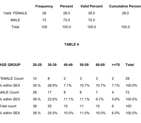
AGE AND SEX DISTRIBUTION
The age of the victims varied from 20 to 90 years. Age groups were classified 10 year interval so as to compare with the previous studies.(Table 1, 2) It was observed that maximum number of cases were in 20‐29 age group which also tells us regarding the age distribution of cases coming for autopsy in MMC,Chennai. There were 38 cases i.e. valid percentage is 38 and cumulative is 38. In 30‐39 groups there were 25 cases, valid percentage is 25 and cumulative is 63%. In 40‐49 age groups, there were 10 cases which accounts for 10% of the total cases and cumulative is 73%. 11% of the cases were constituted by 50‐59 age group. (Cumulative percentage is 84). There were 10 cases in 60‐69 age groups. It constituted 10% of cases and cumulatively 94%. There were only 6 cases in 70 years and above which is about 6% of total and
cumulative percentage is 100. The minimum age of case is 20 and maximum age is 90, mean is age is 38.36, standard deviation is 16.794 and median age is 34.50.
TABLE 1
Age groups Frequency Percent Valid Percent Cumulative Percent
20-29 38 38.0 38.0 38.0
40-49 10 10.0 10.0 73.0
50-59 11 11.0 11.0 84.0
60-69 10 10.0 10.0 94.0
>=70 6 6.0 6.0 100.0
TABLE 2
N Minimu
m
Maximu m
Rang e
Mea n
Std. Deviation
Media n
Std. Error of
Mean
100 20 90 70 38.6 16.794 34.50 1.679
SEX DISTRIBUTION
TABLE 3
Frequency Percent Valid Percent Cumulative Percent
Valid FEMALE MALE Total 28 72 100 28.0 72.0 100.0 28.0 72.0 100.0 28.0 100.0 TABLE 4
AGE GROUP 20-29 30-39 40-49 50-59 60-69 >=70 Total
FEMALE Count % within SEX MALE Count % within SEX Total count % within SEX
SEX DISTRIBUTION
In our study there were 28 females and 72 males out of 100 cases which also represented cases coming to mortuary of our department.
SEX DISTRIBUTION
DISTRIBUTION OF CASES –ACCORDING TO AGE GROUP
Maximum numbers of case were seen in 20‐29 age groups, least in above 70 years age group.
[image:36.612.69.562.136.512.2]Graph 3
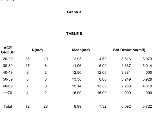
TABLE 5
AGE
GROUP N(m/f) Mean(m/f) Std Deviation(m/f)
20-29 30-39 40-49 50-59 60-69 >=70 Total 28 17 8 8 7 4 72 10 8 2 3 3 2 28 2.93 11.06 12.50 13.38 15.14 16.00 8.99 4.50 5.00 12.00 8.00 13.33 16.00 7.32 3.516 4.337 3.381 3.249 2.268 .000 6.092 3.979 5.014 .000 6.928 4.619 .000 5.722
When comparison between male and female subjects were made, females have late closure, mean for total cases 8.99 vs 7.32 (except in20‐29 age groups). Where closure was seen earlier in females (table 5).
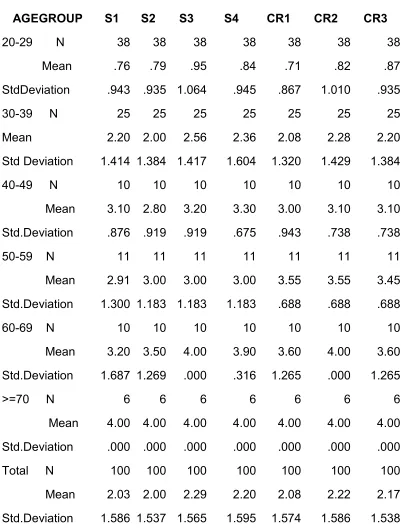
ENDOCRANIAL CLOSURE OF SAGITAL SUTURE
ENDOCRANIAL CLOSURE CORONAL SUTURE
Graph 5
In coronal suture near complete closure occurs by the age of 40-49 age and total closure occurs by 50-59 years. (Mean value 3.10 in 40-49 years and 3.55 by 50-59 years of age).
a
ENDOCRANIAL CLOSURE OF LAMBDOID SUTURE
Over all coronal suture closes earlier followed by sagital and lambdoid suture endocranially. (Mean values 2.18,2.13,and 2.10)
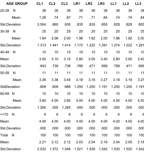
[image:40.612.105.507.203.733.2]TABLE 6
AGEGROUP S1 S2 S3 S4 CR1 CR2 CR3
TABLE 7
AGE GROUP CL1 CL2 CL3 LR1 LR2 LR3 LL1 LL2 LL3
20-29 N Mean Std.Deviation 30-39 N Mean Std.Deviation 40-49 N Mean
Std.Deviation 50-59 N Mean StdDeviation 60-69 N Mean Std.Deviation >=70 N Mean
Std.Deviation Total N Mean Std.Deviation 38 1.26 3.554 25 1.84 1.313 10 3.00 .943 11 3.36 .809 10 3.60 1.265 6 4.00 .000 100 2.21 2.532 38 .74 .860 25 2.08 1.441 10 3.10 .738 11 3.36 .809 10 4.00 .000 6 4.00 .000 100 2.12 1.572 38 .87 .935 25 2.00 1.414 10 3.10 .738 11 3.45 .688 10 3.60 1.265 6 4.00 .000 100 2.12 1.546 38 .71 .835 25 1.96 1.172 10 2.80 .789 11 3.18 1.250 10 4.00 .000 6 4.00 .000 100 2.03 1.521 38 .71 .835 25 1.92 1.222 10 3.00 .471 11 3.18 1.250 10 4.00 .000 6 4.00 .000 100 2.04 1.530 38 .84 .855 25 2.00 1.291 10 3.40 .699 11 3.27 1.191 10 4.00 .000 6 4.00 .000 100 2.16 1.542 38 .74 .828 25 1.96 1.274 10 2.80 .789 11 3.18 1.250 10 4.00 .000 6 4.00 .000 100 2.04 1.530 38 .74 .828 25 1.92 1.222 10 3.00 .471 11 3.18 1.250 10 4.00 .000 6 4.00 .000 100 2.05 1.520 38 .84 .855 25 2.00 1.291 10 3.40 .699 11 3.27 1.191 10 4.00 .000 6 4.00 .000 100 2.16 1.542
In lambdoid suture L3 (pars
esterica) closes earlier fallowed by L2 (pars intermedia) and L1 (pars lambdica). (Mean value of 2.16 ,2.05 and 2.04).
BILATERAL VARIATION IN ENDOCORONAL SUTURE
Graph7
In coronal sutures, there was minor difference in closure among right and left side endocranially, which cans be safely ignored. When sexes were taken separately also there were minimal difference bilaterally.
BILATERAL VARIATION IN ENDOLAMBDOID SUTURE
In lambdoid suture also there were no difference in closure between right and left side. Right and left side of coronal and lamdoid sutures were compared endocranially, there were no significant difference after applying student test (p value>0.05). It implies that there is no bilateral variation in endocranial suture closure. (graph 8)
ENDOCRANIAL SAGITAL CLOSURE-SEXUAL VARIATION.
ENDOCRANIAL CORONAL SUTURE CLOSURE-SEXUAL VARIATION.
Graph10
ENDOCRANIAL LAMBDOID SUTURE CLOSURE-SEXUAL VARIATION
Graph11
DISCUSSION
In later years of life all the teeth have erupted, practically all the epiphyses have united with the diaphysis, the height and weight are of no significance to determine the age. Gustafson has done the work in which he has given the idea to determine the age on the basis of changes that occur in teeth. Literature is full of certain changes such as lipping of the bones, graying of the hair, appearance of arcus senilis in the cornea, opacity in len atherosclerotic changes in the arteries, wrinkling of the skin especially of the face. They are too vague to be considered for determination of age in Medico‐legal work.
Obliteration of skull sutures in late age, practically when all the teeth have erupted and epiphysis have fused i.e. after 21 years of age, gives a fairly accurate idea but here also we find that the determination of age can only be in decades, based on sole criterion of suture obliteration.
Sagittal Suture
In our present study we have found that the sagittal suture, endocranially, starts fusing at the end of 20‐29 years and completion is perfected at the age of 60‐69 years, and this observation conforms with that reported by Todd & Lyon (1924), while it is in contrast to the observation reported by Pommerol (1869), and Topinard (1885), who indicated endocranial commencement of sagittal suture at a much later age at about 40 years. These
latter workers have reported on very scanty specimens so it can’t by considered as authentic.
Coronal Suture
as 20‐29 years and completion by the age of 50‐59 years, other workers like Pommerol (1869), Topinard (1885), Ribbe (1885) reported closure between 40‐50 years. Their study does not indicate whether it was ecto‐cranial or endocranial or it was commencement or termination.
Lambdoid
Lambdoid endocranially, starts fusing at the age of 20‐29 years in the present context which shows that it is a year earlier than that reported by Todd and Lyon (1924), while completion in our study is 50‐59 years. The other workers have not reported on lambdoid suture.
Our Indian data compare well with those of the male whites (Todd & Lyton 1925). Negro skulls however show an earlier date of commencement and closure.
Dwight.(25,28)
All the previous work was done in France, Germany and United States of America, under different climatic conditions and in diverse racial groups. Though consensus of opinion in our country is that the obliteration of the skull
sutures in females is some what earlier than that of males, in the present study no substantial difference was noticed. This finding is in conjunction with Meindl and Lovejoy (10).
CONCLUSIONS
1. There is some correlation between endocranial suture closure and age upto 40‐50 year’s age group, there after there is no significant correlation. Any attempt to derive a reliable formula to estimate the age from score of suture closure was met with failure for the following reasons: 1) the trend of correlation is neither increasing nor decreasing with age, 2) the sample size is too small to derive a formula 3) unequal distribution of males and females in the study sample.
2 Though there is some difference in suture closure in males and females, it is not significant statistically. (Females showed earlier union than males in the age group 20‐
29, in the other age groups suture closure occurred earlier in males).
3 There is no significant variation in suture closure of right and left sides of coronal and lambdoid sutures;
4. Endocranially: coronal suture closes earlier followed by sagittal and lambdoid.
5. Within sagittal suture segment, pars obelica(S3) closes earlier followed by pars lambdica(S4), pars vertices(S2), pars bregmatica(S1). In the coronal suture, pars complicata(C2) unites earlier followed by pars bregmatica(C1) and pars pterica(C3). In lambdoid suture pars
asterica(L3) closesearlier followed by pars intermedia(L2) and pars lambdica(L1).
7. Although cross‐sectional in nature, suture obliteration patterns (totally open, totally closed, partially open, and partially closed) are not temporary progressive stages on an age scale, but rather independent permanent phenomena.
8. Commencement and complete obliteration of a segment of the entire suture is so erratic that it is not amenable for estimating the age.
RECOMMENDATIONS
1. Sutures like basilar suture and metopic suture, lateral sutures, palatal sutures etc.
should be included.
2. As in present study difference between each age group is 10 years and so it should be
reduced to get better results.
3. In order to have a more accurate and better assessment of suture closure various
other modalities like radiology (x‐ray, CT scan, MRI), histology may have to be
combined.
4. In our study age (38% of cases in 20‐29 age group) distribution and sex (72% males)
is not uniform. Hence, this (sampling error) has to be taken into consideration in future
studies.
5. In present study there are only 100 cases which may not be the representation of
whole population. Hence the study population (sample size) has to be increased in
future studies.
6. Age estimation from morphological changes in bone has always been a matter of
debate as it is very erratic and affected by various factors such as climatic, dietetic,
hereditary, nutritional, sociological, racial, environmental, geographical etc. Cranial
sutures are no exception to that. So, much study is needed in estimation of age from
REFERENCES
1. Vij K.Identification.In: Textbook of Forensic Medicine and Toxicology Principles and Practises. Elsevier; 2005. P.50‐63.
2. Reddy KSN. Identification. In: The Essentials of Forensic Medicine and Toxicology. K Sugna Devi; 2005. P.67‐68.
3. Buchner A. The identification of human remains. Int. Dent. J. 1985 dec; 35(4): 307‐
311.
4. Knight Bernard .The establishment of identity of human remains. In: Forensic Pathology. Arnold Publication; 1996 .p.121‐122.
5. Galera V, Ubelaker DH, Hayek LA. Comparison of macroscopic cranial methods of age estimation applied to skeletons from the Terry collection. J. Forensic Sci. 1998 Sep; 43(5) : 933‐9
6. Sahni D, Jit I, Neelam , Sanjeev. Time of closure of cranial sutures in North‐West Indian adults. Forensic Sci. Int. 2005 Mar 10; 148(2‐3): 199‐205.
7. Bednarek J, Bloch‐Boguslawska E, Engelgardt P, Wolska E, Sliwka K . The degree of closure of cranial sutures as a quick method for adult age evaluation in autopsy. Arch. Med. Sadowej Kryminol. 2005 Jul‐Sep; 55(3): 185‐9.
9
Med. Sadowej Kryminol. 2002 Jul‐Sep; 52(3): 221‐7.
9. Hershkowitz I, Latimer B ,Dutour O,Jellema L M, Wish‐Bartaz S, Rothschild C,etal. Why do we fail in aging the skull from the sagittal suture? .Am. J. Phys. Anthropol. 1997 Jul; 103(3): 393‐9.
10. Meindl RS, Lovejoy CO. Ectocranial suture closure: a revised method for the determination of skeletal age at death based on the lateral anterior sutures. Am J Phys Anthropol. 1985 Sep; 68(1):57‐66.
11. Perzonius WRK. Closing and non‐closing sutures in 256 crania of known age and sex from Amsterdam (AD 1883‐1909). J Human Evolution. 1984; 13: 201‐216.
12. Acsady G, Nemeskeri J. History of human life span and mortality. Akademiai Kiado. 1970.
13. Krogman WM, Iscan MY.Skeletal age:cranium.In: The Human skeleton in Forensic Medicine.Charles C Thomas Publishers 1986; 2nd edn : p.103‐132.
14. S.M.Hepworth.On determination of age in Indians: Study of ossification of the epiphysis of the long bones.Ind.Med.Gaz.1929; 64:128.
e
15. G.Galstaun.Some notes on the union of epiphyses in Indian girls. Ind.Med.Gaz.1930;65:191‐192
17. R.Lall, B.S.Nat. Ages of epiphyseal union at the elbow and wrist joints amongst Indians. Ind.J.Med.Res.1934; 21:683‐689.
18. D.Narayan, I.D.Bajaj. Ages of epiphyseal union in long bones of inferior extremity in U.P.subjects. Ind.J.Med.Res.1957; 45:645‐649.
19. I.D.Bajaj, O.P.Bhardwaj, S. Bhardwaj. Appearance and fusion of important ossification centres: a study in Delhi population. Ind.J.Med.Res.1967; 55:1064‐1067.
20. M.J.S.Pillai. The study of epiphyseal union for determining the age of south Indians. Ind.J.Med.Res.1936; 23:1015‐1017.
21. T.W.Mckern, T.D. Stewart. Skeletal age changes in young American males: Environmental Protection Research Division, Technical Report EP‐45, Headquarters Quartermaster Research and Development Command, Natrick, MA.1957; 26‐28.
22. F.E.Johnston. Sequence of epiphyseal union in a prehistoric Kentucky population.Indian Knoll.Hum.Biol.1961; 33:66‐81.
8
23. F.G.Parsons, C.R. Box. The relation of cranial sutures to age. J.Anthropol.Inst.Lond.1905; 35:30‐38.
24. W.M.Cobb. Skeleton in.A.I. Lansing (Ed). Cowdry’s problem of ageing, biological and medical aspects. Williams and Wilkins, Baltimore.1952;804.
26. T.W. Todd, D.W.Lyon jr. Suture closure‐Its progress and age relationship part4. Ectocranial closure in Adult males of Negro stock. Am. J.Phys.Anthropol.1925; 8:149‐
168.
27. A.Hardlicka. Estimation of age in;T.D.Stewart (Ed). Hardlicka’s Practical Anthropometry, 4th ed. Wistar Institute of Anatomy and Biology. Philadelphia.1952:54.
28. T. Dwight. The closure of the cranial sutures as a sign of age. Boston Med. Surg.J.1890; 122:389‐392.
29. T.D.Stewart. Sequence of epiphyseal union suture closure in Eskimo and American Indians. Am.J.Phys.Anthropol.1934; 19:433‐452.
30. R.Powers. The disparity between known age and sex as estimated by cranial suture closure.1962; 84:52‐54.
5
31. R.Singer. Estimation of age from cranial suture closure. A report of its unreliability. J.Foren.Med. (S.Afr).1953; 1:52‐59.
32. A.K. Moondra Age assessment from vault suture closure in elderly persons (an autopsy study in Haroti Region); Desertation for M.D.: University of Rajasthan; 2000.
33. A.Nandy.Principles of Forensic Medicine .1st Ed.New central book agency (p) ltd, Kolkata; 1995:77‐83.