The 3ⴕnontranslated region of the genomes of Sindbis virus (SIN) and other alphaviruses carries several
repeat sequence elements (RSEs) as well as a 19-nucleotide (nt) conserved sequence element (3ⴕCSE). The
3ⴕCSE and the adjoining poly(A) tail of the SIN genome are thought to act as viral promoters for negative-sense
RNA synthesis and genome replication. Eight different SIN isolates that carry altered 3ⴕCSEs were studied in
detail to evaluate the role of the 3ⴕCSE in genome replication. The salient findings of this study as it applies
to SIN infection of BHK cells are as follows: i) the classical 19-nt 3ⴕCSE of the SIN genome is not essential for
genome replication, long-term stability, or packaging; ii) compensatory amino acid or nucleotide changes
within the SIN genomes are not required to counteract base changes in the 3ⴕ terminal motifs of the SIN
genome; iii) the 5ⴕ1-kb regions of all SIN genomes, regardless of the differences in 3ⴕterminal motifs, do not
undergo any base changes even after 18 passages; iv) although extensive addition of AU-rich motifs occurs in
the SIN genomes carrying defective 3ⴕCSE, these are not essential for genome viability or function; and v) the
newly added AU-rich motifs are composed predominantly of RSEs. These findings are consistent with the idea
that the 3ⴕterminal AU-rich motifs of the SIN genomes do not bind directly to the viral polymerase and that
cellular proteins with broad AU-rich binding specificity may mediate this interaction. In addition to the
classical 3ⴕCSE, other RNA motifs located elsewhere in the SIN genome must play a major role in template
selection by the SIN RNA polymerase.
Viruses carrying RNA genomes cause devastating human illnesses, such as AIDS, rabies, poliomyelitis, hepatitis, and hemorrhagic fevers. There is little doubt that these viruses evolve rapidly in response to environmental and genetic pres-sures (25, 57). For example, hundreds of different genotypes of recombinant human immunodeficiency virus (HIV) genomes contribute to the growing AIDS epidemic (7). Viruses such as influenza undergo continuous genetic changes and escape host immune mechanisms (63). The polymerases and cellular fac-tors that act upon RNA genomes are central to the survival and evolution of RNA viruses. Despite our expanding knowl-edge of the biology of RNA viruses, understanding the anat-omy and biochemistry of the enzymes and factors that copy and modify the RNA genomes continues to be challenging. RNA genomes in general are believed to carrycis-acting RNA motifs that regulate RNA synthesis and genome amplification. Many RNA genomes, including those of all retroviruses, carry these regulatory RNA motifs at their genome termini (12, 57). How-ever, results obtained from some animal and plant RNA viral systems suggest the occurrence of internalcis-acting motifs (2, 16, 19, 38, 40, 41, 50). Since thecis-acting RNA motifs pre-sumably interact with viral and cellular factors, the evolution of these RNA motifs is thought to be constrained. In fact, the sequence and/or structure of these RNA motifs are well conserved among members of a given RNA virus family and even among members of closely related families. Mutational changes in these conserved sequence elements (CSEs) could
have lethal effects on virus replication unless compensatory genetic changes occur within the protein factors that bind to these CSEs.
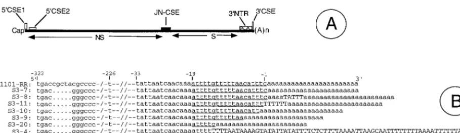
Alphaviruses are mosquito-transmitted animal and human RNA viruses and are the predominant component of the To-gaviridaefamily (29). Alphaviruses cause fever, arthritis, skin rashes, and encephalitis in humans and livestock. Alphaviruses are extensively distributed in both the old and new worlds and are responsible for periodic outbreaks of human illnesses (29, 59). All alphaviruses carry a 12-kb positive-sense RNA that encodes a very similar set of polymerase and structural pro-teins from two open reading frames (53, 59). Alphavirus ge-nomes resemble eucaryotic mRNAs in that they possess 5⬘cap structures and 3⬘poly(A) tails (Fig. 1A). In addition, the 5⬘and 3⬘ proximal sequences of alphavirus genomes carry differing lengths of nontranslatable regions (NTRs) that are believed to carrycis-acting motifs which regulate viral RNA synthesis. At least four CSEs, or RNA motifs, are found in all alphaviruses (Fig. 1A) (59). These CSEs are thought to bind to viral and/or cellular proteins and regulate viral RNA synthesis. Genetically engineered DNA copies of alphavirus RNA genomes have been used extensively to study the roles of RNA motifs and proteins in virus replication and pathogenesis (20, 23, 29, 33, 39, 46, 47, 51, 65). The use of alphavirus vectors in nucleic acid-based gene and vaccine delivery applications has stimu-lated much interest in the biology of alphavirus vectors (17, 18, 26, 37, 54).
Sindbis virus (SIN) is one of the best-studied alphaviruses at the molecular level (59). The SIN genome carries a 0.3-kb 3⬘NTR and a poly(A) tail, whereas the sizes of the 3⬘NTRs of other alphavirus genomes range from 80 to 610 bases (45). Despite this wide size difference, all 27 known alphavirus ge-nomes carry a very highly conserved 19-nucleotide (nt) motif at * Corresponding author. Mailing address: Department of
Microbi-ology, Rm. 4126, Basic Sciences Building, 1005 D. B. Todd Blvd., Meharry Medical College, School of Medicine, Nashville, TN 37208. Phone: (615) 327-6687. Fax: (615) 327-6602. E-mail: rramasamy @mmc.edu.
9776
on November 9, 2019 by guest
http://jvi.asm.org/
the 3⬘end of the genome just upstream of the poly(A) tail (45). In addition, several repeat sequence motifs (RSEs) and strong secondary structures also occur in the 3⬘NTR in alphaviruses (32, 43, 45). The 3⬘19-nt CSE, along with the poly(A) tail, has long been believed to function as the RNA promoter for the initiation of negative-sense RNA synthesis from the positive-sense genome (32, 35, 36). Contrary to these expectations, we recently showed that SIN genomes lacking a part or all of the 19-nt 3⬘CSE were able to initiate replication and produce in-fectious virus (24, 46). Surprisingly, analysis of the 3⬘NTR sequences of the progeny virus genomes demonstrated the presence of either the parental mutant 3⬘CSE or differing lengths of new AU-rich motifs at the initial site of mutation (46). In essence, upon transfection into BHK cells, a given species of in vitro-synthesized SIN RNA carrying a defined deletion in its 3⬘CSE gave rise to a population of viruses that differed in the size of the AU-rich 3⬘terminal region of their genomes (46). It was not clear how the viral polymerase was able to recognize the progeny genomes carrying AU-rich mo-tifs of heterogeneous size at their 3⬘termini. In this report, we present further information on these novel SIN genomes, in-cluding the stability of the altered 3⬘NTRs, growth properties, RNA synthetic abilities, and genome organization and se-quences of representative SIN isolates.
Virus isolates and their growth properties. Previously we
reported the isolation of a library of infectious SIN mutants that carried significant deletions and/or additions in the 19-nt CSE of the 3⬘NTR of these RNA genomes (46). The library was obtained by transfecting BHK cells with the in vitro-tran-scribed SIN RNAs carrying deletions within the 3⬘CSE. The culture supernatants recovered from the BHK cells were sub-jected to a single round of plaque purification, and the indi-vidual plaque suspensions were stored frozen. These plaque suspensions were assigned the passage zero (p-0) designation: they had a virus titer of 103to 104PFU in each suspension. Representative plaque suspensions were used to infect BHK cells at a multiplicity of infection of 0.1 PFU/cell, and virus stocks were prepared. These virus stocks were designated pas-sage 1 (p-1). The virus titers of p-1 stocks of all isolates were in the range of 107PFU/ml. To understand the biological
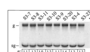
prop-erties and mechanism of the origin of these viruses, we chose to analyze in depth eight isolates, whose 3⬘NTR sequences are shown in Fig. 1B. These isolates carried different lengths of the 3⬘NTRs due to the addition of different sizes of AU-rich motifs adjacent to the poly(A) tail. The plaque sizes of these isolates were similar, except that the plaque size of the isolate S3-4 was ca. 20% smaller than that of other isolates (Fig. 2). To test the stability of these variant 3⬘NTRs and to evaluate the growth properties of these virus isolates, duplicate BHK cell cultures were infected with 3 to 5 PFU of the different SIN isolates/cell and adsorbed at 30°C for 1 h, the inoculum was removed, and 1 ml of fresh medium was added. The cytopathic effect (CPE) was monitored every 2 h for the first 10 h of infection. The final CPE was recorded around 24 to 26 h postinfection (p.i.), the culture supernatant was recovered, and the titer of virus was determined. The virus stocks recovered from the p-1 were used to infect fresh BHK cultures, and the extent of CPE and virus titer were determined. Eighteen such passages were carried out, and CPE and virus titer were determined. As shown in Table 1, the overall difference in virus titer between the isolates and passages varied only up to three- to fourfold. All the isolates except S3-20 produced significant CPE around 7 to 8 h p.i. All the isolates produced extensive CPE and cell detach-ment of BHK cells when observed around 24 to 26 h p.i. Since the total amounts of virus released from all the isolates did not differ significantly, the kinetics of virus release was determined using the p-1 virus isolates. As shown in Fig. 3, the isolate S3-20, which carried a 13-nt deletion in the 3⬘CSE, showed a significant delay in release of virus particles early during virus infection, but around 9 to 10 h p.i., virus release paralleled that of the other isolates. These results demonstrated that the ge-nomes of the SIN isolates carrying diverse 3⬘NTR sequences with drastically altered 3⬘CSE retain a high level of infectivity and release comparable levels of virus particles from BHK cells for all the 18 passages tested.
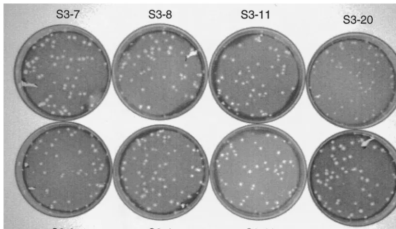
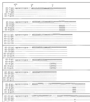
The level of virus-specific RNA synthesized in the infected cells depends heavily on the nature of its gene regulatory elements. Since the 3⬘CSE and the 3⬘NTR are thought to play major roles in polymerase recognition, RNA synthesis, and genome translation, it was thought likely that the level of FIG. 1. (A) Organization of the SIN genome. NS, coding region for nonstructural proteins; S, coding region for structural proteins; 5⬘CSE-1, the 44-nt 5⬘conserved sequence element at the 5⬘end; 5⬘CSE-2, the 51-nt 5⬘conserved sequence element located within the 5⬘translatable region; JN-CSE, the 21-nt-long conserved sequence element located at the junction of the NS and S coding regions that serves as a promoter for subgenomic RNA synthesis; 3⬘NTR, the 0.3-kb 3⬘nontranslated region that carries several repeat sequence elements; 3⬘CSE, the 19-nt conserved sequence element located at the 3⬘end adjoining the poly(A) tail. The drawing is not according to scale. (B) Sequences of the 3⬘NTR domain of eight SIN isolates. These eight SIN isolates, which were recovered from individual plaques (46), were used to infect BHK cells to generate virus stocks. The virus-specific RNAs obtained from infected BHK cells were reverse transcribed with 18TSac⫺and amplified by PCR using the primers T11200 and 18TSac⫺as previously described (46). The single species of PCR product obtained for each of the isolates was purified and sequenced. Each isolate is identified on the left of each sequence. The sequence 1101-RR corresponds to the SIN derived from the parental clone Toto 1101 (39, 47, 51). The control isolate S3-7 was generated from the SIN clone Tapa (51). Since the Tapa plasmid was derived from Toto 1101, it was expected to carry identical protein-coding regions. The Tapa plasmid carries an intact 3⬘NTR and anApaI restriction site at the 3⬘end of the S-coding region. The location of theApaI site (gggccc) and the stop codon (tga) are shown. Notations used: lowercase letters, original sequence of the template used for virus production; underline, bases corresponding to the 3⬘CSE; uppercase letters, bases inserted during genome repair in vivo; dots, base deletions; hyphens, base identity; back-slashes, discontinuity in sequence used for drawing purposes.
on November 9, 2019 by guest
http://jvi.asm.org/
[image:2.612.67.522.70.202.2]virus-specific RNA synthesized from test isolates would be different. To investigate this possibility, BHK cells were in-fected with each of the p-1 virus isolates at a multiplicity of infection (MOI) of 4 to 6 PFU/cell, and the level of virus-specific RNA synthesized between 5 and 9 h p.i. was deter-mined by metabolic labeling. As shown in Fig. 4, the steady-state levels of genomic RNA varied up to 40%, and the ratio of genomic RNA levels to subgenomic RNA levels ranged from 1.3 to 1.5. Since these differences are small, the terminal se-quences of the 3⬘NTRs as found in the eight virus isolates appear to have had little effect on the steady-state levels of virus-specific RNAs. Although the growth kinetics of the iso-late S3-20 was significantly impeded (Fig. 3), the virus-specific RNA levels were unaffected. Similar virus-specific RNA levels
were found in BHK cells infected with p-18 virus isolates (data not shown).
Sequence changes in genomes of different SIN isolates.
Re-sults reported above indicated that the majority of the p-1 virus isolates carrying different 3⬘terminal sequences grew well and synthesized comparable levels of virus-specific RNAs. Al-though these results were unexpected, we Al-thought that addi-tional compensatory sequence changes could have occurred elsewhere in the genome. Therefore, we sequenced the entire genome of all eight virus isolates. In brief, cytoplasmic RNA isolated from infected BHK cells was reverse transcribed with an oligo(dT) primer and amplified by PCR with 12 sets of primer pairs to generate overlapping DNA fragments of ca. 1 kb. These DNA fragments were purified to remove the
[image:3.612.98.507.72.308.2]oligo-FIG. 2. Plaque sizes of SIN isolates. Confluent Vero cultures were infected with p-1 stocks of the eight virus isolates, overlaid with agar, and incubated at 37°C. Around 50 to 54 h p.i., plaques were fixed with paraformaldehyde, stained with crystal violet, and photographed. Note that the plaques of isolates S3-7 and S3-23 were larger, whereas those of S3-10 and S3-20 were smaller. The plaques of isolate S3-10 were clear, but the plaques of all other isolates were somewhat diffused.
TABLE 1. Sindbis virus isolates and their growth propertiesa
Isolate 3⬘CSEb
Passage 1 Passages 2–18
CPEc
Virus titer (107)
CPEc
Virus titer (107)d
8–10 h 24–26 h 8–10 h 24–26 h
S3-7 ⫺1 to⫺19 ⫹ ⫹⫹⫹⫹⫹ 4.92 ⫹ ⫹⫹⫹⫹⫹ 2.92–6.72
S3-8 ⫺1 to⫺19e ⫹ ⫹⫹⫹⫹⫹ 5.68 ⫹ ⫹⫹⫹⫹⫹ 3.15–8.17
S3-11 ⫺2 to⫺19 ⫹ ⫹⫹⫹⫹⫹ 3.14 ⫹ ⫹⫹⫹⫹⫹ 3.83–7.46
S3-10 ⫺3 to⫺19 ⫹ ⫹⫹⫹⫹⫹ 3.16 ⫹ ⫹⫹⫹⫹⫹ 2.45–5.28
S3-9 ⫺7 to⫺19 ⫹ ⫹⫹⫹⫹⫹ 2.43 ⫹ ⫹⫹⫹⫹⫹ 1.96–7.29
S3-20 ⫺14 to⫺19 ⫹ ⫹⫹⫹⫹⫹ 3.01 ⫹ ⫹⫹⫹⫹⫹ 2.11–6.88
S3-4 ⫺15 to⫺19f ⫺ ⫹⫹⫹⫹⫹ 2.15 ⫹ ⫹⫹⫹⫹⫹ 3.17–5.86
S3-23 Noneg ⫹ ⫹⫹⫹⫹⫹ 3.38 ⫹ ⫹⫹⫹⫹⫹ 2.97–6.21
aThe name of each isolate, the composition of the 3⬘terminal domain of their genomes, the extent of CPE observed, and the virus titers are given.
bThe classical 3⬘CSE is denoted by positions⫺1 to⫺19. The⫺1 position corresponds to the first nucleotide adjacent to the poly(A) tail. The remaining portion
of the 3⬘NTR is as given in Fig. 1B.
cCPE as shown by cell death, cell rounding, and formation of weblike structures.⫹, low level of CPE;⫺, no CPE;⫹⫹⫹⫹, maximal CPE. dThe lowest and the highest levels of virus titer found among passages 2 to 18 are given.
eThe 3⬘NTR of this isolate (S3-8) carried the intact 3⬘CSE but also carried an insertion of the UAUUU motif in the poly(A) tail (Fig. 1B). fThe genome of this isolate carried five bases of the 3⬘CSE (⫺15 to⫺19) and a 64-nt-long AU-rich insertion adjacent to the poly(A) tail. gThe absence of 3⬘CSE is accompanied by a 72-nt AU-rich extension adjacent to the poly(A) tail.
on November 9, 2019 by guest
http://jvi.asm.org/
[image:3.612.52.558.540.663.2]nucleotides and sequenced using two to four primers specific to each of the DNA fragments. To determine the precise sequence of the 5⬘-terminal region of the genomes, the first-strand cDNA products corresponding to the 5⬘ 1-kb region were tailed with dC and were amplified by PCR and se-quenced. The sequence data obtained from this analysis were compared with the published sequence of a SIN strain known as HRsp (58). As shown in Table 2, several base changes be-tween the genome of the HRsp strain and that of the SIN lates reported here were demonstrated. Since all the SIN iso-lates used in this study were derived from the parental SIN cDNA clone Toto 1101 or its derivatives (39, 51), it was im-portant to establish whether or not the corresponding base changes could be found in the Toto 1101 DNA. It was also possible that some of these base changes could have occurred during the in vitro synthesis of RNA from these DNA plates. To explore these possibilities, two parental DNA tem-plates, namely Toto 1101 and T3⬘15 (46), were used as con-trols. These two plasmids were transcribed in vitro as described previously (46) and treated with DNase I to remove template DNA, and the resulting RNA preparations were used as tem-plates for reverse transcription and amplification by PCR. PCR products were then sequenced to establish the identity of the bases reported in Table 2. Finally, the nucleotide positions 353, 2992, 2579, and 5702 of Toto 1101 plasmid DNA were se-quenced using Sequenase as a test to rule out possible artifacts of automated sequencing.
[image:4.612.142.453.67.395.2]Table 2 summarizes the positions and base changes in the
FIG. 3. Time course of virus release. Duplicate BHK cell cultures were infected with 6 PFU of p-3 SIN isolates/cell and incubated at 37°C for 1 h, the inoculum was removed, and the cells were replenished with 1 ml of medium containing 2% fetal bovine serum. At 3, 5, 7, 9, and 11 h p.i., culture supernatants were completely removed, and 1 ml of fresh medium was added to the cells. The amount of infectious virus found in the recovered medium was determined by plaque assay. Each value represents the average of two experiments. In the two experiments, the isolate S3-20 showed very similar growth kinetics.
FIG. 4. Levels of virus-specific RNA expressed by SIN isolates. BHK cell cultures were infected with 6 PFU of each virus isolate/cell and labeled with [3H]uridine between 5 and 9 h p.i. as previously described (46, 47). In brief,
infected cells were replenished with 0.6 ml of minimal essential medium con-taining 3g of dactinomycin per ml at 6 h p.i. Twenty minutes later, 50Ci of [3H]uridine (Dupont-NEN) was added to each plate, and the infection was
continued at 37°C for an additional 3 h. At the end of the labeling period, cells were harvested in phosphate-buffered saline and disrupted with 1% NP-40, and the cytoplasmic supernatants were recovered by centrifugation. Cytoplasmic RNA was purified by phenol-chloroform extraction and precipitation with two volumes of ethanol (46, 47). The amount of RNA recovered was determined by UV spectrophotometry. Six micrograms of the isolated RNA was denatured with glyoxal, analyzed on a 1.25% agarose gel, and then fluorographed as described previously (46, 47). The radioactivity corresponding to each of the bands was recovered, solubilized in a biodegradable solvent (BCS; Amersham) and quan-titated. For comparison, the intensity of the RNA bands was also determined by densitometry using the Gel-doc 2000 apparatus and Quantity One image analysis software (Bio-Rad). The top of each lane indicates the identity of the isolate. UI, uninfected; g, genomic RNA, 11.7 kb; sg, subgenomic RNA, 4.2 kb; g/sg, ratio of genomic RNA to subgenomic RNA. The values were determined from two experiments. RNA samples derived from each experiment were loaded twice on the same gel for densitometry and determination of radioactivity.
on November 9, 2019 by guest
http://jvi.asm.org/
[image:4.612.336.525.448.557.2]protein coding sequences of isolates that were sequenced. Overlapping DNA fragments corresponding to the entire ge-nomes of all eight isolates (S3-7 to S3-23) were sequenced and assembled using the DNA assembly software Sequencher (Gene Codes Corp). In brief, 5g of the infected cell RNA from each of the eight SIN isolates was reverse transcribed with the primer 18T Sac, and the first-strand cDNA was puri-fied by digestion with RNase H and RNase T1 enzymes fol-lowing the manufacturer’s protocol for the 5⬘ RACE kit (GIBCO/BRL). The purified products were used as a template for PCR amplification. The primer pairs used to amplify the approximately 1-kb region of the SIN genome from the 5⬘end to the 3⬘end were the following: T32, T1050⫺; T950, T2050⫺; T1950, T3100⫺; T3050, T4150⫺; T4050, T5250⫺; T5200, T6350⫺; T6300, T7350⫺; T7300, T8300⫺; T8200, T9050⫺; T9000, T10050⫺; T10000, T11000⫺; and T10950, 18TSac⫺
(Table 3). The single species of DNA product obtained from these PCRs was purified by a DNA purification kit (Mo Bio Laboratories) and sequenced using the Bigdye terminator kit (ABI). In addition to the primers used in the individual PCRs, additional oligonucleotides were used in sequencing to verify the overlapping regions of the PCR products. To determine the exact 5⬘ terminal sequence of the SIN genomes, the in-fected cell RNA was reverse transcribed with a negative-sense primer, T1050⫺, and the cDNA was purified using the 5⬘
RACE kit (GIBCO/BRL), tailed with dCTP, and amplified by PCR using the abridged anchor primer provided in the kit and the primer T1050⫺ (Table 3). The PCR products obtained from these reactions were purified and sequenced using the primers T200⫺, T500⫺, and T1050⫺ (Table 3). The mis-matched bases between the published genome of the HRsp strain of SIN (58) and those of isolates reported in this study are shown. At least nine base changes were found within the protein-coding sequences of the Toto 1101-RR strain com-pared to the HRsp strain. The nucleotide sequences of the protein-coding region of isolates S3-7, S3-8, S3-11, S3-20, S3-4, S3-23, and Toto 1101-RR were identical (Table 2). Isolate S3-10 carried a C to G (Leu to Val) change at position 2133 in the nsp2-coding region. Isolate S3-9 carried a C to U change at
position 4874, with no amino acid changes. As shown in Fig. 1B, all isolates carried an identical 3⬘NTR, except for the terminal AU-rich motifs. S3-23 carried a C residue instead of U at position⫺226. As shown in Fig. 5, no base changes were found within the 5⬘NTR domains of any of these isolates
com-10288 E1 GaC (Asp) GGC (Gly) GGC (Gly) G G G G G G G G
aHeat-resistant, small-plaque variant of the SIN genome (57). The lowercase letters in the triplet codons denote the base that differs from those of the 1101-RR and
other isolates. The encoded amino acid is indicated in parentheses.
bToto 1101 (38, 50) is the parental SIN DNA clone from which all of the SIN isolates reported here were derived. The underlined base in each of the triplet codons
denotes a base change with respect to the published HRsp sequence (57). To determine if a corresponding base change could be traced back to the Toto 1101 clone, the plasmid was transcribed in vitro with SP6 RNA polymerase, treated with DNase I, and purified. The resulting RNA was subjected to reverse transcription-PCR and sequenced to confirm the base changes at the indicated positions.
cT3⬘15/Xho (49) is a derivative of Tapa that was again derived from Toto 1101. T3⬘15/Xho carries a 4-nt deletion within the 3⬘CSE. T3⬘15/Xho was one of the 3⬘
deletion mutants of the SIN genome that was used in transfection studies to generate some of the SIN isolates studied here. The plasmid T3⬘15/Xho was transcribed in vitro, treated with DNase I, and amplified by reverse transcription-PCR, and the mutant positions in question were sequenced.
dThe base changes observed between the published HRsp strain of the SIN genome (57) and the eight isolates sequenced here are indicated. Note that all the eight
[image:5.612.53.553.83.213.2]isolates were derived from the parental clone Tapa, which is a derivative of Toto 1101. The base differences between 1101-RR and HRsp are mirrored in all of the eight isolates studied here.
TABLE 3. Selected oligonucleotides used in this studya
No. Designation Sequence
1 T32 ACAGCCGACCAATTGCACT
2 T1050⫺ CCGGGATGTACGTGCACA
3 T950 ACGGGAGAAACCGTGGGA
4 T2050⫺ GTTTCTGCAAGCTCTGCC
5 T1950 TAGTGTACAACGAAAGAGP
6 T3100⫺ ACGTTGGTCTTGCAGCTG
7 T3050 AAGGGAATAATTGCTGCA
8 T4150⫺ GCTGCGTTGACAACTGCT
9 T4050 ATTGCGTGATTTCGTCCG
10 T5250⫺ TACTAGTAATAGAGTTGT
11 T5200 TATGGATGACAGTAGCGA1
12 T6350⫺ GGCAATGAGCACATTTTG
13 T6300 GAGCCCCGAATATCCGCA
14 T73050⫺ GTTTACCCAACTTAAACA
15 T7300 AGATTCGGTTACTTCCAC
16 T8300⫺ ACCGGAGTTATCCATGAT
17 T8200 CGAAGGATTCTATAACTG
18 T9050⫺ GTGAACGGGAGGTAGATC
19 T9000 GTACACTGGCCCGCAAGA
20 T10050⫺ CGTAGGCGTCTACCTTCG
21 T10000 CTGCTGCTCCTGCTGCC
22 T11000⫺ CAGGGTGGCCATCCCGCC
23 T10950 TCTCAACAGTCAAATGTG
24 18T Sac⫺ CCTAGAGCTCAG(T)18
25 T286 CGAGCACCAGTATCATTG
26 T2923 TGTTGCTCACCCGCACTG
27 T3503 TAACCTGGTCCCGGTGAA
28 T5623 CCGAACCCGTCCTGTTTG
29 T500⫺ CTTATAAGGCAGTACGTC
aAll of the sequences are indicated in the 5⬘to 3⬘orientation. A minus sign at
the end of a designation denotes negative polarity. In general, the numbers associated with the names of the oligonucleotides denote their approximate locations in the SIN genome.
on November 9, 2019 by guest
http://jvi.asm.org/
[image:5.612.312.550.426.695.2]pared to that of the wild-type isolate S3-7. These results dem-onstrated that the SIN isolates described in this study were derived from the Toto 1101-based SIN sequence and that com-pensatory amino acid coding changes within the protein-coding regions or base changes within noncoding regions are not es-sential to restore biological activity to SIN genomes carrying AU additions or deletions in their genome termini.
Evolution of the 5ⴕ and 3ⴕ proximal sequences of various
SIN genomes.Although the stability and infectivity of these
novel SIN isolates did not require any compensatory base changes in the SIN genomes, it is possible that these genomes may evolve into new and more efficient genomes. Since the 5⬘
and 3⬘ proximal regions were believed to carry RNA motifs that regulate RNA synthesis, we analyzed the base changes in these regions during passage in BHK cells. To determine the sequences of the 5⬘proximal region of SIN genomes, the in-fected cell cytoplasmic RNA corresponding to p-1 and p-18 of all isolates and a control SIN RNA corresponding to Toto 1101-RR were reverse transcribed with the primer T1050⫺, and the resulting first-strand cDNA was tailed with dCTP and amplified by PCR. The PCR products were purified and se-quenced using primers T200⫺, T500⫺and T1050⫺(Table 3). The first 962 bases corresponding to the 5⬘proximal region of all isolates corresponding to p-1 and p-18 were identical (data
not shown). As expected, nt 353 was found to be a U in all the isolates, including that of Toto 1101, whereas the HRsp strain carried a C residue in that position (58).
[image:6.612.143.460.69.426.2]Since the 3⬘NTR domain of these isolates underwent various extents of the repair process during their generation in the RNA-transfected cells, we anticipated additional base changes during evolution. Therefore, we chose to examine virus-spe-cific RNA derived from passages 1, 2, 6, 10, and 18 of all isolates (Fig. 5). Four of the isolates, S3-7, S3-11, S3-10, and S3-20, did not undergo any base changes within the terminal 470 bases in any of the passages tested. Isolate S3-8, which carried an intact 3⬘CSE and an insertion of a short motif, UAUUU, within the poly(A) tail, underwent deletion events during passages 3 to 6, resulting in the formation of wild-type 3⬘NTR. This deletion event could have occurred by a simple polymerase skipping event during synthesis of new RNA. Al-ternatively, the initiation of RNA synthesis could have oc-curred just upstream of this UAUUU motif, using the short oligo(A) motif and the 3⬘CSE as a promoter. Isolate S3-9, which carried only 13 of the 19 bases, acquired the motif CAUUAC within its poly(A) tail, and this modified 3⬘terminus was stable for the remaining passages as tested. Finally, isolates S3-4 and S3-23, which carried extensive AU-rich insertions just adjacent to the poly(A) tail, underwent base changes of A to U. FIG. 5. Evolution of the 3⬘proximal region of SIN isolates. Infected cell cytoplasmic RNA obtained from the indicated passages of all virus isolates was reverse transcribed with 18TSac⫺and amplified by PCR with T11200 and 18TSac⫺. The PCR products were purified and sequenced using the primer T11200. The 3⬘CSE (⫺1 to⫺19) and its remnants are identified by underlining. The bases newly added during the repair process are identified by uppercase letters. The hyphens denote nucleotide identity. Slashes and vertical lines denote discontinuity in sequence, used for drawing purposes.
on November 9, 2019 by guest
http://jvi.asm.org/
These unusually long 3⬘NTRs showed little or no tendency to reduce their size or composition during multiple passages. These results demonstrated the long-term stability and func-tional integrity of different types of 3⬘ AU-rich terminal ele-ments of the genomes of the SIN isolates in BHK cells.
RSEs in the 3ⴕNTR of the new SIN isolates.Previous work
from other laboratories demonstrated the presence of RSEs within the 3⬘NTR (43, 45), but the occurrence of RSEs at other regions of the alphavirus genome has not been reported. Given the multitude of 3⬘ terminal motifs that are compatible with efficient SIN genome replication, the formation and function of the RSEs of 3⬘NTRs may have a direct relationship with genome repair. To evaluate the significance of the RSE in the repair and evolution of alphavirus genomes, the occurrence and distribution of RSEs within the 5⬘and 3⬘proximal regions of all the isolates were determined. Although numerous RSEs of 4 to 7 bases occurred within the genomes of the SIN isolates, those motifs which contained eight or more bases were chosen for analysis. As shown in Table 4, RSE2, RSE3, RSE4, RSE5, RSE7, and RSE8 were found in duplicates within the 3⬘NTR
of all SIN isolates studied. RSE1 was found in both the 5⬘and 3⬘proximal regions of all SIN isolates studied. RSE6 was found in duplicate copies only in the 5⬘proximal region of all the SIN isolates. As shown in Table 4, the 3⬘NTR of the genomes of isolates S3-4 and S3-23 carried many new additional RSEs. Four of these RSEs (k, l, m, and n), corresponding to the isolate S3-4, were found in both p-1 and p-10 genomes. The RSEo and RSEp were found only in the p-10 genome of the isolate S3-4. Likewise, the 3⬘NTR of the p1 and p10 genomes of the isolate S3-23 also carried six RSEs (r, s, t, u, v, and x) whose sequences were different from that of the isolate S3-4. The p-10 genome of the isolate S3-9 carried one new RSE (z) within its 3⬘NTR. The genomes of isolates S3-7, S3-8, S3-10, S3-11, and S3-20 did not undergo any AU additions during their biogenesis (46) and therefore did not carry any of the additional RSEs found in the isolates S3-4 and S3-23. The arrangement and order of the RSEs found in the genomes of isolates S3-4 and S3-23 are shown in Fig. 6. Detailed analysis revealed that the entire AU-rich region that replaces the 3⬘CSE was composed of RSEs of 4 nt or more (data not
12 RSEn AATTTTTTTTAA 2 s3-4/p-1,p-18 3⬘NTR
13 RSEo TTTTAAAA 2 s3-4/p-18 3⬘NTR
14 RSEp TTTTTTTTAA 2 s3-4/p-18 3⬘NTR
15 RSEr TTTTTCTT 3 s3-23/p-1,p-18 3⬘NTR
16 RSEs TTCTTTTT 2 s3-23/p-1,p-18 3⬘NTR
17 RSEt TTTTTCTTTT 2 s3-23/p-1,p-18 3⬘NTR
18 RSEu TTTATTATTT 2 s3-23/p-1,p-18 3⬘NTR
19 RSEv AAATTTTTTTT 2 s3-23/p-1,p-18 3⬘NTR
20 RSEx TTTTTTTTAAA 2 s3-23/p-1,p-18 3⬘NTR
21 RSEz ATTACAAA 2 S3-9/p-18 3⬘NTR, 5⬘prox(384)
aUsing DNAsis software (Hitachi), the RSEs with 100% match that occur within the 5⬘and 3⬘proximal 400-nt region of all of the SIN isolates studied here were
[image:7.612.55.556.84.294.2]determined. Using the same software, the SIN genome was searched for the occurrence of these motifs at internal locations and found to be absent. SIN denotes all of the Sindbis virus genomes studied here, including Toto 1101. p-1, genome sequence corresponding to the passage 1; p-18, genome sequence corresponding to the passage 18; location, the numbers correspond to the nucleotide positions starting from the 5⬘end of the SIN genomes; 5⬘prox, nucleotide position 384, located at the 5⬘proximal region of all the SIN genomes.
FIG. 6. Physical map of the AU-rich RSEs. Each RSE found within the AU-rich terminal motif of isolates S3-23 (A) and S3-4 (B) was arranged and identified by the same letters as indicated in Table 2. Bars of the same kind indicate the same RSE. For example, bars denoting the motif x (panel A) occur very close to each other, whereas those denoting p (panel B) are well separated.
on November 9, 2019 by guest
http://jvi.asm.org/
[image:7.612.99.505.584.699.2]shown). Therefore, it is likely that the 3⬘ repair pathway in-volving the extensive addition of AU-rich motifs may involve reiterative copying of short AU-rich motifs from within the SIN genome.
Kuhn et al. reported that some mutations within the 3⬘CSE of the SIN genome were lethal (32). Results of previous studies by members of our group (24, 46) and the results reported here establish that SIN isolates lacking the classical 3⬘CSE and isolates carrying novel AU-rich motifs are highly infectious for all the 18 passages tested in BHK cells. Increased RNA trans-fection efficiencies and the nature of transtrans-fection methodolo-gies adopted by us might have helped us to recover these novel virus isolates. Despite the differences in their 3⬘terminal se-quences, most of the isolates showed very similar growth pat-terns in BHK cells. It is puzzling to note that the isolate S3-20, which lacked 13 bases of the 3⬘CSE, displayed a drastic de-crease in virus release during the early phase of infection, whereas other isolates, such as S3-9, which lacked 6 bases of the 3⬘CSE, displayed a normal rate of virus release. The de-creased rate of virus release could be due to an early defect in virus assembly or to decreased levels of virus-specific RNA during the first 3 to 6 h of infection. Although the steady-state levels of virus-specific RNA were similar for all the isolates tested, it is possible that the time course of RNA synthesis and decay differ for different isolates. The growth properties and virus-specific RNA levels of isolates S3-4 and S3-23, which carry extensive AU additions at the 3⬘end of their genomes, were comparable to that of the S3-7 isolate, which carries the wild-type 3⬘NTR. Preliminary results indicated that even the kinetics of RNA synthesis was similar among these three iso-lates (unpublished data). Therefore, it is possible that the clas-sical 3⬘CSE, the deletion versions of 3⬘CSE, as found in the isolates S3-9, S3-10, and S3-11, and the AU-rich motifs, as found in the isolates S3-4 and S3-23, serve as equally strong promoters for RNA synthesis. How the SIN polymerase could initiate RNA synthesis from a disparate set of 3⬘ termini re-mains to be determined. As described for picornaviruses (19, 38, 40) and coronaviruses (50), an internal or 5⬘RNA motif of the alphavirus genome may be important for genome replica-tion. We propose that this alternate RNA motif yet to be identified may recruit the polymerase to the SIN genome in the absence of an authentic 3⬘CSE. Once the polymerase is re-cruited to the SIN genome, 3⬘ end recognition may occur by polymerase movement on the template RNA. The 3⬘poly(A) tail, AU-rich motifs, and proteins associated with these se-quences may serve as signals for initiation of RNA synthesis once the polymerase is selectively loaded on the template.
No base changes were found in the genomes of five of eight isolates studied. Importantly, isolates S3-4 and S3-23, which carried long AU-rich 3⬘terminal motifs in their genomes in all of the 18 passages, were also found to carry no base changes elsewhere in the genome. Given the rapid evolution of RNA genomes (25, 31) and the strict conservation of the 3⬘CSE of the alphavirus genome, it is difficult to reconcile the fact that there are no compensatory base changes or evolutionary pres-sures for acquiring the classical 3⬘CSE. The simplest explana-tion is that the 3⬘CSE or the AU-rich elements do not bind directly to the viral polymerase. A case in point is that the highly conserved 98-nt 3⬘ terminal motif of hepatitis C virus failed to bind to the viral polymerase, whereas a 3⬘-coding region with a conserved stem-loop structure specifically bound to the viral polymerase (11). One or more of the known RNA binding proteins (15, 56) may bind to these different 3⬘ termi-nal sequences, and the viral polymerase may be recruited to the 3⬘terminus of the alphavirus genome through interaction with these cellular proteins. Since several AU-rich 3⬘ motifs
were able to support RNA synthesis, RNA binding proteins are expected to possess a broad specificity for adenylates and uridylates, and all of these AU-binding proteins must carry a common motif that binds to the viral polymerase. Since mos-quitoes are vectors for alphaviruses, it is also likely that these broad-specificity RNA binding proteins are present in inverte-brates. It is likely that infectious cycles involving vertebrate and invertebrate cells may be essential to restore the classical 3⬘CSE during virus passage.
What are the determinants that regulate repair and rear-rangement of sequences within the 3⬘NTR? The evolution of the 3⬘NTR of the SIN genome of isolate S3-8 indicates that in the presence of an intact 3⬘CSE, foreign motifs inserted within the poly(A) tail are removed, probably by polymerase jumping events (6, 28, 34). The 3⬘terminal base preceding the poly(A) tail is a C residue in all alphaviruses (45). The 3⬘terminal C of the 3⬘CSE, although not essential for virus replication, appears to serve as a 3⬘ boundary for the alphavirus genome. For example, the absence of the terminal C residue in the isolate S3-11 might be responsible for allowing the 3⬘NTR to retain the stretch of U residues as part of the genome (Fig. 5C). Similarly, the absence of the 3⬘CSE appears to have caused retention of the long AU-rich motifs in isolates S3-4 and S3-23 (Fig. 5G and H). SIN genomes lacking the 3⬘terminal bases of the 3⬘CSE actually stimulated acquisition of AU-rich motifs (46). Isolate S3-9, which lacked six bases from the 3⬘terminus of the 3⬘CSE, acquired a new motif, CAUUAC, at its 3⬘ ter-minus during passage in BHK cells (Fig. 5E). Our previous studies (46) and the work reported here demonstrate that SIN genomes carrying several different 3⬘NTRs with and without the classical 3⬘CSE replicate as well as wild-type SIN. Deletion versions of the 3⬘CSE, as are found in genomes of the isolates S3-9 and S3-20, were fully functional even in the absence of any RNA repair or 3⬘ terminal AU additions (Fig. 1B). In vivo processed and repaired SIN genomes, such as S3-4 and S-23, which carried extensive AU-rich motifs at their 3⬘ends, repli-cated efficiently and maintained these AU-rich motifs through all the 18 passages tested (Fig. 5G and H). These results argue that repair of the 3⬘terminus of the 3⬘NTR of the SIN genome with AU-rich motifs may be coincidental to events such as RNA transport and localization and that the addition of new AU-rich motifs is not required for genome replication. As proposed in a previous section, an alternate RNA motif within the SIN genome might serve to recruit the viral polymerase, and the modified 3⬘termini described for the genomes of these new SIN isolates may serve as accessory signals that indirectly regulate genome replication. The 3⬘NTR of many eucaryotic mRNAs carry AU-rich domains, which are thought to regulate RNA stability, localization, and translation (9, 15). How the addition or the removal of AU-rich elements precisely affects the biology of the SIN replication remains to be determined.
The RSEs found in the 5⬘and 3⬘proximal regions of the SIN genome may regulate RNA synthesis. Duplication of these motifs clearly occurs at a much higher frequency within short 5⬘ and 3⬘ proximal regions. Since polymerase jumping and recombination events are well known among RNA viruses (6, 28, 34), including alphaviruses (21, 24, 47, 64), these motifs might have been copied by the viral polymerase and introduced in the growing polynucleotide chain. Since a major portion of newly formed 3⬘ terminal motifs of isolates such as S3-4 and S3-23 (Fig. 6) was composed of 85 to 95% AU-rich RSE, it is likely that formation of AU-rich RSEs is due predominantly to viral polymerase activity. It appears that the AU-rich domains within the 3⬘NTR or other parts of the genome are copied during synthesis of new RNA to repair the defective 3⬘termini of the SIN genomes. Other motifs containing G or C residues
on November 9, 2019 by guest
http://jvi.asm.org/
rich 3⬘terminus. In this context, it is interesting that the 53-nt 3⬘terminal region, excluding the poly(A) tail, of the wild-type SIN genome is 87% AU-rich. The 3⬘CSE, which is highly conserved in all of the 27 alphaviral genomes, carries an 85 to 90% AU-rich sequence (45). Therefore, it appears that the 3⬘NTR of the alphavirus genome and its repair and the repli-cation machinery have coevolved to maintain the AU-rich 3⬘
terminus. In contrast, the 3⬘NTR of rubella virus, which carries little or no AU-rich 3⬘terminus, undergoes a 3⬘repair process involving few or no AU-rich terminal additions (10). The te-lomerase activity (5) of eucaryotic cells, which adds repeat sequence elements to the termini of chromosomes, and the alphavirus 3⬘ repair machinery, which adds repeat sequence elements to the 3⬘ end of the genome, may have evolved to accomplish similar functions.
Several forms of RNA editing (3) and 3⬘repair of viral RNA genomes (4, 8, 30, 42, 49, 52, 61) have been documented. It is possible that these various RNA-modifying machineries may have common structural and functional domains (14). In fact, an AU-rich sequence element in the 3⬘NTR of apolipoprotein B mRNA was shown to bind to the classical RNA editing enzyme Apobec-1 (1). The 3⬘repair processes observed in the SIN genome constitute the only known example that involves de novo polyadenylation and addition of different sizes of AU-rich motifs (46). Both rubella virus (10) and beet necrotic yellow vein virus (30) appear to add a short stretch of U residues adjacent to the poly(A) tail. The size and kind of repeat motifs added to the 3⬘terminus of RNA genomes may be regulated by the sequence of the template RNA and the composition of the repair machinery, including the polymer-ase. Although many cellular RNA binding proteins (11, 55, 56), such as poly(A) binding proteins and polypyrimidine binding proteins, may play a role in transport, localization, repair, and replication of the SIN genome RNA, the enzymatic activity that introduces AU-rich motifs on the 3⬘terminus of the ge-nome is likely to involve the viral RNA-dependent RNA poly-merase. As proposed for some eucaryotic, procaryotic, and RNA viral systems (22, 27), the SIN polymerase complex, along with the nascent RNA, may undergo sliding, jumping, and stuttering events during new RNA synthesis, resulting in the introduction of novel motifs during chain elongation and 3⬘
end formation. Identification and characterization of the RNA motifs and protein factors that regulate alphavirus genome repair may give new insights into the mechanism of template selection in virus-infected cells.
This work was supported by NIH grant GM 57439.
We acknowledge the use of the DNA Sequencing Core Facility of the Vanderbilt-Ingram Cancer Center. We thank Joel Trupin, Kolari Bhat, and Andy Briscoe and members of our laboratory for critical reading of the manuscript and fruitful discussions.
genetic RNA-RNA recombination in brome mosaic virus. Adv. Virus. Res. 43:275–302.
7.Burke, D. S.1997. Recombination in HIV: an important viral evolutionary strategy. Emerg. Infect. Dis.3:253–259.
8.Carpenter, C. D., and A. E. Simon.1996. In vivo restoration of biologically active 3⬘ends of virus-associated RNAs by nonhomologous RNA recombi-nation and replacement of a terminal motif. J. Virol.70:478–486. 9.Chen, C. Y., and A. B. Shyu.1995. AU-rich elements: characterization and
importance in mRNA degradation. Trends Biochem. Sci.20:465–470. 10. Chen, M. H., and T. K. Frey.1999. Mutagenic analysis of the 3⬘cis-acting
elements of the rubella virus genome. J. Virol.73:3386–3403.
11. Cheng, J.-C., M.-F. Chang, and S. C. Chang. 1999. Specific interaction between the hepatitis C virus NS5B RNA polymerase and the 3⬘end of the viral RNA. J. Virol.73:7044–7049.
12. Coffin, J. M.1996. Retroviridae: the viruses and their replication, p. 1767– 1848.InB. N. Fields, D. M. Knipe, and P. M. Howley (ed.), Fields virology, 3rd ed. Lippencott-Raven Publishers, Philadelphia, Pa.
13. Decker, C. J., and R. Parker.1995. Diversity of cytoplasmic functions for the 3⬘untranslated region of eukaryotic transcripts. Curr. Opin. Cell Biol.7:386– 392.
14. Delarue, M., O. Poch, N. Tordo, D. Moras, and P. Argos.1990. An attempt to unify the structure of polymerases. Protein Eng.3:461–467.
15. Derrigo, M. A., E. Cestelli, G. Savettieri, and I. Di Liegro.2000. RNA-protein interactions in the control of stability and localization of messenger RNA. Int. J. Mol. Med.5:111–123.
16. French, R., and P. Ahlquist.1987. Intercistronic as well as terminal se-quences are required for efficient amplification of brome mosaic virus RNA. J. Virol.61:1457–1465.
17. Frolov, I., T. A. Hoffman, B. M. Pragai, S. A. Dryga, H. V. Huang, S. Schlesinger, and C. M. Rice.1996. Alphavirus-based expression vectors: strategies and applications. Proc. Natl. Acad. Sci. USA93:11371–11377. 18. Garoff, H., and K. J. Li.1998. Recent advances in gene expression using
alphavirus vectors. Curr. Opin. Biotechnol.9:464–469.
19. Goodfellow, I., Y. Chaudhry, A. Richardson, J. Meredith, J. W. Almond, W. Barclay, and D. J. Evans.2000. Identification of acis-acting replication element within the poliovirus coding region. J. Virol.74:4590–4600. 20. Griffin, D. E.1998. A review of alphavirus replication in neurons. Neurosci.
Biobehav. Rev.22:721–723.
21. Hajjou, M., K. R. Hill, S. V. Subramaniam, J. Y. Hu, and R. Raju.1996. Nonhomologous RNA-RNA recombination events at the 3⬘nontranslated region of the Sindbis virus genome: hot spots and utilization of nonviral sequences. J. Virol.70:5153–5164.
22. Hausmann, S., D. Garcin, C. Delenda, and D. Kolakofsky.1999. The versa-tility of paramyxovirus RNA polymerase stuttering. J. Virol.73:5568–5576. 23. Hertz, J. M., and H. V. Huang.1995. Evolution of the Sindbis virus
subge-nomic mRNA promoter in cultured cells. J. Virol.69:7768–7774. 24. Hill, K. R., M. Hajjou, J. Y. Hu, and R. Raju.1997. RNA-RNA
recombina-tion in Sindbis virus: roles of the 3⬘ conserved motif, poly(A) tail, and nonviral sequences of template RNAs in polymerase recognition and tem-plate switching. J. Virol.71:2693–2704.
25. Holland, J. J., K. Spindler, F. Horodyski, E. Grabau, S. Nichol, and S. Vandepol.1993. Rapid evolution of RNA genomes. Science215:1577–1585. 26. Huang, H. V.1996. Sindbis virus vectors for expression in animal cells. Curr.
Opin. Biotechnol.7:531–535.
27. Jacques, J. P., and D. Kolakofsky.1991. Pseudo-templated transcription in prokaryotic and eukaryotic organisms. Genes Dev.5:707–713.
28. Jarvis, T. C., and K. Kirkegaard.1991. The polymerase in its labyrinth: mechanisms and implications of RNA recombination. Trends Genet.7:186– 191.
29. Johnston, R. E., and C. J. Peters.1996. Alphaviruses, p. 843–898.InB. N. Fields, D. M. Knipe, and P. M. Howley (ed.), Fields virology, 3rd ed. Lip-pencott-Raven Publishers, Philadelphia, Pa.
30. Jupin, I., S. Bouzoubaa, K. Richards, G. Jonard, and H. Guilley.1990. Multiplication of beet necrotic yellow vein virus RNA 3 lacking a 3⬘poly(A) tail is accompanied by reappearance of the poly(A) tail and a novel short U-rich tract preceding it. Virology178:281–284.
on November 9, 2019 by guest
http://jvi.asm.org/
31.Koonin, E. V., and A. E. Gorbalenya.1989. Evolution of RNA genomes: does the high mutation rate necessitate high rate of evolution of viral proteins? J. Mol. Evol.28:524–527.
32. Kuhn, R. J., Z. Hong, and J. H. Strauss.1990. Mutagenesis of the 3⬘
nontranslated region of Sindbis virus RNA. J. Virol.64:1465–1476. 33. Kuhn, R. J., H. G. M. Niesters, H. Zhang, and J. H. Strauss.1991. Infectious
RNA transcripts from Ross River virus cDNA clones and the construction and characterization of defined chimeras with Sindbis virus. Virology182: 430–441.
34. Lai, M. M. C.1992. RNA recombination in animal and plant viruses. Mi-crobiol. Rev.56:61–79.
35. Lemm, J. A., A. Bergovist, C. M. Read, and C. M. Rice.1998. Template-dependent initiation of Sindbis virus RNA replication in vitro. J. Virol. 72:6546–6553.
36. Levis, R., B. G. Weiss, M. Tsiang, H. Huang, and S. Schlesinger.1986. Deletion mapping of Sindbis virus DI RNAs derived from cDNAs defines the sequences essential for replication and packaging. Cell44:137–145. 37. Liljestrom, P., and H. Garoff.1991. A new generation of animal cell
expres-sion vectors based on the Semliki Forest virus replicon. Biotechnology (NY) 9:1356–1361.
38. Lobert, P. E., N. Escriou, J. Ruelle, and T. Michiels.1999. A coding RNA sequence acts as a replication signal in cardioviruses. Proc. Natl. Acad. Sci. USA96:11560–11565.
39. Lustig, S., A. C. Jackson, C. S. Hahn, D. E. Griffin, E. G. Strauss, and J. H. Strauss.1988. Molecular basis of Sindbis virus neurovirulence in mice. J. Virol.62:2329–2336.
40. McKnight, K. L., and S. M. Lemon.1998. The rhinovirus type 14 genome contains an internally located RNA structure that is required for viral rep-lication. RNA4:1569–1584.
41. Mills, D. R., C. Priano, P. A. Merz, and B. D. Binderow.1990. Q beta RNA bacteriophage: mappingcis-acting elements within an RNA genome. J. Vi-rol.64:3872–3881.
42. Nagy, P. D., C. D. Carpenter, and A. E. Simon.1997. A novel 3⬘-end repair mechanism in an RNA virus. Proc. Natl. Acad. Sci. USA94:1113–1118. 43. Ou, J. H., D. W. Trent, and J. H. Strauss.1982. The 3⬘-non-coding regions
of alphavirus RNAs contain repeating sequences. J. Mol. Biol.156:719–730. 44. Pardigon, N., E. Lenches, and J. H. Strauss.1993. Multiple binding sites for cellular proteins in the 3⬘end of Sindbis alphavirus minus-sense RNA. J. Virol.67:5003–5011.
45. Pfeffer, M., R. M. Kinney, and O. R. Kaaden.1998. The alphavirus 3⬘ -nontranslated region: size heterogeneity and arrangement of repeated se-quence elements. Virology240:100–108.
46. Raju, R., M. Hajjou, K. R. Hill, V. Botta, and S. Botta.1999. In vivo addition of poly(A) tail and AU-rich sequences to the 3⬘terminus of the Sindbis virus RNA genome: a novel 3⬘-end repair pathway. J. Virol.73:2410–2419. 47. Raju, R., S. V. Subramaniam, and M. Hajjou.1995. Genesis of Sindbis virus
by in vivo recombination of nonreplicative RNA precursors. J. Virol.69: 7391–7401.
48. Raju, R., and D. Kolakofsky.1987. Unusual transcripts in La Crosse virus-infected cells and the site for nucleocapsid assembly. J. Virol.61:667–672. 49. Rao, A. L. N., T. W. Dreher, L. E. Marsh, and T. C. Hall.1989. Telomeric
function of the tRNA-like structure of brome mosaic virus RNA. Proc. Natl. Acad. Sci. USA86:5335–5339.
50. Repass, J. F., and S. Makino.1998. Importance of the positive-strand RNA secondary structure of a murine coronavirus defective interfering RNA in-ternal replication signal in positive-strand RNA synthesis. J. Virol.72:7926– 7933.
51. Rice, C. M., R. Levis, J. H. Strauss, and H. V. Huang.1987. Production of infectious RNA transcripts from Sindbis virus cDNA clones: mapping of lethal mutations, rescue of a temperature-sensitive marker, and in vitro mutagenesis to generate defined mutants. J. Virol.61:3809–3819. 52. Sarnow, P.1989. Role of 3⬘end sequences in the infectivity of poliovirus
transcripts made in vitro. J. Virol.63:467–470.
53. Sawicki, D. L., and S. G. Sawicki.1998. Role of the nonstructural polypro-teins in alphavirus RNA synthesis. Adv. Exp. Med. Biol.440:187–198. 54. Schlesinger, S., and T. W. Dubensky.1999. Alphavirus vectors for gene
expression and vaccines. Curr. Opin. Biotechnol.10:434–439.
55. Schnapp, B. J.1999. A glimpse of the machinery. Curr. Biol.9:R725–R727. 56. Siomi, H., and G. Dreyfuss.1997. RNA-binding proteins as regulators of
gene expression. Curr. Opin. Genet. Dev.7:345–353.
57. Strauss, E. G., J. H. Strauss, and A. J. Levine.1996. Virus evolution, p. 153–172.InB. N. Fields, D. M. Knipe, and P. M. Howley (ed.), Fields virology, 3rd ed. Lippencott-Raven Publishers, Philadelphia, Pa. 58. Strauss, E. G., C. M. Rice, and J. H. Strauss.1984. Complete nucleotide
sequence of the genomic RNA of Sindbis virus. Virology133:92–110. 59. Strauss, J. H., and E. G. Strauss.1994. The alphaviruses: gene expression,
replication, and evolution. Microbiol. Rev.58:491–562.
60. Sullivan, M. L., and P. Ahlquist.1999. A brome mosaic virus intergenic RNA3 replication signal functions with viral replication protein 1a to dra-matically stabilize RNA in vivo. J. Virol.73:2622–2632.
61. Todd, S., J. S. Towner, D. M. Brown, and B. L. Semler.1997. Replication-competent picornaviruses with complete genomic RNA 3⬘noncoding region deletions. J. Virol.71:8868–8874.
62. Weaver, S. C., R. Rico-Hesse, and T. W. Scott.1992. Genetic diversity and slow rates of evolution in New World alphaviruses. Curr. Top. Microbiol. Immunol.176:99–117.
63. Webster, R. G., S. M. Wright, M. R. Castrucci, W. J. Bean, and Y. Kawaoka. 1993. Influenza—a model of an emerging virus disease. Intervirology35:16–25. 64. Weiss, B. G., and S. Schlesinger.1991. Recombination between Sindbis virus
RNAs. J. Virol.65:4017–4025.
65. Xiong, C., R. Levis, P. Shen, S. Schlesinger, C. M. Rice, and H. V. Huang. 1989. Sindbis virus: an efficient, broad host range vector for gene expression in animal cells. Science243:1188–1191.