DISSERTATION
ON
“A STUDY OF 150 CASES OF POSTERIOR
CIRCULATION STROKE”
Submitted in partial fulfilment of
requirements for the degree of
D.M. NEUROLOGY (
BRANCH - I
)
of
THE TAMILNADU Dr. M.G.R. MEDICAL UNIVERSITY
CHENNAI
MADURAI MEDICAL COLLEGE
MADURAI – 625 020
CERTIFICATE
This is to certify that this dissertation entitled “A STUDY OF 150 CASES OF POSTERIOR CIRCULATION STROKE” submitted by
Dr. C.J.SELVAKUMAR appearing for D.M. Neurology Degree (Branch - I) examination in August 2010 is a bonafide record of work done by him under my direct guidance and supervision in partial fulfilment of regulations of the Tamil Nadu Dr. M.G.R. Medical University, Chennai. I forward this to the Tamil Nadu Dr.M.G.R. Medical University, Chennai, Tamil Nadu, India.
Dr. M .CHANDRA SEKARAN, M.D., D.M.,
DEAN
Professor and Head, Madurai Medical College, Department of Neurology & Neurosurgery, Madurai - 625 020. Madurai Medical College,
Madurai - 625 020.
DECLARATION
I solemnly declare that the dissertation titled “A STUDY OF 150 CASES OF POSTERIOR CIRCULATION STROKE” is done by me at Department of Neurology, Madurai Medical College & Govt. Rajaji Hospital, Madurai , during 2008-2009 under the guidance and supervision of Prof. M.CHANDRA SEKARAN, M.D., D.M.
This dissertation is submitted to The Tamil Nadu Dr. M.G.R. Medical University towards the partial fulfilment of requirements for the award of D.M., degree in Neurology.
Place: Madurai, Dr.C.J.SELVAKUMAR,
Date: 31-05-2010. Postgraduate Student in D.M. (Neurology), Department of Neurology, Madurai Medical College,
ACKNOWLEDGEMENT
I gratefully acknowledge and sincerely thank the DEAN, Madurai Medical College, and Government Rajaji hospital, Madurai for allowing me to do this Dissertation and utilize the Institutional facilities.
I would like to express my sincere gratitude to
Prof. M.CHANDRA SEKARAN, M.D, D.M., Professor and Head of the Department of Neurology and Neurosurgery, for his motivation, guidance, and encouragement in fulfilling this dissertation work.
I would like to thank our Assistant Professors Dr.T.R.Gnaneswaran, Dr.P.K.Murugan, Dr.R.Kishore, Dr.C.Justin, Dr.D.Chezhian and
Dr.K.Ganesan for their cooperation and guidance.
I am thankful to all my postgraduate colleagues for their constant support and sharp constructive criticism.
I should thank each & every patient who participated in this study for their whole hearted cooperation in making this study successful.
CONTENTS
S.No Title Page No.
1. INTRODUCTION 1
2. AIM OF THE STUDY 6
3. REVIEW OF LITERATURE 7
4. MATERIALS AND METHODS 47
5. OBSERVATIONS AND RESULTS 50
6. CHARTS AND FIGURES
7. DISCUSSION 58
8. SUMMARY & CONCLUSION 67
9. BIBLIOGRAPHY
10. PROFORMA
INTRODUCTION
Stroke is known to human race since antiquity. The seventh century great Indian
physician, Charaka lucidly described the symptoms of stroke which he called
`Pakshaghat` meaning hit one half of body. The other synonyms are ardhang or
lakwa. According to Charaka, stroke affects either left or right side of body
leading to impaired mobility and function of that half of body (hemiparesis) and
difficulty in speaking which may be inability to talk at all (aphasia) or slurred
speech (dysarthria). He had also identified head as the seat of vital organ,
controlling the senses and nerve centers of the whole body. These meticulous
observations of stroke symptoms are relevant even today.
Stroke is one of the major causes of death disability and dependency among all the
neurological disorders. The World Health Organization (WHO) defines stroke as
rapidly developing clinical symptoms and / or signs of focal, at times global loss of
cerebral function, with symptoms lasting more than 24 hours or leading to death,
with no apparent cause other than of vascular origin.
The global prevalence of stroke is estimated to be 5 to 8 /1000. Globally stroke
incidence was also variable according to the ethnic differences in a common
geographical location and ranged from 93 to 223/1,00,000 population.
Nevertheless, many investigators have addressed this question in various regions of
India, which may be considered representative of the whole population. The crude
prevalence rate was 220/1,00,000(range : 44-843/1,00,000).
The incidence rate of stroke in India was estimated to be 13/1,00,000 in a study
done at Vellore on a population sample of 2,58,576 followed over two years,
while another study conducted at Rohtak found the stroke incidence to be
33/1,00,000 (27/1,00,000 for first ever stroke). The stroke risk increases steeply as
the age advances. In a study from Kashmir, prevalence rate of stroke was 41 per
1,00,000 population in the age range of 15-39 years, which increased to 1,075 per
1,00,000 for the age group of 50-59 years.
The term stroke is applied to a sudden focal neurologic syndrome1, specifically the
type caused by cerebrovascular disease. The term cerebrovascular disease
designates any abnormality of the brain resulting from a pathologic process of the
blood vessels, including occlusion of the lumen by embolus or thrombus, rupture
of a vessel, an altered permeability of the vessel wall, or increased viscosity or
other change in the quality of the blood flowing through the cerebral vessels. The
vascular pathologic process may be considered not only in its grosser aspects—
embolism, thrombosis, dissection, or rupture of a vessel—but also in terms of more
basic or primary disorder, i.e., atherosclerosis, hypertensive arteriosclerotic
cerebrovascular diseases are manifest by the abrupt onset of a focal neurologic
deficit, as if the patient was "struck by the hand of God"2. Stroke is one of the
most common neurological disorders in clinical practice. An estimated 5.7 million
patients died from stroke in 2005, with 87% of these deaths occurring in
underdeveloped countries. Globally, stroke is the third leading cause of death and a
major cause of adult disability. It poses serious medical, socioeconomic, and
rehabilitation problems. With the prevalence of disability resulting from stroke is
expected to rise as populations increase, this burden will increase greatly over the
next 20 years, particularly in developing countries. Stroke physicians are faced
with the challenge of providing effective stroke care and reducing the mortality,
disability, and dependency of stroke survivors.
Stroke is the most important single cause of long term disability in a community
setting as about 30 to 50% of stroke patients are left with residual deficits. The
hospital based studies had shown that 2% of all, 4 to 5 % of medical and 20% of
neurological admissions were due to stroke. Not only this, survivors of a transient
ischemic attack (TIA) or stroke are at an increased risk of another stroke. The new
American Heart Association (AHA) guidelines estimated that about 28% of the
prevalent strokes comprised recurrent strokes. This reflects the magnitude of the
The cost of stroke is difficult to calculate but the disability-adjusted life
years(DALY) lost in India due to stroke in 1990 were 62,48,000 and estimates of
deaths and DALYs lost due to stroke by 2020 are expected to be 5,98,000 and
52,23,000. Though research is ongoing to identify the distribution and
determinants of stroke for a long time, a breakthrough still awaits. Stroke in India
is then a major public health problem.
Strokes occur either in anterior circulation or posterior circulation. Posterior
circulation supplies approximately one-fifth of the total brain. The area includes
brainstem, cerebellum, temporal lobes,occipital lobes and thalamus and is supplied
by 2 vertebral arteries, 1 basilar artery and 2 posterior cerebral arteries3. Posterior
circulation strokes comprise 10-15% of all strokes, 80% of them being ischemic
strokes. Despite its much small size the posterior circulation contains the
brainstem, a midline strategically critical structure without which consciousness,
movement and sensations cannot be preserved. Posterior circulation ischemia can
range from fluctuating brainstem symptoms, caused by intermittent insufficiency
of the posterior circulation (VBI), to the ’locked-in syndrome’ which is caused by
basilar artery or bilateral vertebral artery occlusion 4-7. Stroke syndromes of the
posterior circulation account for approximately 20% all strokes, with upto 20-60%
represents 8-14% of all posterior circulation strokes and carries mortality of over
90%8.
The etiology of posterior circulation strokes has been thought to be primarily due
to local arterial atherosclerosis (large artery disease) and penetrating artery disease
(lacunes). However there is increasing evidence that cardiogenic embolization is
more common than previously suspected and is responsible for 20-50% of
posterior circulation strokes.
The posterior circulation, unlike the intracranial portions of the anterior circulation,
is prone to atherosclerosis as much as other systemic arteries. In the case of one
vertebral artery being occluded, collateral flow comes from the opposite vertebral
artery, from muscular cervical artery branches, and from posterior communicating
artery.
The intracranial branches of the vertebral artery and basilar artery were minutely
studied and a syndrome was described for each prompting a cynic to remark the
AIM OF THE STUDY
To study the demographic profile and symptoms
To study the risk factors
To study the pattern of posterior circulation stroke
To prognosticate the posterior circulation stroke based on clinical and
REVIEW OF LITERATURE
The posterior circulation is constructed quite differently from the anterior
circulation and consists of vessels from each side which unite to form midline
arteries that supply the brainstem and spinal cord. Within the posterior circulation,
there is a much, higher incidence of asymmetric, hypoplastic arteries, and retention
of fetal circulatory patterns.
The proximal portions of the posterior circulation on the two sides differ. On the
right, the subclavian artery arises from the innominate artery, a common channel
supplying the anterior and posterior circulation. On the left side, the subclavian
artery usually arises directly from the aortic arch after the origin of the left
common carotid artery.
The first branch of each subclavian artery is the vertebral artery (VA). The
Vertebral arteries course upward and backward until they enter the transverse
foramens of C6 or C5 and run with in the intravertebral foramen exiting to course
behind the atlas before the piercing the duramater to enter the foramen magnum10.
Their intracranial portions end at the ponto-medullary junction, where the two
vertebral arteries join to form the basilar artery. The first portion of vertebral artery
before entry into the bony vertebral column is V1, the portion within the vertebral
the atlas and before entry into the cranium is V3 and the intracranial portion is V4.
In the neck vertebral arteries have many small muscular and spinal branches. The
intracranial portion of the vertebral arteries gives off posterior and anterior spinal
arteries, penetrating arteries to the medulla and the large posterior inferior
cerebellar arteries (PICAs). Anastomotic channels exist among the ascending
cervical arteries, the thyro cervical arteries, the occipital artery and the second
segment of the vertebral artery.
The Posterior Inferior Cerebellar Artery (PICA) is the largest branch of the
vertebral artery, arising most commonly from its intradural segment. The PICA is
divided into five segments according to Rhoton: an anterior medullary segment,
beginning at the origin of the PICA and terminating at the level of the inferior
olive, continues with the second or lateral medullary segment which ends at the
level of the lower cranial nerves. The third, or tonsilomedullary, segment is closely
related to the tonsils, forming a caudal loop. The fourth, or retrotonsilar, segment
starts at the midlevel of the tonsil and ends where the artery exits to become
hemispheric. The last segment, the hemispheric segment, supplies the occipital
surface of the vermis and cerebellar hemisphere. The vascular territory of the PICA
is highly variable and reflects the high degree of variability of this artery. It
appears to be in balance with other major vessels in the posterior fossa. It supplies
globose and emboliform nuclei. The posterior inferior cerebellar artery (PICA) in
its proximal segment supplies the lateral medulla and its distal branches supply the
inferior surface of the cerebellum.
The basilar artery runs in the midline along the clivus, giving off bilateral anterior
inferior cerebellar artery (AICA) and superior cerebellar artery (SCA) branches
dividing at the pontomesencephalic junction into terminal posterior cerebral artery
branches. The anterior inferior cerebellar artery (AICA) originates from the basilar
artery at the level of the ponto-medullary sulcus and curves in a caudal direction
around the pons towards the cerebellopontine angle. At this level it divides into
superior and inferior trunks. The inferior trunk passes below the flocculus and
vascularizes the inferior portion of the petrosal surface of the cerebellar
hemisphere. The superior trunk has an upward curve and anastomoses with the
superior cerebellar artery.
The superior cerebellar artery (SCA) originates from the superior segment of the
basilar artery, usually a few millimeters before it divides into the posterior cerebral
arteries. Duplication of the SCA is common and noted in 80% of cases. The course
of the SCA is parallel to the posterior cerebral artery and is separated from the
latter by the third nerve. Both arteries sweep around the brain stem towards the
quadrigeminal plates. There, the SCA makes a sharp upward turn to reach the
nucleus
Large
through
circumf
lateral t
and the te
paramedia
h the bas
ferential ar egmentum Cir entorial sur an arteries al portion rteries cou m.
rcle of Wil
rface of the
and sma
n of the
urse aroun
llis and Ve
e cerebellar
aller short
brain ste
nd the brai
ertebrobas
r hemisphe
circumfer
em into t
instem giv
silar arter ere.
rential arte
the tegme
ving off br
rial system
eries pene
entum. L
ranches to
m
etrate
Long
In 75% of cases, both posterior cerebral arteries arises from the bifurcation of the
basilar artery, in 20% one has its origin from the ipsilateral internal carotid artery
(fetal origin of the PCA), in 5% of cases, both originate from the respective,
ipsilateral internal carotid arteries. The precommunal, or P1 segment of the true
posterior cerebral artery is atretic in such cases.
The peduncular or P1 segment of the true posterior cerebral artery gives rise to
small brainstem penetrating branches that supply the medial part of cerebral
peduncles, the substantia nigra, red nucleus, oculomotor nucleus, midbrain
reticular formation, subthalamic nucleus, decussation of the superior cerebellar
peduncles, medial longitudinal fasciculus and the medial lemniscus. The artery of
Percheron arises from either the right or the left precommunal segment of the
posterior cerebral arteries and it divides in the subthalamus to supply the
inferomedial and anterior portions of the thalamus and subthalamus bilaterally.
The ambient or P2 segment of the posterior cerebral arteries gives off small
circumferential branches that course around the midbrain to supply the lateral part
of cerebral peduncles, medial lemniscus, tegmentum of the midbrain, superior
colliculi, lateral geniculate body, posterolateral nucleus of the thalamus, choroid
plexus and hippocampus. The thalamogeniculate branches, which originate from
the P2 segment of the posterior cerebral artery, supply the thalamus, geniculate
choroidal artery arising from proximal P2 supplies the colliculi, posterior thalamus,
pineal gland and part of midbrain. The lateral posterior choroidal artery supplies
the choroidal plexus of the third and fourth ventricles.
The quadrigeminal or P3 segment runs within the quadrigeminal cistern behind the
brainstem. Its branches are the inferior temporal arteries that supply the inferior
surface of the temporal lobe. The occipital artery travels in the
parieto-occipital fissure and supplies most of the posterior one third of the brain’s medial
surface and a small area of the lateral surface. The calcarine artery runs in the
calcarine fissure and supplies the visual cortex and the occipital pole. The splenial
artery is where the posterior pericallosal artery anastomoses with the pericallosal
artery from the ACA, runs adjacent to the corpus callosum at the splenium, and
supplies the posterior part of the corpus callosum.
Physiology of blood flow in brain
The brain is metabolically active organ. The brain uses glucose as its sole
substrate for energy metabolism. A constant supply of Adenosine Tri Phosphate
(derived from glucose metabolism) is needed to maintain neuronal integrity and to
keep the major extra-cellular cations Ca++ and Na++ outside the cells and the
intracellular cation K+ within the cells.
glucose each minute and a total of 125g of glucose each day11. The brain uses
approximately 20% of cardiac output when the body is resting. Cerebral blood
flow is normally approximately 55ml for each 100 g of brain tissue per minute and
cerebral oxygen consumption is normally approximately 3.5 cc / 100g / minute.
Reduction of CBF below 10 to 12 mL/100 g/min causes infarction, almost
regardless of its duration. The critical level of hypoperfusion that abolishes
function and leads to tissue damage is therefore a CBF between 12 and 23 mL/100
g/min. At these levels of blood flow the electroencephalogram (EEG) is slowed,
and below this level it becomes isoelectric. In the region of marginal perfusion, the
K+ level increases (as a result of efflux from injured depolarized cells) and
adenosine triphosphate (ATP) and creatine phosphate are depleted. These
biochemical abnormalities are reversible if the circulation is quickly restored to
normal. Disturbance of calcium ion homeostasis and accumulation of free fatty
acids interfere with full recovery of cells. A CBF of 6 to 8 mL/100 g/min causes
marked ATP depletion, increase in extracellular K+, increase in intracellular Ca,
and cellular acidosis, invariably leading to histologic signs of necrosis. These
changes do not become apparent for several hours. Free fatty acids (appearing as
phospholipases) are activated and destroy the phospholipids of neuronal
membranes. Prostaglandins, leukotrienes, and free radicals accumulate, and
cellular, or cytotoxic, edema. Similar abnormalities affect mitochondria even
before other cellular changes are evident.
Brain energy use and blood flow depend on the degree of neuronal activity. PET
and functional magnetic resonance imaging show that using the right hand
increases, metabolism and cerebral blood flow in motor cortex. The capacity of the
cerebral circulation to maintain relatively constant level of cerebral blood flow
despite changing blood pressure has traditionally been termed autoregulation.
Cerebral blood flow remains constant when mean arterial blood pressures are
between 50 and 150 mm of Hg12.
The survival of the brain regions at risk depends on a number of factors13
1. Adequacy of collateral circulation
2. The state of systemic circulation
3. Serologic factors
4. Changes within the obstructing vascular lesions and
5. Resistance within the micro circulatory bed.
Distribution of Vascular pathology - Thrombosis
Normally, vertebral artery is about half the diameter of the internal carotid artery.
is not altered. In some cases, when two vertebral arteries are of normal size they
do not join in the usual fashion. One vertebral artery continues as basilar artery
and the other as the posterior inferior cerebellar artery. The vertebral artery gives
numerous branches as it ascends through the neck, it has a rich series of
anastomoses with the thyrocervical trunk, the vertebral artery of the opposite side
and the occipital branch of the external carotid artery. Any of these anastomoses
may bypass segmental occlusion.
Sites of predilection for atherosclerotic narrowing in the posterior circulation
include the proximal origins of vertebral arteries and the subclavian arteries, the
proximal and distal ends of intracranial vertebral arteries, the basilar artery, and the
origin of the posterior cerebral arteries14-17. Atherosclerotic narrowing rarely
affects the distal superficial branches like posterior inferior cerebellar artery,
anterior inferior cerebellar artery and superior cerebellar arteries.
Lipohyalinosis and medial hypertrophy secondary to hypertension affect mainly
the thalamogeniculate penetrators from the posterior cerebral arteries and
paramedian perforating vessels to the pons, midbrain and thalamus from the basilar
artery. Atheroma formation or emboli that lodge at the top of the basilar artery or
along the P1 segment may cause symptoms by occluding one or more of the small
brainstem – penetrating branches. Occlusion in the posterior cerebral artery distal
circumferential branches.
Atherothrombotic lesions have predilections for V1 and V4 segments of the
vertebral artery. The first segment may become diseased at the origin of the vessel
and may produce posterior circulation emboli. Collateral flow from the
contralateral vertebral arteries or the ascending cervical, thyrocervical or occipital
arteries is usually sufficient to prevent low flow transient ischemic attacks or
stroke. Whether infarction is single or multiple depends on the interplay of the
atherosclerotic lesion in the collateral vessels, the leptomeningeal arteries and the
posterior communicating artery that connects the carotid with the vertebrobasilar
system.
When one vertebral artery is atretic and an atherothrombotic lesion threatens the
origin of the other, the collateral circulations, which may also include retrograde
flow down the basilar artery is often insufficient. In this setting, low flow transient
ischemic attacks may occur, consisting of syncope, vertigo, and alternating
hemiplegia.
Disease of the distal fourth segment (V4) of the vertebral artery can promote
thrombus formation manifest as embolism or with propagation of basilar artery
thrombosis. Stenosis proximal to the origin of the posterior inferior cerebellar
cerebellum.
If the subclavian artery is occluded proximal to the origin of the vertebral artery,
there is reversal in the direction of blood flow in the ipsilateral vertebral artery.
Exercise of the ipsilateral arm may increase demand on vertebral flow, producing
posterior circulation TIA’s, `subclavian steal syndrome’. Although atheromatous
disease rarely narrows the V2 and V3 segments of the vertebral artery, this region
is subject to dissection18, fibromuscular dysplasia and rarely encroachment by
osteophytic spurs within the vertebral foramina.
Atheromatous lesions can occur anywhere along the basilar trunk but are most
frequent in the proximal basilar and distal vertebral segments. Embolisms from
heart or proximal vertebral or basilar segments are more commonly responsible for
`Top of the basilar` syndromes. Temporal arteritis affects the vertebral arteries just
before they pierce the duramater to enter the cranial cavity19.
Emboli can block any artery depending on the size and the nature of the embolic
materials. In posterior circulation, emboli preferably block the intracranial
vertebral artery, distal basilar artery and the posterior cerebral arteries.
The extent and size of the infarct depend on the rate of occlusion, adequacy of
Intracranial posterior circulation recipient sites for embolism
Risk Factors
The risk factors for posterior circulation strokes are the same as for other forms of
cardiovascular disease. These are either modifiable or nonmodifiable. The
non-modifiable risk factors include age, gender, race, and family history of stroke or
TIA. The modifiable risk factors include hypertension, heart disease such as atrial
fibrillation, ischemic heart disease, rheumatic heart disease, hypercoagulable
states, antiphospholipid antibody syndrome, homocystinemia etc.,. Stroke risk
Past history of stroke or a TIA increases the risk of having another stroke upto ten
times. 35% patients of those who experience TIAs have a stroke within five years.
Heart disease like atrial fibrillation increases the stroke risk upto six times.
Smoking doubles stroke risk. The most common causes for vertebrobasilar
occlusion are arteriosclerosis in the elderly and trauma in the younger population20.
Trauma to the vertebral artery inflicted by hyperextension or sudden rotatory
movements of the neck can produce disaster brainstem disease. The most common
causes of such trauma seem to be hyperextension or whiplash injuries of the neck,
over enthusiastic chiropractic manipulation, freek accidents occurring during
football and swimming. A rarer cause is extension of the head on the neck for
dental extraction or for intubation during general anaesthesia.
Puncture of the mid cervical portion for arteriography occasionally results in neural
deficit. This procedure has been known to cause subintimal dissection leading to
acute occlusion of the vessel. It can also initiate vasospasm which may result in
signs and symptoms of cerebrovascular insufficiency. Osteoarthritis of cervical
spine, congenital anomalies of cervical spine such as the Klippel-Feil syndrome,
basilar impression, cervical rib and fibrous bands. Compression by slips of muscle
from the insertion of the longus colli and scalenus anticus at C6 vertebra can also
Clinical features
Posterior circulation is not a homogenous entity. Many patients are severely
disabled or die, where as others suffer only transient or minor disability. The
prognosis varies and is dependent on multiple factors including
1. The nature, locus and severity of vascular lesions.
2. The presence of coexisting vascular lesions elsewhere.
3. Hemodynamic, circulatory and coagulation factors.
4. Congenital constitution of the individual vascular bed.
Patients usually present with a wide variety of symptoms of neurological
dysfunction includes hemi or quadriparesis, cranial nerve defects (III – XII),
respiratory difficulty, altered sensorium, vertigo and ataxia. Multiple cranial nerve
signs indicate involvement of more than one brain stem level. Patients may
present with only hemiparesis, which may progress rapidly to quadriparesis or a
locked in syndrome. The onset of symptoms may not be as abrupt as with anterior
circulation strokes21. As the posterior circulation supplies the brainstem,
cerebellum, and occipital cortex, the symptoms frequently involve were dizziness,
diplopia, dysarthria, dysphasia and dystaxia. The hallmark of posterior circulation
motor, sensory findings on the opposite side. The exact symptoms depend on the
precise location of infarct.
THROMBOTIC STROKES
Posterior cerebral artery syndrome
Historically, the French neurologist Charles Foix22 in 1923 first described the
syndrome of infarction in the PCA territory as a thalamocapsular deficit. Embolic
occlusion is the usual cause of stroke in this territory. Two clinical syndromes are
commonly observed with occlusion of the PCA:
(1) P1 syndrome: midbrain, subthalamic, and thalamic signs, which are due to
disease of the proximal P1 segment of the PCA or its penetrating branches
(thalamogeniculate, Percheron, and posterior choroidal arteries)
(2) P2 syndrome: cortical temporal and occipital lobe signs, due to occlusion of
the P2 segment distal to the junction of the PCA with the posterior communicating
artery.
P1 syndrome
If the proximal P1 segment is occluded infarction usually occurs in the ipsilateral
subthalamus and medial thalamus and in the ipsilateral cerebral peduncle and
syndrome or with contralateral hemiplegia – Weber’s syndrome may result. The
ataxia indicates involvement of the red nucleus or dentatorubrothalamic tract and
hemiplegia is localized to the cerebral peduncles. If the subthalamic nucleus is
involved, contralateral hemiballismus may occur. Occlusion of the artery of
Percheron causes paresis of upward gaze, drowsiness and often abulia.
Extensive infarction in the midbrain and subthalamus occurring with bilateral
proximal posterior cerebral artery occlusion presents as coma, unreactive pupils,
bilateral pyramidal signs and decerebrate rigidity.
The occlusion of penetrating branches of thalamic and thalamogeniculate arteries
produce less extensive thalamic and thalamocapsular lacunar syndromes. The
features of thalamic Dejerine – Roussy syndrome are contralateral hemisensory
loss followed later by an agonizing tearing or burning pain in affected areas.
Associated signs include hemiparesis, hemiballismus, choreoathetosis, intention
tremor, inco-ordination and posturing of the hand and arm particularly while
walking.
P2 syndrome
Occlusion of the distal PCA segments cause infarction of the medial temporal and
the usual presentation. Occasionally only the upper quadrant of the visual fields is
involved.
Medial temporal lobe and hippocampal involvement may cause an acute
disturbance in memory particularly if it occurs in the dominant hemisphere. The
defect usually clears, because memory has bilateral representation.
If the dominant hemisphere is affected and the infarct extends to the splenium of
corpus callosum, the patient may demonstrate alexia without agraphia. Occlusion
of posterior cerebral arteries produces peduncular hallucinosis.
Bilateral infarction in the distal posterior cerebral arteries produces cortical
blindness and the patient is often unaware of it, or may even deny it – Anton’s
syndrome. A constellation of symptoms termed Balint’s syndrome can occur,
usually with bilateral visual association area lesions. Bálint's syndrome involves
deficits in the orderly visuomotor scanning of the environment (oculomotor
apraxia) and in accurate manual reaching toward visual targets (optic ataxia). The
third and most dramatic component of Bálint's syndrome is known as
simultanagnosia and reflects an inability to integrate visual information in the
center of gaze with more peripheral information. The patient gets stuck on the
detail that falls in the center of gaze without attempting to scan the visual
environment for additional information. The patient with simultanagnosia "misses
the forest for the trees." Balint’s syndrome occurs most often with infarctions
secondary to low flow in the watershed between the distal posterior and middle
cerebral artery territories. Patients may even experience persistence of a visual
image for several minutes despite gazing at another scene (palinopia).
Vertebral and posterior inferior cerebellar arteries syndrome
The occlusion of these arteries produces two syndromes
1. Lateral medullary syndrome which presents with vertigo, numbness of the
hoarseness of voice, dysarthria, dysphagia and ipsilateral Horner’s
syndrome.
2. Rarely, a medial medullary syndrome occurs with infarction of the pyramid
causing contralateral hemiparesis of the arm and leg, sparing the face. If the
medial lemniscus and emerging hypoglossal nerve fibers are involved
contralateral loss of joint position sense and ipsilateral tongue weakness
occur.
Basilar artery syndrome
Basilar artery (BA) occlusive disease is a highly life threatening condition first
atherosclerosis is most prevalent in the sixth and seventh decades of life. Occlusion
of the distal basilar artery is usually secondary to embolism and is most frequent in
the fourth decade. The picture of basilar artery occlusion is easily recognized by a
constellation of bilateral long tract signs (sensory and motor) with signs of cranial
nerve and cerebellar dysfunction. Disturbance of eye movements occur because of
infarction of the lateral gaze centers in the paramedian pontine tegmentum, e.g. the
medial longitudinal fasciculus (internuclear ophthalmoplegia), the parapontine
reticular formation (PPRF), which generates lateral gazes, or the combination of
both, resulting in the so called “one and a half syndrome”. Infarction of the medial
pontine tegmentum will cause coma and is a poor prognostic sign. In most patients
with BA thrombosis, obstruction is limited to the mid portion of the basilar artery.
Embolic occlusion rather than thrombotic occlusion mainly blocks the distal part of
the BA when it divides into the PCAs. The distal BA supplies the midbrain and the
diencephalon by small perforating arteries. Occlusion of branches at the bifurcation
(top) of the basilar artery results in a remarkable number of complex syndromes
that include, in various combinations, somnolence or coma, memory defects,
akinetic mutism, visual hallucinations, ptosis, disorders of ocular movement
(convergence spasm, paralysis of vertical gaze, retraction nystagmus,
pseudoabducens palsy, retraction of upper eyelids, skew deviation of the eyes), an
supranuclear vertical-gaze defect, and amnesia (the so-called “paramedian
diencephalic syndrome”) is typically due to bilateral paramedian thalamic strokes
in the territory of the anterior thalamic/subthalamic (or thalamoperforating) arteries
(“en ailesde papillon”)22.
The most dreaded posterior circulation infarction is the “Locked-In Syndrome”,
which is caused by basilar artery occlusion, resulting in bilateral findings due to
midbrain infarction. The syndrome is characterized by a progression of symptoms
leading to quadriplegia with paralysis of horizontal gaze and bilateral facial and
oropharyngeal palsy. The patient is awake and is only able to move his or her eyes
vertically. This is often preceded by brief brainstem TIAs occurring several times
a day. Patients become stuporous or comatose as the reticular activating system
becomes involved.
Superior cerebellar artery syndrome
Occlusion of the superior cerebellar artery results in severe ipsilateral cerebellar
ataxia, nausea and vomiting, dysarthria, and contralateral loss of pain and
temperature sensation over the extremities, body, and face (spino and
trigeminothalamic tract). Partial deafness, ataxic tremor of the ipsilateral upper
Anterio
Occlusi
infarctio
or Inferior
ion of the
on because
Ve
r cerebella
anterior in
e the size o
rtebrobas
ar artery s
nferior cer
of this arte
silar arteri
syndrome
rebellar ar
ery and the
ial supply
rtery produ
e territory i
uces variab
it supplies
ble degree
vary inver es of
with those of the PICA. The principal symptoms include: (1) ipsilateral deafness,
facial weakness, vertigo, nausea and vomiting, nystagmus, tinnitus, cerebellar
ataxia, horner's syndrome, and paresis of conjugate lateral gaze; and (2)
contralateral loss of pain and temperature sensation. An occlusion close to the
origin of the artery may cause corticospinal tract signs.
Lacunar stroke
Refers to infarction following atherothrombotic or lipohyalinotic occlusion of one
of the small vessels. They range in size from 3-4 mm to 1-2 cm. Hypertension and
age are the principal risk factors. Lacunar infarcts cause approximately 20% of all
strokes.
Brainstem lacunar infarcts produce a wide range of symptoms and signs.
1. Pure motor hemiparesis from an infarct in the posterior limb of internal
capsule or basis pontis.
2. Pure sensory stroke from an infarct in ventrolateral thalamus.
3. Ataxic hemiparesis from an infarct in the base of the pons.
4. Dysarthria and a clumsy hand or arm due to infarction in the base of the
pons or in the genu of internal capsule.
Syndromes resulting from occlusion of the penetrating arteries of the basilar artery
horizontal gaze palsy, and hemiparesis with crossed sixth nerve palsy. Lower
basilar branch syndromes include internuclear ophthalmoplegia, horizontal gaze
palsy and appendicular cerebellar ataxia.
Location of infarcts in posterior circulation
HAEMORRHAGIC STROKES
The incidence of spontaneous ICH may be higher among black persons than white
persons, since a higher incidence of hypertension is seen in black persons younger
A-Lobar of MCA
Men h
spontan
The art
are the p
r hge - penet A, C-Thalam
Basilar a
have a
neous intrac
eries in th
perforator
Locati
trating. br of mic hge- thal artery, E-Cer
5-20%
cerebral he
he brain da
arteries, w
on of haem
f ACA , MC lamogenicul rebellar
hge-higher i
emorrhage
amaged by
which serve
morrhages
CA , PCA , B late br. of PC - penetrating
incidence
patients, 9
y exposure
e the basal
s in the br
B-Basal gang CA, D- Pont g br. of PICA
of ICH
90% are ol
e to chroni
ganglia, th rain
glial hge - len ine hge- par A , AICA , S
H than
der than 45
ic hyperten halamus, an nticulostriate ramedian br SCA. women. 5 years. nsion typic
nd pons. O e br. . of
Of
cally
areas that also may be affected include the centrum semiovale and, occasionally,
the cerebellum.
Thalamic hemorrhages also produce a contralateral hemiplegia or hemiparesis
from pressure on, or dissection into, the adjacent internal capsule. A prominent
sensory deficit involving all modalities is usually present. Aphasia, often with
preserved verbal repetition, may occur after hemorrhage into the dominant
thalamus, and constructional apraxia or mutism occurs in some cases of
nondominant hemorrhage. There may also be a homonymous visual field defect.
Thalamic hemorrhages cause several typical ocular disturbances by virtue of
extension inferiorly into the upper midbrain. These include deviation of the eyes
downward and inward so that they appear to be looking at the nose, unequal pupils
with absence of light reaction, skew deviation with the eye opposite the
hemorrhage displaced downward and medially, ipsilateral Horner's syndrome,
absence of convergence, paralysis of vertical gaze, and retraction nystagmus.
Patients may later develop a chronic, contralateral pain syndrome
(Déjerine-Roussy syndrome).
In pontine hemorrhages, deep coma with quadriplegia usually occurs over a few
minutes. There is often prominent decerebrate rigidity and "pin-point" (1 mm)
evoked by head turning (doll's-head or oculocephalic maneuver) or by irrigation of
the ears with ice water. Hyperpnea, severe hypertension, and hyperhidrosis are
common. Death often occurs within a few hours, but small hemorrhages are
compatible with survival.
Hypertensive hemorrhages in the cerebellum tend to occur adjacent to the dentate
nucleus or in the deep white matter, depending on the perforating branches of the
superior cerebellar or posterior inferior cerebellar arteries. Cerebellar hemorrhages
usually develop over several hours and are characterized by occipital headache,
repeated vomiting, and ataxia of gait. In mild cases there may be no other
neurologic signs other than gait ataxia.
Dizziness or vertigo may be prominent. There is often paresis of conjugate lateral
gaze toward the side of the hemorrhage, forced deviation of the eyes to the
opposite side, or ipsilateral sixth nerve palsy. Less frequent ocular signs include
blepharospasm, involuntary closure of one eye, ocular bobbing, and skew
deviation. Dysarthria and dysphagia may occur. As the hours pass, the patient often
becomes stuporous and then comatose from brainstem compression or obstructive
hydrocephalus; immediate surgical evacuation before brainstem compression
occurs may be lifesaving. Hydrocephalus from fourth ventricle compression can be
essential for survival. If the deep cerebellar nuclei are spared, full recovery is
common. The major neurologic deficit with an occipital hemorrhage is hemianopia
and with a left temporal hemorrhage, aphasia and delirium.
Vertebrobasilar insufficiency (VBI) is a term used to describe fluctuating
brainstem symptoms, such as dizziness associated with cranial nerve symptoms or
cerebellar dysfunction over a period of days to weeks. This indicates insufficient
flow through the posterior circulation and is essentially a brainstem TIA. Rarely
VBI will present as vertigo alone.
TIAs occur for at least 2 weeks prior to posterior circulation stroke presentation in
50% of patients. In a study of 85 cases of angiographically proven basilar artery or
bilateral vertebral artery occlusion, prodromal or progressive symptoms occurred
in 75% of cases. The most common prodromal symptoms included vertigo, nausea
and headache ranging from days to months before the stroke. Only 5% of patients
had isolated vertigo as a prodromal syndrome. In 63% of patients the onset of
symptoms was gradual and progressive.
The most common presenting symptoms were vertigo, nausea, headache,
dysarthria and cranial nerve palsies. Hemiparesis or tetraparesis was present in
61% of the patients. 36% presented awake, while 23% were somnolent and 30%
Investigations
CT scan was the initial procedure of choice. But it is ineffective in showing
brainstem and cerebellar infarcts. But it is used mainly to exclude brainstem
hemorrhages. Scans obtained in the first several hours after an infarction generally
show no abnormality, and the infarct may not be reliably seen for 24 to 48 hr.
Even later CT may fail to show small ischemic strokes in the posterior fossa
because of the bone artifact.
In patients presenting with a high pretestprobability of posterior circulation stroke
based on clinical symptoms, the presence of the HDBA sign (hyperdense basilar
artery) on unenhanced CT is a strong predictor of basilar artery thrombosis, and
both short-and long-term outcome25. Contrast enhanced CT scans adds specificity
by showing contrast enhancement of subacute infarcts and allows visualization of
venous structures coupled with newer generation scanners. Administration of
intravenous contrast allows visualization of large cerebral arteries. Such ` CT
angiograms’ may be useful in acute stroke management to reveal the presence or
absence of large vessel pathology.
The advent of MRI, with its ability to image posterior fossa structures clearly,
made investigation of vertebrobasilar territory infarcts more feasible. Magnetic
areas of the brain, including the posterior fossa and cortical surface, if appropriate
imaging sequences, are obtained. It also identifies intra cranial haemorrhage and
other abnormalities. The higher the field strength, the more reliable and precise the
image.
Diffusion- weighted imaging is more sensitive for early brain infarction than
standard magnetic resonance sequences as is FLAIR (fluid – attenuated inversion
recovery) imaging. MR angiography is highly sensitive for extracranial internal
carotid plaque as well as intracranial stenosis of large vessels with higher degrees
of stenosis. MR angiography tends to overestimate the degree of stenosis when
compared to conventional X-ray angiography. Magnetic resonance angiography
has a sensitivity of 97% and a specificity of98.9%26.
MRI is less sensitive for acute blood products than CT and is more expensive and
less readily available. Claustrophobia also limits its application. Most acute stroke
protocols use CT because of these limitations. However, MRI may be useful
outside the acute period by more clearly defining the extent of tissue injury and
discriminating new from old regions of brain infarction. MRI may have particular
utility in patients with TIA: it is also more likely to identify new infarction, which
is a strong predictor of subsequent stroke. The appearance of haemorrhages in MRI
Evolution of Intraparenchymal Hematoma
Phase Time Hemoglobin, Location Appearance
T1-Weighted
MRI
T2-Weighted
MRI
Hyperacute <24 h Oxyhemoglobin,
intracellular
Isointense or
hypointense
Hyperintense
Acute 1-3 d Deoxyhemoglobin,
intracellular
Hypointense Hypointense
Early
subacute
>3 d Methemoglobin,
intracellular
Hyperintense Hypointense
Late
subacute
>7 d Methemoglobin,
extracellular
Hyperintense Hyperintense
Chronic >14 d Ferritin and
hemosiderin,
extracellular
Hypointense Hypointense
Diffusion-weighted imaging is more effective than T2-weighted imaging in
patients with acute posterior-circulationstrokes27. MRI with fat suppression is an
sensitive technique, images clotted blood within the dissected vessel wall and has
revealed carotid or vertebral dissection as the cause of stroke in a sizable fraction
of young patients (age<45). Stroke with neck, jaw or retroauricular pain, with or
without Horner’s syndrome, should prompt this imaging modality or conventional
X-ray angiography.
Transcranial Doppler ultrasound and MRA provided methods of studying the
vertebral and basilar arteries safely and quickly. Extracranial ultrasound was more
used to define lesions within the extra cranial subclavian arteries and the vertebral
arteries.
Conventional X-ray cerebral angiography is the gold standard for identifying and
quantifying atherosclerotic stenosis of vertebral arteries and also to look for other
pathologies including aneurysm, vasospasm, intraluminal thrombus, fibromuscular
dysplasia, A-V fistula, vasculitis and collateral channels of blood flow.
Other techniques
Both xenon techniques {xenon and positron emission tomography (PET)} can
quantify cerebral blood flow. These tools are generally used for research, but can
be useful for determining the significance of arterial stenosis and planning for
revascularization surgery. Single photo emission tomography (SPECT), CT –
currently are research tools. Cardiac investigations also improved with better
echocardiography, which showed lesions in the aorta as well as the heart. Hence it
becomes possible to investigate the brain and cardiovascular lesions, and stroke
mechanism quickly and noninvasively in patients with posterior circulation
ischemia.
Treatment
Blood pressure should be lowered if there is malignant hypertension or
concomitant myocardial ischemia or if blood pressure is >185/110 mmHg and
thrombolytic therapy is anticipated. In other patients blood pressure reduction is
necessary when the BP>220/120mmHg in ischaemic stroke and BP>185/105 in
haemorrhagic stroke.When faced with the competing demands of myocardium and
brain, lowering the heart rate with a β1-adrenergic blocker (such as esmolol) can be
a first step to decrease cardiac work and maintain blood pressure. Fever is
detrimental and should be treated with antipyretics and surface cooling. Serum
glucose should be monitored and kept at <6.1 mmol/L (110 mg/dL) using an
insulin infusion. Intravenous mannitol in doses of 1 g/kg, then 50 g every 2 or 3 h,
may forestall further deterioration, but most of these patients, once comatose, are
likely to die unless drastic measures are taken. In such instances, controlled
probably of little value; several trials have failed to demonstrate their efficacy. In
the past several years there has been renewed interest in hemicraniectomy as a
means of reducing the mass effect and intracranial pressure in these extreme
circumstances.
The National Institute of Neurological Disorders and Stroke (NINDS) recombinant
tPA (rtPA) Stroke Study showed a clear benefit for IV rtPA in selected patients
with acute stroke. The NINDS study used IV rtPA (0.9 mg/kg to a 90-mg max;
10% as a bolus, then the remainder over 60 min) vs. placebo in patients with
ischemic stroke within 3 h of onset. A recent study extended the acceptable
interval for drug administration to 4.5 h.
Warfarin and heparin had been used extensively to prevent TIAs and reduce the
chances of an impending stroke. These anticoagulants may halt the advance of a
progressive thrombotic stroke, but they are clearly not effective in all cases. The
two situations in which the immediate administration of heparin has drawn the
most support from our own clinical practice are in fluctuating basilar artery
thrombosis and in impending carotid artery occlusion from thrombosis or
dissection. Aspirin (325 mg daily) has proved to be perhaps the most consistently
useful drug in the prevention of thrombotic and possibly in embolic strokes. In
a similar drug (such as ticlopidine or dipyridamole) can be substituted. The
European Stroke Prevention Study (ESPS) II showed efficacy of both 50 mg/d of
aspirin and extended-release dipyridamole in preventing stroke, and a significantly
better risk reduction when the two agents were combined. Control of blood
pressure, diabetes mellitus and the administration of a lipid-lowering drug are
advisable, even if lipid levels are normal. In the most comprehensive study of
statins to date, the institution of high doses of drug reduced the incidence of
subsequent stroke after a TIA or first stroke by 2 percent over 5 years ( Stroke
Prevention by Aggressive Reduction in Cholesterol Investigators [SPARCL trial]).
Endovascular mechanical thrombectomy has recently shown promise as an
alternative treatment of acute stroke in patients who are ineligible for, or have
contraindications to, thrombolytics or in those who have failed to have vascular
recanalization with IV thrombolytics.
The MERCI (Mechanical Embolus Removal in Cerebral Ischemia) single-arm trial
investigated the ability of a novel endovascular thrombectomy device to restore
patency of occluded intracranial vessels within 8 h of ischemic stroke symptoms28.
There is growing evidence that intraparenchymal hemorrhage may be exacerbated
by acutely elevated blood pressure, and current recommendations are to lower
with nonvasodilating IV drugs such as nicardipine, labetalol, or esmolol. For
cerebellar hemorrhages, a neurosurgeon should be consulted immediately to assist
with the evaluation; most cerebellar hematomas >3 cm in diameter will require
surgical evacuation. If the patient is alert without focal brainstem signs and if the
hematoma is <1 cm in diameter, surgical removal is usually unnecessary. Patients
with hematomas between 1 and 3 cm require careful observation for signs of
impaired consciousness and precipitous respiratory failure. Patients with cerebellar
hemorrhages or with depressed mental status and radiographic evidence of
hydrocephalus should undergo urgent neurosurgical evaluation. Stuporous or
comatose patients generally are treated presumptively for elevated ICP, with
tracheal intubation and hyperventilation, mannitol administration, and elevation of
the head of the bed while surgical consultation is obtained.
Patient care in comprehensive stroke units followed by rehabilitation services
improves neurologic outcomes and reduces mortality. Use of clinical pathways and
staff dedicated to the stroke patient can improve care. Proper rehabilitation of the
stroke patient includes early physical, occupational, and speech therapy. It is
directed toward educating the patient and family about the patient's neurologic
deficit, preventing the complications of immobility (e.g., pneumonia, DVT and
pulmonary embolism, pressure sores of the skin, and muscle contractures), and
rehabilitation is to return the patient to home and to maximize recovery by
providing a safe, progressive regimen suited to the individual patient. Additionally,
the use of restraint therapy (immobilizing the unaffected side) has been shown to
improve hemiparesis following stroke, even years following the stroke, suggesting
that physical therapy can recruit unused neural pathways. This finding suggests
that the human nervous system is more adaptable than originally thought and has
stimulated active research into physical and pharmacologic strategies that can
enhance long-term neural recovery.
Outcome measures of stroke
Stroke scales enable the exact measurement of severity, disability and recovery29,30
of patients over a given time period. During a follow up stroke study, it is essential
to have prescribed assessment methods. Ideal stroke scale should be valid, reliable
and simple to administer. One of the first and previously most frequently used
scales, the Mathew stroke scale, was first reported in 1972 in an evaluation of
glycerol therapy31. Subsequently other scales like Toronto stroke scale,
Scandinavian stroke scale, Canadian Neurological scale, Orgongozo scale,
European stroke scale were used. National Institutes of Health Stroke Scale
(NIHSS) was presented in 198932, and is based on several previous scales – the
the Boston diagnostic aphasia examination. Over recent years, the NIHSS has been
the instrument most widely used in clinical trials. It is used in acute phase of
ischemic stroke to assess severity of stroke, during follow up to note the recovery
of patients, particularly after intervention strategies and to predict outcome. It
contains 13 items which assess consciousness level, conjugate gaze, visual fields,
motor function in extremities, sensory function, facial palsy, language assessment
and neglect are included. Maximal score is 42 and minimum is zero. For practical
purpose score of 0 to5 indicates mild deficit, 6 to 15 denotes moderate deficit and
score of more than 15 is suggestive of severe neurologic deficit. National Institutes
of Health Stroke Scale (NIHSS) score was lower (less severe) among Posterior
circulation stroke than Anterior circulation stroke patients. Recently, a modified
version of this scale (mNIHSS) has been presented, in which items with poor
reliability or redundance were excluded and the choices for the sensory item
collapsed. So this version contains 11 items33. Although simpler, this has to be
tested in a prospective fashion.
Modified Rankin Scale (MRS) presented in 1957 is a 5-point global assessment
categorization of a patient’s function based on the ability to perform ADL’s34. The
Rankin Scale is more oriented towards handicap, in terms of dependence on others,
irrespective of the nature of the disability. It measures independence of a stroke
to severe disability requiring constant care. Grade 6 is given for patients who have
expired.
Barthel index (BI) is a widely used measure of functional outcome35,36 .The Barthel
Index is more oriented towards assessing the activities of daily living, and
concentrates on motor handicaps, ignoring communication skills. It measures
performance of a stroke patient on ten activities of daily living. These activities
involve self-care and mobility. The score range is zero to 100. A patient having
MATERIALS AND METHODS
This study was conducted during the period of January 2008 To December 2009.
All patients admitted with clinical features suggestive of stroke were taken. All
were subjected to CT scan brain and MRI Scan brain with MRA. Patients with
evidence of posterior circulation stroke in clinical features & imaging were taken
up for the study.
Inclusion criteria
1. All patients with clinical features suggestive of brainstem stroke.
2. Adult patients in the age range of 21-80 years.
3. Evaluated within 7 days from the onset of last symptoms.
4. All patients should have CT Brain, MRI brain with MRA done within 7 days
of stroke.
5. MRI brain showing infarcts and haemorrhages within the posterior
circulation territory.
6. Patients willing to come for regular follow up visits for clinical outcome
assessment.
Exclusion criteria
1. Patients of <21 or >80 years of age.
3. Transient ischaemic attacks.
4. Patients with severe metabolic disturbances complicating the stroke.
5. Patients having evidence of infarcts in other areas i.e., territory of anterior
circulation, border zone infarcts, venous infarcts, head injury, tumors..
6. Patients unable to come for follow up periodically at stipulated time.
Patient’s details regarding age, sex, risk factors like hypertension, diabetes
mellitus, atrial fibrillation, ischemic heart disease, hypercholesterolemia, smoking,
past H/O TIA were recorded. The onset of symptoms and signs were recorded.
CT Brain, MRI brain with MRA and DWI was done in all patients.
We subdivided the posterior circulation territories into regions – proximal, middle
and distal9. The intracranial vertebral artery joins at the ponto-medullary junction
to form the basilar artery. The territory supplied by intra cranial vertebral arteries
includes the medulla and cerebellum supplied by the posterior inferior cerebellar
artery. This region is designated as proximal intracranial posterior circulation
territory.The basilar artery bifurcates at the ponto- mesencephalic junction. The
territory supplied by basilar artery including the pons and portion of the cerebellum
supplied by AICA branches is designated as middle intracranial posterior
The portion of the posterior circulation supplied by the distal basilar artery,
superior cerebellar artery, posterior cerebral artery and its penetrating branches is
referred to as distal intracranial posterior circulation territory. The distal territory
includes the mid brain, thalamus, SCA supplied cerebellum, occipital and temporal
OBSERVATION AND RESULTS
150 patients admitted with clinical features suggestive of posterior circulation
stroke were enrolled in this study.
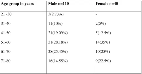
Age and sex distribution
Among these 150 patients there were 110 males (73.33%) and 40 females
(26.67%).
The age group distributions were shown in table 1. The age group ranges from 21
years to 80 years. The maximum numbers of cases were in the age group of 51-60
[image:55.612.69.555.460.715.2]years in both sexes.
Table 1
Age group in years Male n=110 Female n=40
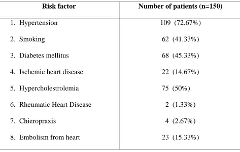
Risk factors
The possible risk factors were studied in all 150 patients, about 72.67% had
hypertension and around 41.33% gave history of chronic smoking. The risk
[image:56.612.70.543.260.559.2]factors identified is shown in Table 2.
Table 2
Risk factor Number of patients (n=150)
1. Hypertension
2. Smoking
3. Diabetes mellitus
4. Ischemic heart disease
5. Hypercholestrolemia
6. Rheumatic Heart Disease
7. Chieropraxis
8. Embolism from heart
109 (72.67%)
62 (41.33%)
68 (45.33%)
22 (14.67%)
75 (50%)
2 (1.33%)
4 (2.67%)
23 (15.33%)
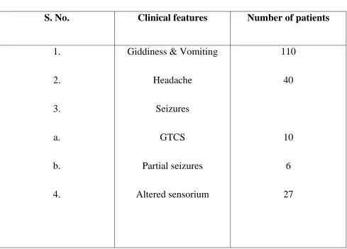
Clinical features
The clinical features at the onset of stroke were studied. Most of our patients
the onset, among which 10 had generalized tonic clonic seizures and 6 had partial
seizures with secondary generalization. The clinical features at the onset were
shown in table 3.
[image:57.612.68.557.205.556.2]Table 3
S. No. Clinical features Number of patients
1.
2.
3.
a.
b.
4.
Giddiness & Vomiting
Headache
Seizures
GTCS
Partial seizures
Altered sensorium
110
40
10
6
27
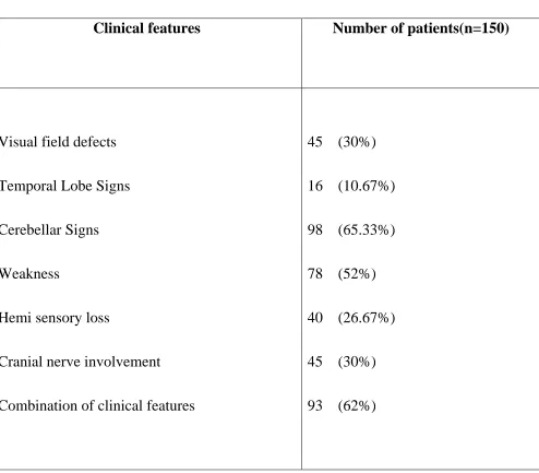
The other clinical features that were present in this study were according to the
territory involved. This included homonymous hemianopia, temporal lobe signs,
IV, V, VI, VII, IX and X). Most of these were combination of clinical features.
[image:58.612.74.568.172.605.2]This was shown in table 4.
Table 4
Clinical features Number of patients(n=150)
Visual field defects
Temporal Lobe Signs
Cerebellar Signs
Weakness
Hemi sensory loss
Cranial nerve involvement
Combination of clinical features
45 (30%)
16 (10.67%)
98 (65.33%)
78 (52%)
40 (26.67%)
45 (30%)
93 (62%)
Past H/O TIA’s were seen in only 42 patients, past H/O stroke is seen in 12
To describe the location of infarcts, we subdivided the posterior circulation in to
proximal, middle and distal intra cranial arteries accordingly described by NEMC
posterior circulation registry.
The clinical features and neuroimaging were taken together to describe the location
of infarct.
Neuro Imaging
CT scan brain plain was done in all patients. 30 patients had haemorrhages and 82
patients had infarcts in CT Brain. CT brain was normal in 38 patients in our study.
All the 38 patients had infarcts in MRI Brain.
MRI brain with MRA was done in 150 patients. 30 patients had haemorrhages and
120 patients had infarcts in MRI Brain.
In MRI Brain 52 patients had infarcts in thalamus, midbrain, temporal or occipital
lobes, 14 patients had infarcts in pons, 9 patients had infarcts in cerebellum or
medulla. 14 patients had infarcts in both temporal or occipital lobes and pons. 14
patients had infarcts in midbrain, thalamus and cerebellum. 5 patients had infarcts
in pons and cerebellum. 12 patients had infarcts in cerebellum, occipital lobes,
In MRI with MRA isolated basilar artery thrombosis was seen in 7 patients and
isolated posterior cerebral artery thrombosis was seen 15 patients. One patient had
vertebral artery dissection. 4 patients had thrombosis in vertebral artery. 2 patients
had thrombosis in vertebral, basilar and posterior cerebral arteries. 2 patients had
thrombosis in vertebral and basilar arteries. 3 patients had basilar and posterior
cerebral artery thrombosis. 5 patients had thrombosis in vertebral and posterior
cerebral arteries. One patient had isolated PICA thrombosis.
In MRI Brain 9 patients had pontine haemorrhages, 4 patients had occipital lobe
haemorrhages, 5 patients had thalamic haemorrhages, 3 patients had medullary
haemorrhages, 2 patients had cerebellar haemorrhages, and 2 patients had
haemorrhages in the midbrain. 2 patients had haemorrhages in the pons and
occipital lobe. 2 patients had haemorrhages in the pons and cerebellum. 1 patient
had haemorrhages in the medulla and cerebellum.
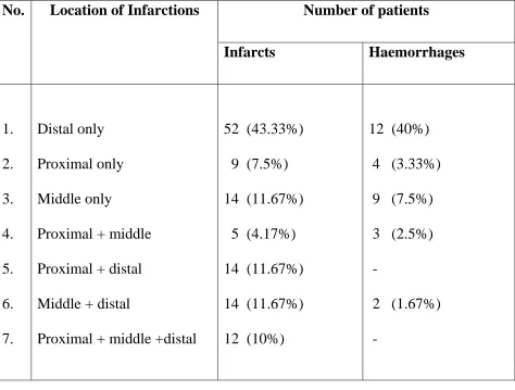
Among these 150 patients, we found that distal territory involvement was more
common. Isolated middle and proximal territory infarcts were less in this study. In
other patients we had varying combinations of proximal, middle and distal territory
infarcts.
Table 5
No. Location of Infarctions Number of patients
Infarcts Haemorrhages
1. 2. 3. 4. 5. 6. 7. Distal only Proximal only Middle only
Proximal + middle
Proximal + distal
Middle + distal
Proximal + middle +distal
52 (43.33%)
9 (7.5%)
14 (11.67%)
5 (4.17%)
14 (11.67%)
14 (11.67%)
12 (10%)
12 (40%)
4 (3.33%)
9 (7.5%)
3 (2.5%)
-
2 (1.67%)
-
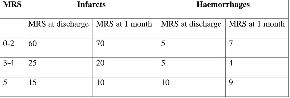
Prognosis was assessed using Modified Rankin Scale and Barthel Index. 20
patients with infarcts and 10 patients with haemorrhages died within one week.
Modified rankin scale and barthel index was assessed at discharge and at one
month. 60 patients with infarcts and 5 patients with haemorrhages were in MRS
0-2, 25 patients with infarcts and 5 patients with haemorrhages were in MRS 3-4, 15
patients with infarcts and 10 patients with haemorrhages were in MRS 5 at
[image:61.612.70.544.114.473.2]
Table 6
MRS Infarcts Haemorrhages
MRS at discharge MRS at 1 month MRS at discharge MRS at 1 month
0-2 60 70 5 7
3-4 25 20 5 4
[image:62.612.64.551.119.523.2]5 15 10 10 9
Table 7
BI Infarcts Haemorrhages
BI at discharge BI at 1 month BI at discharge BI at 1 month
0-50 40 35 10 7
55-90 45 50 8 11
95-100 15 15 2 2
40 patients with infarcts and 10 patients with haemorrhages were in BI 0-50 (full
dependency), 45 patients with infarcts and 8 patients with haemorrhages were in BI
55-90 (moderate dependency), 15 patients with infarcts and 2 patients with
0 5 10 15 20 25 30 35
2
21-30 331-40 4 26.
SEX
41-50 5
AGE DIST
.67%
X DISTRIBU
51-60 6
TRIBUTION
73.33%
UTION
1-70 7
N
1-80