A STUDY ON EVALUATION OF MOTOR, COGNITIVE
AND BEHAVIORAL MANIFESTATIONS OF
BASAL GANGLIA INFARCTS
Dissertation submitted to
THE TAMILNADU DR.M.G.R. MEDICAL UNIVERSITY
in partial fulfilment of the requirements for the award of the degree of DM (NEUROLOGY) – BRANCH – I
THE TAMILNADU DR.M.G.R MEDICAL UNIVERSITY CHENNAI
CERTIFICATE
This is to certify that the Dissertation entitled, “A STUDY ON EVALUATION OF MOTOR, COGNITIVE AND BEHAVIORAL MANIFESTATIONS OF BASAL GANGLIA
INFARCTS” is the bonafide record work done by
Dr.G.Gnanashanmugam, under our guidance and supervision in the Institute of Neurology, Rajiv Gandhi Government General Hospital, Madras Medical College, Chennai, submitted as partial fulfilment for the requirements of D.M. Degree examination Branch I NEUROLOGY, AUGUST 2011, under The Dr.M.G.R. Medical University, Chennai.
DR. R.M.BHOOPATHY. D.M.,
PROFESSOR OF NEUROLOGY, INSTITUTE OF NEUROLOGY, MADRAS MEDICAL COLLEGE, CHENNAI-3
DR. V.SUNDAR. Mch.,
PROFESSOR AND HEAD OF THE DEPARTMENT, INSTITUTE OF NEUROLOGY,
MADRAS MEDICAL COLLEGE, CHENNAI-3
Dr. V.KANAGASABAI,M.D.,
THE DEAN,
DECLARATION
I solemnly declare that this dissertation titled “A STUDY ON EVALUATION OF MOTOR, COGNITIVE AND BEHAVIORAL MANIFESTATIONS OF BASAL GANGLIA INFARCTS” is done by me in the Institute of Neurology, Madras Medical College & Rajiv Gandhi Government General Hospital, Chennai under the guidance and supervision of Prof. Dr. R.M. Bhoopathy, M.D., D.M., Professor of Neurology, Institute of Neurology, Madras Medical College & Rajiv Gandhi Government General Hospital, Chennai. This dissertation is submitted to the Tamil Nadu Dr.MGR Medical University, Chennai in partial fulfilment of the university requirements for the award of the degree of D.M.Neurology.
Place : Chennai Date : 09.06.2011
ACKNOWLEDGEMENT
It gives me great pleasure to acknowledge all those who guided, encouraged and supported me in the successful completion of my dissertation.
First and foremost, I express my gratitude to, the Dean Dr.V.Kanagasabai M.D. for having permitted me to carry out this dissertation work at Rajiv Gandhi Government General Hospital, Madras Medical College, Chennai.
I am extremely thankful to Prof. Dr. V.Sundar. M.ch., Professor of Neurosurgery, Head of the department, Institute of Neurology, Rajiv Gandhi Government General Hospital Chennai for his constant encouragement, valuable guidance and support.
I express my sincere thanks and gratitude to our Professors Dr.C.Mutharasu.D.M., Dr.K.Bhanu.D.M., Dr.R.Lakshmi Narasimhan. D.M., Dr. S. Balasubramanian. D.M., for their valuable suggestions and support
I express my sincere thanks to Prof. Dr. C. Rajendiran M.D., Director of Institute of General Medicine, Rajiv Gandhi Government General Hospital Chennai for allowing me to collect the case materials and for his guidance.
I am extremely thankful to our Assistant Professors Dr.V.Kamaraj.D.M., Dr.S.Arunan.D.M., Dr.M.Jawahar.D.M., Dr.P.Muthukumar.D.M., Dr. V.Kannan.D.M., for their valuable guidance and support.
CONTENTS
S. No. Title Page No.
1 INTRODUCTION 1
2 REVIEW OF LITERATURE 3
3 AIM AND OBJECTIVES 20
4 MATERIALS AND METHODS 21
5 RESULTS 27
6 DISCUSSION 45
8 CONCLUSION 66
9 REFERENCES
1
INTRODUCTION
Isolated basal ganglia infarcts are rare. Basal ganglia infarcts are often associated with infarcts in other structures like cerebral cortex, thalamus and fronto parietal white matter. Clinical consequences of basal ganglia infarcts are often masked by infarcts in other areas. When basal ganglia infarct extends into the adjacent internal capsule, more concern and priority will be given to the hemiplegia and associated behavioral and cognitive features may be overlooked.
Several studies have been conducted to analyze the clinical consequences of isolated basal ganglionic infarcts. Most of the studies were conducted in several countries outside India. So, we decided to analyze the clinical consequences of isolated basal ganglia infarcts in our patient population.
Normal functions of many neurological structures were identified by the consequences of destruction of these structures. The pathologies that damage the human brain are rarely restricted to single anatomical structures. Stroke, trauma and tumour do not respect functional anatomical boundaries.
2
anatomical pathology, and even more importantly do not show the distant functional effects (diaschisis) of such lesions.
Nevertheless, the consequences of lesions identified by CT or MRI give some clues as to function. The introduction of CT and MRI has produced many reports of clinicopathological correlation based upon single cases or small series of patients with lesions in particular brain structures.
3
REVIEW OF LITERATURE
Infarction limited only to the basal ganglia is rare. Several studies have been conducted so far to analyze the manifestations of basal ganglionic infarcts. In most of these studies, movement disorders were a selection criterion. But, in our study, case selection depended on the localization of infarction in the basal ganglia without involvement of other structures.
Understanding the organization and physiology of basal ganglia is essential to analyze the manifestations of basal ganglia infarcts.
ORGANIZATION AND CIRCUITS OF BASAL GANGLIA:
Basal ganglia include the corpus striatum, the substantia nigra (pars compacta and a pars reticularis), the subthalamic nucleus of Luys, and the ventral tegmental area. The corpus striatum comprises the striatum proper (or neostriatum), made up of the putamen, caudate nucleus, and nucleus accumbens, and the globus pallidus (or paleostriatum), with its medial or internal (Gpi) and lateral or external (Gpe) segments and the ventral pallidum, with its internal and external portions1.
4
substantia nigra pars reticularis. The main outputs from the medial globus pallidus and substantia nigra pars reticulata are to the thalamus, and thence to the premotor (e.g., supplementary motor area, anterior cingulate motor area, and lateral premotor cortex) and frontal lobe structures.
ANATOMY OF BASAL GANGLIA:
INPUTS INTO THE STRIATUM:
5
into the striatum. Most striatal efferents project to the globus pallidus. Other striatal efferents go to substantia nigra.
PALLIDAL AFFERENTS AND EFFERENTS:
The globus pallidus receives ascending afferent fibers from the substantia nigra and subthalamus (mainly to the medial or internal pallidum). Both the external and internal globus pallidus also receive afferents from the striatum.
The major outflow from the globus pallidus arises from the internal portion and projects to the ventral anterior (VA) and ventral lateral (VL) nuclei of the thalamus. These thalamic nuclei also receive afferents from the pars reticularis of the substantia nigra. Because the VL thalamic nucleus projects to the motor cortex and the VA thalamic nucleus projects to the premotor cortex, the major basal ganglia efferents influence the motor system.
6
NIGRAL AFFERENTS AND EFFERENTS:
The pars reticularis of the substantia nigra receives fibers from the cerebral cortex, the striatum, the globus pallidus, and the subthalamic nucleus. Pars reticularis efferents project to the VA and VL thalamic nuclei and to the reticular formation and superior colliculus. The pars compacta of the substantia nigra sends dopaminergic fibers to the caudate nucleus and putamen. This output is excitatory for the striatal neurons of the direct pathway and inhibitory to the striatal neurons of the indirect pathway.
It can thus be seen that the basal ganglia exert their influence mainly by way of the cerebral cortex (i.e., they do not send fibers that connect directly with brainstem and spinal cord structures). They provide a subcortical network by which the entire cerebral cortex can influence the motor system (motor and premotor cortex.
7
In the indirect system, the putamen and caudate receive inhibitory input from the pars compacta of the substantia nigra and project inhibitory fibers to the lateral globus pallidus which, in turn, inhibits (through GABA) the subthalamic nucleus. The subthalamic nucleus stimulates (through glutamate) the medial globus pallidus, inhibitory over the ventrolateral nucleus of the thalamus. Stimulation of this system inhibits the ventrolateral nucleus of the thalamus and results in cortical inhibition.
8
LESIONS OF BASAL GANGLIA:
Pathologic processes affecting the basal ganglia are often diffuse. When discrete, they usually also affect neighboring structures, such as the internal capsule, the hypothalamus, and the white matter of the cerebral hemispheres. Therefore, except for hemiballismus often associated with damage to the contralateral subthalamic nucleus, correlation between basal ganglia lesions and clinical motor dysfunction tends to be obscure.
The literature concerning behavioral effects of lesions of the basal ganglia in experimental animals is often conflicting, and these lesions rarely produce models of human movement disorders. In general, stimulation and destructive lesions of the caudate, putamen, and globus pallidus produce inhibition of movement or contralateral body turning.
Lesions of the subthalamic nucleus produce contralateral hemiballismus. Small unilateral lesions of the anteroventral portion of the caudate cause contralateral choreoathetosis. Unilateral lesions of the globus pallidus may cause contralateral hemidystonia, hemiparkinsonism, or tremor, whereas bilateral globus pallidus lesions may cause dystonia, parkinsonism, abulia, or akinesia.
9
falls are distinctly slow, tilting motions in a stereotyped lateral or diagonal trajectory and occur with the eyes open but are exacerbated by eye closure.
In a study2 of behavioral and movement disorders with lesions affecting the basal ganglia, lesions of the caudate nucleus rarely caused motor disorders (e.g., chorea or dystonia) but were more likely to cause behavioral problems, especially abulia (apathy with loss of initiative and of spontaneous thought and emotional responses) or disinhibition. Lesions of the putamen and globus pallidus rarely caused abulia and did not produce disinhibition but commonly caused dystonia, particularly when the putamen was involved.
Bilateral lesions of either the putamen or the globus pallidus caused parkinsonism or dystonia-parkinsonism infrequently. The prominence of behavioral disturbances with caudate lesions emphasizes the more complex cognitive role of this structure, whereas the frequent occurrence of dystonia and less commonly parkinsonism with lesions of the putamen and globus pallidus emphasizes the motor roles of these structures.
10
disturbances) resulting from subcortical lesions affecting the deep frontal and paraventricular white matter (subcortical aphasias).
Movement disorders can be defined as neurologic dysfunctions in which there is either an excess of movement (Abnormal Involuntary Movements, or AIMs; hyperkinesias; dyskinesias) or a paucity of voluntary and automatic movements (akinesia, bradykinesia, or hypokinesia) unassociated with weakness or spasticity. Paucity of movement characterizes the disorder known as parkinsonism.
STUDIES ON BASAL GANGLIA INFARCTS:
Khailash P Bhatia and C.David Marsden analyzed the behavior and movement disorders in basal ganglionic lesions2. The behavioral and movement disorders reported in 240 patients described in the literature with lesions affecting the caudate nucleus, putamen and the globus pallidus (lentiform nucleus) have been analyzed. Amongst the 240 cases, dystonia was the most frequent movement disorder recorded (36%); chorea (8%) and parkinsonism (6%) or dystonia-parkinsonism (3%) were uncommon.
11
lesions was the syndrome of abulia, sometimes alternating with disinhibition (11%).
Lesions of the lentiform nuclei rarely caused abulia (10%) and did not produce disinhibition, but they commonly caused dystonia (49%), particularly when the putamen was involved (63%). Bilateral lesions of the lentiform nuclei, either of the globus pallidus or of the putamen, caused parkinsonism (19%) or dystonia- parkinsonism (6%) infrequently.
The prominence of the behavioral disturbance of abulia with caudate lesions emphasizes the more complex cognitive role of this basal ganglia structure. The frequent occurrence of dystonia and less commonly of parkinsonism with lentiform lesions emphasize the motor roles of putamen and globus pallidus.
12
pallidus. (v) Bilateral lesions of the lentiform nucleus, usually involving the globus pallidus, infrequently cause parkinsonism or dystonia-parkinsonism.
Emre Kumral et al evaluated the clinical profile of acute caudate vascular lesions3. 31 patients were analyzed. Caudate infarct was present in 25 patients and caudate hemorrhage in 6. The most frequent neurological abnormalities were abulia and psychic akinesia (48%), frontal system abnormalities (26%), speech deficits in patients with left-sided lesions (23%), and neglect syndromes in those with right-sided lesions (10%). Patients with infarct in the territory of the lateral lenticulostriate arteries extending to neighboring structures showed more frequent motor and neuropsychological deficits than those with infarct in the territory of the anterior lenticulostriate arteries.
Heike Russman et al analyzed the clinical features and topographic correlation of acute lentiform infarct in 13 patients4. All had faciobrachiocrural hemisyndrome, while none showed acute or delayed parkinsonism or abnormal movement. Nine patients had a lesion restricted to the putamen. Two of them had ataxic motor hemisyndrome and 7 had sensorimotor hemisyndrome (with ataxia in 4, left hemineglect in 1, and deep pain in the arm and leg in 1).
13
the territory of the medial perforating branches of the medial cerebral artery. Presumed cause of stroke was small-artery disease in 5, artery-to-artery embolism in 4, cardioembolism in 3 and undetermined in 1. Movement disorders were not described.
M Giroud et al analyzed the clinical features5 of unilateral lenticular infarct in 20 patients. Two distinct clinical syndromes were identified corresponding to the two anatomical areas of the lenticular nucleus: behavioral and cognitive disorders were associated with infarcts within the globus pallidus, whereas both motor disorders (dystonia) and cognitive disorders were associated with infarcts within the putamen.
Outcome was excellent in all the patients for motor function, but slight cognitive disorders, problems with short term memory, and dysphasia persisted for several months. The size of the lesion did not explain these symptoms. The author proposed that slight reduction in cerebral blood flow found in the adjacent frontotemporal area6 may explain them by a deafferentation or a diaschisis phenomenon.
14
Other motor disorders, such as unilateral chorea, hemichorea-hemiballism, asterixis, acute stereotypies, acute focal dystonia8, and subacute parkinsonism, have also been reported after unilateral lesions of the lentiform nucleus but often also involve the caudate nucleus or the internal capsule. Lesions involving the globus pallidus may cause behavioral and speech disorders9, as well as motor disorders including delayed contralateral hemidystonia or subacute choreoathetosis. In most cases, there is a delay of months to years between the onset of the actual lesion10 and the development of the motor disorders.
Hemichorea-hemiballismus11 secondary to lacunar infarct in the basal ganglia region is rare. Goldblatt et al12described a patient with what was called hemichorea in whom lesions were found to be restricted to the contralateral head of the caudate nucleus and putamen at autopsy, and Kase et al13 described a 54-year-old man with hemichorea-hemiballismus in whom CT scan revealed a lacunar infarct in the contralateral putamen and caudate nucleus. It has therefore been suggested that the syndrome of hemichorea-hemiballismus should be included in the group of clinical syndromes that are most commonly caused by lacunar infarcts.
15
structures to the globus pallidus and substantia nigra14. On the other hand, ischaemia may have stimulated neurons that were left intact, activating dopamine production and release and thus reducing the inhibitory influences of the substantia nigra and globus pallidus on cortical motor function.
The pathologic process responsible for the dystonia during the initial recovery period is unknown. Some authors have compared the interval of delayed-onset dystonia in patients of perinatal anoxia and stroke. They emphasized the age of hypoxic injury in deciding the duration of delay in spite of the differences of etiology.
Delayed onset dystonia is a rare sequelae of stroke. The anatomical basis and pathogenesis of delayed-onset dystonia is uncertain. Mitchell suggested that the delay in the onset of hemichorea or athetosis following hemiplegia was caused by progressive changes in the original brain lesion. Burke (1980)15 had hypothesized that the mechanism of delayed-onset dystonia occurring a year or more after the insult may be due to aberrant neuronal sprouting.
16
However, the hypothesis of Pettrigrew has limitations. Some patients with immediate onset could not be explained with neuronal regeneration.
Young Chul Chai et al17 analyzed 34 patients with cerebro vascular disease in the literature with delayed onset dystonia. Dystonia following stroke almost always appeared within 1-12 months (mean 6.5 months). There were four patients with contralateral basal ganglia lesions, who had a short delay despite the early age of hypoxic insult.
One patient with dystonia following stroke in young age had a shorter interval compared to other elderly patients with dystonia following stroke. The pathological lesions in the patient of delayed-onset dystonia following stroke have a variety of anatomical lesion sites.
Of 34 patients reported in the literature, 21 (61.7%) had basal ganglia lesion, 13 (38%) internal capsule lesion, 11 (32%) thalamic lesion, 2 (5.8 %) cortical lesion and 2 (5.8%) without any lesions. Damage to the neuronal circuit connecting caudate, putamen, globus pallidus and thalamus seems to be responsible for the dystonia following stroke. However, it can occur without radiological evidence of striatal lesion. Thalamic degeneration following striatal lesion or cortical lesion has also been reported.
17
the anterior cerebral artery. In previous studies, there is no mention of arterial territories involved in patients with caudate infarcts. Actually, there is considerable overlap between the 3 arteries supplying the head of the caudate nucleus: the lateral lenticulostriate, anterior lenticulostriate, and Heubner’s recurrent artery.
Anterior lenticulostriate arteries primarily circumscribed the MCN, LCN, and VCN and partially involved the anterior limb of the capsule. Infarctions in the territory of the lateral lenticulostriate arteries were limited to the MCN, LCN, VCN, anterior part of the internal capsule, and putamen19. From a clinical standpoint, infarcts in the territory of the anterior lenticulostriate arteries yield only mild neuropsychological deficits, while those with infarcts in the territory of the lateral lenticulostriate arteries presented prominent motor and neuropsychological deficits.
The most prominent clinical features of basal ganglionic vascular lesions were behavioral and cognitive abnormalities, as in previous studies. Behavioral changes may have occurred as a result of loss of function in cortical zones, caused by loss of striatal efferent projections from the caudate nucleus. The caudate nucleus is the principal crossing area of basal ganglia–thalamo cortical loops.
18
anatomic structures by cortico-pallido-nigra-thalamocortical loops20. These loops are multiple, discrete, but partially overlapping and are integrated through their passage in pallidum and substantia nigra to the circumscribed nuclei of the thalamus, and from there they are projected back to their original lobar areas.
In a study by Emre Kumral et al on acute caudate vascular lesions in 31 patients, one half of the patients had abulia, characterized by decreased spontaneous activity and speech and prolonged latency in responding to questions and other stimuli. Three patients had psychic akinesia, characterized by severe mental and affective stagnation and lack of initiative for action and speech. Trillet et al observed psychic akinesia in 3 patients with apathy, flattened affect, lack of initiative for usual daily activities, stereotyped behaviors, and prolonged akinetic attacks. Moreover, these features were previously reported in patients with bilateral globus pallidus or putaminal lesions.
19
20
OBJECTIVES AND AIMS OF THE STUDY
1. To evaluate the motor features of basal ganglia infarcts such as the hyperkinetic and hypokinetic movement disorders.
2. To evaluate the cognitive impairment and it’s severity in basal ganglia infarcts.
21
MATERIALS AND METHODS
STUDY DESIGN AND PERIOD:
Our study was a prospective observational study, conducted in Government General Hospital, Chennai, from January 2009 to March 2011. Patients were selected from Neurology OP, emergency ward, general medical wards and Neurology ward. Patients were properly informed about the study and the Institutional Ethics Committee clearance was obtained. We studied all the patients with basal ganglia infarcts either acute or chronic with the following eligibility criteria. Patients were diagnosed with the aid of CT or MRI. Patients who were not affordable for MRI scan were subjected only to CT study of brain.
INCLUSION CRITERIA:
1. Patients who were admitted in Medical and Neurological wards for acute ischemic stroke with CT or MRI evidence of acute or chronic basal ganglia (putamen, caudate, globus pallidus or subthalamic nucleus) infarcts with or without infarcts in the internal capsule.
22
EXCLUSION CRITERIA:
1. Patients with CT or MRI evidence of infarcts in a site other than basal ganglia and internal capsule.
2. Patients with past history of intracerebral hemorrhage.
3. Past history of head injury or encephalitis.
4. Previous history of psychiatric, mood or personality disorder and longstanding antipsychotic drug intake.
5. Evidence of coexisting neurodegenerative dementia or epilepsy.
6. Presence of chronic medical illness like heart failure, chronic kidney disease, cirrhosis of liver and hypothyroidism.
7. Evidence of systemic connective tissue disorder like, SLE and Rheumatoid arthritis
23
CLINICAL ASSESSMENT:
All the patients selected for the study underwent detailed neurological examination including higher mental functions, motor, sensory, cranial nerve and cerebellar functions. Patients were carefully assessed for the presence of hypokinetic or hyperkinetic movement disorder. Rapid alternating movement of fingers, finger tapping, foot tapping and gait assessment was done to diagnose bradykinesia. Patients were carefully examined for the presence of rigidity, dystonia, choreo athetotic movements.
Detailed higher mental function assessment was done. Attention span, language function (fluency, repetition, comprehension, naming, reading, and writing) and memory function (immediate, recent and remote memory) were assessed.
Frontal lobar functions were assessed by trail making test, Luria’s motor series, rhythmic tapping test, stroop test, alternate sequence tasks, and assessment of judgment, abstract thinking and reasoning power.
24
Temporal lobar functions were assessed by detailed verbal, visual memory examination, paired associate learning and story recalling ability. Occipital lobar functions were assessed by analysis of gnosis for familiar objects and faces.
With detailed history taking, patients were probed for the presence of behavioral, mood and sleep disturbances. Standard rating scales and questionnaire were used to assess the mood and behavior disturbance. Beck Depression Inventory II and Hamilton Anxiety Scale were used to assess the depression and anxiety respectively. Probing questions were asked to ascertain whether the patient had behavioral disturbances like delusions, hallucinations, irritability, apathy and disinhibition. Patients were assessed for the presence of behavior disorder by Neuropsychiatric Inventory (NPI) and scoring was given.
25
INVESTIGATIONS:
All the patients underwent routine biochemical investigations like blood sugar, urea, creatinine, lipid profile, complete hemogram, liver function tests, blood VDRL, HIV serology. CT brain was performed for all the patients using horizontal orbitomeatal sequences with 0.5mm sections.
MRI brain was performed for the patients who can afford to pay the cost of MRI imaging. Those who cannot pay for the MRI imaging underwent only CT imaging of brain. MRI brain was performed on a 1.5 Tesla MRI machine with conventional sagittal T1, axial T2 and FLAIR sequences. TOF MR Angiogram was also performed during MRI procedure.
All the patients underwent four vessel Doppler studies. ECG, X-ray chest PA view and echocardiogram was done for all the patients to search for the cardiac source of embolism like mural thrombus, wall hypokinesia or akinesia, aneurysm and atrial fibrillation
26
Small artery disease was considered in the presence of only lacunar infarcts with diameter less than 1.5mm, hypertension, diabetes, absence of large vessel disease in Doppler study and absence of cardiac source of embolism by echocardiography.
Large artery disease was considered in the presence of stenosis of more than 50% of appropriate large artery on Doppler study and larger size of the infarct (>1.5cm).
27
RESULTS
51 patients fulfilled the eligible inclusion and exclusion criteria and were analyzed in this study.
SEX:
Among the 51 patients who were included in the study, 35 patients were males and 16 patients were females.
AGE:
The average age of disease presentation in this study was 54 years. The youngest patient was a 14 year old girl and eldest patient was 71 year old male. Most of the patients were in the range of 51 to 60 years. The following table shows the age distribution of the patients.
TABLE 1: AGE DISTRIBUTION OF PATIENTS:
AGE IN YEARS NUMBER OF
PATIENTS PERCENTAGE
61 – 70 8 15.%
51 – 60 26 51%
41 – 50 15 29.4%
31 – 40 1 2%
28
CEREBROVASCULAR RISK FACTORS:
Among 51 patients in this study, 36 patients were hypertensive, 23 patients were diabetics, 14 patients were hypertensive and diabetic, 10 patients had hyperlipidemia and 10 patients had past history of stroke. 30 patients were smokers and 24 were alcoholics.
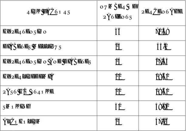
TABLE 2: CEREBROVASCULAR RISK FACTORS:
RISK FACTORS NUMBER OF
PATIENTS PERCENTAGE
HYPERTENSION 36 70.59
DIABETES MELLITUS 23 45.1
HYPERTENSION AND DIABETES 14 27.45
HYPERLIPEDEMIA 10 19.61
PAST H/O STROKE 10 19.61
SMOKING 30 58.82
ALCOHOLISM 24 47.06
SOURCE OF BASAL GANGLIA INFARCTS:
29
vessel occlusion in the ipsilateral carotid artery. The remaining 3 patients had cardiac source of embolism; 2 patients (48 and 40 years) suffered from rheumatic heart disease with severe mitral stenosis and atrial fibrillation; 1 patient (50 year old male, diabetic) suffered from ischemic heart disease with evidence of left ventricular mural thrombus in transthoracic echocardiogram.
30 patients had basal ganglionic infarcts due to presumed small vessel disease. These patients had lacunar infarcts in the unilateral or bilateral basal ganglia with infarct diameter less than 1.5cm and had normal carotid and vertebral Doppler study and echocardiogram. Of these 30 patients, 1 patient (42 year old male) had left basal ganglionic and capsular infarct due to small vessel vasculitis. He had evidence of systemic vasculitis in the form of bilateral retinal vasculitis, vasculitic leg ulcers, elevated ESR and young onset systemic hypertension.
30
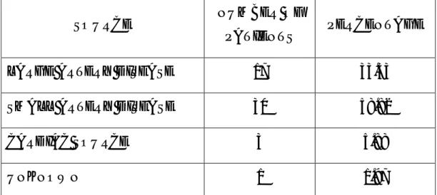
TABLE 3: SOURCE OF BASAL GANGLIA INFARCTS:
SOURCE NUMBER OF
PATIENTS PERCENTAGE
LARGE ARTERY DISEASE 17 33.33
SMALL ARTERY DISEASE 30 58.82
CARDIAC SOURCE 3 5.88
UNKNOWN 1 1.97
PATIENT CHARACTERISTICS:
After selection into the study, the 51 patients were analyzed with detailed clinical assessment and laboratory investigations including neuro imaging. They were categorized into 4 groups according to their presenting symptomatology.
GROUP I:
31
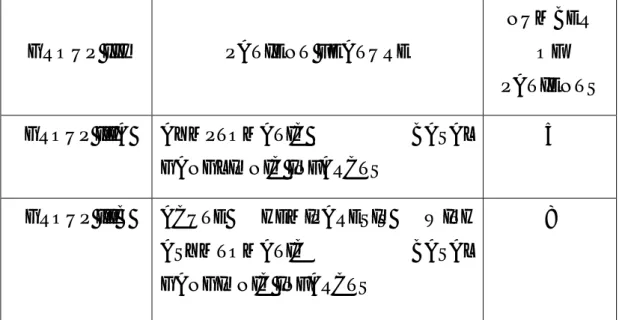
This group comprised of 18 patients. Of these 18 patients, 8 patients presented with hemiparesis; Among the 8 patients, 3 patients had motor aphasia, 2 patients had hemidystonia. These 8 patients in addition had signs of behavioral and cognitive impairment on examination. 7 patients presented with acute onset of hemichorea or choreoathetosis. 2 patients presented acutely with hemiballismus. 1 patient (60 year old male) presented with acute onset dementia, in the form of disinhibited behavior, memory impairment, visuospatial disorientation and emotional incontinence.
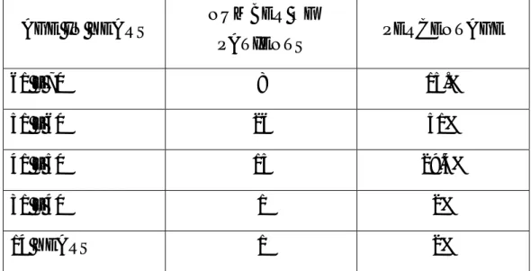
TABLE 4: GROUP I:
PRESENTING FEATURE NUMBER OF PATIENT
HEMIPARESIS ONLY 3
L HEMIPARESIS WITH APHASIA 3
HEMIPARESIS WITH DYSTONIA 2
HEMICHOREA 7
HEMIBALLISMUS 2
DEMENTIA 1
GROUP II:
32
detailed clinical assessment, all their symptoms and signs were referable to basal ganglia infarcts.
This group comprised of 14 patients. Of 14 patients, 7 patients had dementia as the major clinical feature. Of these 7 patients, 2 patients in addition had parkinsonian features. In 2 patients, bilaterally symmetrical parkinsonism was the dominant clinical feature. These 2 patients had also cognitive impairment. In 5 patients, dystonia was the major clinical feature. Of 5 patients, 1 patient had parkinsonian feature and 2 patients had cognitive impairment.
TABLE 5: GROUP II:
MAJOR FEATURE NUMBER OF PATIENTS
DEMENTIA 7
PARKINSONISM 2
DYSTONIA 5
GROUP III:
33
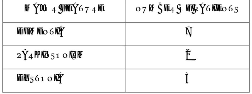
IIIA: Patients, who presented to the Neurology OPD for some other non specific complaints like headache and giddiness, underwent neuro imaging and imaging showed basal ganglia infarcts were included in this group. On detailed clinical assessment, they had no symptoms or signs referable to basal ganglia infarcts. This group consisted of 5 patients.
IIIB: Patients who were admitted with acute onset hemiparesis with imaging evidence of basal ganglionic and capsular infarcts were included in this group. They also had no symptoms or signs referable to basal ganglia infarcts. This group consisted of 8 patients.
TABLE 6: GROUP III:
GROUP III PATIENT FEATURE
NUMBER OF PATIENTS GROUP IIIA AYMPTOMATIC BASAL
GANGLIONIC INFARCTS
5
GROUP IIIB ACUTE HEMIPARESIS WITH ASYMTOMATIC BASAL GANGIONIC INFARCTS
34
GROUP IV:
Patients, who presented to the Neurology OPD with some other non specific symptoms, underwent neuro imaging and imaging showed basal ganglionic infarcts were included in this group. On detailed clinical assessment, they had some signs referable to basal ganglionic infarcts, either cognitive impairment or motor feature. They were not aware of their cognitive or motor impairment.
This group comprised of 6 patients. All these patients had evidence of cognitive impairment and mood disorder on detailed clinical assessment. 1patient (55 year old male) in addition had right hand dystonia.
CLINICAL FEATURES:
MOTOR FEATURES:
Of 51 patients, 22 patients had motor features related to basal ganglia infarct. Of these 22 patients, 7 patients had hemichorea, 2 patients had hemiballismus, 8 patients had dystonia, 2 patients had parkinsonism and 3 patients had parkinsonism with dystonia. All the 7 patients with chorea and 2 patients with hemiballismus presented acutely to the emergency ward (group 1). All 5 patients with parkinsonism presented non acutely to the Neurology OPD (group II) with bilaterally symmetric akinetic rigid syndrome.
35
hand dystonia along with hemiparesis and another patient had hemidystonia with hemiparesis. For 8 patients, dystonia confined predominantly to the unilateral hand; 2 patients had hemidystonia and 1 patient had blepharospasm. 2 patients developed dystonia in the limbs which suffered hemiparesis several years back; they had chronic infarct in the contralateral putamen, pallidum and capsule; they were diagnosed as post hemiplegic dystonia.
All patients with hemichorea had contralateral infarct in the caudate nucleus. 2 patients with hemiballismus had contralateral infarct in the sub thalamic nucleus. All 5 patients with parkinsonism had the evidence of bilateral basal ganglia infarcts; 2 patients had bilateral pallidal infarct and 3 patient had bilateral infarcts in the lentiform nucleus; among these 5 patients, 2 patients had bilateral caudate infarct and 2 patients had unilateral caudate infarct. Of 11 patients with dystonia, all had either unilateral or bilateral putaminal or pallidal infarct and 4 patients had caudate infarct.
TABLE 7: MOTOR FEATURES:
MOTOR FEATURES NUMBER OF
PATIENTS PERCENTAGE
HEMICHOREA 7 31.82
HEMIBALLISMUS 2 9.09
DYSTONIA 8 36.36
PARKINSONISM 2 9.09
36
COGNITIVE FEATURES:
Of 51 patients, 27 patients had evidence of cognitive impairment on detailed higher mental function examination. Decreased attention span, executive dysfunction and impaired verbal memory recall were the common cognitive problems in these patients. Some of the patients also had language disturbance, visuospatial disorientation and calculation difficulty.
Of 27 patients with cognitive impairment, 11 patients belonged to Group I (acute presentation). Among these 11 patients, cognitive impairment was the presenting feature in one patient; in all other patients motor impairment was the presenting feature and they had cognitive impairment on examination. 10 patients belonged to Group II (non acute presentation); and 6 patients belonged to Group IV.
Decreased attention span was the most common abnormality noted; 24 patients had evidence of reduced attention span on digit repetition test, go no go test, letter cancellation test and trail making test. 17 patients had evidence of executive dysfunction on alternate sequence tasks, trail making, stroop test, Luria’s motor series and rhythmic tapping test. 19 patients had memory impairment on verbal memory recall (improving with cues), paired associate learning, story recall and tests of orientation to time and place.
37
aphasia with reduced word fluency, word finding difficulty, grammatical errors, impaired repetition, naming, reading, writing and preserved comprehension. These 3 patients had acute large infarct involving left caudate, putamen, pallidum and capsule.
10 patients had difficulty in performing complex calculation (oral and wrote); 2 patients had visuospatial disorientation with way finding difficulty, difficulty in identifying cities in the map and defective visuospatial tasks on Addenbrooke’s cognitive assessment; 4 patients had constructional disability in copying diagrams.
Cognitive functions were scored with Mini Mental Scale Examination (MMSE) and Addenbrooke’s cognitive scoring. Of 27 patients with cognitive impairment, MMSE scale was in the range of 16 to 27 and Addenbrooke’s cognitive scoring was in the range of 61 to 97.
38
TABLE 8: COGNITIVE FEATURES:
COGNITIVE IMPAIRMENT NUMBER OF
PATIENTS PERCENTAGE
POOR ATTENTION SPAN 24 47.06
EXECUTIVE DYSFUNCTION 17 33.33
MEMORY IMPAIRMENT 19 37.25
APHASIA 3 5.8
CALCULATION ERRORS 10 19.61
VISUOSPATIAL DISORIENTATION
2 3.92
CONSTRUCTIONAL DISABILITY
4 7.84
BEHAVIORAL FEATURES:
39
patients had suicidal ideas. Of 27 patients, 4 patients had severe depression with score more than 29. 9 patients had moderate depression with score 20 to 29. 8 patients had mild depression with score between14 to 20. 6 patients had mild depression with score less than 14.
8 patients had features of anxiety as assessed by Hamilton’s Anxiety Scale. 7 patients had disinhibited behavior in the form of unconcerned micturition, undressing in front of others and abusing bad words. 4 patients had delusions and 4 patients had formed visual hallucinations. 22 patients had evidence of sleep disturbance. The most common sleep disturbance was insomnia with delayed sleep onset. 11 patients had irritability and recurrent outburst of anger.
40
TABLE 9: BEHAVIORAL FEATURES:
BEHAVIORAL ABNORMALITY
NUMBER OF
PATIENTS PERCENTAGE
APATHY 25 49.02
DEPRESSION 27 52.94
ANXIETY 8 15.69
DISINHIBITION 7 13.73
DELUSION 4 7.84
HALLUCINATION 4 7.84
INSOMNIA 22 43.14
IRRITABILITY 11 21.57
INFARCT CHARACTERISTICS:
41
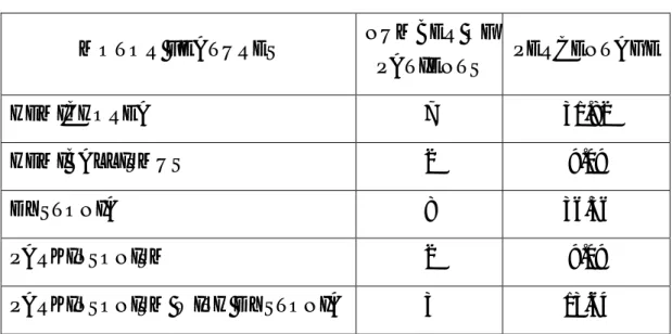
Of 51 patients, 25 patients had infarcts confining only to the caudate nucleus (either unilateral or bilateral). 13 patients had isolated lentiform infarcts (either unilateral or bilateral putamen or pallidum). 11 patients had infarcts in both caudate and lentiform nucleus. 2 patients had infarct in the unilateral subthalamic nucleus.
Of 51 patients, 16 patients had larger size of the infarct occupying more than one basal ganglionic structure and internal capsule with infarct size greater than 1.5cm. 35 patients had smaller size of the infarcts with infarct diameter less than 1.5cm.
TABLE 10: INFARCT CHARACTERISTICS:
INFARCT LOCATION NUMBER OF
PATIENTS PERCENTAGE
UNILATERAL CAUDATE 25 49.02
BILATERAL CAUDATE 11 21.57
UNILATERAL PUTAMEN 16 31.37
BILATERAL PUTAMEN 1 1.96
UNILAT PALLIDUM 20 39.22
BILATERAL PALLIDUM 5 9.80
SUBTHALAMIC NUCLEUS 2 3.92
42
TABLE 11: DISTRIBUTION OF INFARCTS:
INFARCTS NUMBER OF
PATIENTS PERCENTAGE
ISOLATED LENTIFORM INFARCTS
13 25.49
ISOLATED CAUDATE INFARCT 25 49.02 CAUDATE AND LENTIFORM
INFARCTS
11 21.57
SUBTHALAMIC NUCLEI INFARCT
2 3.92
CORRELATION OF IMAGING AND CLINICAL FEATURES:
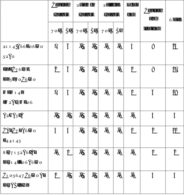
TABLE 11: MOTOR DISORDERS PRODUCED BY BASAL GANGLIONIC INFARCTS: Caudate Infarct Putamen Infarct Pallidal infarct Lenti form Caudate And Lenti form Sub Thal amic Total UNI BI UNI BI UNI BI
Hemichorea 6 -- -- -- -- -- -- 1 -- 7
Hemiballismus -- -- -- -- -- -- -- -- 2 2
Dystonia -- -- -- -- -- -- 6 2 -- 8
Parkinsonism -- -- -- -- -- -- -- 2 -- 2
Dystonia with parkinsonism
43
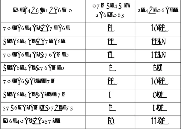
TABLE 12: COGNITIVE IMPAIRMENT PRODUCED BY BASAL GANGLIA INFARCTS: Caudate Infarct Putamen Infarct Pallidal Infarct Lenti form Caudate and lentiform Total UNI BI UNI BI UNI BI
POOR ATTENTION SPAN
7 6 -- -- -- -- 3 8 24
EXECUTIVE DYSFUNCTION
2 6 -- -- -- -- 1 8 17
MEMORY IMPAIRMENT
7 5 -- -- -- -- 1 6 19
APHASIA -- -- -- -- -- -- -- 3 3
CALCULATION ERRORS
3 4 -- -- -- -- 1 2 10
VISUOSPATIAL DISORIENTATION
-- 1 -- -- -- -- -- 1 2
CONSTRUCTIONAL DISABILITY
44
TABLE 13: BEHAVIORAL DISORDER PRODUCED BY BASAL GANGLIA INFARCTS:
Caudate
Infarct
Putamen
Infarct
Pallidal
Infarct Lenti
Form
Caudate
And
Lenti-form
Sub
Thal-amic
Total
UNI BI UNI BI UNI BI
APATHY 6 7 -- -- -- -- 3 9 -- 25
DEPRESSION 7 7 -- -- -- -- 2 10 1 27
ANXIETY 1 1 -- -- -- -- 2 4 -- 8
DISINHIBITION -- 4 -- -- -- -- -- 3 -- 7
DELUSION -- 2 -- -- -- -- -- 2 -- 4
HALLUCINATION -- 2 -- -- -- -- -- 2 -- 4
INSOMNIA 6 6 -- -- -- -- 3 6 1 22
15.00%
51%
29.40%
2% 2%
0.00% 10.00% 20.00% 30.00% 40.00% 50.00% 60.00%
61 – 70 51 – 60 41 – 50 31 – 40 14 YEARS
P
e
rcentage
AGE DISTRIBUTION OF PATIENTS
33%
59% 6%
2%
SOURCE OF BASAL GANGLIA INFARCTS
LARGE ARTERY DISEASE SMALL ARTERY DISEASE
0 5 10 15 20 25 30 35 40
0 10 20 30 40 50 60 APA T HY D EPR ESS ION A NXI ET Y D IS IN H IB IT IO N DELU S ION H AL L U CI NA TI ON IN SO MN IA IR R IT AB ILIT Y 49.02 52.94 15.69 13.73 7.84 7.84 43.14 21.57 Percentag e BEHAVIORAL FEATURES 25% 49% 22% 4%
DISTRIBUTION OF INFARCTS
Fig. 1
Fig . 2
CASE 16 : INFARCT IN LEFT CAUDATE AND LENTIFORM NUCLEUS IN A PATIENT WITH R HEMIPARESIS AND MOTOR
APHASIA
CASE 1: BILATERAL CAUDATE AND PALLIDAL INFARCT
Fig.3
Fig. 4
CASE 5: ACUTE INFARCT LEFT PALLIDUM AND PUTAMEN WITH ACUTE RIGHT HEMIPARESIS
45
DISCUSSION
Pure basal ganglionic infarcts are rare. Often the acute ischemic infarct extends into the adjacent internal capsule or white matter structures, resulting in neurological deficit referable to the internal capsule. The profound hemiparesis and dysarthria often dominate the clinical picture and mask the neurological impairment produced by basal ganglia infarcts. The cognitive and behavioral abnormalities of basal ganglia infarcts are often overlooked and may disable the activities of daily living. In our study, we analyzed the motor, cognitive and behavioral abnormalities of basal ganglia infarcts.
MOTOR FEATURES:
In our study, among 51 patients with basal ganglionic infarcts, 28 patients had infarcts extending to the adjacent internal capsule (acute and chronic). 18 patients had hemiparesis on clinical examination either as an acute presenting manifestation or as a longstanding residual deficit due to past stroke. As hemiparesis is not directly related to the basal ganglia lesion, we analyzed motor features other than hemiparesis to assess the impact of basal ganglia infarct.
DYSTONIA:
46
involving one hand, 2 patients had hemidystonia and 1 patient had blepharospasm. 2 patients had acute dystonia. 2 patients had delayed dystonia several years after the insult. Hemidystonia was rare in our study, only 2 patients had hemidystonia. Generalized dystonia was not observed in our patients, although several patients had bilateral caudate or lentiform infarcts.
Among 11 patients with dystonia, 7 patients (63.6%) had isolated lentiform nucleus infarct with; 6 patients had unilateral and 1 patient had bilateral lentiform infarct. 4 patients had combined caudate and lentiform infarcts. No patients with isolated caudate infarct developed dystonia. Infarcts in the lentiform nucleus were more frequently associated with dystonia.
Though exact mechanism of dystonia is little known, it has been proved in various studies on basal ganglionic lesions that most lesions causing dystonia were in the lentiform nucleus, particularly the putamen24. It seems that crude destruction of the putamen is more likely to cause dystonia than any other form of movement disorder. Such crude putaminal lesions might be expected to destroy both the direct striato-pallidal (nigral) pathway supporting cortically generated movement, and the indirect pathway via lateral globus pallidus and subthalamus which eventually inhibits thalamo cortical excitation of premotor cortical areas25.
47
disorder observed in basal ganglionic lesions. M.Giroud et al, in his study on unilateral lentiform infarcts, reported that dystonia occurred in 63% of the putaminal lesion and in 37% of the globus pallidus lesions. In this study, most lesions inducing dystonia were in the lentiform nucleus, mostly when putamen was involved. In our study, patients with dystonia had combined involvement of both putamen and globus pallidus.
2 patients in our study developed dystonia several years after the initial neurological insult. These 2 patients had chronic infarct in the contralateral lentiform nucleus and internal capsule. They had past history of hemiparesis several years (10 and 7 years) ago. Suggested pathophysiological mechanisms for delayed dystonia are aberrant neuronal sprouting, ephaptic transmission after the insult, remyelination and late inflammatory changes26.
CHOREA:
Next to dystonia, chorea was the common movement disorder. 7 patients had chorea in this study (13.7%). All patients had chorea involving one side limbs (hemichorea). 6 patients had isolated infarct involving the contralateral caudate nucleus. 1 patient had large infarct involving contralateral caudate, lentiform nucleus and internal capsule and also had mild hemiparesis in addition to hemichorea.
48
only 18 of the 240 patients (8%); most patients with chorea (15 of 18; 83%) had involvement of the caudate nucleus. Only three of 68 (4%) lesions affecting the lentiform nucleus, but sparing caudate, were associated with chorea.
Striatal projections to the globus pallidum externum are probably more severely involved in patients with chorea and basal ganglionic lesion. Injection of GABA antagonists into the lateral pallidum, which block striatal inhibition of the lateral globus pallidus, can provoke contralateral hemichorea in monkeys (Crossman et al, 1988)27. 2-Deoxyglucose activity was greatly increased in the subthalamus indicating increased lateral pallidal inhibition of this nucleus (Mitchell et al., 1985)28. This was associated with 2-DG evidence of decreased subthalamomedial pallidal excitation and reduced pallido-thalamic inhibition29.
Hemichorea or hemiballism can be produced reliably by lesions of the subthalamus or subthalamo-pallidal pathways in primates and man30 (Martin, 1957; Whittier and Mettler,
49
PARKINSONISM:
In our study, 5 patients had parkinsonism (9.8%). All these 5 patients had bilaterally symmetric parkinsonism with akinetic rigid phenotype. All of them belonged to group II (chronic presentation). No patients in the acute presentation group (group I) had parkinsonism. No patients had hemi parkinsonism. In these 5 patients, bradykinesia involved the upper and lower limbs equally without any preferential distribution to the lower limbs.
Interestingly, 1 patient (case 52) had akinetic rigid syndrome, eyelid stare, vertical gaze paresis and frequent backward falls; the initial working diagnosis was progressive supranuclear palsy. But, subsequently MRI brain revealed right caudate and bilateral pallidal infarct. His parkinsonian features improved well with levo dopa.
All the 5 patients had bilateral basal ganglionic infarcts; 4 patients had bilateral pallidal and caudate infarcts and 1 patient had bilateral putaminal and unilateral pallidal infarct.
50
Our findings are comparable with the Meta analysis of Khailash Bhatia and Marsden. In their analysis, parkinsonism also was relatively uncommon, being seen in only 21 of the 240 patients (9%). In 14 cases, the parkinsonism was relatively pure, but seven others had additional dystonic features. Nineteen of these 21 parkinsonian patients had lesions involving the lentiform nuclei.
Striatal lesions causing loss of both its excitatory effect on the direct striato pallidal pathway, and its inhibitory effect on the indirect pathway, thus withdrawing striato pallido thalamo cortical support of cortically generated movement, could explain the parkinsonism33. Lesions of the human globus pallidus cause parkinsonism infrequently, and have to be bilateral to do so34. In our study, 2 patients with parkinsonism had bilateral pallidal infarct without involvement of putamen, but they had associated caudate infarct.
BEHAVIORAL FEATURES:
51
APATHY:
Apathy was the most common behavioral abnormality noticed. 25 patients (49%) in this study had apathy with lack of mental and motor drive. The severity of apathy was assessed with NPI (Neuropsychiatric Inventory). Apathy was seen both in acute and chronic basal ganglionic infarcts. Most of these patients had infarcts in the caudate nucleus. 6 patients had unilateral and 7 patients had bilateral caudate infarcts. 9 patients had combined caudate and lentiform infarct; 3 patients had isolated lentiform infarcts.
Most of the patients (88%) with apathy had caudate infarcts. Apathy (12%) was rare in isolated lentiform infarcts. In Meta analysis by Khailash Bhatia, the commonest behavioral disorder was abulia, which manifested in 30 of the 240 (13%) cases. In 23 of these 30 (77%) patients with abulia, the lesion involved the caudate nucleus.
The loss of mental and motor initiative and drive that characterizes abulia, causing apathy and blunting of responses, does seem to be most commonly due to damage to the caudate and sometimes to the globus pallidus, which may normally be involved in these functions35.
52
had bilateral caudate with lentiform infarct and one had unilateral caudate with lentiform infarct. In contrast to apathy which occurred in both unilateral and bilateral caudate lesions, disinhibited behavior was rare in unilateral caudate lesions and occurred predominantly in bilateral caudate lesions.
Abulia, and less frequently disinhibition, does seem to be a significant consequence of caudate damage. The massive projection from prefrontal cerebral cortex to the head of the caudate nucleus and the reciprocal striato-pallido thalamo- cortical projections back to prefrontal cortex via the 'complex' basal ganglia circuits (Alexanderet al., 1986, 1990)36 emphasize the strength of prefrontal-caudate systems. Indeed abulia and disinhibition are recognized clinical consequences of damage to prefrontal cortex in man. Both behavioral syndromes can therefore be produced by damage either to prefrontal cortex or to the caudate nucleus37.
Apathy is considered to be due to damage to the anterior cingulate frontal basal ganglionic circuit38. The anterior cingulate gyrus projects to the ventral striatum, thence to ventral and rostrolateral globus pallidus and rostrodorsal substantia nigra, the paramedian medial dorsal thalamic nucleus, which projects back to the anterior cingulate cortex.
53
rostromedial substantia nigra, medial ventral anterior and dorsal thalamic nuclei, which project back to the orbito-frontal cortex40.
Depression was observed in 27 patients (53%). But, only 12 patients had severe depression on Beck’s Depression Inventory. Most of these patients had sadness, pessimistic attitude, loss of pleasure, self dislike, loss of interest in household and occupational activities, worthless feeling and crying. No patients had suicidal ideas. Of these 27 patients, no patients had volunteered their depressive symptoms. All these patients presented to the hospital only because of their motor disability or other behavioral impairment.
14 patients with depression had isolated caudate infarcts, 7 had unilateral and 7 had bilateral caudate infarct. 10 patients had caudate and lentiform infarct. Only 2 patients with isolated lentiform infarct had depression.
54
4 patients (7.8%) had delusion and 4 patients (7.8%) had hallucination. In all 4 patients, the delusion was in the form of suspecting the life partner whether she had illegal contact with some other person. 4 patients had formed visual hallucination in the form of viewing close relatives or grandparents though nobody actually existed. No patients had auditory hallucinations. All these patients with delusion and hallucination had severe dementia with cognitive impairment.
All 4 patients with delusion had bilateral caudate infarct, 2 patients in addition had lentiform infarct. Among 4 patients with visual hallucinations, 2 patients had isolated caudate infarct and 2 patients had caudate and lentiform infarct.
McMurtray et al43 in his study on patients with lacunar infarction in the caudate nucleus, found that patients with caudate infarcts and delusions had greater frontal-executive impairments on neuropsychological measures and decreased metabolism of the inferior prefrontal cortex on PET imaging. These findings support the hypothesis that development of delusions was consequent to disruption of inferior prefrontal lobe functions, which may include alterations in reality monitoring and testing.
55
the patients with behavioral and mood disorder had caudate infarcts. Isolated lentiform infarcts rarely produced behavioral or mood disorder. This shows the importance of frontal basal ganglionic circuitry in producing behavioral abnormality.
This study shows that behavioral and mood disorders are very common in patients with basal ganglionic infarcts. All the patients with basal ganglionic infarcts should be thoroughly explored for the presence of behavioral disorder. Identification and appropriate treatment of behavioral disorder44 and depression in these patients will reduce the morbidity and will definitely improve the quality of life.
COGNITIVE FEATURES:
Of 27 patients with cognitive impairment, only 10 patients had cognitive impairment as their presenting feature; remaining patients presented with motor features or some other non specific complaints, but cognitive impairment was identified on detailed examination. So, cognitive impairment in basal ganglionic infarct is more than what we see, again like the tip of iceberg (like behavioral abnormality).
POOR ATTENTION SPAN:
56
attentional tasks like digit repetition test, go no go test, serial subtraction test, letter cancellation test and trail making test.
13 patients had isolated caudate infarcts, 7 had unilateral and 6 had bilateral infarcts. 8 patients had combined caudate and lentiform infarcts. Only 3 patients had isolated lentiform infarcts.
Though poor attention span has no specific localizing value, brain regions involved in attentional mechanisms can be separated into two categories: activated sensory areas and brain structures that activate them. The brain regions activated are inferior parietal lobule, premotor areas, cingulate gyrus and posterior temporo occipital areas. Activation of these cortical areas requires input from the basal ganglia through the thalamus. Several studies have shown that basal ganglionic lesions can cause poor attention span45.
EXECUTIVE DYSFUNCTION:
\17 patients (33.33%) had executive dysfunction as evidenced by alternate sequence tasks, trail making, stroop test, Luria’s motor series and rhythmic tapping test. Of these, 8 patients had isolated caudate infarcts, 2 with unilateral and 6 with bilateral infarcts. 8 patients had caudate and lentiform infarcts. 1 patient alone had isolated lentiform infarct.
57
The dorsolateral prefrontal syndrome is characterized by defects of executive function and motor programming. Such patients are unable to generate hypotheses and cannot maintain or shift set. They also exhibit reduced verbal and design fluency, with defective strategic learning and motor execution of alternating or sequential tasks47. The dorsolateral prefrontal cortex projects primarily to the dorsolateral head of caudate nucleus, thence to dorsomedial globus pallidus and rostral substantia nigra, ventral anterior and medial dorsal thalamic nuclei, which project back to the dorsolateral prefrontal region48.
MEMORY IMPAIRMENT:
19 (38%) patients had evidence of memory impairment and defective new learning tasks. Of these, 12 patients had isolated caudate infarcts, 7 with unilateral and 5 with bilateral infarcts. 6 patients had caudate and lentiform infarcts. Only 1 patient had isolated lentiform infarct. These patients had impairment on tasks like verbal memory recall, paired associate learning and word category fluency tasks.
58
The caudate has been hypothesized to play a distinct role in memory. In monkeys, for example, stimulation of the caudate has been shown to impair memory50. Mendez et al reported on 11 patients with unilateral or bilateral caudate infarction and 1 patient with a healed abscess who came to medical attention because of an acute behavioral disturbance51. Among these patients, 1 patient with bilateral lesions was judged to have a "global dementia" with an MMSE score of 18. Moreover, among 7 patients with unilateral lesions, there were significant impairments in tasks requiring planning and sequencing compared with age-matched control subjects. They also had short attention spans and decreased free recall of episodic and semantic items.
59
Pozzilli et al54 suggested that the deficit in verbal comprehension and verbal memory may have been due to dysfunction of the cortico caudate connections. On the basis of these prior studies, the cognitive impairment in our patients might be assumed to indicate that caudate lesions lead to chronic deficits in frontal lobe function. This chronic frontal lobe dysfunction gradually leads to dysfunction in other connected cortical regions, and this is manifested by cognitive decline. Other explanations such as chronic dysfunction of the caudate might also be proposed as an explanation for these findings.
Emre Kumra et al3 studied the behavioral features in acute caudate stroke. Among 31 patients with acute caudate infarct, 3 patients with unilateral caudate infarct had verbal memory impairment on Rey auditory verbal learning test and 3 patients had combined verbal and visual memory impairment. This may be due to disconnection of caudate nucleus from frontal lobe.
APHASIA:
3 patients had language disturbance in the form of motor aphasia. All 3 patients presented acutely with large infarct involving left caudate, lentiform nucleus and internal capsule. These patients had hesitant and interrupted speech with word finding difficulties, with impaired repetition, reading, writing, naming and intact comprehension. They also had literal paraphasia.
60
transcortical motor aphasia and 1 patient had global aphasia; 5 patients had left caudate infarct and 1 patient with non fluent aphasia had bilateral caudate infarct.
It is well known that different types of aphasia, such as transcortical, nonfluent aphasia, characterized by semantic and verbal paraphasias and perseverations without comprehension impairment, occur in patients with left caudate or anterior lentiform vascular lesions55. Alexander et al observed that patients with subcortical lesions involving the caudate nucleus, anterior limb of the internal capsule, and putamen had word-finding difficulty or hesitancy without severe aphasic abnormalities. It is probable that acute disconnection of linguistic pathways between anterior and posterior speech areas, which are connected with the left caudate nucleus and anterior limb of the internal capsule, may yield a different type of aphasia56.
Though transcortical aphasia has been described frequently in basal ganglionic lesions, in our study no patient had transcortical aphasia; all had non fluent aphasia (Broca’s aphasia).In most of the patients with aphasia due to dominant striatocapsular infarct, the infarct also extends into the adjacent white matter of insula and temporal lobe57.
61
without any impairment of comprehension, and preserved repetition in five right handed patients with left putaminal infarction.
VISUOSPATIAL DISORIENTATION AND CONSTRUCTIONAL DISABILITY:
In our study, 2 patients had visuospatial disorientation and 4 patients had constructional disability. Of 2 patients with visuospatial disorientation, 1 patient had isolated bilateral caudate infarct and 1 patient had caudate and lentiform infarct. Of 4 patients with constructional disability, 1 patient had isolated caudate infarct and 3 had caudate and lentiform infarct.
62
CALCULATION DIFFICULTY:
10 patients in this study had calculation difficulty. Of these, 7 had isolated caudate infarct; 4 had bilateral caudate infarct and 3 had left caudate infarct. 1 patient had isolated lentiform infarct and 2 patients had combined caudate and lentiform infarct.
In a PET study by Stanislas et al60, multiplication activities yielded superior activity in the left lentiform nucleus, left inferior parietal gyrus and bilateral fusiform and lingual gyri. In his study, he concluded that left basal ganglia would be involved in storing and retrieving rote verbal multiplication facts.
63
calculations and that procedural knowledge relies on a visuo-spatial sketchpad that contains a representation of each sub-step of the procedure.
ASYMPTOMATIC PATIENTS WITH BASAL GANGLIONIC INFARCTS:
Of 51 patients, 13 patients had asymptomatic basal ganglionic infarcts. They had no symptoms or signs referable to basal ganglia. Of 13 patients, 5 patients presented to the Neurology OPD with non specific complaints like head ache or giddiness. CT brain showed basal ganglionic infarcts. 8 patients presented to the Emergency ward with acute hemiparesis. They had acute infarct in the internal capsule and basal ganglia. But, they had no signs or symptoms referable to basal ganglia. All these patients had at least one of the cerebro vascular risk factors like hypertension, diabetes mellitus, hyperlipidemia or cigarette smoking.
64
Though small vessel disease is the common risk factor for asymptomatic basal ganglionic infarcts, we observed large vessel disease in up to 50% of patients as the cause of silent infarct. Toshiyuki et al62 analyzed the risk factors in silent cerebral infarcts in 219 patients. In his study MRA showed cerebral arterial stenosis ipsilateral to the side of the infarct in 21% of patients.
Carotid artery stenosis63 was a significant and independent predictor of SCIs in the BG. This finding was consistent with the findings of previous reports. In studies of symptomatic lacunar infarction64, it has been pointed out that ipsilateral carotid stenotic lesions are potential embolic sources associated with lacunar infarction in the territory of deep perforating arteries. Ghika et al65 reported that 28 of 100 patients with symptomatic lacunar infarction in the territory of the deep perforators of the carotid system had ipsilateral carotid artery stenosis. Stenotic lesions of the ICA may also play a role in the pathogenesis of lacunes through hemodynamic effects. In an animal model study, diffuse cerebral ischemia from carotid occlusion caused infarction only in the striatum, and a possible toxic effect of dopamine release in the ischemic zone has been assumed to be related to the damage.
65
in the 147 patients. This community-based study shows that silent infarction in stroke patients is more related to certain stroke risk factors than others and that silent infarction does not seem to influence the prognosis of stroke.
LIMITATIONS OF THE STUDY:
1. Proper follow up for at least 6 months was available only for 23 patients. The remaining patients could not be followed up subsequently. So, We could not ascertain that how many percentage of patients with motor, behavior or cognitive impairment had subsequent improvement and how many were static. So, prognosis was not accurately analyzed in this study.
2. MRI brain with MRA was done only for 18 patients. Remaining patients underwent only CT imaging of the brain. Patients with only CT brain could not be analyzed accurately for the source of infarct. Though extracranial stenosis was diagnosed with Doppler study in patients with large vessel disease, the possibility of intracranial stenosis could not be ruled out in patients with assumed small vessel disease.
66
CONCLUSION
Motor, cognitive and behavioral consequences of basal ganglia infarcts are more common than expected. The current concepts of basal ganglia organization and physiology do not fully explain the disorders observed in man when the striatum and globus pallidus are damaged by crude pathology like infarction.
In our study, dystonia was the most common movement disorder observed in patients with basal ganglionic infarcts, followed by chorea. Mild to moderate depression and apathy is very common in basal ganglionic infarcts; it should be identified and treated effectively to improve the quality of life. Reduced attention span, impaired verbal memory recall and executive dysfunction was the common cognitive impairments identifies which signifies the important role of frontal basal ganglionic subcortical circuitry in higher cognition like executive function and basic cognition like attention and memory.
67
BIBLIOGRAPHY
1. Brazis Paul W, Masdeu Jose C, Biller Jose. Localization in Clinical
Neurology, 5th edition, 2007; 421-425.
2. Kailash P. Bhatia, C. David Marsden. The behavioural and motor
consequences of focal basal ganglia lesions in man. Brain 1994; 117:
859-876.
3. Emre Kumral, Dilek Evyapan, Kaan Balkir. Acute Caudate Vascular
Lesions. Stroke 1999; 30:100-108.
4. Heike Russmann, Francois Vingerhoets, Joseph Ghika, Philippe
Maeder, Julien Bogousslavsky. Acute infarction limited to lentiform
nucleus. Arch Neurol. 2003;60:351-355.
5. M Giroud, M Lemesle, G Madinier, Th Billiar, R Dumas. Unilateral
lenticular infarcts: radiological and clinical syndromes, aetiology and
prognosis. Journal of Neurology, Neurosurgery, and Psychiatry 1997;
63:611–615.
6. Laplane D, Levasseur M, Pillon B, Dubois B, Baulac M, Mazoyer B,
et al. Obsessive-compulsive and other behavioural changes with
bilateral basal ganglia lesions. Brain 1989; 112:699–725.
7. Marsden CD, Obeso JA, Zarranz JJ, Lang AE. The anatomical basis of
symptomatic hemidystonia. Brain. 1985; 108:463-483.
8. Russo LS Jr. Focal dystonia and lacunar infarction of the basal ganglia:
9. Strub RL. Frontal lobe syndrome in a patient with bilateral globus
pallidus lesions. Arch Neurol. 1989; 46:1024-1027.
10.Pettigrew LC, Jankovic J. Hemidystonia: a report of 22 patients and
a review of the literature. J Neurol Neurosurg Psychiatry. 1985; 48:
650-657.
11. Sandyk R. Hemichorea- Hemiballismus caused by lacunar infarction in
the basal ganglia. A case report. S Afr Med J 1983; 63: 739-740.
12. Goldblatt D, Markesbery W, Reeves AG. Recurrent hemichorea
following striatal lesions. Arch Neurol 1974; 31: 51-54.
13. Kase CS, Maulsby GO, deJuan E, Mohr JP. Hemichorea-hemiballism
and lacunar infarction in the basal ganglia. Neurology 1981; 31:
452-455.
14. Johnson WF, Fahn S. Treatment of vascular hemiballism and
hemichorea. Neurology 1977; 27: 634--636.
15. Burke RE, Fahn S, Gold AP. Delayed-onset dystonia in patients with
“static” encephalopathy. J Neurol Neurosurg Psychiatry 1980; 43:
789–97.
16. Pettigrew L, Jankovic J. Hemidystonia: a report of 22 patients and a
review of the literature. J Neurol Neurosurg Psychiatry 1985; 48:650–7.
17. Young Chul Choi, Myung Sik Lee, Saing Choi. Delayed onset focal