0022-538X/95/$04.0010
Copyrightq1995, American Society for Microbiology
The Herpes Simplex Virus 1 U
L11 Proteins Are Associated
with Cytoplasmic and Nuclear Membranes and
with Nuclear Bodies of Infected Cells
JOEL D. BAINES,1ROBERT J. JACOB,2LINDA SIMMERMAN,2
ANDBERNARD ROIZMAN1*
The Marjorie B. Kovler Viral Oncology Laboratories, The University of Chicago, Chicago, Illinois 60637,1 and Department of Microbiology and Immunology and Nano Probe Laboratory,
Markey Cancer Center, University of Kentucky, Lexington, Kentucky 405362
Received 16 August 1994/Accepted 19 October 1994
Earlier studies have shown that the UL11 gene of herpes simplex virus encodes a myristylated virion protein
and that the UL11 gene enables efficient virion envelopment and export from infected cells. A rabbit polyclonal
antibody directed against an affinity-purified UL11–glutathione-S-transferase fusion protein was made and
used to study the properties of the UL11 protein and its distribution in infected cells. We report the following:
(i) UL11 protein formed up to five bands (apparentMrs, 17,000 to 22,000) in denaturing polyacrylamide gels;
(ii) fluorescent-antibody studies revealed the presence of UL11 protein in the perinuclear space and in sites
within the nucleus; (iii) immune electron microscopic studies indicated that the UL11 gene products were
associated with the inner nuclear membrane, with cytoplasmic membranes and ribbon-like cytoplasmic struc-tures resembling membranous organelles, with nuclear bodies shown by fluorescence microcopy to be different from nucleoli in which US11 protein accumulates, and with enveloped virions but not with nuclear capsids; and
(iv) the nuclear bodies containing UL11 protein were reminiscent both of type IV morphotypes consisting of an
electron-dense core containing the UL11 proteins surrounded by a more electron-transluscent core and of type
V morphotypes consisting of material homogenous in electron opacity. We conclude that (i) the UL11 protein
is processed after synthesis; (ii) the localization of UL11 protein with virions and membranes is consistent with
the hypothesis that UL11 plays a role in the transport of virions to the extracellular space; and (iii) although
the significance of the association of UL11 proteins with nuclear bodies is unknown, the results indicate that
nuclear bodies differ with respect to their morphologies and contents of viral protein and suggest that UL11
protein may have more than one function in the infected cell.
Herpes simplex virus 1 (HSV-1) encodes at least 60 genes in the unique sequences of the long component (UL), 13 genes in the unique sequence of the short component (US), and 4 genes in the sequences which flank ULand US(1, 5, 6, 12, 13, 17, 19, 20, 21, 25, 26). This report concerns one gene, UL11, and its product, UL11 protein.
A previous report has shown that an intact UL11 gene is required for normal release of infectious virions from infected cells (2). Electron microscopic examination of cells infected with a UL11 deletion mutant revealed an increase in unenvel-oped virions within the cytoplasm and a concomitant decrease of extracellular virions relative to those found in cells infected with wild-type viruses. In addition, these studies revealed that cells infected with the UL11 deletion mutant exhibited in-creased numbers of particles containing electron-dense cores abutting the inner lamellae of the nuclear membrane, which were presumably arrested in the process of becoming envel-oped compared with what has been observed in cells infected with wild-type virus. These observations suggested that the UL11 protein served to facilitate capsid envelopment and translocation to the extracellular space. Relevant to the issues raised by these observations are the following points.
(i) Capsids are assembled in the nuclei of infected cells and become enveloped at the inner lamellae of the nuclear mem-brane. In the space between inner and outer lamellae, the
enveloped capsids are incorporated into transport vesicles de-rived from the membranes forming the outer lamellae of the nuclear membrane and the contiguous endoplasmic reticulum (reviewed in references 32 and 33). These vesicles transport the virions through the Golgi apparatus, or a modification thereof, to the extracellular space.
(ii) The products of several genes have been shown to play a role in the transport of capsids from the site of envelopment to the extracellular space. Thus, in the absence of UL20 pro-tein, enveloped nucleocapsids are not enclosed in transport vesicle membranes derived from the outer lamellae and the contiguous endoplasmic reticulum (4). Retention of virus par-ticles in the cytoplasm has also been observed in cells infected with glycoprotein D and H mutants (10, 14). Precisely how defects in or the absence of these gene products results in failure to transport or in retention of virus particles is not known.
(iii) The UL11 protein is myristylated, probably at the N terminus (22). One possible method by which the UL11 gene products could facilitate HSV virion transport is that the my-ristyl moiety might serve to anchor the protein in cellular membranes and thus localize them in sites to facilitate envel-opment and transport. An alternative possibility is that the UL11 gene products are located on the surface of the nucleo-capsids and that this association serves to render nucleo-capsids com-petent for envelopment. It has been reported that UL11 pro-tein is a virion component (22). To differentiate between these and other possibilities, we have undertaken studies to localize the UL11 encoded within infected cells.
* Corresponding author. Mailing address: The Marjorie B. Kovler Viral Oncology Laboratories, The University of Chicago, 910 E. 58th St., Chicago, IL 60637. Phone: (312) 702-1898. Fax: (312) 702-1631.
825
on November 9, 2019 by guest
http://jvi.asm.org/
MATERIALS AND METHODS
Cells and viruses.HSV-1 strain F [HSV-1(F)] is the prototype HSV-1 strain used in the University of Chicago laboratory (16). Recombinant viruses de-scribed in this report were derived from HSV-1(F)D305, a mutant derived from HSV-1(F) by deletion of 500 bp from the domain of the thymidine kinase gene (27). Virus stocks were made, and titers were determined, in Vero cells. Viral DNA was made from extracts of infected Vero cells. Viral proteins analyses were done with extracts of infected HEp-2 cells, whereas immunofluorescense studies were done with both HEp-2 and the human 143TK2cells. All cell lines were maintained in Dulbecco’s modified Eagle medium supplemented with 5% new-born calf serum. The recombinant virus R7219, lacking solely the majority of the UL11 gene, contains a fragment of the thymidine kinase gene within a truncated
UL11 open reading frame (Fig. 1A), as described previously (2). The R7237
recombinant virus was derived from R7219 and contains a restored UL11 gene
(2). The R7202 recombinant virus, containing a lacZ gene inserted in place of a large fraction of the sequences encoding glycoprotein E, has also been described previously (3).
Production of UL11 polyclonal antiserum.The plasmid pRB455 contains the
entire UL11 open reading frame within the pGEM3Z (Promega) vector (2). The
HSV DNA of pRB455 was cloned into the plasmid pGEM3Zf(1) containing an f1 origin of replication, and single-stranded DNA was prepared by using the R408 helper system as previously described (37). The DNA oligomer ATG GGC CTC TCG CGA TCC GGG (altered bases are underlined) was synthesized in an Applied Biosystems 380B DNA synthesizer, phosphorylated in the presence of ATP and T4 polynucleotide kinase (U.S. Biochemical), and used to prime sec-ond-strand DNA synthesis with T4 DNA polymerase according to the directions of the manufacturer of a kit designed for this purpose (Bio-Rad). The mutagen-esis reaction introduced an NruI site within the UL11 open reading frame and
changed the fifth predicted amino acid from phenylanine to arginine. This plas-mid, designated pRB893, was cut with HindIII, the ends were filled with T4 DNA polymerase in the presence of deoxynucleoside triphosphates, and the resulting plasmid was digested with NruI. A DNA fragment encoding the bulk of the UL11
open reading frame was then purified and cloned into the similarly blunted
EcoRI and SmaI sites of the pGEX3X fusion vector (Pharmacia) to produce
pRB4501. Thus, DNA encoding arginine followed by the last 90 of the 95 UL11
codons was inserted in frame with the gene encoding glutathione-S-transferase (GST) (Fig. 1B). The region of the DNA containing the junction between GST and UL11 protein was sequenced to ensure that the sequences encoding the
fusion protein remained in frame (not shown). The soluble fusion protein mi-grated with an apparent Mrof 43,000 in denaturing polyacrylamide gels (not
shown) and was affinity purified from bacterial cell lysates by virtue of its gluta-thione binding activity as described previously (3). Antibody to the fusion protein was raised in a New Zealand White rabbit given five biweekly injections of 250 mg of the affinity-purified fusion protein emulsified in Freund’s complete adju-vant for the first injection and in Freund’s incomplete adjuadju-vant for subsequent injections.
Immunoblots.Immunoblots were done essentially as described previously (3). Briefly, lysates of HEp-2 cells infected with 5.0 PFU of various viruses per cell were electrophoretically separated on a polyacrylamide gel, transferred electri-cally to a nitrocellulose sheet, and reacted with the UL11 specific polyclonal
antiserum diluted 1:1,000 in 5% skim milk in phosphate-buffered saline (PBS) (150 mM NaCl, 10.0 mM Na2HPO4, 2.0 mM KH2PO4) for 1 h at room
temper-ature or overnight at 48C. The blots were then washed in excess 5% skim milk, reacted with goat anti-rabbit immunoglobulin conjugated to alkaline phos-phatase, washed again, and reacted with alkaline phosphatase substrate (Bio-Rad).
Fluorescent-antibody studies.Replicate 1 cm2Lab Tek (Nunc) cultures were
mock infected or infected with the R7202 virus. At 8 or 15 h after infection, the cells were washed and fixed with methanol at2208C. Cells were then reacted in a humidity chamber for 1 h at 378C either with anti-US11 monoclonal antibody
(clone 28 [34]) diluted 1:1,500 in dilution buffer (1% bovine serum albumin [BSA] in PBS) and the polyclonal rabbit anti-UL11 protein (diluted 1:100 in
dilution buffer) or with anti-gC monoclonal antibody HC-1 (diluted 1:1,000 in dilution buffer) and polyclonal antiserum directed against the UL11-GST fusion
protein (diluted 1:100 in dilution buffer). After extensive washing in PBS, the cells were reacted with dilution buffer, rhodamine-conjugated goat anti-mouse immunoglobulin G (IgG), or fluorescein-conjugated goat anti-rabbit IgG (Bio-Rad). The cells were then washed extensively in PBS, overlaid with 50% glycerol, and viewed and photographed with a fluorescence microscope equipped with appropriate filters to discriminate between rhodamine and fluorescein fluores-cence. Photography was done with Kodak TMX 400 black-and-white print film. The HC-1 antibody was purchased from the Goodwin Institute, Plantation Fla.
Immunogold electron microscopy.HEp-2 cells exposed to 5.0 PFU of R7202 per cell were harvested at 18 h after infection, washed with PBS, fixed in 4% paraformaldehyde–0.1% glutaraldehyde in PBS for 1 to 2 h at 48C, pelleted by low-speed centrifugation, washed with and resuspended in PBS, dehydrated, infiltrated, and low-temperature embedded (18) in Lowicryl K4M (Polysciences Inc., Warrington, Pa.) by the progressive low-temperature technique as described in detail elsewhere (11). Sections, 80 to 90 nm, were cut on a Reichart Ultracut 4E ultramicrotome (Reichart, Vienna, Austria) with a diamond knife (Diatome, Fort Washington, Pa.) and collected onto 300-mesh nickel grids (Ted Pella Inc., Redding, Calif.). The grids were floated on microdrops (26 to 60 ml) for the following procedures except where extensive washing was involved. Sections collected on grids were quenched with 0.5 M ammonium chloride (pH 5.5) for 1 h and then transferred directly for blocking in 10% normal goat serum in PBS supplemented with 1% BSA for 15 min at room temperature. Grids prepared in this manner were placed on drops of dilutions (e.g., 1:10 to 1:10,000 in PBS with 1% BSA) of anti-UL11 polyclonal serum overnight in a humidity chamber at 48C,
washed by passage at room temperature over a series of seven microdrops of PBS with 1% BSA, and reacted for 1.5 h in a humidity chamber at room temperature with a 1:75 dilution of goat anti-rabbit IgG conjugated to 15-nm gold particles (Ted Pella, Inc.) in PBS with 1% BSA. The grids were then washed exhaustively three times in 50-ml beakers of distilled water and air dried at room temperature prior to being stained with 5% uranyl acetate for 10 min. Samples were examined in a Hitachi H7000/STEM electron microscope (Hitachi, Tokyo, Japan) equipped with a Lab6 electron source, on-line frame store, and advanced image processing (Fisons, Foster City, Calif.). Photography was done with Kodak 4489 film. Plates shown are without image processing.
RESULTS
Production and testing of the anti UL11 polyclonal rabbit
serum.In order to localize the UL11 gene products in infected cells, we produced a polyclonal rabbit serum against the UL11 protein. To attain this objective, 90 of the 95 codons of the UL11 gene were cloned in frame with the gene encoding GST as detailed in Materials and Methods (Fig. 1B). The chimeric gene was expressed in Escherichia coli, and the affinity-purified fusion protein was then used to immunize a rabbit for the production of polyclonal antiserum.
[image:2.612.58.296.69.183.2]The UL11 open reading frame predicts a protein of 95 amino acids with a translated Mrof 10,459 (25). To test the specificity of the antiserum, lysates of replicate HEp-2 cell cultures mock infected or infected with the UL11 deletion virus R7219, virus R7237 containing a restored UL11 gene, or wild-type virus were electrophoretically separated on a 15% acrylamide gel and electrically transferred to a sheet of nitrocellulose. The nitrocellulose was then reacted with the UL11-GST-specific antiserum. The results (Fig. 2) were as follows. The UL11 antiserum reacted with a broad, heterogeneous protein mass in lanes containing lysates of cells infected with HSV-1(F) and R7237 viruses. In this mass approximately five distinct bands, ranging in Mrfrom 17,000 to 22,000, were discernible. These bands were not present in electrophoretically separated lysates of mock-infected cells or cells infected with the UL11 deletion virus R7219. In both infected and uninfected cells, single bands with Mrs of approximately 100,000 and 65,000 were visible. We
FIG. 1. Schematic diagrams of the sequence arrangements of the wild-type virus [HSV-1(F)] and the R7219 recombinant virus from which the UL11 gene had been deleted (A) and of the plasmid DNA encoding UL11 fusion protein
(B). (A) Rectangular boxes in the top line represent the inverted repeats flanking the unique sequences. Arrows indicate the directions and lengths of the indicated open reading frames. The UL11 deletion-insertion virus R7219 contains a
trun-cated thymidine kinase (tk) gene driven by the HSV-1a27 promoter in place of the UL11-GST fusion protein. (B) The majority (90 of 95 codons) of the UL11
open reading frame was cloned in frame with the gene encoding GST. The positions of the UL11 sequences (filled rectangle) in this construct are shown as
colinear with the UL11 sequences in panel A. B, BamHI; H, HindIII; C, carboxyl
terminus; N, amino terminus.
826 BAINES ET AL. J. VIROL.
on November 9, 2019 by guest
http://jvi.asm.org/
conclude that the bands with apparent Mrs of 17,000 to 22,000 represent the UL11 gene products, inasmuch as the bands were not present in immunoblots of mock-infected cell lysates or lysates of cells infected with a virus lacking the UL11 gene.
Localization of UL11 protein in infected cells by
immuno-fluorescence.Three series of experiments were done. In the first, mock-infected or infected replicate cultures of 143TK2 or HEp-2 cells were fixed in cold methanol at 8 or 15 h after infection with R7202 virus, reacted with the rabbit antiserum directed against the UL11-GST fusion protein, and visualized with goat anti-rabbit IgG conjugated to fluorescein. The R7202 virus lacks the glycoprotein E gene (3) and was chosen for these studies to avoid false-positive immunofluorescence due to high-affinity binding of rabbit IgG to the Fc receptor ex-pressed by glycoprotein E. Examination of the infected cells (data not shown) revealed the presence of a fluorescence ring around the nuclei, a single patch or multiple bright patches which were present at 8 h postinfection and which extended around the perinuclear space at 15 h postinfection, and large fluorescent granules in the nuclei at 15 h postinfection. This pattern of fluorescence was not observed in mock-infected cells or cells infected and stained with the secondary antibody only. The second series of experiments was designed to determine whether the cytoplasmic localization of the UL11 protein co-incided with that of viral glycoproteins inasmuch as UL11 pro-tein is myristylated and therefore could be expected to bind to membranes. To this end HEp-2 cells were fixed in cold meth-anol 15 h after mock infection or infection with the R7202 virus and were reacted first with either the mouse anti-gC monoclo-nal antibody HC-1, the UL11 polyclonal rabbit serum, or both antibodies and subsequently with rhodamine-conjugated goat anti-mouse immunoglobulin and fluorescein-conjugated goat anti-rabbit immunoglobulin. The results (Fig. 3A and B) were as follows. In some infected cells gC was distributed through-out the cytoplasm, whereas in other cells fluorescence was especially concentrated within a perinuclear region (Fig. 3B). In cells reacted with both anti-gC and anti-UL11 primary an-tibodies, the cytoplasmic sites that reacted with the gC mono-clonal antibody as demonstrated by rhodamine fluorescence also exhibited fluorescein fluorescence following excitation
with light from appropriate filters. These results suggested that at least in the cytoplasm, the general distributions of glyco-protein C and UL11 appeared to be similar. Although this suggested that UL11 proteins were associated with cellular membranes, further studies to investigate the precise localiza-tion of UL11 protein in the infected cell were warranted.
The third series of experiments addressed the nature of the nuclear structures containing the UL11 protein. Earlier studies (24) have shown that the US11 protein localized in nucleoli, and therefore, the question arose as to whether US11 and UL11 colocalize in the nucleus. To determine whether UL11 and US11 proteins colocalize, HEp-2 cells were fixed in cold methanol 15 h after mock infection or infection with the R7202 virus and were reacted with either the anti-US11 mouse mono-clonal antibody (24, 34), the UL11 polyclonal rabbit serum, or both antibodies. The bound anti-US11 and anti-UL11 antibod-ies were visualized by reacting the cells with rhodamine-con-jugated goat and mouse immunoglobulin and fluorescein-con-jugated goat anti-rabbit immunoglobulin, respectively. The results (Fig. 3C and D) showed that whereas the US11 protein localized in nucleoli of virtually all cells infected with this virus (Fig. 3D), the infected-cell nuclei exhibiting structures contain-ing the UL11 protein were less frequent (Fig. 3C) and more punctate in appearance. Moreover, the structures illuminated by the anti-UL11 and anti-US11 antibodies did not coincide (Fig. 3C and D). The secondary antibodies failed to react with infected or uninfected cells in the absence of the primary antibodies, and the UL11 or US11 primary antibodies did not react with uninfected cells counterstained with the conjugated secondary antibodies (data not shown).
Immunogold electron microscopy.In the preceding section we showed that the UL11 protein appeared to colocalize with gC in the cytoplasm. The purpose of this series of experiments was twofold, i.e., to visualize the nuclear structures reacting with the anti-UL11 antibodies and to more precisely define the site(s) of UL11 protein localization in the cytoplasm. HEp-2 cells were harvested 18 h after mock infection or infection with 5 PFU of R7202 virus per cell, fixed, sectioned, reacted with the anti-UL11 polyclonal rabbit serum, and counterstained with goat anti-rabbit IgG conjugated to gold particles, as de-scribed in Materials and Methods. Cells infected with HSV-1(F) were also examined by immune electron microscopy, with similar results (data not shown). Examination of thin sections of (i) infected cells reacted with the anti-UL11 antibody (Fig. 4 and 5A) or antibody recognizing GST (not shown) and (ii) uninfected cells reacted with the anti-UL11 antibody (Fig. 5B and C) revealed the following.
(i) Gold particles were present in intracytoplasmic virions (Fig. 4E and F), including virions juxtaposed to the cytoplasmic side of the inner nuclear membrane (Fig. 4D). The majority of the gold particles were inside virions, often juxtaposed next to the electron-dense core. However, inasmuch as the electron microscopic studies were done with thin sections which cut virions in different planes, the position of the gold particle is not always indicative of the location of the protein in the virion. Thus, a gold particle located near the core may represent a particle situated between the capsid and the envelope rather than at the core.
(ii) Gold particles in association with intranuclear capsids were not observed (data not shown).
(iii) Gold particles were strongly associated with ribbon-like intracytoplasmic structures of infected cells (Fig. 4G) but not uninfected cells (Fig. 5B and C). Although these structures resemble remnants of membranous organelles, their exact identity is not known.
(iv) Gold particles indicative of the presence of UL11
pro-FIG. 2. Photograph of immunoblot probed with UL11-GST rabbit polyclonal
antiserum. Lysates of mock-infected and infected HEp-2 cells were electro-phoretically separated on a 15% polyacrylamide gel and transferred electrically to a nitrocellulose sheet. The sheet was probed with a rabbit polyclonal antibody directed against affinity-purified UL11-GST fusion protein. Bound IgG was
iden-tified by reaction with a goat anti-rabbit alkaline phosphatase conjugate followed by fixation of substrate. The positions of protein standards are indicated, and their respective Mrs are indicated in thousands. Lanes contain lysates from cells
infected as follows: lane 1, mock infected; lane 2, HSV-1(F); lane 3, UL11
deletion-insertion mutant R7219; lane 4, virus derived from R7219 containing a restored UL11 gene (R7237).
on November 9, 2019 by guest
http://jvi.asm.org/
[image:3.612.130.224.72.213.2]tein were particularly numerous in intranuclear structures which resemble previously described dense bodies, whereas others are best described as nuclear bodies (7, 15) (Fig. 4A to C, 5A, and 6). Gold particles associated with intranuclear dense bodies were not seen in thin sections of cells reacted with antisera recognizing GST (not shown). Gold particles indica-tive of the presence of UL11 protein were associated with at least two types of nuclear bodies. One type resembled the type IV morphotype (8), characterized by a microfibrillar cortex surrounding an electron-dense granular core (Fig. 4A and B, 5A, and 6C). These nuclear bodies consisted of an internal electron-opaque core surrounded by a wide band of less elec-tron-opaque material which had a loose, reticular appearance (Fig. 4B). In these nuclear bodies UL11 protein localized solely within the electron-dense core and was virtually excluded from the more electron-translucent cortex surrounding it. The sec-ond type resembled type V nuclear bodies, consisting of a homogenous microfibrillar cortex or dense homogenous com-ponent (Fig. 4C and 6B). In these nuclear bodies the gold particles were particularly numerous and randomly dispersed (Fig. 4C).
UL11-specific gold labeling was either infrequent in or ab-sent from other types of intranuclear bodies (Fig. 5A). The appearance of these bodies was less homogenous, and they consisted of electron-translucent material. These bodies are considered distinct structures and are not currently included in the nuclear body morphotypes (8).
[image:4.612.132.482.72.437.2]In immune electron microscopy, the antigenicity of the pro-teins is preserved at a cost of ultrastructural detail inasmuch as osmium fixatives and lead stains are not used. To verify some of the observations made in the studies described above, thin sections fixed and stained by conventional electron microscopy protocols were also examined. In these experiments Vero cells infected with 5.0 PFU of HSV-1(F) per cell were fixed at 24 h after infection and were embedded, sectioned, and examined in an electron microscope as previously described (4). Figure 6 illustrates a sampling of intranuclear structures (i.e., dense bodies and nuclear bodies) found. While some of the struc-tures shown could be related to each other, it is evident from the cursory study shown here that the nuclei contain a large number of condensated materials whose function and compo-sition are not known. Of the structures shown in Fig. 6, only the
FIG. 3. Photographs of infected cells reacted with anti-UL11, anti-gC, and anti-US11 antibodies. As described in Materials and Methods, cells were fixed in cold
methanol 15 h after infection with gE2R7202 virus. (A and B) Cells were reacted first with anti-UL11 polyclonal rabbit serum and anti-glycoprotein C mouse
monoclonal antibody HC-1, then with an anti-rabbit serum and anti-glycoprotein C mouse monoclonal antibody HC-1, and then with an anti-rabbit IgG antibody conjugated to fluorescein and an anti-mouse IgG antibody conjugated to rhodamine. Panels A and B show the same field photographed to show fluorescein (UL11)
2- and rhodamine (glycoprotein C)-specific fluorescence, respectively. (C and D) Cells were reacted first with the anti-UL11 rabbit polyclonal serum and the anti-US11
mouse monoclonal antibody and then with goat anti-mouse IgG antibody conjugated to rhodamine or goat anti-rabbit IgG conjugated to fluorescein. Panels C and D show the same field photographed to show fluorescein (UL11)- and rhodamine (US11)-specific fluorescence, respectively.
828 BAINES ET AL. J. VIROL.
on November 9, 2019 by guest
http://jvi.asm.org/
FIG. 4. Electron photomicrograph of thin sections of infected HEp-2 cells reacted with anti-UL11 rabbit polyclonal serum and anti-rabbit F(ab)2fragment antibody
conjugated to gold particles. Cells were harvested 24 h after infection with 5.0 PFU of R7202 virus per cell, fixed, embedded, sectioned, and reacted with the anti-UL11
antiserum as described in Materials and Methods. The sections were then reacted with goat anti-rabbit F(ab)2fragments, conjugated to 15-nm gold particles, and viewed
in a Hitachi electron microscope. (A and C) Intranuclear electron-opaque dense body displaying reactivity with the anti-UL11 serum. (B) Intranuclear dense body
displaying an electron-translucent halo surrounding a more electron-opaque core. The reactivity with the anti-UL11 serum was restricted to the electron-dense core.
(D) Gold particles bound to cytoplasmic virions and virions associated with inner lamellae of the nuclear membrane. Some gold particles were also seen associated with the nuclear membrane in the absence of virions. (E and F) Gold particles associated with intracytoplasmic virions. (G) Gold particles associated with ribbon-like structures in the cytoplasm. cyto, cytoplasm; nm, nuclear membrane; nuc, nucleus. Bars in panels A, B, C, and G, 500 nm; bars in panels D, E, and F, 200 nm.
829
on November 9, 2019 by guest
FIG. 5. Electron photomicrographs of thin sections of mock-infected and infected cells reacted with the anti-UL11 rabbit polyclonal serum followed by anti-rabbit
F(ab)2antibody conjugated to gold particles. The procedures were the same as described in the legend to Fig. 4. (A) Gold particles are present in an electron-opaque
dense body but not in a more electron-translucent or mixed-density intranuclear body. (B and C) Mock-infected cells reacted with the anti-UL11 polyclonal rabbit serum
and then with the anti-rabbit F(ab)2antibody conjugated to gold particles. cyto, cytoplasm; nuc, nucleus. Bars, 500 nm.
830
on November 9, 2019 by guest
http://jvi.asm.org/
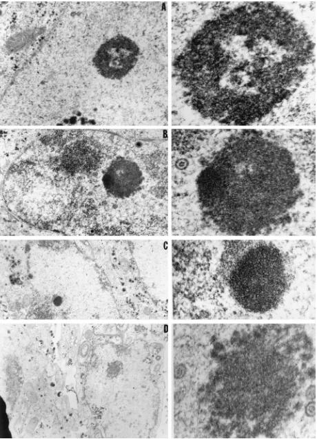
FIG. 6. Electron photomicrographs of intranuclear dense bodies obtained by conventional electron microscopy. Cells were fixed 24 h after infection with HSV-1(F) and embedded. Thin sections were examined in a Siemens electron microscope. Left panels, photographs taken at low magnification; right panels, the dense bodies of interest photographed at higher magnification. We interpret the bodies in panels A and D to be mixed-density bodies and those in panels B and C to be type V and IV nuclear bodies. The sizes of the dense bodies can be estimated by comparing them with viral capsids, which are approximately 105 nm in diameter.
831
on November 9, 2019 by guest
electron-dense structure in panels B and C resembled the type V and type IV nuclear bodies, respectively, reacting with the anti-UL11 polyclonal rabbit serum.
DISCUSSION
Previous studies have shown that UL11 protein is myris-tylated, that in the absence of the UL11 gene empty capsids and capsids containing DNA accumulate at the nuclear mem-brane, and that little infectious virus is released from infected cells. Thus, the apparent net function of the UL11 protein is to enable efficient transport of virions through the cytoplasm into the extracellular space (2, 22). The properties of the protein and its apparent function suggested the hypothesis that it in-teracts with membranes. The objective of the studies described in this report was to determine the localization of the UL11 protein in infected cells. To this end we raised antibody to the protein and determined its localization by immune light and electron microscopy. The salient features of the results were as follows.
(i) The polyclonal rabbit serum to the UL11 protein reacted with electrophoretically separated polypeptides with apparent Mrs of 17,000 to 22,000, i.e., considerably larger than the pre-dicted Mrof 10,249 and, as also reported by others (22), much more dispersed than would have been predicted for a post-translationally unmodified protein. The predicted UL11 gene product is rich in proline, which contributes to the aberrant migration of proteins in denaturing polyacrylamide gels. The observation that the proteins reacting with the rabbit anti-UL11 serum formed multiple bands differing in electrophoretic mobility is consistent with the results of others and suggests that the UL11 protein may undergo extensive posttranslational modifications involving, at least in part, phosphorylation by a virion-associated protein kinase (23).
(ii) Two series of studies support the hypothesis that UL11 protein interacts with membranes or membrane-associated proteins. The immune light microscopy suggested that UL11 is associated with the nuclear membrane and that its distribution in the infected cells is similar to that of gC. Immune electron microscopy studies indicate that the UL11 protein is found in connection with three relevant structures, i.e., virions, nuclear membranes, and undefined long ribbon-like structures. It is useful to consider each of these in detail.
UL11 protein localized with virions appeared most fre-quently juxtaposed to structures interior to the surface of the envelope. While we are unable to localize it precisely, the results support the hypothesis that UL11 protein is associated with the underside of the envelope or the tegument. The ab-sence of UL11 protein in association with intranuclear capsids is consistent with the view that UL11 does not bind to capsids directly. The association of the virion with nuclear membranes is concordant with the hypothesis that UL11 is associated with either membrane or tegument proteins.
The nature and origin of the cytoplasmic ribbon-like struc-tures are unknown. As noted in Results, preservation of the antigenic reactivity of cellular proteins for immune electron microscopy is at the expense of the use of fixation and staining procedures that optimally preserve ultrastructural detail. The ribbon-like structures may well be remnants of cytoplasmic membranes, but this will require further verification.
(iii) The observation that UL11 protein associated with in-tranuclear dense bodies was unexpected. Nuclear dense bodies have been observed in uninfected (7, 15) and HSV-infected (29, 32, 35) cells. They could consist of aggregated viral or cellular proteins and, as has been suggested, components of nucleoli. A recent study has redefined the electron-opaque nuclear
struc-tures that are round and in the size range of 0.20 to 2.0mm as nuclear bodies and have organized them into at least five different morphotypes (8) based on their electron densities and visible ultrastructures (7). We have shown that UL11 proteins localize within the core of nuclear bodies typical of morpho-type IV. These structures consist of an electron-opaque core surrounded by a slightly more electron-translucent cortex. The cortex does not react with the anti-UL11 antibodies. A second structure that reacts with the anti-UL11 antibodies resembles nuclear bodies of type V morphotype in that they appear uniform in density and consist of a microfibrillar-like material. Figure 6 illustrates several intranuclear dense bodies differing in morphology as observed by conventional electron micro-scopic techniques. Another intranuclear dense body of mixed density is not at present included in the nuclear body morpho-types and can be recognized in Fig. 5A and 6A and D. This intranuclear body may have its origin in the nucleolus, since it has been shown to be present in HSV-infected cells and to contain a nucleolar protein with an apparent Mrof 100,000 (30).
The suggestion that nuclear bodies represent functionally distinct entities rests on the observation that several viral pro-teins, i.e., UL11, UL12, US8.5, and US11, have been reported to localize in intranuclear dense bodies. These nuclear bodies are probably distinct from intranuclear viral replication compart-ments containing the UL29 and UL30 gene products (9, 17, 24, 28, 31, 34, 35). The product of the HSV-1 UL12 gene has been reported (30) to localize to what appeared to be the mixed-density bodies and type V nuclear bodies. Although both were seen in the studies reported here, only the latter are similar (Fig. 4C) to those found in association with UL11. The nuclear structures to which US11 and possibly US8.5 localize have been identified as nucleoli or nucleolar products (17, 24, 28, 30).
(iv) Among other proteins reported to facilitate the egress of virus from infected cells is the alkaline DNase product of the UL12 gene (36). As noted above, this protein also localizes in morphotype V nuclear bodies. Questions that remain unre-solved are whether these bodies are biochemically as well as morphologically identical and whether there is a functional interaction between the products of the UL11 and UL12 proteins. The studies described in the report suggest that UL11 may have multiple functions. Although there are other possibilities, the possibility that a small protein of 95 amino acids may have multiple functions is of interest but may not be surprising. As reviewed elsewhere, this appears to be a common property of herpesvirus proteins (33).
ACKNOWLEDGMENTS
We thank Richard Roller for monoclonal antibodies to US11 and
Gabriella Campadelli-Fiume for making the antibody to the UL
11-GST fusion protein.
The studies at the University of Chicago were aided by grants from the National Cancer Institute (CA47451) and the National Institute of Allergy and Infectious Diseases (AI24009), the Public Health Service, and by an unrestricted grant from Bristol-Myers Squibb Program in Infectious Diseases. The studies at the University of Kentucky were aided by the Llewellyn H. May fund and the University of Kentucky major instrumentation program in the purchase of the electron micro-scope equipment (7E-7H23 and 7E-7H24) and the initiation of the Nano Probe Lab of the Markey Cancer Center.
REFERENCES
1. Ackermann, M., J. Chou, M. Sarmiento, R. A. Lerner, and B. Roizman. 1986. Identification by antibody to a synthetic peptide of a protein specified by a diploid gene located in the terminal repeats of the L component of herpes simplex virus genome. J. Virol. 58:843–850.
2. Baines, J. D., and B. Roizman. 1992. The UL11 gene of herpes simplex virus
1 encodes a function that facilitates nucleocapsid envelopment and egress from cells. J. Virol. 66:5168–5174.
832 BAINES ET AL. J. VIROL.
on November 9, 2019 by guest
http://jvi.asm.org/
3. Baines, J. D., and B. Roizman. 1993. The UL10 gene of herpes simplex virus
1 encodes a novel glycoprotein, gM, which is present in the virion and in the plasma membrane of infected cells. J. Virol. 67:1441–1452.
4. Baines, J. D., P. L. Ward, G. Campadelli-Fiume, and B. Roizman. 1991. The UL20 gene of herpes simplex virus 1 encodes a function necessary for viral
egress. J. Virol. 65:6414–6424.
5. Baradaran, K., C. E. Dabrowski, and P. A. Schaffer. 1994. Transcriptional analysis of the region of herpes simplex virus type 1 genome containing UL8, UL9, and UL10 genes and identification of a novel delayed-early gene product, OBPC. J. Virol. 68:4251–4261.
6. Barker, D., and B. Roizman. 1992. The unique sequence of the herpes simplex virus 1 L component contains an additional open reading frame designated UL49.5. J. Virol. 66:562–572.
7. Bouteille, M., S. R. Kalifat, and J. Delarue. 1967. Ultrastructural variations of nuclear bodies in human disease. J. Ultrastruct. Res. 19:474–486. 8. Brasch, K., and R. L. Ochs. 1992. Nuclear bodies (NBs): a newly
‘‘rediscov-ered’’ organelle. Exp. Cell Res. 202:211–223.
9. Bush, M., D. R. Yager, M. Gao, K. Weisshart, A. I. Marcy, D. Coen, and
D. M. Knipe.1991. Correct intranuclear localization of herpes simplex virus DNA polymerase requires the viral ICP8 DNA binding protein. J. Virol.
65:1082–1089.
10. Campadelli-Fiume, G., F. Farabegoli, S. D. Gaeta, and B. Roizman. 1991. Origin of unenveloped capsids in the cytoplasm of cells infected with herpes simplex virus 1. J. Virol. 65:1589–1595.
11. Carlemalm, E., R. M. Garavito, and W. Villiger. 1982. Resin development for electron microscopy and an analysis of embedding at low temperature. J. Microsc. 126:123–143.
12. Chou, J., and B. Roizman. 1986. The terminal a sequence of the herpes simplex virus genome contains the promoter of a gene located in the repeat sequences of the L component. J. Virol. 57:629–637.
13. Chou, J., and B. Roizman. 1990. The herpes simplex virus 1 gene for ICP34.5, which maps in inverted repeats, is conserved in several limited-passage isolates but not in strain 17syn1. J. Virol. 64:1014–1020. 14. Desai, P. J., P. A. Schaffer, and A. C. Minson. 1988. Excretion of
noninfec-tious virus particles lacking glycoprotein H by a temperature-sensitive mu-tant of herpes simplex virus type 1: evidence that gH is essential for virion infectivity. J. Gen. Virol. 69:1147–1156.
15. De The, G., M. Riviere, and W. Bernhard. 1960. Examination by electron microscopy of the VX2 tumor of the domestic rabbit derived from the Shope papilloma. Bull. Assoc. Fr. Etude Cancer 47:570–584.
16. Ejercito, P. M., E. D. Kieff, and B. Roizman. 1968. Characterization of herpes simplex virus strains differing in their effects on social behavior of infected cells. J. Gen. Virol. 2:357–364.
17. Georgeopoulou, U., A. Michaelidou, B. Roizman, and P. Mavromara-Nazos. 1993. Identification of a new transcriptional unit that yields a gene product within the unique sequences of the short component of the herpes simplex virus 1 genome. J. Virol. 67:3961–3968.
18. Kellenberger, E., E. Carlemalm, W. Villiger, J. Roth, and R. M. Garvito. 1980. Low denaturation embedding for electron microscopy of thin sections, p. 1–59. Chemische Werke Lowi GmbH, Waldkralburg, Germany. 19. Lagunoff, M., and B. Roizman. 1994. Expression of a herpes simplex virus 1
open reading frame antisense to theg134.5 gene and transcribed by an RNA
39coterminal with the unspliced latency-associated transcript. J. Virol. 68: 6021–6028.
20. Liu, F., and B. Roizman. 1991. The promoter, transcriptional unit, and coding sequences of herpes simplex virus 1 family 35 proteins are contained within and in frame with the UL26 open reading frame. J. Virol. 65:206–212.
21. Mackem, S., and B. Roizman. 1980. Regulation of herpesvirus macromolec-ular synthesis: transcription-initiation sites and domains of alpha genes. Proc. Natl. Acad. Sci. USA 77:7122–7126.
22. MacLean, C. A., B. Clark, and D. J. McGeoch. 1989. Gene UL11 of herpes simplex virus type 1 encodes a virion protein which is myristylated. J. Gen. Virol. 70:3147–3157.
23. MacLean, C. A., A. Dolan, F. E. Jamieson, and D. J. McGeoch. 1992. The myristylated virion proteins of herpes simplex virus type 1: investigation of their role in the virus life cycle. J. Gen. Virol. 73:539–547.
24. MacLean, C. A., F. J. Rixon, and H. S. Marsden. 1987. The products of gene US11 of herpes simplex virus type 1 are DNA-binding and localize to the nucleoli of infected cells. J. Gen. Virol. 68:1921–1937.
25. McGeoch, D. J., M. A. Dalrymple, A. J. Davison, A. Dolan, M. C. Frame, D.
McNab, L. J. Perry, J. E. Scott, and P. Taylor.1988. The complete DNA sequence of the long unique region in the genome of herpes simplex virus type 1. J. Gen. Virol. 69:1531–1574.
26. McGeoch, D. J., A. Dolan, S. Donald, and F. J. Rixon. 1985. Sequence determination and genetic content of the short unique region in the genome of herpes simplex virus type 1. J. Mol. Biol. 181:1–13.
27. Post, L. E., S. Mackem, and B. Roizman. 1981. Regulation ofagenes of herpes simplex virus: expression of chimeric genes produced by fusion of thymidine kinase withagene promoters. Cell 25:227–232.
28. Puvion-Dutilleul, F. 1987. Localization of viral-specific 21 kDa protein in nucleoli of herpes simplex infected cells. Eur. J. Cell Biol. 43:487–498. 29. Puvion-Dutilleul, F., J. Pedron, M. I. Aithier, and P. Sheldrick. 1982.
Ul-trastructural studies on the nucleus of herpes simplex virus type 1-infected cells. Biol. Cell 44:249–260.
30. Puvion-Dutilleul, F., and E. Pichard. 1986. Viral alkaline nuclease in in-tranuclear dense bodies induced by herpes simplex infection. Biol. Cell. 58: 15–22.
31. Rixon, F. J., M. Atkinson, and J. Hay. 1983. Intranuclear distribution of herpes simplex virus type 2 DNA synthesis: examination by light and electron microscopy. J. Gen. Virol. 64:2087–2092.
32. Roizman, B., and D. Furlong. 1974. The replication of herpesviruses, p. 229–403. In H. Fraenkel-Conrat and R. R. Wagner (ed.), Comprehensive virology, vol. 3. Plenum Press, New York.
33. Roizman, B., and A. E. Sears. 1993. The replication of herpes simplex viruses, p. 11–68. In B. Roizman, C. Lopez, and R. J. Whitley (ed.), The human herpesviruses. Raven Press, New York.
34. Roller, R., and B. Roizman. 1992. The herpes simplex virus 1 RNA binding protein US11 is a virion component and associates with ribosomal 60S
subunits. J. Virol. 66:3624–3632.
35. Schwartz, J., and B. Roizman. 1969. Similarities and differences in the development of laboratory strains and freshly isolated strains of herpes simplex virus in Hep-2 cells: electron microscopy. J. Virol. 4:879–889. 36. Shao, L., L. M. Rapp, and S. K. Weller. 1993. Herpes simplex virus 1 alkaline
nuclease is required for efficient egress of capsids from the nucleus. Virology
196:146–162.
37. Vieira, J., and J. Messing. 1987. Production of single-stranded plasmid DNA. Methods Enzymol. 153:3–13.
![FIG. 1. Schematic diagrams of the sequence arrangements of the wild-typevirus [HSV-1(F)] and the R7219 recombinant virus from which the UL11 gene](https://thumb-us.123doks.com/thumbv2/123dok_us/1288053.81628/2.612.58.296.69.183/fig-schematic-diagrams-sequence-arrangements-typevirus-recombinant-virus.webp)