0022-538X/94/$04.00+0
Copyright ( 1994, American SocietyforMicrobiology
Stimulation
of the Adenovirus
Major
Late Promoter In
Vitro
by
Transcription Factor USF
Is Enhanced
by
the Adenovirus
DNA
Binding
Protein
DIEDERIKC.ZIJDERVELD,' FABRIZIOD'ADDADI FAGAGNA,2 MAURO GIACCA,2
H. T. MARCTIMMERS,' ANDPETERC.VANDERVLIET1*
Laboratory for Physiological Chemistry, University of Utrecht, 3508 TA Utrecht, TheNetherlands,1
andIntemationalCentreforGeneticEngineeringandBiotechnology, I-34012 Trieste, Italy2
Received 13 June1994/Accepted 21September1994
Previous studies have shown that the sequence-independent adenovirus DNA binding protein (DBP)
increases transcriptionfromseveral promoters,notablyfrom theadenovirusmajorlatepromoter(MLP) and
theadeno-associated virus P5 promoter, both of which containaUSF/MLTFbindingsite.Inordertostudythis
mechanism,wehaveinvestigatedthe eflectsofDBPonthebindingofUSF/MLTFtoMLPandontranscription
from MLPby a reconstituted invitro system.As shown by gel retardation and DNase Ifootprinting, upon
saturation ofDNA, DBPenhances the binding affinity ofUSF43 to the promoter three- to fourfoldwithout
changingthefootprintpattern.In contrast,thebindingof the TATA boxbinding proteintothepromoter is not
influenced by DBP.Noprotein-proteininteractionsbetween DBP and USF43 could be observed in the absence
ofDNA, suggestingthatenhancedbindingis causedbyachangein DNAstructureinducedbytheDBP-DNA
complex.Employingatranscription systemreconstituted withpurified generaltranscription factors,weshow
that USF43 enhances basaltranscriptionandthatUSF43-dependent transcriptionis further increasedby DBP,
while DBP alone does not have an effect on basal transcription. Our results suggest that transcription
enhancement byDBPis based on a specific increase in thebinding ofa transcriptionfactor toa promoter
throughsubtlechangesinDNA structure, similartothe mechanismbywhich DBPstimulates theinitiationof
DNAreplication.
One of themajor transcription factors involved in
adenovi-ruslatetranscription is the cellularprotein upstream
stimula-toryfactor USF (MLTF) (4, 10, 25-27, 34-36, 52). Itcanbind
to the adenovirus major late promoter (MLP) upstream
se-quence between -63 and -58, relative to the transcription
startsite,andactivate transcriptionfrom MLP(10, 34, 35).In
addition,USF is also involved in regulatingtheexpression of
several cellular genes, such as the human growth hormone
gene(31), the mousemetallothionein I gene (5), and therat
-y-fibrinogengene(9).Furthermore,aUSFbindingsitecanbe
found inaregion encompassingtheputative human origin of replication B48 (13, 15).
USF consists oftwopolypeptideswith molecularmassesof 43 and 44 kDa (36). Both polypeptides show independent
DNA binding, and each binds either as a homo- or
het-erodimer to the palindromic CACGTG motif (34). The
43-kDa component (USF43) is a member of the basic region
helix-loop-helix leucinerepeat(B-HLH-LR) class of
transcrip-tionfactors(16). Deletion mutagenesis identifiedtwodomains
N terminal of the B-HLH-LR domain(amino acids 15 to59
and 93 to 156) that contribute to transcriptional activation
(20). The 44-kDa component is less well characterized.
Re-cently, afull-lengthcDNAencodingUSF44wascloned,
show-ing that USF44 and USF43 aremembers of the sameprotein
familywith highlyconserved DNA binding and dimerization
domainsbutquitedivergent N-terminalamino acidsequences
(37). A comparison of the protein sequence with previously
*Corresponding author. Mailing address: Laboratory for
Physio-logical Chemistry, University of Utrecht, P.O. Box 80042, 3508 TA Utrecht,The Netherlands. Phone:31-30-538989. Fax: 31-30-539035.
published sequences showed that USF44 is identical to Fos
interacting protein (1).
USF boundtoitsconsensusrecognition siteupstreamof the
TATAboxcanstimulate transcription,but itcanalsobindto
initiator elementsencompassingthetranscriptionstartsiteand
transactivatetranscription (14, 24, 33).In invitrotranscription assays, crudepreparations of USFcanstimulatetranscription upto 10-fold. Asaresult ofpurification, partof itsactivating
potential is lost, and highly purified preparations can only
activate transcriptionapproximately threefold(34).
Recombi-nant USF43 is also ableto activate transcription in a manner
indistinguishable fromthat ofhighly purified USF (32). USF
interacts directly with the TFIID complex and binds DNA
synergistically with this complex (34, 35). In the presence of
TFIID, USF inhibits nucleosome assembly on promoter
se-quences and thereby facilitates theformation of preinitiation
complexes during in vitro chromatin assembly (51). During
adenovirus infection, the activation oftranscription by USF
requires DNA replication, which suggeststhat early in
infec-tion,adenoviruschromatin isnotaccessibletoUSF(45). Early
ininfection, adenovirusDNAis still complexedtoproteinVII,
whereaslate in infection, the situation is different and DNA
may becomplexed withthe adenovirusDNA binding protein
(DBP) (8).
DBP is a multifunctional protein of529 amino acids that
consistsoftwo domains(22). The N-terminaldomain (amino
acids 1 to 173) is not well conserved among serotypes and
harbors the nuclearlocalization signal (12, 28). The C-terminal
domain is well conserved, contains DNA binding properties,
andharborsmost of thebiological functions ascribedtoDBP
(21).
DBPbindsRNA, single-strandedDNA(ssDNA), and
dou-ble-stranded DNA(dsDNA)in asequence-independent man-8288
on November 9, 2019 by guest
http://jvi.asm.org/
DBP ENHANCEMENT OF TRANSCRIPTION STIMULATION BY USF 8289 ner. Binding to ssDNA is cooperative, and the monomeric
binding site is 11 to 15 bases, varying slightly for different
polynucleotides (23). Recently, the crystal structure of DBP
waselucidated and DBP was shown to be a roughly globular
protein with a striking 17-amino-acid C-terminal extension. This C-terminal hook can interact with a neighboring DBP molecule, which can lead to the formation of a protein chain. The deletion of this hook destroys cooperativity of ssDNA binding (46). Binding to dsDNA is not cooperative, and both association and dissociation are very rapid (39). Hydroxyl radical footprinting and electron microscopy show that DBP changes the structure of dsDNA. This DNA acquires a rigid structure; at the same time, DBP introduces changes in base-to-base positions and is able to remove higher-order structure from DNA. Cryoelectron microscopy suggests that under saturating conditions, DBP may form two interwound chains around the DNA helix (39).
DBP is intimately involved in the viral life cycle. It functions
inboth the initiation and elongation phases of DNA
replica-tion (for reviews, see references 18, 38, and 47), in virus assembly, in the stability of mRNA, and in the replication of adeno-associatedviruses (for reviews, see references 7, 18, 19, and 48). DBP is also involved in transcriptional regulation. DBP is able to enhance its own synthesis. Mutant analysis suggeststhat DBP enhances its own expression only when it is present in a highly phosphorylated form (29). By in vitro transcription runoff assays and transfection assays, DBP was found to specifically repress transcription from the adenovirus
E4 promoter (6, 17). Furthermore, transfection experiments
with DBP expression constructs showed that DBP enhances
theexpression of reporter genes controlled by several different
adenovirus or adeno-associated virus promoters, with the strongest effect on genes controlled by the adenovirus MLP and the adeno-associated virus P5 promoter, both of which containaUSF binding site (6). The mechanism by which DBP modulates transcription is unknown.
We and others reported previously that DBP is able to
enhancethe binding of the cellular transcription factor nuclear factor I (NFI) to its recognition site in the adenovirus auxiliary origin of replication, which leads to enhanced initiation of DNA replication in the presence of DBP (11, 40). In this work, weinvestigatedthe influence of DBP on transcription from the adenovirus MLP. We find that DBP enhances the binding of USF43 toits recognition site upstream of the adenovirus MLP.
Furthermore,
we show that DBP does not interact directly withUSF43, suggesting that the stimulation of USF binding is the result of structural alterations of the binding site induced by
DBP.Finally, we show that DBP enhances the stimulation of
transcriptionby
USF43
in a reconstituted in vitro transcription assay.MATERIALS AND METHODS
Purification of proteins. The coding region of
USF43
was cloned 3' of the glutathione S-transferase region in the bacte-rialexpressionvector pGEX2T (13a). The resulting expression plasmidpGST-USF43 was transformed into the SF8 strain ofEscherichia
coli.Transformed bacteria (500 ml) were grown at37°C until they reached an optical density at 600 nm of 0.6.
Next,protein expression was induced by the addition of IPTG
(isopropyl-13-D-thiogalactopyranoside)
(final concentration, 1mM). After 4 h, the culture was centrifuged at 5,000 xg for 5
min at4°C, the bacterial pellet was resuspended in 12.5 ml of
50 mMTris-HCl (pH 8.0)-i mM EDTA-0.4 mM
Na2S2O5-4
mMdithiothreitol (buffer A), and 10%
(wt/vol)
sucrose wasadded. The solution was frozen and thawed once, lysozyme was
added to
0.25
mg/ml,
and
the solution was frozen
and thawedtwice more before
being
sonicatedthree times
for 30 s each.NaCl was added to 500
mM,
and
the solution was
centrifugedat
35,000
rpm
in a Sorvall SW41 rotor for 90min
at4°C.
Thesupematant
was diluted to 150 mM
NaCl with buffer
A, and 2ml of
50%
glutathione-agarose
(GA)
beads, equilibrated
inbuffer A
and
containing
150 mM
NaCl,
was added.
Bindingwasfor 60
min at4°C
on arotating
wheel.
Subsequently,
theslurrywas
packed
ina
column,
washed,
and eluted
with 20 mMglutathione
inbuffer A.
DBPwas
purified
as described
previ-ously
(53).
Bothproteins
were more than95%
pureas judgedby
sodium
dodecyl
sulfate
(SDS)-polyacrylamide
gelelectro-phoresis and Coomassie staining.
The TATA
box
binding protein
(TBP)
preparation
used ingel
retardationassays
contained recombinant,
histidine-taggedTBP,
which was
purified
by
Ni2+-NTA
chromatography
(44)and was 30 to 50%
pure
as
judged
by
Coomassie
staining.The
coding region
of the DNA
binding domain
ofNFI
(NFI-BD)
was
cloned in the bacterial
expression
vectorpET-l5b.
Histidine-tagged
NFI-BD
(His-NFI-BD)
was
purified byNi2+-NTA
chromatography
and was
homogeneous.Gel retardation.
Binding
reactions were carried
outon ice orat
room
temperature
for 60
min
in
a total volume
of 20[lI
containing
25 mM HEPES
(N-2-hydroxyethylpiperazine-N'-2-ethanesulfonic
acid)-KOH
(pH
7.5),
60 mM NaCl, 2 mMMgCl2,
10%
glycerol,
50
,ug
of bovine
serum
albumin
(BSA)per
ml, 10,000
cpm
of TATA or UBS DNA, and indicated
amounts
ofproteins.
UBS
DNA consisted of the 140-bpPvuII-XbaI
fragment
from
pMLTF
(3),
which was
Klenowendlabelled with
[ca-32P]dCTP.
TATA DNA
consisted of the133-bp
EcoRI-HindIII
fragment
from
pBS-MLP,
which wasKlenow end labelled with
[ot-32P]dATP.
pBS-MLP
contains theMLP
sequence
from -51 to +33 cloned in the
SmaI
site ofpBS-.
After 60
min,
2.5
,ul
of
loading
buffer containing
0.09%Nonidet P-40 was added. When
USF43
binding
was assayed,400
ng
ofpoly(dI-dC)
-poly(dI-dC)
was added tothe loadingbuffer added to the
sample
at the moment of
loading thesample
onto the
gel.
Since
DBP
dissociates rapidly
fromdsDNA
(39),
this
procedure
prevents
the
occurrence
of shiftedbands because of DBP
binding
without
affecting
USF43
bind-ing.
When TBP
binding
was
assayed,
no
poly(dI-dC)
*
poly(dI-dC)
was
added,
since this addition
may
also affect TBP
binding.The
binding
conditions for
both
USF43
and TBP were
identicalup
to the moment of
loading,
and
therefore,
the resultscan becompared directly.
When DBP
binding
was assayed, UBSDNA was denatured
by
boiling
for 2
min.
Free
DNA andprotein-DNA
complexes
were resolved on a native
5%poly-acrylamide gel
which was run in
0.5x
Tris-borate-EDTA
buffer-0.01%
Nonidet P-40 for 2 h at 8 V/cm.
Gels weresubsequently
dried and
autoradiographed.
Results werequan-tified with an LKB Ultrascan XL gel
scanner.DNase
I
footprinting.
Binding
reactions were carried
out atroom
temperature
for 60
min
in the same
buffer
usedfor gelretardation,
containing 10,000
cpm
of UBS DNA and indicated
amounts of
proteins.
After 60
min,
0.1
U of DNase
I togetherwith 250
ng
of
poly(dI-dC)
-poly(dI-dC)
was added, anddigestion
was
allowed for 90 sec at
30°C.
Reactions
wereterminated
by
the addition
of 3
RI
of 0.2 M EDTA-10% SDS.Samples
were extracted once
withphenol-chloroform,
precip-itated
with
ethanol,
and
analyzed
on
a10%
denaturing poly-acrylamide gel.Affinity
chromatography.
Threemicrograms
ofUSF43
in 100,u
of buffer
A
containing
60
mM NaCl(buffer
B) wasincubated
with 100
pAof GA
beads,
which were equilibrated inthe
same
buffer,
for
1 h at4°C.
Subsequently,
the beads werepacked
in
a column. Ten
micrograms
of
DBPin 1 mlof buffer VOL.68, 1994on November 9, 2019 by guest
http://jvi.asm.org/
B
A
-1-
DBP27
1 2 3 4 5 6 7 8 9* _ __--_USF-DNA
_i
freeDNA--- USF --- USF
DBP + __ _
1 2 3 4 5 6 7 8 9 1011 12 13 1415 16 17 18
&A_
*w1 -USF-DNA_hih~i~~
_
~jjk~hi~
~
-
free DNA
k,
-+- +
DBP
-A- DBPTB..P..
DBPL
<+
-1 2 3 4 5 6 7 8 9 10
TBP-DNA _ mw ;,
DBP-DNA
-freeDNA
-id
n
G(0lu
-U..
0 10 20 30 40 50 60 70 80amoLrnt 73P (ng)
FIG. 1. DBP enhances thebindingofUSF43toitsrecognitionsite. ThebindingofUSF43toUBSDNA and ofTBP toTATA DNAwasassayed by gel retardation. (A) Autoradiographofa5%nondenaturinggel;10,000cpmofUBSDNAwasincubated withincreasing amounts of DBPin thepresenceof0.8ngofUSF43.Lanes 1to9, 0, 2, 5, 10, 20, 40, 80, 160,and320ngofDBPadded,respectively. (B) Autoradiograph ofa5% nondenaturing gel; 10,000cpmofUBS DNAwasincubated withincreasingamountsofUSF43inthepresence(lanes1to9)orabsence(lanes10 to18) of150ngofDBP. TheamountsofUSF43 (in nanograms)addedwere0(lanes1and10),0.5(lanes2 and11),1(lanes3and12),2(lanes
4 and13),4(lanes5and14),8(lanes6and15), 16(lanes7 and 16),32(lanes8 and 17),and 64(lanes9and 18).(C) Autoradiograph ofa5% nondenaturinggel; 10,000cpmofTATA DNAwasincubated withincreasingamounts ofTBP in thepresence(lanes 1 to 5)orabsence(lanes6
to10) of300ngofDBP. Theamountsof TBP(innanograms)addedwere0(lanes1 and6),10(lanes2and7),20(lanes3 and8),40(lanes4
and9),and80(lanes5and10). (D) Graphicrepresentationof the results ofpanelC. Theamountsofshiftedprobeasdeterminedbyscanning theautoradiograph (arbitrary units) areplotted againsttheamountof TBP added in the absenceorpresence of DBP.
Bwas passed over this column four times. The column was
washed with 1 ml of buffer B and eluted with 20 mM
glutathioneinbuffer B.One-hundred-microliterfractionswere
collected. USF"3 and DBP contents were assayed by gel
retardation, withUBSDNAandheat-denaturedUBS DNAas
theprobe,respectively.
Reconstituted transcription. Reactions were carried out,
and sampleswere processedas described previously (43, 44).
Briefly,reactionmixtureswereassembledoniceandcontained
12mMHEPES-KOH (pH 7.9), 60 mM KCl, 12% glycerol,0.6
mM EDTA, 0.3 mM phenylmethylsulfonyl fluoride, 2 mM
dithiothreitol,5mMMgCl2, 100 ,ug of BSAperml, and 10U
of RNAguard (Promega). The transcription factors present
wereTFIID, TFIIA, and TFIIF, whichwerepartially purified
fractions from a HeLa cell extract; RNA polymerase II,
purified fromanAmal CHO cellextractandmorethan90%
pure asjudged bysilver staining; andTFIIBand TFIIE,both
of which were bacterially expressed recombinant proteins
purifiedtohomogeneityasjudged by Coomassie staining. The
transcription templateswerepAML(C2AT)200, which contains
a210-nucleotide(nt)G-lesscassettepreceded bythesequence
-53 to +10 from the adenovirusMLP, andpML112(C2AT),
which contains a 380-nt G-less cassette preceded by the
sequence -112to +10from theadenovirus MLP. Incubation
wasfor60minat30°C.Productswereresolvedonadenaturing
5%polyacrylamide gel.
RESULTS
DBPenhances thebindingofUSF"3toitsrecognition site.
We testedthe effect of DBPon the binding ofsubsaturating
amountsofUSF43 to its recognition site in a gel retardation
assay.Figure 1Ashows that thebinding ofUSF"3 ismarkedly
increased when DBPispresent.This effect isdependentonthe
DBPconcentrationandreachesaplateauatapproximately 80
to 160 ngof DBP (Fig. 1A, lanes 7 and 8). In these experi-ments, there was an approximately 100-fold molar excess of
DBPoverUSF43. Thereasonforthisis that there isonlyone
USFbindingsiteonthisDNA,whereas thesaturation of this
DNA by DBP requires approximately 10 to 15 molecules of
C
D
on November 9, 2019 by guest
http://jvi.asm.org/
[image:3.612.88.530.73.409.2]DBP ENHANCEMENT OF TRANSCRIPTION STIMULATION BY USF 8291
DBP. Furthermore, DBP binds unstably to dsDNA and high
concentrations are required for saturated binding. Interest-ingly, and stressing the relevance of the conditions chosen, high concentrations of DBP (approximately 2x
107
molecules percell) accumulate within the infected cell, resulting in an
approximately 1,000-fold molar excess of DBP over USF (49).
In areciprocal experiment, when theUSF43 concentration was
varied in the absence or presence of 150 ng of DBP, the concentration ofUSF43 at which half of the DNA was bound
waslowered threefold when DBP was present (Fig. 1B).USF43
wasstable for at least 3 h at4°Cand for 1 h at
37°C,
regardlessof the presence of DBP, indicating that the effect of DBP on
the binding affinity ofUSF43was not due to stabilization (data
notshown). To determine if this enhancement of binding was
specific for USF43, we tested to see if the binding of TBP was similarly affected by DBP. Figure 1C shows that TBP binding
is notincreased when DBP is present, even at 300 ng. In this
particular experiment, the binding of DBP to the double-stranded probe is also observed, in contrast to the results in
Fig.1A, lane 9, when the same amount of DBP was added. The
reason for this is that we did not addpoly(dI-dC) -poly(dI-dC)
tothesample at the moment of loading, since it also competes
for TBP binding. The enhancement of TBP binding by DBP
wasnotobserved when the DBP concentration was varied and
theconcentration of TBP was fixed (Fig. 1D) or when binding
wasassayed by DNase I footprinting (data not shown).
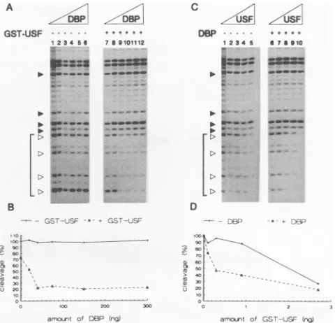
Subsequently, we tested if DBP changes the binding of USF43 qualitatively by DNase I footprint analysis. No
differ-ences in theprotection pattern of the USF binding site could
be detected when DBP was present during incubation (Fig. 2C,
compare lanes 5 and 9), although the lower border of the
footprint is not clearly visible in this figure. Bands below the
lowest band of thefootprint were faint, but upon overexposure,
it wasobvious that the lowest band shown represents the lower
border of the footprint in the absence or presence of DBP.
Again,
USF43
was found to bind more efficiently in thepresence of DBP. Figure 2B shows that with a fixed amount of USF43, the protection of the USF binding site is increased from 25 to 80% by the addition of DBP, and Fig. 2D shows that
thebinding affinity ofUSF43is increased two- to sixfold in the
presence of DBP. DBP alone showed no protection of the USF bindingsite.
DBPdoes notinteract directly withUSF43.The stimulation
of
USF43
binding by DBP can in principle be mediated throughdifferentmechanisms. DBP might interact directly with USF43
andtether it to DNA, or DBP might change the structure of
dsDNA, leading to more efficient binding by
USF43.
The construction of a USF43 fusion protein containing a
GST tagenabled us to immobilizeUSF43on GA beads and test
whether DBP can bind to the bead-protein complex.
Figure 3 shows thatUSF43can be immobilized on GA beads
and eluted with 20 mM glutathione (lanes 6 to 8). Using
conditions under which DBP stimulated USF43 binding to
DNA, we testedwhether a direct interaction between
USF43
and DBP could beobserved. In this experiment, approximately
1 p,g of
USF43
was immobilized on a GA column. WhereasDBP could be detected in the flowthrough and first wash
fraction (Fig. 3, lanes 14 to 16), no DBP was present in the
eluatecontainingUSF43.Since the detection limit for DBP in
this assay was approximately0.3ng, we can conclude that less
than 0.3 ng of DBP per 100 ng ofUSF43was complexed.
DBP is also known to stimulate the binding of another
transcription factor, NFI, to its recognition site (11, 40). The
binding of NFI-BD is stimulated by DBP in a manner
indis-tinguishable from that of the intact NFI protein (2). In a way
similar to that used for USF43 but employing Ni2+-NTA
A
GST-USF 1!~3S456
3
>
>
B s
7 86101112
'i.
c
ZUSFI
D8P ---* 12S345
z
.-:3=
- -, --
->W> -
->M. w.
>' ..
D
..c- -o
2 7 ^\> .
U0 >..__.'IO
a-ourt of 36P (rg) amooLr¶ of GS--,SF (-g) FIG. 2. DBP increases the binding affinity but does not change the footprintpattern ofUSF43. (A and C) DNase I footprint analyses of USF43 binding in the presence or absence of DBP. The protected binding site is shown in brackets. (A) UBS DNA (10,000 cpm) was incubated with increasing amounts of DBP in the absence (lanes 1 to 6) or presence (lanes 7 to 12) of 12 ng ofUSF43.The amounts of DBP (in nanograms) added were 0 (lanes 1 and 7), 20 (lanes 2 and 8), 40 (lanes 3 and 9), 80 (lanes 4 and 10), 150 (lanes 5 and 11), and 300 (lanes 6 and 12). (B) Graphic representation of the results in panel A. Average cleavage at the positions of the open triangles in panel A (corresponding to the USF binding site) relative to cleavage at positions of the closed triangles (which isameasure of the degree of occupancy of the USF binding site) is plotted as a function of the amount of DBP added. Relative cleavage in lane 1 is set up as 100%.
(C)UBS DNA (10,000 cpm) was incubated with increasing amounts of
USF43in the absence (lanes 1 to 5) or presence (lanes6to 10) of 150
ng of DBP. The amounts ofUSF43(in nanograms) added were0(lanes
1and 6),1(lanes 2 and 7), 3 (lanes 3 and 8), 9 (lanes 4 and 9), and 27 (lanes 5 and 10). (D) Graphic representation of the results in panel C.
Relative cleavage is plotted as a function of the amount of USF43
added in thepresence or absence of DBP.
affinity chromatography, we tested for possible direct interac-tion between NFI-BD and DBP. We immobilized
His-NFI-BD on a
Ni2+-NTA
column and found that DBP was notretained on such a column under conditions at which DBP stimulates the binding of NFI-BD (data not shown).
Since we were unable to show direct interaction between
DBP and either
USF43
or His-NFI-BD, NFI-BD and USF43are unrelated proteins, and DBP is known to induce extensive structural alterations in dsDNA, we favor the explanation that DBP enhances the binding of transcription factors by affecting the general structure of DNA.
DBP enhances transcription stimulation by
USF43.
Tran-scription in vivo from the adenovirus MLP is stimulated by the
binding of USF43 to its recognition site upstream of the
promoter. We checked if transcription in a reconstituted in
vitro system could also be stimulated by USF43. The in vitro
system employed contained homogeneous preparations of recombinant basal transcription factors TFIIB and TFIIE and purified preparations of TFIIA, TFIID, TFIIF, and RNA
polymerase II. In in vitro transcription reactions, we used the
following two templates: pML112(C2AT), in which a 380-nt VOL.68, 1994
on November 9, 2019 by guest
http://jvi.asm.org/
[image:4.612.315.555.68.299.2]8292 ZIJDERVELD ET AL.
~-cv
glutathione
eluate fractions
1 2 3 4 5 6 7 8 9 10 11
glutathione Suae
12f131 7 ons
12 13 14 15 16 17 18 19 20 21 2-2
USF-DNA-- _
[image:5.612.119.508.71.271.2]dsDNA -_
FIG. 3. DBP and USF43 do not form a stable complex. Gel retardation assay of column fractions with UBS DNA (lanes 1 to 11) or heat-denatured UBS DNA(lanes 12to22).Ten-microlitersampleswereassayed.FT1, sampletaken after DBPwaspassedoverthe column once;
FT2, sampletakenafterfour columnpassages;wash1,first 100,ul of thewash;wash2,theremaining900[L1.Thepositionsof free ds- andssUBS DNAand ssUBSDNA-DBPand dsUBS DNA-USF43complexes areindicated.
G-lesscassetteispreceded bythesequence -112to +10 from the adenovirus MLP and which containsoneUSFbinding site;
andpAML(C2AT)200, which served as aninternal reference,
contains a 210-nt G-less cassette preceded by the sequence
-53 to +10 from the adenovirus MLP, and lacks this USF
binding site.Whenincreasing amountsofUSF43were added,
transcription of the 380-nt cassette was increased relative to
transcription of the 210-ntcassette in a
concentration-depen-dent fashion (Fig. 4A, lanes 1 to 5). Maximal stimulation
wasabout 2.5-foldin the absenceof DBP (lane 4). (An addi-tional band is always seenjust below the 380-ntproduct; this
band presumably arises from reinitiation and premature
ter-mination rather than internal initiation, since this band is also increased in thepresenceofUSF43 and itsintensity
com-pared with that of the 380-nt band is always approximately
10%, regardlessof addedproteins.)When DBP is added(lanes
6 to 10), the level of transcription of the 380-nt cassette,
relative to transcription ofthe 210-ntcassette, is consistently
higher than in the absence of DBP. DBP alone does not
influence transcription atthe concentrations used here (Fig. 4A, lanes 11 to 15). The enhancement of USF43-stimulated
transcription at a suboptimal USF43 concentration increases
with an increasingDBP concentration andreaches aplateau
at 1,000 to2,000ngof DBP (Fig. 4A, lanes 16to20, and C).
Thisconcentration is 16to32 times higher than that required
for maximal stimulation of USF43 binding. The most likely
explanation forthisis that in the transcriptionreaction,200ng
of DNA is present as opposed to 0.1 to 0.2 ng of DNA in
binding reactions. This excess DNA effectively lowers the
concentrationof unbound DBP,and therefore, moreDBP is
necessaryinthetranscription assaytoreach thesamelevel of
stimulation.
DISCUSSION
Inthispaper,wehaveshownthatDBP enhances the binding
ofUSF43 toits recognition site. We have also shown that this
increase inbinding affinity in thepresenceof DBPresults inan
enhanced stimulation oftranscriptioninvitro by USF"3.
Previously, DBP had been shown to increase the binding
affinity of NFI-BD (2, 11, 40). Although the structure of
NFI-BD isnotyet known,on the basis ofsequence
compari-son,it doesnotbelongtothe class of B-HLH-LRtranscription
factorsandmostlikelyitsstructureis unrelatedtoUSF43 (30).
The increase in thebindingaffinityof bothtranscriptionfactors doesnotappeartobe the result of stable interactionwithDBP
but is mediated throughstructuralchanges imposedon DNA
uponDBPbinding. DBPdoesnotenhance thebindingofall
transcriptionfactors totheir recognition sites. Previous work
showed that thebindingof thePOU domain ofNFIII/Oct-1is unaffectedbyDBP(40),andweobserved thesameresultsfor
thebindingof TBPto the TATA box.
At present,we do not know what determines whether the
bindingofatranscriptionfactorcanbe enhancedbyDBP. The
saturation of dsDNA with DBP doesnotchangethelengthof the DNA, which excludes the possibility that the DNA is
extensively wrapped around theprotein, unlike the situation
for the ssDNA-DBP complex. Circular dichroism measure-mentsindicate the introduction ofbase-to-base distortions in
complexed DNA, consistent with altered helical pitch or
distance between base pairs. Furthermore, hydroxyl radical
footprintingshowsa disappearance ofpositions of
hypersen-sitivitypresent in naked DNA. Since circular dichroism
mea-surements indicate the introductions of base-to-base
distor-tions, the regular structure, as suggested by hydroxyl radical
footprinting,isinterpretedtobe theresult ofalargenumber of
short-lived DBP-mediated distortions. Because interactions
arebrief, the net result is a regular dsDNA structure in the
presence of DBP, brought about as a consequence of an increase inthedynamicflexibility ofDNA. Sucha
conforma-tion, devoid of stable secondary structure, maybe
preferen-tiallyboundbysometranscription factors.
Thestimulation ofUSFbindingtoDNA byDBPisstrongest
at saturating DBP concentrations. We know that within
in-fectedcells,DBPaccumulates tohighlevels(approximately2
x 107 molecules per cell), presumably reaching very high
concentrations within discrete clusters (41). Therefore, it is consideredverylikelythat adenovirus dsDNA is coated with
.- DBP-DNA
4- USF-DNA
*MMiuI
+- ssDNA----
d-sDNA..-**-
WoAto.
dsDNAJ. VIROL.
on November 9, 2019 by guest
http://jvi.asm.org/
DBP ENHANCEMENT OF TRANSCRIPTION STIMULATION BY USF
GST-USF43
DBP - +
1 2 3 4 5 6 7 8 9 10
_. . ._
_1111 11 1+
11 12 1314 15 16171t11, m
380n --_'- mm"- ____ ....____ 30
210nt B
2 75 E 2.25
, 200 '
0 150 /
o 25 '
O S 20 33D 40z 50 60
USF (ng)
C
rJ2
?'S-225
200
00 _____
o 500 '000 50o 2000
[image:6.612.162.451.71.331.2]DP (ng)
FIG. 4. Stimulationof reconstituted in vitro transcriptionbyUSF43is enhanced by DBP. (A) Reconstituted in vitro transcriptionwasperformed
withincreasingamountsof USE43 in thepresence (lanes1 to5)orabsence (lanes 6to10) of 1,000ngofDBPand with purified proteins and
increasingamountsof DBP in thepresence(lanes 11to15)orabsence (lanes16to20)of 15ng ofUSF3.TheamountsofUSF43(in nanograms)
were0(lanes1 and6),7.5(lanes2 and7), 15(lanes 3 and 8), 30 (lanes4 and9),and 60(lanes5and 10). TheamountsofDBP(in nanograms)
were0(lanes11and 16),150(lanes12 and17), 500 (lanes13and 18),1,000(lanes14and 19), and 2,000 (lanes 15 and 20). Graphic representation of theresults from lanes 1to10(B)and 11to20(C) of panelA.The level oftranscription of the 380-ntcassetterelativetothat of the 210-nt
cassetteis plottedas afunction of theamountofUSF43added in thepresence orabsence of DBP, with the relative level in lane 1setup as 1,
(B)or as afunctionof theamountof DBP added in thepresence orabsence ofUSF43,with the relative level in lane 11setup as1 (C).
DBP late ininfection (39), which underlines the relevance of
these datafor the in vivo situation.
Althoughweobserveonlyatwo- tofourfold increase in the
binding affinityofUSFinthepresenceof DBP, thismayresult
inamuch larger increaseintheoccupancyof USF binding sites
invivo and hence inamuch larger increase inUSF-activated
transcription. This should be the case when the intracellular
concentration ofUSF, estimated at approximately 104 mole-cules per cell, is limiting. Such a situation may arise late in
infectionwhen104to 10 progenyDNAmoleculesperinfected
cell have accumulated. Furthermore, USF maybe recruited
specificallytoviral DNA since in vivofootprintingshows that
USF, although presentatthesameconcentration throughout
the infection cycle, can bind only to replicated viral DNA.
Upon enteringthe nucleusshortly afterinfection,adenovirus
DNA is still complexed to viral protein VII (8). The DNA
structure is changed after replication, and DNA may be
complexedtoDBPatthisstage, sincenoprotein VII,whichis
made late in infectionasaprecursorand which is cleavedata late step in virusassembly,ispresenttobindnewly synthesized
DNA. Thisimpliesthat USFbindingtoDNA is inhibited when DNA iscomplexedwithproteinVII but thatcomplex forma-tion of DNA with DBP allows and stimulates USF binding
(45). Interestingly, the stimulation oftranscription byDBP in
vivo is strongest forgenescontrolledbythe adenovirusMLP
and the adeno-associated virus P5 promoter, both of which
contain afunctional USFbindingsite (6).
We showedthat DBP does not stimulate basaltranscription
invitro. Thismeansthatatleast invitro,theunwinding activity
of DBP, whichwedemonstratedrecently (54), doesnotleadto anincrease intranscription.
Several sequence-independent DBPs have been shown to
alter thebindingofregulatory transcriptionfactors. However,
a relationbetween modulations ofbinding and transcription
has not been documented in most instances. For example,
the high mobility group (HMG) proteins can influence the
bindingofsequence-specificDBPs.HMG 1 and 2 increase and
slightlyalter the bindingofpurifiedUSF/MLTF, ashas been
shown by gel retardation and DNase I footprinting analysis,
but whether HMG 1 and 2 also stimulate USF-activated
transcription has not been determined (50). HMG I(Y)
in-creases the binding of NF-KB to the PRDII site in the beta interferonpromoter, while it leaves the bindingof NF-KB to
IGKB and H-2 sites unaffected (42). Since both NF-KB and
HMG I(Y)bend DNA, by introducing abend at the PRDII
site, HMG I(Y) might lower the free energy required for
NF-KB bindingtoPRDII(42).Since there isnoindication that
DNA is severely bent when complexed to DBP, a similar
mechanism for the enhancement oftranscriptionfactor
bind-ing byDBP isunlikely.
The enhancement oftranscription factorbinding to DNA
maybeageneralmechanism forachieving specificityofgene
activation. In the case of DBP, viral transcription may be
specifically enhanced. Adenovirus uses cellular transcription
factors to promote its own transcription as well as DNA
replication, and in order to avoid competition with cellular
binding sites, theviralbinding sites are converted into
high-affinitysitesuponcomplexformation with DBP.
A
VOL.68, 1994 8293
21 0 rTt--.* 1% .w'o- ..- ftk.-Mdo .Mft- ..-. .0,-
on November 9, 2019 by guest
http://jvi.asm.org/
ACKNOWLEDGMENTS
Wethank F. C. P.Holstegeforhelpwith invitrotranscriptionassays and for a critical reading of themanuscript, F. E. J. Coenjaerts for
providingpurifiedDBP,J.Dekker forproviding purified His-NFI-BD,
and W.van Driel for expert technical assistance.
Thisworkwasfinancially supported bythe Netherlands
Organiza-tion forScientificResearchthroughtheCouncilforMedical Research
and by grant 8203-48 from the AIDS program of the Instituto
SuperiorediSanita,Rome,Italy.F.D.D.issupported byaSISSA/ISAS
(Trieste, Italy) predoctoralfellowship.
REFERENCES
1. Blanar, M. A., and W. J. Rutter. 1992. Interaction cloning: identification of a helix-loop-helix zipper protein that interacts with c-fos. Science 256:1014-1018.
2. Bosher,J., I. R.Leith, S. M.Temperley, M. Wells, and R. T. Hay. 1991.TheDNA-binding domainofnuclearfactor I issufficientto cooperatewith theadenovirustype 2DNA-binding proteininviral
DNAreplication. J.Gen. Virol. 72:2975-2980.
3. Carr, C. S., and P. A. Sharp. 1990. A helix-loop-helix protein
related totheimmunoglobulinEbox-binding proteins.Mol. Cell. Biol. 10:4384-4388.
4. Carthew,R.W.,L. A.Chodosh,and P. A. Sharp. 1985. An RNA
polymerase II transcription factorbinds an upstream element in
the adenovirusmajor late promoter.Cell 43:439-448.
5. Carthew,R.W.,L.A.Chodosh,and P. A.Sharp. 1987. Themajor
late transcription factorbinds to and activates the mouse
metal-lothionein Ipromoter. Genes Dev. 1:973-980.
6. Chang, L.-S.,and T. Shenk. 1990. The adenovirus DNA-binding protein stimulates the role of transcription directed by adenovirus andadeno-associated viruspromoters. J.Virol. 64:2103-2109. 7. Chase, J. W., and K. R. Williams. 1986. Single-stranded
DNA-binding proteins required for DNAreplication.Annu. Rev.
Bio-chem. 55:103-136.
8. Chatterjee, P.KI,M. E.Vayda, and S.J. Flint. 1986.Adenoviral
proteinVIIpackagesintracellularviralDNAthroughout the early
phaseofinfection. EMBO J. 5:1633-1644.
9. Chodosh,L.A.,R. W.Carthew,J. G. Morgan, G. R. Crabtree, and P. A.Sharp.1987. The adenovirusmajorlatetranscription factor
activates theraty-fibrinogenpromoter. Science 238:684-688. 10. Chodosh, L.A., R. W.Carthew,and P. A.Sharp. 1986. Asingle
polypeptide possesses thebinding and transcription activities of
the adenovirus major late transcription factor. Mol. Cell. Biol. 6:4723-4733.
11. Cleat, P. H., and R. T. Hay. 1989. Co-operative interactions
between NFI and the adenovirus DNA binding protein at the adenovirusoriginofreplication.EMBO J. 8:1841-1848. 12. Cleghon, V., K. Voelkerding, N. Morin, C. Delsert, and D. F.
Klessig. 1989.Isolationandcharacterizationof a viable adenovi-rus mutant defective in nuclear transport of the DNA-binding
protein.J. Virol.63:2289-2299.
13. Csordas, E., L. Marusic, A. Ochem, A. Patthy, S. Pongor, M.
Giacca,and A.Falaschi. 1993.Interactionsof USF and Ku antigen withahumanDNAregion containingareplication origin. Nucleic
AcidsRes. 21:3257-3263.
13a.d'AddadiFagagna,F., et al.Unpublisheddata.
14. Du, H.,A. L. Roy, and R. G. Roeder. 1993. Human transcrip-tion factor USF stimulates transcription through the initiator elements of the HIV-1 and the Ad-ML promoters. EMBO J. 12: 501-511.
15. Giacca, M.,M. InesGutierrez, S. Menzo, F.d'AddadiFagagna, and A. Falaschi. 1992. A human binding site for transcription factor USF/MLTF mimics the negative regulatory element of
human immunodeficiencyvirus type 1. Virology 186:133-147. 16. Gregor, P. D., M. Sawadogo, and R. G. Roeder. 1990. The
adenovirusmajorlate transcription factor USF is a member of the
helix-loop-helixgroup ofregulatory proteins and binds to DNA as a dimer. GenesDev.4:1730-1740.
17. Handa, H.,R. E. Kingston, and P. A. Sharp. 1983. Inhibition of
adenovirusearly regionIVtranscriptionin vitro by a purified viral DNAbinding protein.Nature (London) 302:545-547.
18. Hay, R.T.,andW. C. Russell. 1989. Recognition mechanisms in
thesynthesisof animal virus DNA. Biochem. J. 258:3-16.
19. Kelly, T. J., M. S. Wold, and J. Li. 1988. Initiation of viral DNA replication. Adv. Virus Res. 34:1-42.
20. Kirschbaum, B. J., P. Pognonec, and R.G. Roeder. 1992. Defini-tion of the transcriptional activation domain of recombinant 43-kilodalton USF. Mol. Cell. Biol. 12:5094-5101.
21. Kitchingman,G. R. 1985.Sequence ofthe DNAbinding protein of
a human subgroup E adenovirus (type 4): comparisons with subgroup A(type 12), subgroupB (type 7)andsubgroupC (type
5). Virology 146:90-101.
22. Kruier, W., F. M. A. Van Schaik, and J. S. Sussenbach. 1981.
Structure and organization of the gene coding for the DNA bindingprotein ofadenovirustype5.Nucleic AcidsRes. 9:4439-4457.
23. Kuil, M. E., H. Van Amerongen, P. C. Van derVliet,and R. Van
Grondelle. 1989. Complex formation between the adenovirus DNA-binding protein andsingle-stranded poly(rA). Biochemistry
28:9795-9800.
24. Mansour, S. L., T. Grodzicker, and R. Tjian. 1986. Downstream
sequences affect transcription initiationfrom theadenovirusmajor late promoter. Mol. Cell. Biol. 6:2684-2694.
25. Miyamoto, N. G., V. Moncollin, J.M.Egly,and P. Chambon. 1985.
Specific interaction between a transcription factor and the up-streamelementof the adenovirus-2 majorlate promoter. EMBO
J. 4:3563-3570.
26. Miyamoto, N. G., V. Moncollin, M.Wintzerith, R. Hen,J. M.Egly, and P. Chambon. 1984.Stimulationof in vitrotranscriptionbythe upstream element of the adenovirus-2 major late promoter in-volves aspecific factor. NucleicAcids Res. 12:8779-8799. 27. Moncollin, V., N. G. Miyamoto, X. M. Zheng, and J. M. Egly.1986.
Purification of a factorspecific for the upstream element ofthe
adenovirus-2major latepromoter. EMBOJ. 5:2577-2584.
28. Morin, N., C. Delsert, and D. F.Klessig.1989.Nuclearlocalization
of the adenovirus DNA-binding protein: requirement of two signals and complementation during viral infection. Mol. Cell.
Biol. 9:4372-4380.
29. Morin, N., C. Delsert, and D. F. Klessig. 1989. Mutations that affect phosphorylation of the adenovirus DNA-binding protein
alterits abilityto enhance its own synthesis. J. Virol.63:5228-5237.
30. Paonessa, G., F. Gounari, R. Frank, and R. Cortese. 1988. Purification of aNFI-like DNA-binding protein from rat liverand cloningof the corresponding cDNA. EMBO J. 7:3115-3123.
31. Peritz, L. N., E. J. B. Fodor, D. W. Silversides, P. A. Cattini, J. D. Baxter, andN. L.Eberhardt. 1988. The human growth hormone
genecontains both positive and negative control elements. J. Biol. Chem. 263:5005-5007.
32. Pognonec, P., and R. G. Roeder. 1991. Recombinant 43-kDaUSF binds to DNA and activates transcription in a manner indistin-guishable from that of natural 43/44-kDa USF. Mol. Cell. Biol. 11:5125-5136.
33. Roy, A. L., M. Meisterernst, P. Pognonec, and R. G. Roeder. 1991. Cooperativeinteraction of an initiator-binding transcription factor
andthehelix-loop-helixactivator USF. Nature (London)
354:245-248.
34. Sawadogo, M. 1988. Multiple forms of the human gene-specific transcription factor USF II. DNA binding properties and tran-scriptional activity ofthepurifiedHeLaUSF. J. Biol. Chem. 263:
11994-12001.
35. Sawadogo, M., and R. G. Roeder. 1985. Interaction of a gene-specific transcription factor with the adenovirus major late pro-moter upstream of the TATA box region. Cell 43:165-175. 36. Sawadogo, M., M. W. Van Dyke, P. D. Gregor, andR.G. Roeder.
1988. Multiple forms of the human gene-specific transcription
factor USF I. Complete purification and identification of USF from HeLacell nuclei. J. Biol. Chem. 263:11985-11993. 37. Sirito, M., Q. Lin, T. Maity, and M. Sawadogo. 1994. Ubiquitous
expression of the 43- and 44-kDa forms oftranscription factor
USF in mammalian cells. Nucleic Acids Res. 22:427-433. 38. Stillman, B. M. 1989. Initiation of eukaryotic DNA replication in
vitro. Annu. Rev. Cell Biol. 5:197-245.
39. Stuiver, M. H., W. G. Bergsma, A. C. Arnberg, H. Van Amerongen, R. Van Grondelle, and P. C. Van der Vliet. 1992. Structural alterations of double-stranded DNA in complex with the adeno-virusDNA-binding protein. Implications for its function in DNA
on November 9, 2019 by guest
http://jvi.asm.org/
DBP ENHANCEMENT OF TRANSCRIPTION STIMULATION BY USF 8295 replication. J.Mol. Biol. 225:999-1011.
40. Stuiver, M. H., and P. C. van der Viiet. 1990. Adenovirus
DNA-binding protein forms a multimeric protein complex with double-stranded DNA and enhances binding of nuclear factorI.J. Virol. 64:379-386.
41. Sugawara, K", Z. Gilead, and M. Green. 1977. Purification and molecular characterization of adenovirus type 2 DNA-binding
protein.J.Virol. 21:338-346.
42. Thanos, D., and T. Maniatis. 1992. The high mobility group protein HMGI(Y)isrequiredforNF-KBdependent virus
induc-tionofthehumanIFN-f gene.Cell71:777-789.
43. Timmers, H. T. M. 1994. Transcription initiation by RNA poly-merase II does notrequire hydrolysis ofthe f--y phosphoanhy-dride bond ofATP.EMBOJ. 13:391-399.
44. Timmers, H. T. M., and P. A. Sharp.1991.The mammalian TFIID protein is present in twofunctionally distinct complexes. Genes
Dev.5:1946-1956.
45. Toth, M., W. Doerfler, and T. Shenk 1992. Adenovirus DNA
replication facilitates binding of the MLTF/USF transcription factor to the viral major late promoter within infected cells. Nucleic AcidsRes.20:5143-5148.
46. Tucker, P. A., D. Tsernoglou, A. D. Tucker, F. E. J.Coenjaerts, H.
Leenders,and P.C. Van der Vliet. 1994. Crystal structure of the adenovirus DNA binding protein reveals a hook-on model for cooperativeDNAbinding. EMBOJ. 13:2994-3002.
47. Van derVliet, P.C. 1990.AdenovirusDNAreplication invitro,p.
1-32. In P. R. Strauss and S. H. Wilson (ed.), The eukaryotic
nucleus,vol. 1. TheTelfordPress, West Caldwell,N.J.
48. Van derVliet, P. C., J. Claessens, E. De Vries, P. A. J. Leegwater, G.J. M. Prujn, and R. T. Van Miltenburg. 1988. Interaction of cellularproteinswiththe adenovirusoriginofDNAreplication.
CancerCells 6:61-70.
49. Van der Vliet, P. C., and A. J. Levine. 1973. DNA bindingproteins
specific for cellsinfectedby adenovirus. Nature NewBiol. 246: 170-174.
50. Watt, F., and P. L. Molloy.1988.High mobilitygroupproteins 1
and 2stimulate binding ofaspecific transcription factor to the adenovirusmajorlatepromoter. Nucleic Acids Res. 16:1471-1486. 51. Workman, J. L., R. G. Roeder, and R. E. Kingston. 1990. An upstreamtranscription factor, USF (MLTF), facilitates the
forma-tionofpreinitiation complexes during in vitro chromatin assembly. EMBOJ.9:1299-1308.
52. Yu, Y.-T., and J. L. Manley. 1984. Generation and functional
analyses for base-substitutionmutantsof the adenovirus 2major latepromoter.NucleicAcids Res.12:9309-9321.
53. Zijderveld, D. C., M. H.Stuiver,and P. C. Van derVliet. 1993. The
adenovirusDNAbinding protein enhances intermolecular
rena-turation but inhibits intramolecular DNA renarena-turation. Nucleic Acids Res.21:2591-2598.
54. Zijderveld, D. C., and P. C. van der Vliet. 1994.Helix-destabilizing
properties of the adenovirus DNA-binding protein. J.Virol. 68: 1158-1164.
VOL.68,1994