Hepadnavirus polymerases initiate reverse transcription in a protein-primed reaction that involves the covalent linkage of the first deoxyribonucleotide to the polymerase polypeptide. Analysis of the initial steps in this reaction as well as certain details of genome replication has been hampered by the difficulties encountered in the expression of functional hepadnavirus polymerases in heterologous systems. We have expressed human hepatitis B virus (HBV) polymerase (pol) in insect cells, using the recombinant baculovirus system. Analysis of immunoaffinity-purified pol indicated that (i) a portion of pol had initiated minus-strand DNA synthesis within infected insect cells; (ii) the pol mRNA appeared to be the template for reverse transcription; (iii) the products were small (100 to 500 nucleotides); (iv) only minus-strand DNA was synthesized; (v) the products were covalently bound to protein; and (vi) the 5* end of the minus-strand DNA mapped to DR1 by primer extension. The purified pol was also active in an in vitro polymerase assay. Analyses suggested that a different fraction of pol was active in the in vitro assays. Incubation of pol with labeled deoxyribonucleotide triphos-phates resulted in the labeling of the pol polypeptide in a reaction that appeared to represent in vitro nucleotide priming. In vitro nucleotide priming was confirmed by the appearance of32
P-labeled phosphoty-rosine on pol following in vitro reactions with32
P-labeled deoxyribonucleotide triphosphates. The ability to purify significant quantities of HBV pol will facilitate functional and physical analysis of this enzyme as well as the search for novel inhibitors of HBV replication.
The members of the family Hepadnaviridae replicate their nucleic acid through a reverse transcription step (15, 46, 49). The reverse transcriptase (RT), designated pol, also has an RNase H domain and an amino-terminal domain involved in the protein priming of first-strand DNA synthesis (4, 38). The mechanism of genome replication has been elucidated by a variety of methods yielding the following presumptive replica-tion scheme. The initial step appears to be the recognireplica-tion of the pregenomic RNA by the polymerase. Recognition occurs best in cis, whereby pol binds to its own mRNA (2, 17–19, 21, 36). More than one RNA sequence of pregenomic RNA may facilitate this recognition step (11, 18), with the essential se-quence being a stem-loop structure termed epsilon that is present at both ends of pregenomic RNA (18, 19, 21, 36). Although epsilon is present on both ends of pregenomic RNA, only the 59copy appears to function in packaging (18, 19). The epsilon sequence in itself is sufficient to induce the packaging of foreign RNA sequences by pol and the viral capsid protein (18, 19). The entire pol open reading frame is required for RNA packaging, even though no known pol enzymatic func-tion is required (2, 12, 17), and the packaging of pol is depen-dent upon an RNA molecule possessing a 59 copy of epsilon (5). Thus, neither pol nor pregenomic RNA can be packaged in the absence of the other. A priming reaction in which a nucleotide becomes covalently linked to pol (4, 9, 33) via a phosphodiester bond with a tyrosine residue ensues (4). The addition of the first nucleotide appears to be templated by a sequence in a bulge in the 59copy of the stem-loop, and the reaction is then extended by three additional nucleotides
tem-plated within the bulge (53, 55). Although it is generally be-lieved that the priming reaction occurs after packaging, the possibility that this reaction occurs prior to packaging has not been formally tested. The primed pol complex is translocated to a complementary sequence in the 39copy of DR1 where the synthesis of minus-strand DNA is initiated (13, 29, 34, 42, 44, 45, 58).
Synthesis of minus-strand DNA terminates at the 59end of pregenomic RNA, yielding a molecule with an 8- or 9-base terminal redundancy (42, 58). The pregenomic RNA is de-graded by the RNase H activity of pol with the exception of 12 to 18 nucleotides at the 59 end. This capped oligoribonucle-otide is translocated, in the second translocation step, to DR2 on minus-strand DNA and serves as the primer of plus-strand DNA (30, 31, 43, 48). Finally, a third strand transfer occurs once plus-strand DNA synthesis has reached the 39end of the minus-strand DNA, resulting in a noncovalently closed, par-tially double-stranded, circular DNA molecule. The synthesis of plus-strand DNA is only partially completed in mature viri-ons, yielding the gapped DNA substrate that is filled in follow-ing infection of susceptible cells or durfollow-ing the endogenous polymerase assay (20, 24, 50).
The events of hepadnavirus genome replication have been elucidated by studies of infected tissues, purified virions, viri-ons expressed from cloned DNA in transfected cell lines, and mutagenesis of these cloned genomes. Attempts to purify an active polymerase have been largely unsuccessful. Some stud-ies have demonstrated that active pol cannot be solubilized from cores and that pol within cores cannot switch to an ex-ogenously supplied template (39), while in other studies, poly-merase released from virions has shown some function in ac-tivity gel assays (6, 7, 35). Expression of pol in heterologous systems has met with limited success as well. pol expressed in bacteria has been reported to retain specific RNA binding properties (22). RT activity in which the DNA products are
* Corresponding author. Mailing address: Department of Virology and Immunology, Southwest Foundation for Biomedical Research, 7620 N. W. Loop 410, San Antonio, TX 78228. Phone: (210) 674-1410. Fax: (210) 670-3329. Electronic mail address: RLANFORD@ DARWIN.SFBR.ORG.
4431
on November 9, 2019 by guest
http://jvi.asm.org/
bound to protein has been detected in Xenopus oocyte lysates in association with the expression of hepatitis B virus (HBV) pol (47); however, the template and products of this reaction have not been fully characterized. Recently, two systems using the duck hepatitis B virus (DHBV) pol have demonstrated RT activity that is template dependent and protein primed. One system utilizes in vitro translation of DHBV pol to obtain a functional pol (54), while the other packages a fusion protein of DHBV pol in a virus-like particle from the yeast retrotrans-poson Ty1 (52). Both systems yield pol that possesses accurate protein-primed RT activity that synthesizes minus-strand DNA originating at DR1 (52, 54). The pol mRNA in each of these systems contains a 39copy of the stem-loop required for initi-ation of nucleotide priming but no 59 copy of this sequence. The realization that nucleotide priming occurs at the stem-loop prior to translocation to DR1 was obtained from studies with these two systems (53, 55). The in vitro translation system has also been used to map the pol tyrosine residue at which nucleotide priming occurs (57, 59). Surprisingly, the in vitro translation system for pol has not been successfully employed for the human counterpart, HBV, nor has DHBV pol been expressed and purified in a functional form by a conventional expression system.
In this report, we describe the expression of HBV pol in insect cells, using the recombinant baculovirus system. pol was rapidly purified by immunoaffinity chromatography by virtue of the FLAG epitope engineered at the amino terminus. A por-tion of the purified pol had correctly initiated the synthesis of minus-strand DNA within infected insect cells, while presum-ably a different portion of the purified pol was active in in vitro assays for nucleotide priming and reverse transcription.
MATERIALS AND METHODS
Cells.The Sf9 cell line derived from Spodoptera frugiperda was cultivated in spinner culture as previously described (28). The cultivation medium was TNMFH (51) supplemented with 5% fetal bovine serum and 0.1% pluronic F68 prior to infection and was changed to Grace’s medium supplemented with 2% fetal bovine serum and 0.1% pluronic F68 after infections. The methods for growth, isolation, and assay of recombinant baculoviruses were as previously described (51).
Viruses.HBV sequences of the ayw subtype are numbered as designated by
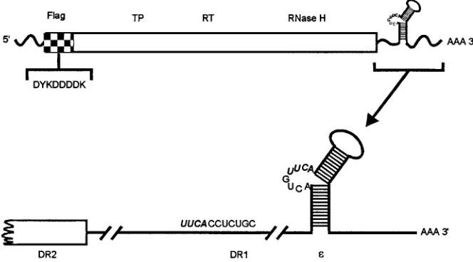
Galibert and coworkers (14). The FLAG–pol–stem-loop (FPL-pol) construct was cloned by the following procedure. The amino terminus of the pol open reading frame was first cloned into the HindIII site of the bacterial expression vector pFLAG-2 (International Biotechnologies Inc., New Haven, Conn.) by using PCR to generate the HindIII site adjacent to the pol AUG such that the pol open reading frame was in frame with the FLAG epitope. This construct encodes a fusion protein having the sequence Met-Asp-Tyr-Lys-Asp-Asp-Asp-Asp-Lys-Leu preceding the polymerase Met at nucleotide 2309 (Fig. 1). The amino terminus of the fusion protein was retrieved from pFLAG-2 and joined to the remainder of pol to create a construct terminating at the Ssp1 site at nucleotide 1638, 13 nucleotides downstream of the polymerase TAG termination codon. This con-struct was designated FP-pol for FLAG-pol. FPL-pol was created from FP-pol by the addition of 350 nucleotides to the BglII site at nucleotide 1988, which results in the addition of DR1 and the stem-loop structure present in the 39untranslated region of the pregenomic RNA. FPL-pol has a mutation changing nucleotide 1524 from G to C, altering pol amino acid 800 from a Gly to an Arg. This mutation introduces a protein kinase A recognition site into the carboxy termi-nus of pol as previously described (3). Both pol constructs were cloned into the baculovirus transfer vector pBacPAC9 (CLONTECH, Palo Alto, Calif.), and recombinant viruses were isolated.
SDS-PAGE and immunoblot analysis.Insect cell lysates and purified pol were disrupted in electrophoresis sample buffer containing 2% sodium dodecyl sulfate (SDS) and 2% 2-mercaptoethanol and were heated to 1008C for 5 min. Proteins were separated by SDS-polyacrylamide gel electrophoresis (PAGE) as previously described (23, 26). Gels from in vitro assays for pol function were stained with Coomassie blue, dried, and autoradiographed. For immunoblot analysis, pro-teins were electrophoretically transferred to a Flurotrans polyvinylidene difluo-ride (PVDF) blotting membrane (Pall Biosupport, Glen Cove, N.Y.), and mem-branes were processed as previously described (27) with first a 1/4,000 dilution of a rabbit antiserum to full-length pol and then125I-protein A (New England
Nuclear [NEN], Boston, Mass.). The rabbit antibody was produced against gel-purified full-length pol derived from the insoluble pellet of baculovirus-infected insect cells. The antiserum recognized epitopes in both the amino and carboxy termini of pol and had an immunoblot titer in excess of 1/32,000.
Immunoaffinity purification of pol.Spinner cultures (250 ml) were harvested at 48 h postinfection with recombinant baculoviruses. Cells were pelleted at 100
[image:2.612.138.470.74.258.2]3g for 10 min and washed three times with phosphate-buffered saline (PBS). The cell pellet was resuspended in 10 ml of PBS containing 10% glycerol, 0.5% Nonidet P-40, protease inhibitors (100mM leupeptin, 1 mM prefablock [Boehr-inger Mannheim, Indianapolis, Ind.], 10mM aprotinin, 10mg of pepstatin per ml, and 1 mM EDTA), 50 U of RNasin (Promega, Madison, Wis.) per ml, and 5 mM dithiothreitol. The cell pellet was extracted for 20 min on ice and clarified at 13,0003g for 15 min at 48C. The clarified extract was passed three times over an affinity column containing M2 monoclonal antibody (International Biotech-nologies Inc.) at a flow rate of 0.5 ml/min. M2 monoclonal antibody recognizes the sequence Asp-Tyr-Lys-Asp-Asp-Asp-Asp-Lys within the FLAG epitope. The column was washed sequentially with four volumes each of TNG (100 mM Tris HCl [pH 7.5], 30 mM NaCl, 10% glycerol), TNG with 1 M NaCl, and TNG at 0.5 ml/min. Bound polymerase was eluted with 0.1 M glycine (pH 3.0)–10% glycerol, collected in 1-ml fractions, and neutralized with 67ml of 0.8 M Tris HCl (pH FIG. 1. Structure of FPL-pol transcript from the baculovirus construct. The HBV pol open reading frame is represented in the top diagram as a rectangular box. At the amino terminus, the hatched section represents the FLAG epitope with its amino acid sequence shown below in single-letter code. The terminal protein (TP), RT, and RNase H domains are indicated above the polymerase open reading frame. The 39end of the transcript is enlarged below to detail the DR2, DR1, and epsilon (ε) regions. The four homologous nucleotides between the bulge in epsilon and the first four nucleotides of DR1 are shown in boldface type. DHBV pol undergoes a nucleotide priming reaction templated by the bulge in epsilon, and then the primed polymerase is translocated to the homologous region in DR1.
on November 9, 2019 by guest
http://jvi.asm.org/
mM Tris [pH 7.4], 10 mM NaCl, 10 mM EDTA, 1% SDS) for 2 h at 658C. DNA was extracted with phenol-chloroform and then chloroform and was ethanol precipitated as previously described (8). DNA from labeled reaction mixtures was analyzed on urea–8% acrylamide gels prepared as for a sequencing gel (41) but which were run in a standard gel unit at 400 V for 1.25 h. The gels were fixed in 7.5% trichloroacetic acid, dried, and autoradiographed. For Southern hybrid-ization, DNA was analyzed on 1% alkaline agarose gels which were run in 30 mM NaOH–1 mM EDTA for 4 h at 50 V (41), was transferred to a GeneScreen Plus membrane (NEN) by capillary transfer in 0.4 M NaOH, and was hybridized with strand-specific HBV riboprobes. Hybridization was conducted in 50% form-amide–7% SDS–0.25 M sodium phosphate–0.25 M NaCl–1 mM EDTA at 428C as previously described (8). The riboprobes were made by in vitro transcription with [a-32
P]UTP (NEN; 3,000 Ci/mmol) and T7 and T3 riboprobe kits (Pro-mega). The vector for riboprobes contained an HBV insert from BamHI nucle-otide 1403 to XbaI nuclenucle-otide 1993 in pBluescript(2) (Stratagene, La Jolla, Calif.). Transcription with T3 and T7 yielded probes complementary to minus-and plus-strminus-and DNAs, respectively.
Primer extension.Primer extension analysis was carried out essentially as described previously (43). The primer was complementary to minus-strand DNA, and the 39end was 20 nucleotides upstream of DR1 and spanned nucleotides 1786 to 1805 (59-TAGGCATAAATTGGTCTGCG-39). The primer was 59end labeled with [g-32P]ATP (NEN; 6,000 Ci/mmol) and T4 polynucleotide kinase.
DNA was purified as described above and annealed to 200 pg of the primer in a 10-ml volume by heating to 958C for 2 min and cooling on ice. Primer extension was conducted in a 15-ml reaction mixture (50 mM Tris [pH 8.0], 40 mM KCl, 6 mM MgCl2, 1 mM dithiothreitol, 100mM dNTPs, and 8 U of avian
myeloblas-tosis virus RT) for 1 h at 428C. The product was purified by extraction with phenol-chloroform and chloroform, ethanol precipitated, and analyzed on a sequencing gel (41) with a sequencing ladder generated with the same oligonu-cleotide.
Phosphoamino acid analysis.Polymerase reactions were conducted in the presence of a single nucleotide ([32P]TTP) for 1 h at 308C, and the labeled pol
band was isolated by SDS-PAGE and electrophoretic transfer to a PVDF mem-brane. The pol band was localized by autoradiography and excised from the membrane, and partial acid hydrolysis was conducted in situ on the membrane. Phosphoamino acid analysis was conducted as previously described (10). The membrane was treated with 6 N HCl in a nitrogen atmosphere for 1 h at 1108C. The products were lyophilized and resuspended in pH 1.9 electrophoresis buffer (88% formic acid, glacial acetic acid, and H2O in a ratio of 50:156:1,794). The
products were mixed with unlabeled phosphoamino acid standards (Sigma) and spotted onto thin-layer cellulose plates (Eastman Kodak Co., Rochester, N.Y.). First-dimension electrophoresis was conducted at 1,500 V for 20 min in pH 1.9 buffer. The second-dimension electrophoresis was conducted at 1,300 V for 16 min in pH 3.5 buffer (pyridine, glacial acetic acid, and H2O in a ratio of 10:100:
1,890). The unlabeled amino acid standards were localized with ninhydrin (0.25% in acetone) and by heating to 608C for 15 min, and labeled phos-phoamino acids were localized by autoradiography.
RESULTS
Expression of HBV polymerase in insect cells.In attempts to develop an in vitro system for the analysis of HBV polymerase function, we have expressed pol in insect cells by using the recombinant baculovirus expression system. In our initial stud-ies, full-length pol and individual pol domains were expressed from different recombinant viruses. Analysis of posttransla-tional processing of pol demonstrated that at least two distinct sites on pol were phosphorylated (1). No definitive polymerase function was demonstrated by using these original constructs despite efforts to biochemically purify a functional polymerase as well as attempts to refold pol with purified cellular chaper-ones (data not shown).
In the current studies, attempts to express a functional poly-merase involved two changes in the pol expression vector.
First, an affinity tag was added to the amino terminus to facil-itate rapid purification. The 10-amino-acid FLAG sequence (Met-Asp-Tyr-Lys-Asp-Asp-Asp-Asp-Lys-Leu) was fused to the amino terminus of pol such that pol could be purified on M2 monoclonal antibody columns that recognize an epitope in this sequence. Second, the 39 terminus of the construct was extended beyond the pol open reading frame to include DR1 and the epsilon stem-loop structure (Fig. 1) shown to be im-portant for function with in vitro-translated DHBV pol (54). The construct containing the affinity tag and the 39extension was designated FPL-pol to denote FLAG–pol–stem-loop.
Sf9 cells were infected with the FPL-pol virus and were harvested at 48 h postinfection. Equivalent amounts of the particulate and soluble fractions of a cell lysate were analyzed by SDS-PAGE and Coomassie blue staining. pol was expressed at high levels in insect cells, but most of the pol was present in the particulate fraction (Fig. 2A). A prominent pol band with a molecular weight of approximately 84,000 was detected by Coomassie blue staining of the gel. Although a band was ob-served at the same position in the soluble fraction, analysis of these fractions by Western blot (immunoblot) suggested that insufficient pol was present in the soluble fraction to account for the band detected by Coomassie blue staining. Although fivefold less of the particulate fraction than of the soluble fraction was analyzed by Western blot, much greater pol reac-tivity was present in this fraction. The band detected by Coo-massie blue staining in the soluble fraction is likely an insect or baculovirus protein.
[image:3.612.319.547.72.219.2]pol was affinity purified from the soluble fraction of a cell lysate derived from FPL-pol-infected Sf9 cells with an affinity resin containing the M2 monoclonal antibody. Western blot analysis of equivalent amounts of the starting material and of the unbound and bound fractions indicated that essentially all of the soluble pol bound to the column. The purified material was analyzed by SDS-PAGE and Coomassie blue staining, and a prominent pol band with a molecular weight of approxi-mately 84,000 was detected (Fig. 2B). Several other prominent FIG. 2. Purification of FPL-pol. Sf9 cells were harvested at 48 h postinfection with the recombinant baculovirus FPL-pol. (A) SDS-PAGE analysis of cell lysate prepared by sonication in PBS into particulate and soluble fractions. The par-ticulate (P) and soluble (S) fractions from the cell extract were analyzed by Coomassie blue (CB; 1/1,000 of each fraction) staining and Western blot (WB; 1/5,000 of the particulate fraction and 1/1,000 the soluble fraction). (B) A cell extract was prepared with detergent extraction. Samples from the purification were analyzed by SDS-PAGE and Western blot (WB). The starting soluble fraction (ST), the unbound fraction (UN), and the eluate (E) of the immunoaf-finity purification were analyzed by Western blot (1/1,000 each). The purified pol was examined by Coomassie blue staining as well (1/140 of the purified fraction). M, molecular weight standards (indicated in thousands by the numbered arrow-heads on the left).
on November 9, 2019 by guest
http://jvi.asm.org/
polypeptides were present in the purified pol preparations. A band of approximately 70,000 molecular weight failed to im-munoblot with either the rabbit antiserum to pol or the M2 monoclonal antibody, as did bands of 56,000 and 46,000 mo-lecular weight. A prominent band with a momo-lecular weight of approximately 110,000 failed to react with the antiserum to pol, but did react with the M2 antibody, suggesting that this band was a cellular protein that contained an epitope cross-reactive with M2. The 110,000-molecular-weight protein could also be purified from insect cells infected with a baculovirus encoding simian virus 40 T antigen by using the M2 column (data not shown). Despite the fact that most pol synthesized in insect cells is insoluble, the high level of expression in insect cells coupled with immunoaffinity purification rendered purification of significant quantities of pol feasible.
Detection of HBV minus-strand DNA covalently linked to purified pol.The purified preparations of pol were examined for the presence of HBV DNA to determine whether nucle-otide priming and minus-strand DNA synthesis had occurred in the baculovirus-infected insect cells. Purified pol was ex-tracted with phenol with or without prior treatment with pro-teinase K to determine whether the minus-strand DNA was covalently linked to pol. The DNA products were analyzed by electrophoresis on denaturing, alkaline agarose gels and by Southern hybridization with strand-specific riboprobes. The results demonstrated that the pol mRNA had been reverse transcribed in the insect cells, since a DNA product was de-tected with a minus-strand-specific probe. No product was detected with a plus-strand-specific probe (Fig. 3). The minus-strand DNA ranged from approximately 100 to 500 nucleo-tides. The lack of proteinase K treatment resulted in the com-plete loss of the DNA products, suggesting that the minus-strand DNA was covalently linked to pol.
The appropriate synthesis of minus-strand DNA not only involved covalent linkage of the DNA to pol but also resulted in a product with 59 ends mapping to DR1. The 59 end of virion-associated minus-strand DNA for HBV maps to either the 3rd or 4th nucleotide of DR1, a G residue at nucleotide 1828 or a T residue at 1829 (40, 58). These residues are within the sequence TGAA, the complement of which is found on pregenomic RNA within DR1 and in the bulge within the epsilon stem-loop. In vitro reactions with DHBV pol have demonstrated that the priming reaction is initiated on the stem-loop by the addition of four nucleotides to pol and this initiation complex is transferred to DR1 for elongation of minus-strand DNA (53, 55).
Primer extension analysis was performed to map the 59end of the minus-strand DNA associated with purified pol. The DNA associated with pol was purified and annealed with a 32P-labeled oligonucleotide complementary to a sequence 24
nucleotides from the 59 end of the viral minus-strand DNA, and the oligonucleotide was extended with avian myeloblasto-sis virus RT. The primer extension products were analyzed on a sequencing gel along with a ladder generated by sequenc-ing of cloned HBV DNA with the primer used in the exten-sion analysis. The results revealed that the most abundant product mapped to a G residue at nucleotide 1828 (a C resi-due on pregenomic RNA), while longer exposure of the gel revealed an additional band and suggested that the 59 end may map to the T residue at nucleotide 1829 (Fig. 4) or this product may reflect the addition of a nontemplated nucleotide by avian myeloblastosis virus RT (13). Our analyses cannot determine the exact 59end of minus-strand DNA. The same 59 extension product was obtained from viral DNA purified from an infectious plasma (data not shown). The T and G residues correspond to the complementary ACUU sequence present in DR1 of pregenomic RNA. The 59 terminus of minus-strand DNA from in vitro DHBV pol nucleotide priming-reverse transcription reactions maps to both DR1 and the bulge within the 39epsilon stem-loop (54). Presumably, minus-strand DNA mapping to the stem-loop reflects a failure to translocate the initiated complex to DR1 (53, 55). Upon prolonged exposure of the gel, we could detect faint bands mapping to the ACUU (nucleotide 1868) present in the bulge of the epsilon stem-loop, as well as to an ACUU sequence at 1854 (part of the lower stem of epsilon), an unrelated sequence at approxi-mately 1930, and an ACUU sequence at 1980.
Nucleotide binding and RT activity.A modification of the in vitro nucleotide priming-reverse transcription assay described for analysis of DHBV pol (54) was utilized to examine in vitro FIG. 3. Analysis of HBV DNA associated with purified polymerase. DNA
[image:4.612.390.479.69.267.2]associated with purified FPL-pol was extracted with phenol with (1) or without (2) prior treatment with proteinase K (Prot. K). The DNA products were analyzed by alkaline agarose electrophoresis, were transferred to a nylon mem-brane, and were hybridized with riboprobes complementary to minus-strand DNA (2DNA) or positive-strand DNA (1DNA). M, labeled HindIIIlDNA markers (indicated in base pairs by the numbered arrowheads at the left).
FIG. 4. Primer extension analysis of pol-associated minus-strand DNA. DNA associated with FPL-pol was purified following treatment with RNase A and proteinase K, and the purified DNA was annealed with a 59-end-labeled oligo-nucleotide spanning oligo-nucleotides 1786 to 1805. The primer was extended by using avian myeloblastosis virus RT as described in Materials and Methods. Primer extension (PE) products (arrowhead) were analyzed on an 8% polyacrylamide sequencing gel adjacent to a sequencing ladder (lanes G, A, T, and C) generated by dideoxysequencing reactions with HBV plasmid DNA and the same primer that was used for the primer extension products. The 11-nucleotide sequence of DR1 on pregenomic RNA is bracketed and labeled with the sequence. The complementary sequence is present on the 59end of viral minus-strand DNA.
on November 9, 2019 by guest
http://jvi.asm.org/
activity of purified HBV pol. When the purified material was examined by SDS-PAGE following the pol assays, a heavily labeled smear appeared (Fig. 5A, lane Con) beginning at the position of the Coomassie blue-stained pol band. Presumably, the labeling at the position of the pol band represents covalent linkage of a single nucleotide to pol in the nucleotide priming reaction, and the labeling of the higher-molecular-weight ma-terial represents the extension of this product by reverse tran-scription.
As would be expected for a hepadnavirus polymerase (49, 54), the reaction was not inhibited by aphidicolin, an inhibitor of cellular DNA polymerases, or actinomycin D, an inhibitor of DNA-template DNA synthesis. The labeling of the pol band in separate reaction mixtures containing these two inhibitors was less intense than that in the control reaction mixture because of the ethanol used as a solvent for the inhibitors, as can be seen when the control reaction is performed at the same eth-anol concentration. Inclusion of phosphonoformic acid (PFA) in the reaction buffer resulted in the labeling of the pol band without labeling of higher-molecular-weight species, suggest-ing that PFA does not inhibit the nucleotide primsuggest-ing reaction but does inhibit elongation (32, 54). Inclusion of EDTA in the buffer blocked all labeling, confirming that both polymerase activity (20) and the priming reaction (54) require Mg21. Treatment of purified pol with RNase A prior to the poly-merase reaction abolished most of the activity (in comparison with that of the control reaction), indicating that the reaction was dependent on an RNA template. Use of increased con-centrations of RNase A completely abolished the pol reaction. Treatment with DNase after the polymerase reaction removed the label present in the upper smear on the gel, but the labeled material at the position of pol was protected from DNase digestion. Treatment of the pol reaction product with protein-ase K eliminated the labeled band at the position of pol, suggesting, as expected, that the label is associated with pro-tein. To demonstrate the specificity of the reaction for HBV
pol, polymerase reactions were conducted with material puri-fied on the M2 column by using a cell extract from insect cells infected with a recombinant baculovirus that expresses simian virus 40 T antigen (25). No labeled product from these reac-tions was detected (Fig. 5B; lane T-ag).
Next, the temperature optimum and time course of the pol reaction were examined. pol reactions were conducted for 30 min at 15, 23, 30, 37, and 428C. The intensity of labeling at the position of the pol band was greatest at 308C. At 378C, the degree of labeling was similar to that at 308C, but the labeled material shifted to a higher apparent molecular weight. Incor-poration of the label appeared to be diminished by incubation at 428C. When the same reactions were performed in the presence of [32P]TTP but in the absence of unlabeled dNTPs, the reaction was most intense at 308C, suggesting that priming may proceed best at 308C, while the temperature optimum for elongation may be 378C. When the pol reaction was conducted at 308C for 5 min to 3 h, a progressive increase in the amount of label in the high-molecular-weight smear was observed through the 3-h time point (Fig. 6).
[image:5.612.345.525.72.226.2]Recent studies with DHBV pol have demonstrated that the first nucleotide of minus-strand DNA (the third nucleotide of DR1, a G residue) is templated by a nucleotide sequence in epsilon (53, 55) and that the in vitro priming reaction has a high degree of specificity for the incorporation of a G residue unless the residue in epsilon is changed by mutagenesis. To determine whether the in vitro priming reaction for HBV pol displayed specificity for a specific residue as the first nucle-otide, the pol reactions were conducted with a single labeled nucleotide in the absence of unlabeled nucleotides. Incorpo-ration of labeled TTP in the presence of dATP, dCTP, and dGTP was much greater than incorporation in the absence of all four dNTPs (Fig. 7A). pol was labeled with each of the four dNTPs in reactions with only a single nucleotide present. In-corporation of the label in the pol band was greatest with TTP, followed by dGTP and dATP. The level of labeling of the pol band with dCTP was very low. This is the same order of appearance of these nucleotides as that in the complementary sequence of DR1 starting at nucleotide 1829 (ACUU) which is also found within epsilon. Whether the increased incorpora-tion of TTP suggests that priming preferentially occurs at the FIG. 5. In vitro polymerase assay. Immunoaffinity-purified FPL-pol was
ex-amined in an in vitro nucleotide priming-reverse transcription assay as described in Materials and Methods. Purified pol was incubated in the presence of [32P]TTP and unlabeled dGTP, dATP, and dCTP for 30 min at 308C under
various conditions. The products of the polymerase assay were analyzed by SDS-PAGE and autoradiography. For these assays, one-third of the amount of purified pol shown by Coomassie blue staining in Fig. 2 was used in each reaction mixture. (A) Analysis of inhibitors in polymerase assays. Lanes: Con, control with standard reaction conditions; Eth, control with 2% ethanol added to the standard reaction mixture; Aph, 20mM aphidicolin and 2% ethanol; Act, 100mg of actinomycin D per ml and 2% ethanol; PFA, 1 mM PFA; EDTA, 20 mM EDTA; RNase, pretreatment with 1mg of RNase A per ml for 10 min at 308C; DNase, posttreatment with 100 U of DNase per ml for 30 min at 378C; PtK, posttreatment with 1 mg of proteinase K per ml in 1% SDS for 2 h at 658C. The pol protein band is indicated by an arrow. (B) Specificity of polymerase reaction. Cell lysates from Sf9 spinner cultures infected with recombinant baculoviruses FPL-pol (Pol) and 941T (which encodes simian virus 40 T antigen [T-ag]) were purified on the M2 affinity column, and polymerase reactions were conducted with the purified proteins under the reaction conditions described above. La-beled polymerase products were detected only in assays using FPL-pol.
FIG. 6. Temperature optimum and time course for polymerase reactions. In vitro nucleotide priming-reverse transcription assays were conducted with puri-fied FPL-pol with [32P]TTP and unlabeled dGTP, dATP, and dCTP. pol reaction
mixtures were incubated for 30 min at 15, 23, 30, 37, and 428C, or alternatively pol reactions were conducted at 308C for 5, 10, or 30 min and 1.5 or 3 h. All reaction products were analyzed by SDS-PAGE and autoradiography. The pol protein band is indicated by an arrowhead.
on November 9, 2019 by guest
http://jvi.asm.org/
[image:5.612.72.281.73.181.2]A residue or whether pol has a higher affinity for TTP than for the other dNTPs cannot be determined from these data. How-ever, these results suggest that HBV pol may be capable of priming with different nucleotides. Alternatively, the first nu-cleotide may have been added to pol in the insect cells and this reaction represents an elongation of a preexisting product. Comparison of the narrow band obtained in reactions with PFA or in reactions with only a single nucleotide to the higher-molecular-weight smear obtained when the standard reactions are performed suggests that in these reactions HBV pol is adding the first nucleotide in vitro (Fig. 7B).
To conclusively demonstrate that nucleotide priming occurs in vitro requires the detection of labeled phosphotyrosine on pol following the in vitro reaction. If the first nucleotide is added to pol in vitro, the32P-labeled phosphate in the alpha position of the labeled dNTP will become covalently attached to tyrosine. In vitro pol reactions were conducted in the pres-ence of a single nucleotide ([32P]TTP), and the pol band was isolated by SDS-PAGE and transfer to a PVDF membrane. Phosphoamino acid analysis was conducted by two-dimen-sional electrophoresis as described in Materials and Methods. A labeled spot was detected at the position of phosphoty-rosine, demonstrating that the purified pol was competent for in vitro nucleotide priming (Fig. 8). Consistent with the above data indicating that the nucleotide priming reaction could oc-cur with different nucleotides, phosphotyrosine was also de-tected when the reaction was conducted with [32P]dGTP in the absence of other nucleotides (data not shown).
Analysis of in vitro-synthesized HBV DNA.To examine the size of the in vitro-synthesized DNA products and to deter-mine whether the DNA products were covalently bound to protein, polymerase reaction mixtures were extracted with phenol with or without prior treatment with proteinase K to remove covalently bound protein (16) and the DNA products were examined by electrophoresis in urea-acrylamide gels prior to autoradiography. In the absence of treatment with proteinase K, the labeled DNA products were almost com-pletely lost in the phenol phase, while the sample treated with proteinase K prior to phenol extraction yielded an intensely labeled smear on the denaturing DNA gel (Fig. 9). The size of
the single-stranded DNA product was estimated by compari-son with denaturedfX174 Hinf1 DNA markers. The products ranged from less than 25 to more than 60 nucleotides. The small size of the in vitro-synthesized DNA products was in part due to the limiting size of the RNA template (see Discussion). No DNA products from reactions conducted in the presence of PFA were detected.
DISCUSSION
[image:6.612.102.255.70.232.2]The expression of a functional HBV polymerase has been especially problematic. Although pol is highly active in an endogenous polymerase assay using secreted mature virions or immature cytoplasmic cores, attempts to purify pol in an active soluble form have for the most part yielded unsatisfactory results. The inability of encapsidated pol to switch to an exog-enously provided template was especially discouraging. Some success has been reported for the refolding of denatured pol in activity gel assays, but these approaches have limited utility for the in vitro analysis of genome replication. The failure to express functional pol led to speculation that encapsidation or some accessory function of the core protein may be required for pol function. Reports of two in vitro models for analysis of DHBV pol (52, 54) and a report on RT activity from Xenopus FIG. 7. Nucleotide preference of HBV pol for in vitro nucleotide priming
reaction. (A) Polymerase reactions were conducted with purified FPL-pol and unlabeled dNTPs with [32P]TTP (1dNTPs, lane T) or without unlabeled dNTPs
and with only the specified32P-labeled dNTP (2dNTPs, lanes T, G, A, and C).
(B) Polymerase reactions were conducted with three unlabeled dNTPs (G, A, and C) and [32P]TTP; without unlabeled dNTPs and with only [32P]TTP; or with
unlabeled dNTPs (G, A, and C), 1 mM PFA, and [32P]TTP (1PFA, lane T). The
pol band is indicated by an arrowhead.
FIG. 8. Phosphoamino acid analysis of in vitro-labeled pol. Polymerase re-actions were conducted with [32
P]TTP in the absence of other unlabeled nucle-otides. The labeled polymerase products were separated by SDS-PAGE and electrophoretically transferred to a PVDF blotting membrane, and the labeled pol band was excised. Phosphoamino acid analysis was conducted as described in Materials and Methods. Partial acid hydrolysis was conducted at 1108C for 1 h, and the products were analyzed by two-dimensional electrophoresis on thin-layer cellulose plates prior to autoradiography. The positions of phosphotyrosine (P-Y), phosphoserine (P-S), and phosphothreonine (P-T) were determined by nin-hydrin staining of unlabeled amino acid standards. First (1dim) and second (2dim) dimensions are indicated, as well as the location of free phosphate (Pi).
FIG. 9. Analysis of DNA synthesized in in vitro polymerase reactions. Poly-merase reactions with purified FPL-pol were conducted, using [32
P]TTP and unlabeled dATP, dGTP, and dCTP either in the absence (2PFA) or presence (1PFA) of PFA. The DNA products were purified with (1) or without (2) prior treatment with proteinase K (Prot. K). The products were analyzed by denatur-ing urea-acrylamide gel electrophoresis and autoradiography. M, denatured,
32
P-labeledfX174 Hinf1 DNA markers (base pairs indicated by the numbered arrowheads).
on November 9, 2019 by guest
http://jvi.asm.org/
purification of pol expressed from an mRNA lacking 59or 39 copies of DR1 and epsilon. Although limited activity was ob-served with an occasional preparation, the results of these studies were mostly negative. The difficulties experienced in the purification of significant quantities of pol led to the inclu-sion of an affinity tag in the pol sequence despite concerns with regard to altering pol function. In addition, we chose to include a 39copy of DR1 and epsilon on the pol mRNA because of the success of such constructs with DHBV pol. The inclusion of an affinity tag at the amino terminus permitted the rapid purifi-cation of large quantities of pol active in nucleotide priming and RT assays.
Our initial studies with FPL-pol detected both minus- and positive-strand HBV DNAs following in vitro pol reactions, and surprisingly, most of the minus-strand DNA was not co-valently linked to pol (data not shown). Further analyses sug-gested that low levels of contaminating HBV DNA from the baculovirus vector were serving as the templates in these re-actions. Modification of the purification procedure reduced contaminating baculovirus DNA to undetectable levels and resulted in the detection of only minus-strand DNA, all of which was covalently linked to pol. These data suggested that pol can utilize templates other than the pol mRNA. Recent studies with DHBV pol indicate that pol possesses polymerase activity in the absence of nucleotide priming (56). This obser-vation would in part explain the high level of activity we ob-served for pol when a baculovirus DNA template was used, as well as the lack of covalent linkage of pol to minus-strand DNA in these reactions. Northern (RNA) hybridization analyses of pol-associated RNA suggest that significant levels of pol mRNA copurify with pol, albeit in a highly degraded state (data not shown). The copurification of pol mRNA with pol is consistent with the cis preference of pol in the packaging of pregenomic RNA (2, 17–19, 21, 36) and may rely on the 39 copy of epsilon in the FPL-pol construct. Taken together, the above data indicate that the template may be limiting in the pol reactions; in support of this assumption, we have observed that pol activity and the size of the DNA products are increased in the presence of exogenously supplied synthetic RNA templates (data not shown).
Our analyses suggest that at least two populations of pol are present in the purified preparations. One population consists of molecules possessing covalently linked minus-strand DNA with the 59end mapping to DR1. These molecules were pre-sumably active in the insect cells but did not appear to be active following purification. The lack of in vitro activity for pol primed in vivo is assumed, since labeling with a single nucle-otide did not label higher-molecular-weight material at detect-able levels, which would be consistent with the addition of a nucleotide to the preexisting minus-strand DNA. Presumably, a second pol population was active in vitro and underwent nucleotide priming in vitro, as judged by the appearance of 32P-labeled phosphotyrosine. A third population which was not
active either in the insect cells or in vitro likely exists as well, since much of the pol band did not shift in mobility following
Attempts are under way to increase the fraction of pol active in in vitro assays by modification of the purification scheme as well as by alteration of the conditions for the in vitro assay, including the addition of exogenous templates.
The in vitro polymerase reaction of HBV pol purified from insect cells differs in several properties from the DHBV in vitro pol systems previously described. DHBV pol appears to display an absolute specificity for the first nucleotide in the priming reaction. For DHBV pol, the priming reaction requires dGTP which is templated by the bulge in epsilon. When the C residue in the bulge is mutated to G, the priming reaction requires dCTP (55). In vitro, HBV pol was labeled with each of the four dNTPs in reaction mixtures with a single nucleotide present, albeit at very low levels with dCTP. For TTP and dGTP, covalent linkage directly to pol was demonstrated by the pres-ence of32P-labeled phosphotyrosine. In reaction mixtures sup-plemented with only a single labeled dNTP, labeling of the pol band was most intense with TTP. Extrapolating the mechanism of priming from DHBV to HBV suggests that the preference for priming with a T residue may be due to priming at the A residue in the sequence ACUU found in the epsilon bulge and DR1. The priming of HBV pol with a T residue would result in the 59end of minus-strand DNA being nucleotide 1829. The 59 end of HBV minus-strand DNA has been mapped to both the 3rd (G at 1828) and 4th (T at 1829) nucleotides of DR1 (40, 58). An alternative explanation is that the increased incorpo-ration of TTP merely reflects an increased affinity for TTP. Mutations in the epsilon bulge are required to further eluci-date the specificity of the in vitro HBV pol priming reaction. A second difference between HBV pol and DHBV pol was observed in the 59extension products from minus-strand DNA. The 59ends of minus-strand DNA from in vitro reactions with DHBV map to two sites, one at the correct end for minus-strand DNA within DR1 and a second abundant product that maps to the priming site within the epsilon bulge (55). Almost none of the 59 extension product of minus-strand DNA mapped to epsilon for the HBV DNA synthesized in the insect cells, indicating either a more efficient strand transfer to DR1 than for DHBV or direct initiation at DR1. The efficiency of in vitro strand transfer for HBV pol cannot be determined, since the 59ends of the in vitro-synthesized DNA cannot be distin-guished from the product synthesized within insect cells.
At this time, pol reactions are conducted with partially pu-rified material that contains several other prominent polypep-tides. At least one of the polypeptides copurifies with pol because of a fortuitous reactivity with the M2 antibody that is reactive with the FLAG sequence. The nature of the other copurifying polypeptides is not known. It is of interest that Pollack and Ganem (37) have recently speculated on the po-tential for cellular proteins involved in DHBV RNA packaging and pol priming. This speculation is based on the observation that certain mutations in the loop domain of DHBV epsilon do not abolish pol binding but severely reduce DNA priming and RNA packaging.
Although our studies are still preliminary, the availability of
on November 9, 2019 by guest
http://jvi.asm.org/
substantial quantities of partially purified HBV pol that is active in nucleotide priming and reverse transcription should permit many types of functional and physical studies not pre-viously possible.
ACKNOWLEDGMENTS
We thank Shannon Snyder for excellent technical assistance and Don Jarvis for assistance with phosphoamino acid analysis.
This work was supported by grants from the National Institutes of Health.
REFERENCES
1. Ayola, B., P. Kanda, and R. E. Lanford. 1993. High level expression and phosphorylation of hepatitis B virus polymerase in insect cells with recom-binant baculoviruses. Virology 194:370–373.
2. Bartenschlager, R., M. Junker-Niepmann, and H. Schaller. 1990. The P gene product of hepatitis B virus is required as a structural component for genomic RNA encapsidation. J. Virol. 64:5324–5332.
3. Bartenschlager, R., C. Kuhn, and H. Schaller. 1992. Expression of the P-protein of the human hepatitis B virus in a vaccinia virus system and detection of the nucleocapsid-associated P-gene product by radiolabelling at newly introduced phosphorylation sites. Nucleic Acids Res. 20:195–202. 4. Bartenschlager, R., and H. Schaller. 1988. The amino-terminal domain of
the hepadnaviral P-gene encodes the terminal protein (genome-linked pro-tein) believed to prime reverse transcription. EMBO J. 7:4185–4192. 5. Bartenschlager, R., and H. Schaller. 1992. Hepadnaviral assembly is
initi-ated by polymerase binding to the encapsidation signal in the viral RNA genome. EMBO J. 11:3413–3420.
6. Bavand, M., M. Feitelson, and O. Laub. 1989. The hepatitis B virus-associ-ated reverse transcriptase is encoded by the viral pol gene. J. Virol. 63:1019– 1021.
7. Bavand, M. R., and O. Laub. 1988. Two proteins with reverse transcriptase activities associated with hepatitis B virus-like particles. J. Virol. 62:626–628. 8. Beames, B., and R. E. Lanford. 1993. Carboxy-terminal truncations of the HBV core protein affect capsid formation and size of the encapsidated HBV RNA. Virology 194:597–607.
9. Bosch, V., R. Bartenschlager, G. Radziwill, and H. Schaller. 1988. The duck hepatitis B virus P-gene codes for protein strongly associated with the 59-end of the viral DNA minus strand. Virology 166:475–485.
10. Boyle, W. J., P. Van der Geer, and T. Hunter. 1991. Phosphopeptide mapping and phosphoamino acid analysis by two-dimensional separation on thin-layer cellulose plates. Methods Enzymol. 201:110–149.
11. Calvert, J., and J. Summers. 1994. Two regions of an avian hepadnavirus RNA pregenome are required in cis for encapsidation. J. Virol. 68:2084– 2090.
12. Chang, L.-J., R. C. Hirsch, D. Ganem, and H. E. Varmus. 1990. Effects of insertional and point mutations on the functions of the duck hepatitis B virus polymerase. J. Virol. 64:5553–5558.
13. Condreay, L. D., T.-T. Wu, C. E. Aldrich, M. A. Delaney, J. Summers, C.
Seeger, and W. S. Mason.1992. Replication of DHBV genomes with muta-tions at the sites of initiation of minus- and plus-strand DNA synthesis. Virology 188:208–216.
14. Galibert, F., E. Mandart, F. Fitoussi, P. Tiollais, and P. Charnay. 1979. Nucleotide sequence of the hepatitis B virus genome (subtype ayw) cloned in E. coli. Nature (London) 281:646–650.
15. Ganem, D., and H. E. Varmus. 1987. The molecular biology of the hepatitis B viruses. Annu. Rev. Biochem. 56:651–693.
16. Gerlich, W. H., and W. S. Robinson. 1980. Hepatitis B virus contains protein attached to the 59terminus of its complete DNA strand. Cell 21:801–809. 17. Hirsch, R. C., J. E. Lavine, L.-J. Chang, H. E. Varmus, and D. Ganem. 1990.
Polymerase gene products of hepatitis B viruses are required for genomic RNA packaging as well as for reverse transcription. Nature (London) 344: 552–555.
18. Hirsch, R. C., D. D. Loeb, J. R. Pollack, and D. Ganem. 1991. cis-acting sequences required for encapsidation of duck hepatitis B virus pregenomic RNA. J. Virol. 65:3309–3316.
19. Junker-Niepmann, M., R. Bartenschlager, and H. Schaller. 1990. A short cis-acting sequence is required for hepatitis B virus pregenome encapsida-tion and sufficient for packaging of foreign RNA. EMBO J. 9:3389–3396. 20. Kaplan, P. M., R. L. Greenman, J. L. Gerin, R. H. Purcell, and W. S.
Robinson.1973. DNA polymerase associated with human hepatitis B anti-gen. J. Virol. 12:995–1005.
21. Knaus, T., and M. Nassal. 1993. The encapsidation signal on the hepatitis B virus RNA pregenome forms a stem-loop structure that is critical for its function. Nucleic Acids Res. 21:3967–3975.
22. Ko¨chel, H., M. Kann, and R. Thomssen.1991. Identification of a binding site in the hepatitis B virus RNA pregenome for the viral pol gene product. Virology 182:94–101.
23. Laemmli, U. K. 1970. Cleavage of structural proteins during the assembly of
the head of bacteriophage T4. Nature (London) 227:680–685.
24. Landers, T. A., H. B. Greenberg, and W. S. Robinson. 1977. Structure of hepatitis B Dane particle DNA and nature of the endogenous DNA poly-merase reaction. J. Virol. 23:368–376.
25. Lanford, R. E. 1988. Expression of simian virus 40 T antigen in insect cells using a baculovirus expression vector. Virology 167:72–81.
26. Lanford, R. E., and J. S. Butel. 1979. Antigenic relationship of SV40 early proteins to purified large T polypeptide. Virology 97:295–306.
27. Lanford, R. E., K. D. Carey, L. E. Estlack, G. C. Smith, and R. V. Hay. 1989. Analysis of plasma protein and lipoprotein synthesis in long-term primary cultures of baboon hepatocytes maintained in serum-free medium. In Vitro Cell. Dev. Biol. 25:174–182.
28. Lanford, R. E., and L. Notvall. 1990. Expression of hepatitis B virus core and precore antigens in insect cells and characterization of a core-associated kinase activity. Virology 176:222–233.
29. Lien, J., D. J. Petcu, C. E. Aldrich, and W. S. Mason. 1987. Initiation and termination of duck hepatitis B virus DNA synthesis during virus maturation. J. Virol. 61:3832–3840.
30. Lien, J.-M., C. E. Aldrich, and W. S. Mason. 1986. Evidence that a capped oligoribonucleotide is the primer for duck hepatitis B virus plus-strand DNA synthesis. J. Virol. 57:229–236.
31. Loeb, D. D., R. C. Hirsch, and D. Ganem. 1991. Sequence-independent RNA cleavages generate the primers for plus strand DNA synthesis in hepatitis B viruses: implications for other reverse transcribing elements. EMBO J. 10: 3533–3540.
32. Mason, W. S., J. Lien, D. J. Petcu, L. Coates, W. T. London, A. O’Connell,
C. Aldrich, and R. P. Custer.1987. In vivo and in vitro studies on duck hepatitis B virus replication, p. 3–16. In W. S. Robinson, K. Koike, and H. Will (ed.), Hepadna viruses. A. R. Liss, New York.
33. Molnar-Kimber, K. L., J. Summers, J. M. Taylor, and W. S. Mason. 1983. Protein covalently bound to minus-strand DNA intermediates of duck hep-atitis B virus. J. Virol. 45:165–172.
34. Molnar-Kimber, K. L., J. W. Summers, and W. S. Mason. 1984. Mapping of the cohesive overlap of duck hepatitis B virus DNA and of the site of initiation of reverse transcription. J. Virol. 51:181–191.
35. Oberhaus, S. M., and J. E. Newbold. 1993. Detection of DNA polymerase activities associated with purified duck hepatitis B virus core particles by using an activity gel assay. J. Virol. 67:6558–6566.
36. Pollack, J. R., and D. Ganem. 1993. An RNA stem-loop structure directs hepatitis B virus genomic RNA encapsidation. J. Virol. 67:3254–3263. 37. Pollack, J. R., and D. Ganem. 1994. Site-specific RNA binding by a hepatitis
B virus reverse transcriptase initiates two distinct reactions: RNA packaging and DNA synthesis. J. Virol. 68:5579–5587.
38. Radziwill, G., W. Tucker, and H. Schaller. 1990. Mutational analysis of the hepatitis B virus P gene product: domain structure and RNase H activity. J. Virol. 64:613–620.
39. Radziwill, G., H. Zentgraf, H. Schaller, and V. Bosch. 1988. The duck hepatitis B virus DNA polymerase is tightly associated with the viral core structure and unable to switch to an exogenous template. Virology 163:123– 132.
40. Saldanha, J. A., H. Qiu, H. C. Thomas, and J. Monjardino. 1992. Mapping of 59ends of virion-derived HBV DNA. Virology 188:358–361.
41. Sambrook, J., E. F. Fritsch, and T. Maniatis. 1989. Molecular cloning: a laboratory manual, 2nd ed. Cold Spring Harbor Laboratory Press, Cold Spring Harbor, N.Y.
42. Seeger, C., D. Ganem, and H. E. Varmus. 1986. Biochemical and genetic evidence for the hepatitis B virus replication strategy. Science 232:477–484. 43. Seeger, C., and J. Maragos. 1989. Molecular analysis of the function of direct repeats and a polypurine tract for plus-strand DNA priming in woodchuck hepatitis virus. J. Virol. 63:1907–1915.
44. Seeger, C., and J. Maragos. 1990. Identification and characterization of the woodchuck hepatitis virus origin of DNA replication. J. Virol. 64:16–23. 45. Seeger, C., and J. Maragos. 1991. Identification of a signal necessary for
initiation of reverse transcription of the hepadnavirus genome. J. Virol.
65:5190–5195.
46. Seeger, C., J. Summers, and W. S. Mason. 1991. Viral DNA synthesis. Curr. Top. Microbiol. Immunol. 168:41–60.
47. Seifer, M., and D. N. Standring. 1993. Recombinant human hepatitis B virus reverse transcriptase is active in the absence of the nucleocapsid or the viral replication origin, DR1. J. Virol. 67:4513–4520.
48. Staprans, S., D. D. Loeb, and D. Ganem. 1991. Mutations affecting hepad-navirus plus-strand DNA synthesis dissociate primer cleavage from translo-cation and reveal the origin of linear viral DNA. J. Virol. 65:1255–1262. 49. Summers, J., and W. S. Mason. 1982. Replication of the genome of a
hepatitis B-like virus by reverse transcription of an RNA intermediate. Cell
29:403–415.
50. Summers, J., A. O’Connell, and I. Millman. 1975. Genome of hepatitis B virus: restriction enzyme cleavage and structure of DNA extracted from Dane particles. Proc. Natl. Acad. Sci. USA 72:4597–4601.
51. Summers, M. D., and G. E. Smith. 1987. A manual of methods for baculo-virus vectors and insect cell culture procedures. Tex. Agric. Exp. Stn. Bull.
1555:1–48. (Plus appendix.)