0022-538X/96/$04.00
1
0
Copyright
q
1996, American Society for Microbiology
Reversion of a Human Immunodeficiency Virus Type 1 Integrase
Mutant at a Second Site Restores Enzyme Function
and Virus Infectivity
BRUNELLA TADDEO,
1* FRANCESCA CARLINI,
1PAOLA VERANI,
1ANDALAN ENGELMAN
2Laboratory of Virology, Istituto Superiore di Sanita
`, Rome, Italy,
1and Division of Human Retrovirology, Dana-Farber
Cancer Institute, and Department of Pathology, Harvard Medical School, Boston, Massachusetts 02115
2Received 15 April 1996/Accepted 15 August 1996
The integration of a DNA copy of the retroviral RNA genome into the host cell genome is essential for viral
replication. The virion-associated integrase protein, encoded by the 3
*
end of the viral
pol
gene, is required for
integration. Stable virus-producing T-cell lines were established for replication-defective human
immunode-ficiency virus type 1 carrying single amino acid substitutions at conserved residues in the catalytic domain of
integrase. Phenotypically reverted virus was detected 12 weeks after transfection with the integrase mutant
carrying the P-109
3
S mutation (P109S). Unlike the defective P109S virus, the revertant virus (designated
P109S
R) grew in CD4
1SupT1 cells. In addition to the Ser substitution at Pro-109, P109S
Rhad a second
substitution of Ala for Thr at position 125 in integrase. Site-directed mutagenesis was used to show that the
P109S T125A genotype was responsible for the P109S
Rreplication phenotype. The T125A substitution also
rescued the in vitro enzyme activities of recombinant P109S integrase protein. P109S integrase did not display
detectable 3
*
processing or DNA strand transfer activity, although 5 to 10% of wild-type disintegration activity
was detected. P109S T125A integrase displayed nearly wild-type levels of 3
*
processing, DNA strand transfer,
and disintegration activities, confirming that T125A is a second-site intragenic suppressor of P109S. P109S
integrase ran as a large aggregate on a size exclusion column, whereas wild-type integrase ran as a monomer
and P109S T125A integrase ran as a mixed population. Pro-109 and Thr-125 are not immediately adjacent in
the crystal structure of the integrase catalytic domain. We suggest that the T125A substitution restores
integrase function by stabilizing a structural alteration(s) induced by the P109S mutation.
Integration of a double-stranded DNA copy of the viral
RNA into the host cell genome is a crucial step in a productive
retroviral infection (35). Genetic studies have shown that two
regions of the viral genome are required for integration: the
long terminal repeat (LTR) ends of linear viral DNA (10, 47,
49) and the 3
9
region of the pol gene, which encodes the
integrase protein (14, 50, 52, 57). Integrase cleaves the LTRs at
conserved CA dinucleotides within U3 and U5 (3
9
processing
reaction) (2, 54) and subsequently joins the recessed viral ends
to the 5
9
phosphates of a staggered double-strand cut in
chro-mosomal target DNA (strand transfer reaction) (2, 25). Both
3
9
processing and DNA strand transfer activities are carried
out in vitro by purified integrase proteins using duplex
oligo-nucleotide substrates that mimic the ends of the viral LTR (4,
11, 34, 36, 58, 63, 68). In addition, purified integrases can
catalyze an apparent reversal of the DNA strand transfer
re-action, a process termed disintegration (9).
Comparison of a number of retroviral integrases,
retrotrans-posases, and bacterial transposases identified two highly
con-served amino acid sequence motifs, HHCC and D,D(35)E (32,
37, 39), important for polynucleotidyl transferase activity.
Hu-man immunodeficiency virus type 1 (HIV-1) integrase proteins
lacking the amino-terminal HHCC motif are defective for 3
9
processing and DNA strand transfer activities but display
ap-preciable levels of disintegration activity (5, 66, 67). The most
highly conserved region of retroviral integrases is the central
part of the proteins containing the D,D(35)E motif in which
the invariable Asp residues are located at positions 64 and 116,
and the Glu residue is at position 152 (39). HIV-1 proteins
carrying certain single amino acid substitutions of the Asp and
Glu residues are defective for 3
9
processing, DNA strand
trans-fer, and disintegration activities as assessed by conventional
oligonucleotide assays (20, 42, 64). Strand transfer activities of
HHCC deletion mutant proteins and D,D(35)E point mutant
proteins, however, can be detected by PCR (26).
The crystal structures of the catalytically active core domains
of HIV-1 and avian sarcoma virus integrases have revealed a
five-stranded
b
sheet flanked by helical regions (3, 17). The
fold-ing topology is conserved in other polynucleotidyl transferases,
including bacteriophage MuA transposase (53), RNase H, and
the Holliday junction-resolving enzyme RuvC (17). The
active-site region in the HIV-1 structure is identified by the positions
of the conserved aspartate residues of the D,D(35)E sequence
motif. Active-site aspartates are located at similar positions in
all members of the polynucleotidyl transferase superfamily.
The active-site residues bind and coordinate divalent metal
ions for catalysis (17).
In addition to analyses of recombinant integrase function in
in vitro biochemical assays, amino acid substitutions of
con-served residues in the catalytic domain have also been assessed
for their effects on HIV-1 replication in tissue culture cells (1,
6, 22, 40, 41, 44, 59, 61, 69). The results indicate the occurrence
of defects in integration, reverse transcription, and virion
par-ticle assembly, suggesting that integrase mutations can
inter-fere with a variety of steps in the early and late stages of the
viral replication cycle.
To further investigate the integration process in
HIV-1-infected cells, we have established T-cell lines chronically
pro-ducing replication-defective viruses containing single amino
* Corresponding author. Mailing address: Laboratory of Virology,
Istituto Superiore di Sanita
`, Viale Regina Elena, 299, 00161 Rome,
Italy. Phone: 39-6-49903222 or 39-6-499903209. Fax: 39-6-4938784 or
39-6-49387183. Electronic mail address: [email protected].
8277
on November 9, 2019 by guest
http://jvi.asm.org/
acid substitutions in the catalytic domain of integrase (59, 61).
In this report we describe the isolation and characterization of
a phenotypically reverted virus which appeared spontaneously
in the supernatant of cells stably producing the P-109
3
S
(P109S) integrase mutant. We previously described that the
P109S virus was replication defective as a consequence of a
block in integration (61). The P109S substitution had no
dis-cernible effects on viral protein synthesis and processing, virion
morphology, and viral DNA synthesis. This amino acid
substi-tution was shown by others to abolish DNA binding (15) and to
reduce the 3
9
processing and DNA strand transfer activities
(16) of recombinant HIV-1 integrase in in vitro biochemical
assays. The P109S revertant virus replicated normally in CD4
1human SupT1 T cells. The reversion event was identified as a
second-site suppressor mutation that restored in vitro enzyme
activity and in vivo viral infectivity to the P109S
integrase-defective mutant.
MATERIALS AND METHODS
Cell culture, transfection, and infection.Human SupT1 cells were maintained in RPMI 1640 supplemented with 10% fetal calf serum. Cells (53106) were
cotransfected with 5mg of integrase-defective HIV-1 proviral DNA and 0.5mg of pBS-Neo (kindly provided by Klaus Uberla, University of Erlangen-Nurnberg, Erlangen, Germany) by electroporation. G418 (0.8 mg/ml) was added to the cells 48 h after transfection, and a G418-resistant population was selected 3 weeks later. Singly pBS-Neo-transfected cells were cleared from the culture via syncy-tium formation with doubly transfected cells, resulting in a G418-resistant cul-ture in which most, if not all, cells were stably expressing the mutant HIV-1 provirus.
COS-1 monkey kidney cells were grown in Dulbecco’s modified Eagle’s me-dium supplemented with 10% fetal calf serum. The cells (23106) were seeded
in 10-cm-diameter plates 24 h before transfection. Cells were transfected with 10
mg of proviral DNA by using DEAE-dextran (55). Virus production was evalu-ated by measurement of reverse transcriptase (RT) activity 48 h posttransfection as previously described (61). SupT1 cells (106) were infected with cell-free virus
equivalent to 105cpm of RT activity. Virus production was monitored by
mea-suring the culture supernatant RT activity every third day.
DNA analysis.Total DNA was prepared from stably transfected and freshly infected SupT1 cells by standard techniques (55). DNAs were subjected to PCR with primers P1 (nucleotides 4137 to 4157 of HXBc2 HIV-1 [46]) and Art5 (nucleotides 5174 to 5151), which were designed to amplify a 1,037-bp fragment encompassing the entire integrase-coding region. PCR mixtures contained 10 mM Tris-HCl (pH 8.3), 50 mM KCl, 0.15 mM MgCl2, 0.2 mM each
deoxynucleo-side triphosphate (dNTP), 0.6mg of each primer, 1mg of total DNA, and 2.5 U of AmpliTaq DNA polymerase (Perkin-Elmer, Norwalk, Conn.) in a volume of 100ml. Reaction mixtures were kept at 948C for 4 min and then subjected to 35 cycles of amplification. Each cycle consisted of 1 min of denaturation (948C), 2 min of annealing (558C), and 3 min of extension (728C). The PCR products were purified following agarose gel electrophoresis and cloned into TA3 pCR II vector DNA under conditions specified by the manufacturer (Invitrogen, San Diego, Calif.). At least four clones from each PCR were sequenced by the chain termi-nation method (56).
Construction of mutant HIV-1.Overlapping PCR was used to introduce site-specific mutations in the 39region of the pol gene in pHXB-SV, which contains infectious HXBc2 proviral DNA and a simian virus 40 origin of replication for episomal propagation in COS-1 cells (13). The T125A mutation was incorpo-rated into the 1.8-kb PflMI restriction site fragment (bases 3486 to 5297), essen-tially as previously described (22, 29). Wild-type and P109S plasmids were used as templates to generate half fragments, each containing a PflMI site near one end and the T125A mutation near the other end. Full-length fragments were generated in a second PCR with the overlapping halves as the template and the two PflMI restriction site primers. The reaction mixtures contained 20 mM Tris-HCl (pH 8.8), 10 mM KCl, 10 mM (NH4)2SO4, 2 mM MgSO4, 0.2 mM each
dNTP, 0.5mg of DNA template, 0.6mg of each primer, and 2 U of Vent DNA polymerase (New England Biolabs, Beverly, Mass.). The reaction mixtures were subjected to 15 cycles of denaturation at 948C (1 min), annealing at 588C (1.5 min), and extension at 728C (2.5 min). The full-length fragments were digested with PflMI and then ligated with PflMI-digested pHXB-SV DNA. The presence of the mutations was confirmed by DNA sequencing.
Expression and purification of recombinant integrase.The mutant integrase-coding regions were amplified by PCR as previously described (5) with the different proviral plasmids as templates. One primer (59-ATACATATGTTTTT AGATGGA-39) contained an NdeI restriction site (underlined) and an ATG codon for initiation of translation in Escherichia coli, and the other primer (59-TGTGGATCCTAATCCTCATCC-39) contained a BamHI restriction site (underlined) and a TAG termination codon. The amplified products were cloned into the T7 expression vector pET-15b (Novagen, Madison, Wis.), which results
in expression of integrase fused to an amino-terminal affinity tag with six adjacent histidine residues. The His tag facilitates purification by metal-chelating affinity chromatography (62). The PCR-amplified regions of the plasmids were analyzed by DNA sequencing.
Plasmids were transformed into E. coli BL21(DE3) (60), and cells were grown at 378C in Terrific broth (21) containing 100mg of ampicillin per ml until the optical density at 600 nm was 0.8 to 1. Protein expression was induced by the addition of isopropyl-1-thio-b-D-galactopyranoside to a final concentration of 0.4 mM, and the culture was grown for an additional 3 h. The cells were harvested and resuspended in 35 ml of ice-cold 25 mM N-2-hydroxyethylpiperazine-N9 -2-ethansulfonic acid (HEPES) (pH 7.6)–0.1 mM EDTA, frozen in liquid nitrogen, and stored at2808C. The remainder of the preparation was done at 48C or on ice. The cells were thawed overnight and resuspended in 35 ml of lysis buffer (20 mM Tris-HCl [pH 8.0], 0.15 M NaCl, 2 mMb-mercaptoethanol [b-ME], 0.1 mM EDTA, 5 mM imidazole, 0.2 mg of lysozyme per ml). After 30 min, the cells were subjected to six cycles of sonication for 25 s at 100 W, allowing 4 min for cooling between each cycle. The lysate was centrifuged at 40,0003g for 35 min, and the supernatant (fraction I) was saved. The pellet was resuspended by homogeniza-tion in 35 ml of buffer A (20 mM Tris-HCl [pH 8.0], 1.5 M NaCl, 2 mMb-ME, 0.1 mM EDTA, 5 mM imidazole). The suspension was centrifuged at 40,0003
g for 35 min, and the supernatant (fraction II) was saved. The pellet was resus-pended by homogenization in 25 ml of buffer B (20 mM Tris-HCl [pH 8.0], 0.5 M NaCl, 6 M guanidine-HCl, 2 mMb-ME, 0.1 mM EDTA, 5 mM imidazole). The suspension was stirred slowly for 30 min and centrifuged at 40,0003g for 35 min, and the supernatant (fraction III) was saved. Integrase-containing frac-tions were identified following sodium dodecyl sulfate-polyacrylamide gel elec-trophoresis (SDS-PAGE).
(i) Purification under native conditions.A 2-ml Chelating Sepharose Fast Flow (Pharmacia Biotech, Piscataway, N.J.) column was precharged with 50 mM NiSO4z6H2O as previously described (12) and then equilibrated with buffer A.
Fraction II was loaded, and the column was washed with approximately 500 ml of buffer A and then with about 500 ml of buffer A containing 60 mM imidazole. The protein was eluted with a linear gradient of 60 mM to 1 M imidazole in buffer A. Integrase-containing fractions were pooled, and EDTA was added to a final concentration of 5 mM. The protein was dialyzed against 20 mM HEPES (pH 7.6)–1 M NaCl–2 mM EDTA–2 mMb-ME–0.3 M imidazole–10% (wt/vol) glycerol. The His tag was removed from the amino terminus of integrase by treatment with human thrombin (Sigma, St. Louis, Mo.). Four vector-derived amino acids, Gly-Ser-His-Met, remain after cleavage. Reaction mixtures (30 National Institutes of Health units per mg of fusion protein) were incubated at 268C for 40 min, the concentration of thrombin was doubled, and the incubation was continued for another 40 min. The extent of cleavage was monitored by SDS-PAGE. Thrombin was removed by adsorption to a Benzamidine Sepharose 6B (Pharmacia Biotech) column. Cleaved protein was dialyzed against 20 mM HEPES (pH 7.6)–1 mM EDTA–1 M NaCl–10% glycerol–1 mM dithiothreitol (DTT) and centrifuged at 19,0003g for 10 min. The supernatant, which con-tained soluble integrase, was frozen in liquid nitrogen and stored at2808C.
Wild-type integrase from the NL4-3 strain of HIV-1 was expressed and puri-fied as previously described (12, 58). The D116N (19, 20) and 50-288 (5, 19) mutant integrase proteins were purified under native and denaturing conditions, respectively, as previously described.
(ii) Purification under denaturing conditions.Fraction III was loaded over a precharged 2-ml Ni21chelating column equilibrated with buffer B. The column
was washed with approximately 500 ml of buffer B and then with about 500 ml of buffer B containing 20 mM imidazole. Protein was eluted with a linear gradient of 20 to 600 mM imidazole in buffer B. Integrase-containing fractions were pooled, and EDTA was added to a final concentration of 5 mM. The pooled fractions were dialyzed against 6 M guanidine-HCl–20 mM HEPES (pH 7.6)–2 mM EDTA–2 mMb-ME. The protein concentration was adjusted to 1 mg/ml in dialysis buffer, and then the mixture was diluted with an equal volume of buffer containing 1 M NaCl, 20 mM HEPES (pH 7.6), 2 mM EDTA, and 2 mMb-ME. The protein was sequentially dialyzed against three buffers which were progres-sively less denaturing: (i) 20 mM HEPES (pH 7.6)–2 M urea–0.5 M NaCl–5 mM EDTA–10 mMb-ME–10 mM [(3-cholamidopropyl)-dimethylammonio]-1-pro-panesulfonate (CHAPS), (ii) 20 mM HEPES (pH 7.6)–1 M NaCl–1 mM EDTA–1 mM DTT–10 mM CHAPS–10% glycerol, and (iii) 20 mM HEPES (pH 7.6)–0.5 M NaCl–1 mM EDTA–1 mM DTT–10 mM CHAPS–10% glycerol (buffer C). The dialysate was centrifuged at 19,0003g for 10 min, and the supernatant containing soluble refolded integrase was treated with thrombin as described above. The flowthrough from the Benzamidine Sepharose column was dialyzed against buffer C and then centrifuged at 19,0003g for 10 min. The P109S T125A and P109S proteins, which were recovered at 0.4 and 0.25 mg/ml, respectively, were frozen in liquid nitrogen and stored at2808C.
Integrase assays.The DNA substrate for assaying 39processing activity cor-responded to the terminal 30 bp of the U5 end of HIV-1 LTR. Substrate DNA was prepared by labeling the 59end of a single-stranded oligonucleotide (AE144) with32P and then annealing the complementary single strand (AE143), as
pre-viously described (21). The preprocessed substrate for assaying strand transfer activity was prepared by labeling AE155 (59-TTTTAGTCAGTGTGGAAAAT CTCTAGCA-39) at the 59end and annealing AE143. The substrate for measur-ing disintegration activity, the Y oligomer, was prepared by labelmeasur-ing AE157 and annealing AE146, AE117, and AE156, as previously described (20). Labeled
on November 9, 2019 by guest
http://jvi.asm.org/
substrates were separated from unincorporated nucleotides as previously de-scribed (21).
Integrase reaction mixtures (16ml) contained 25 mM morpholinepropanesul-fonic acid (MOPS) (pH 7.2), 0.1 mg of bovine serum albumin per ml, 10 mM
b-ME, 10% glycerol, 25 nM labeled DNA substrate, 0.7 mM CHAPS, 63 mM NaCl, 7.5 mM MnCl2, and the indicated integrase protein at either 0.25 or 0.5 mM. The in vitro complementation reactions were identical except for the con-centrations of CHAPS (1.4 mM), NaCl (36 mM), and integrase (0.4 mM). Mutant proteins (7.7mM) were premixed in protein storage buffer and then added to reaction mixtures such that the total concentration of integrase was 0.4
mM.
Reactions were terminated after 60 min at 378C, and the products were analyzed by electrophoresis on 15% polyacrylamide–urea gels as previously de-scribed (12). The results were visualized by autoradiography and quantitated by densitometry (IS-1000 Digital Imaging System; Alpha Innotech Corp., San Le-andro, Calif.).
Gel filtration chromatography.Proteins were dialyzed against buffer C and centrifuged at 19,0003g for 10 min prior to size exclusion chromatography. Estimates of molecular mass were based on retention times with a 24-ml Super-dex 200 HR 10/30 column (Pharmacia Biotech) and a Pharmacia fast protein liquid chromatograph at 48C. The column was calibrated at 0.5 ml/min in buffer containing 20 mM HEPES (pH 7.6), 0.5 M NaCl, 1 mM EDTA, 1 mM DTT, 1 mM CHAPS, and 10% glycerol with molecular mass standards (Bio-Rad, Her-cules, Calif.). Integrase proteins (0.25 mg/ml) were injected (200ml) and ana-lyzed under the same conditions.
RESULTS
Isolation of a revertant virus.
Stable expression of
integrase-defective viruses was achieved in SupT1 cells by cotransfecting
integrase mutant proviruses and a selectable marker. The cells
were passaged repeatedly, and the culture medium was
col-lected and tested for the presence of replication-competent
virus by infecting fresh SupT1 cells and measuring the RT
activity of the infected-cell supernatants. Following the initial
release of virus displaying the expected replication-defective
phenotype, infectious virus appeared after 12 weeks in the
supernatant of cells stably transfected with the P109S integrase
mutant. The revertant virus, which we refer to as P109S
R,
exhibited a 7-day delay in peak virus production compared
with wild-type HIV-1 (Fig. 1A). P109S
Rwas analyzed to
iden-tify the genetic basis for the reversion.
Identification of a second-site suppressor mutation.
Total
DNA extracted from the G418-resistant cell line, as well as
from SupT1 cells freshly infected with P109S
R, was analyzed by
DNA sequencing, as described in Materials and Methods.
Eight of eight sequenced clones contained the original P109S
mutation in the 3
9
end of the pol gene. A second, single A-to-G
base change was identified in each clone at position 4601
(codon ACG to GCG), resulting in a predicted substitution of
Ala for Thr at position 125 in the integrase protein (T125A).
No other mutations were identified in the integrase-coding
regions of these eight clones.
To directly test if this second mutation was responsible for
the revertant phenotype, the T125A change was introduced
into wild-type and P109S proviral DNAs by site-directed
mu-tagenesis. The plasmids were transfected into COS-1 cells, and
the replication potentials of the COS-1 cell supernatants were
assessed by infecting SupT1 cells. Parallel cultures of SupT1
cells were infected with equivalent RT counts per minute of
wild-type and mutant viruses. The T125A substitution had no
discernible effect on HIV-1 replication, as the growth curve of
this virus was indistinguishable from that of the wild type (Fig.
1B). The parental P109S virus showed no sign of replication
over the 21-day observation period (Fig. 1B) (61). The virus
containing both the P109S and T125A changes was able to
replicate in SupT1 cells, showing a 3-day delay in appearance
of peak RT activity compared with the wild type (Fig. 1B).
These data show that the T125A amino acid substitution
restores infectivity to the P109S integrase mutant virus. The
T125A mutant virus grew like the wild type, indicating that the
suppressor mutation alone does not affect HIV-1 replication.
To further investigate the loss of integration activity in the
P109S mutant and the mechanism of second-site reversion, the
in vitro enzymatic activities of purified integrase proteins
con-taining the P109S, T125A, and P109S and T125A amino acid
substitutions were compared with those of the wild-type
pro-tein.
Biochemical activities of the mutant integrase proteins.
The
T125A integrase protein was purified by nickel affinity
chro-matography after extraction of lysed E. coli cells with high salt
concentrations. Wild-type (NL4-3) integrase was purified
un-der native conditions as previously described (12, 58). P109S
and P109S T125A integrase proteins were purified by nickel
affinity chromatography under denaturing conditions and were
subsequently refolded in the presence of detergent. No
differ-ences have been detected in the activities of wild-type and
mutant HIV-1 integrase proteins purified by using the
alterna-tive approaches (5, 12, 26, 27). The 3
9
processing, DNA strand
transfer, and disintegration activities of the proteins were
as-sayed under identical reaction conditions.
T125A integrase displayed Mn
21-dependent 3
9
processing,
strand transfer, and disintegration activities (Fig. 2, lanes 8 and
9) indistinguishable from those of wild-type integrase (Fig. 2,
FIG. 1. Replication of wild-type (WT) and integrase mutant viruses in SupT1 cells. (A) Cells were infected with the P109SRvirus recovered from the T-cell
line stably transfected with P109S proviral DNA and with the supernatant of COS-1 cells transfected with WT proviral DNA. The culture medium was changed and assayed for RT activity every 3 days. (B) Cells were infected with the supernatants of COS-1 cells transfected with the indicated proviral plasmid DNAs. The growth curve for wild-type HIV-1 was from the experiment with the results shown in panel A. The cultures were analyzed as described for panel A.
on November 9, 2019 by guest
http://jvi.asm.org/
lanes 2 and 3). The 3
9
processing and DNA strand transfer
activities of the P109S integrase mutant were undetectable
(Fig. 2, upper panel, lanes 4 and 5), even when 1
m
M protein
was tested (Fig. 2, upper panel, lane 10). However, this mutant
did display 5 to 10% of wild-type disintegration activity (Fig. 2,
lower panel, lanes 4 and 5). The T125A second-site
substitu-tion partially restored activities to the P109S mutant protein
(Fig. 2, lanes 6 and 7): at 0.5
m
M, the P109S T125A integrase
showed 3
9
processing (Fig. 2, upper panel, lane 7) and
disin-tegration (Fig. 2, lower panel, lane 7) activities comparable to
those of the wild type (Fig. 2, lane 3). The DNA strand transfer
activity of the P109S T125A mutant, assayed at 0.5
m
M protein
with the precleaved U5 substrate, was also similar to the level
of wild-type integrase activity (data not shown).
We have shown that the T125A amino acid substitution is
a second-site intragenic suppressor of P109S as assayed by
HIV-1 replication and the in vitro biochemical activities of
recombinant integrase protein. It is generally believed that
multimerization of integrase subunits is an essential step in the
formation of an enzymatically active complex with the DNA
substrate (8, 19, 65). We therefore wondered if the P109S
substitution might affect integrase multimerization. Wild-type
integrase exists in a concentration-dependent equilibrium
be-tween monomeric, dimeric, and tetrameric species under
con-ditions of high ionic strength (27, 30, 63, 66). We analyzed
wild-type, P109S, T125A, and P109S T125A integrases by size
exclusion chromatography to assess the multimeric states of
the proteins.
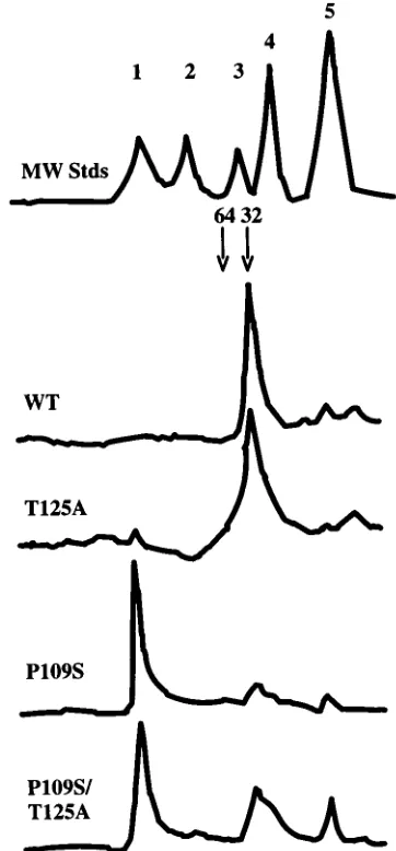
Size exclusion chromatography.
Wild-type and T125A
[image:4.612.70.283.69.443.2]inte-grase proteins migrated predominantly as single species on a
gel filtration column. The retention times of these proteins
compared with those of molecular mass standards were
con-sistent with monomeric integrase (Fig. 3). The T125A protein
also showed evidence of dimeric and higher-order aggregates.
P109S integrase also ran predominantly as a single species, but
in this case the protein migrated as a large aggregate at the
void volume of the column, showing only a trace of monomer
(Fig. 3). P109S T125A integrase eluted as a mixture of the
FIG. 2. 39processing and disintegration activities of wild-type (WT) and mutant integrase proteins. Each protein was assayed for 39processing (top) and disintegration (bottom) activities at two different concentrations: 0.25mM (lanes 2, 4, 6, and 8) and 0.5mM (lanes 3, 5, 7, and 9). The 39processing activity of P109S integrase was also assayed at 1mM (upper panel, lane 10). Integrase was omitted from the reactions in lane 1. The other lanes contained reaction mixtures with the indicated mutant proteins. SUB, substrate DNAs; 39PRO, 39processing product; 30, disintegration product.
FIG. 3. Size exclusion profiles of wild-type (WT), T125A, P109S, and P109S T125A integrase proteins. The chromatogram of the molecular mass standards (MW Stds) is also shown (1,670 kDa; 2, 158 kDa; 3, 44 kDa; 4, 17 kDa; 5, 1.35 kDa). The predicted migration positions of monomeric and dimeric integrase are marked 32 and 64, respectively, by arrows.
on November 9, 2019 by guest
http://jvi.asm.org/
[image:4.612.342.523.293.682.2]two forms, containing similar levels of aggregates and
mono-mers (Fig. 3). These results show that the P109S substitution
can affect the multimeric state of recombinant integrase and
that the T125A second-site mutation is capable of correcting,
at least partially, the alteration induced by the P109S
muta-tion. Similar results were detected following digestion with
V8 protease. Both the wild-type and T125A proteins
yield-ed the expectyield-ed protease-resistant catalytic and
carboxyl-ter-minal fragments (20). The P109S mutant, in contrast, displayed
an aberrant pattern, most likely because the protein
aggre-gates prohibited access to some of the protease sites (data not
shown). The cleavage pattern of the P109S T125A protein was
more similar to the pattern of wild-type integrase than it was to
that of the P109S protein (data not shown).
These results show that the P109S amino acid substitution
induces the aggregation of recombinant HIV-1 integrase. The
P109S protein was purified under denaturing conditions and
was subsequently refolded. The P109S T125A protein was also
refolded and contained both aggregated and monomeric
inte-grase (Fig. 3). We were concerned that the P109S T125A
aggregates might be inactive, accounting for the lower specific
3
9
processing and DNA strand transfer activities of this protein
compared with the T125A and wild-type integrases (Fig. 2). To
test this, the P109S T125A monomers were isolated from the
aggregated material by gel filtration chromatography. The
spe-cific 3
9
processing and DNA strand transfer activities of the
purified monomers were identical to those of the starting
P109S T125A aggregate-monomer mixture (data not shown).
If the P109S T125A aggregates were inactive, we would have
expected the purified monomers to display a higher specific
activity than the aggregate-monomer mixture. We conclude
that the aggregated forms of P109S T125A are most likely
active. The P109S mutant protein was next analyzed in in vitro
complementation assays to further investigate the defective
nature of this protein.
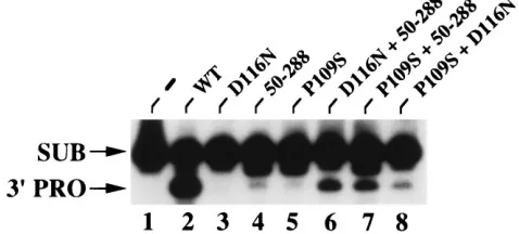
In vitro complementation.
We and others have previously
shown that mixtures of certain defective integrase proteins
display 10 to 50% of wild-type 3
9
processing and DNA strand
transfer activities in vitro (19, 65). For example, the 50-288
integrase deletion mutant protein, which lacks the
amino-ter-minal HHCC domain, does not display detectable 3
9
process-ing (Fig. 4, lane 4) or DNA strand transfer activity (5) on its
own. Full-length integrase containing the single amino acid
substitution of Asn for active-site residue Asp-116 (D116N)
does not display detectable 3
9
processing (Fig. 4, lane 3) or
DNA strand transfer or disintegration activity (20). An
equi-molar mixture of these two proteins, however, displays
approx-imately 10 to 20% of wild-type 3
9
processing (Fig. 4, lane 6)
and DNA strand transfer (19) activities. In this reaction
mix-ture, both 3
9
processing and DNA strand transfer are catalyzed
by the intact active site of the 50-288 protomer, while the
es-sential amino-terminal domain function(s) is supplied in trans
by the D116N protomer. P109S integrase was next tested in
place of the D116N protein. Remarkably, this reaction mixture
yielded levels of 3
9
processing (Fig. 4, compare lane 7 with lane
6) and DNA strand transfer (data not shown) activities similar
to those of the mixture containing the 50-288 and D116N
proteins. An equimolar mixture of the P109S and D116N
mu-tant proteins also displayed detectable 3
9
processing and DNA
strand transfer activities, although these levels were reduced
approximately fivefold compared with those of the mixture of
the P109S and 50-288 proteins (Fig. 4, compare lane 8 with
lane 7).
DISCUSSION
A number of conserved residues in the catalytic domain of
HIV-1 integrase have been targeted by site-directed
mutagen-esis. Substitution of the active-site residue Asp-64, Asp-116, or
Glu-152 abolishes HIV-1 replication in human macrophages
(24, 69) and T-cell lines (1, 6, 22, 40, 41, 44, 59, 61, 69) and can
abolish the 3
9
processing, DNA strand transfer, and
disinte-gration activities of recombinant integrase protein in vitro (20,
42, 64). Substitution of less well conserved residues in the
catalytic domain of integrase can also affect HIV-1 replication
(30, 41, 59, 61, 69) and in vitro enzyme activities (16, 39, 42,
64). However, in these cases recombinant proteins display
some polynucleotidyl transferase activity as assessed by using
conventional oligonucleotide assays.
A common defect of integrase active-site mutant proteins is
likely improper divalent metal ion coordination. For example,
recombinant integrase proteins containing conservative
charge-to-charge substitutions support partial activities under
condi-tions in which conservative charge-to-neutral substitucondi-tions
de-stroy activity (20). Defects caused by changing less well
con-served residues in the catalytic domain are less likely to occur
through a common mechanism. Indeed, such mutations have
been shown to affect reverse transcription (41, 59) and particle
assembly (30) in addition to integration (41, 61, 69) in vivo. To
further investigate the mechanistic bases for these pleiotropic
defects in HIV-1-infected cells, we established T-cell lines
chronically producing replication-defective viruses for several
of our previously characterized integrase mutants (59, 61).
A second amino acid change in integrase, T125A, restores
replication to the defective P109S mutant virus.
In this study,
we analyzed a T-cell line chronically producing the P109S
integrase mutant virus, which is replication defective because
of a block in integration (61). We identified and characterized
a phenotypically reverted virus (P109S
R) which appeared while
passaging the stably transfected T-cell line. An intragenic
sec-ond-site amino acid substitution, T125A, that restored both
viral infectivity and recombinant enzyme function to the P109S
integrase-defective mutant was identified. The biological
mech-anism(s) underlying the emergence of the revertant virus is
unclear. Assuming that the appearance of the second-site
mu-tation occurred during reverse transcription, we suspect that
the original virus bearing the P109S substitution is able to
integrate viral DNA to a minimum extent. This level of
repli-cation must be below the detection limit of the exogenous RT
(Fig. 1B) and p24 antigen capture (61) assays.
Virus recovered following transfection with molecularly
cloned P109S T125A proviral DNA showed a replication
pro-FIG. 4. In vitro complementation with P109S integrase. Integrase was omit-ted from the reaction in lane 1. The reaction in lane 2 contained wild-type (WT) integrase, and those in lanes 3 through 8 contained the indicated mutant pro-teins. The low level of 39processing activity detected in lanes 4 and 5 is due to contaminating E. coli exonuclease activity (23), not integrase. Other labeling is the same as in Fig. 2.
on November 9, 2019 by guest
http://jvi.asm.org/
[image:5.612.58.297.76.184.2]file similar to that of the P109S
Rvirus (Fig. 1). The 4-day delay
in peak RT activities in cells infected with P109S
Ris most likely
due to the presence of the parental P109S mutant in this viral
stock. This mixture would effectively reduce the number of
infectious particles when equal RT counts per minute for the
P109S
Rand P109S T125A viruses are compared. Consistent
with this interpretation, virus harvested from the peak of
P109S
Rreplication (day 14 in Fig. 1A) showed the same
rep-lication kinetics as the P109S T125A virus (data not shown).
The T125A substitution restores function to recombinant
P109S integrase.
The T125A amino acid substitution also
res-cued the in vitro biochemical activities of recombinant HIV-1
integrase protein. P109S integrase was defective for 3
9
process-ing and DNA strand transfer activities but displayed about 5 to
10% of wild-type disintegration activity. The P109S T125A
integrase displayed type disintegration and nearly
wild-type levels of 3
9
processing and DNA strand transfer activities
(Fig. 2).
The in vitro activities of the recombinant integrase proteins
correlated well with the replication profiles of the different
viruses. In both cases, P109S was defective, T125A was
equiv-alent to the wild type, and P109S T125A displayed nearly
wild-type activities; the P109S T125A virus showed a 3-day
delay in peak virus production compared with the wild type
(Fig. 1B), and P109S T125A recombinant integrase displayed
approximately 50% of wild-type 3
9
processing and DNA strand
transfer activities (Fig. 2). These results establish that T125A is
an intragenic second-site suppressor of P109S in the context of
both HIV-1 replication and the in vitro biochemical activities
of recombinant integrase protein. To our knowledge, this is the
first example of an intragenic second-site suppressor of a
retro-viral integrase mutation.
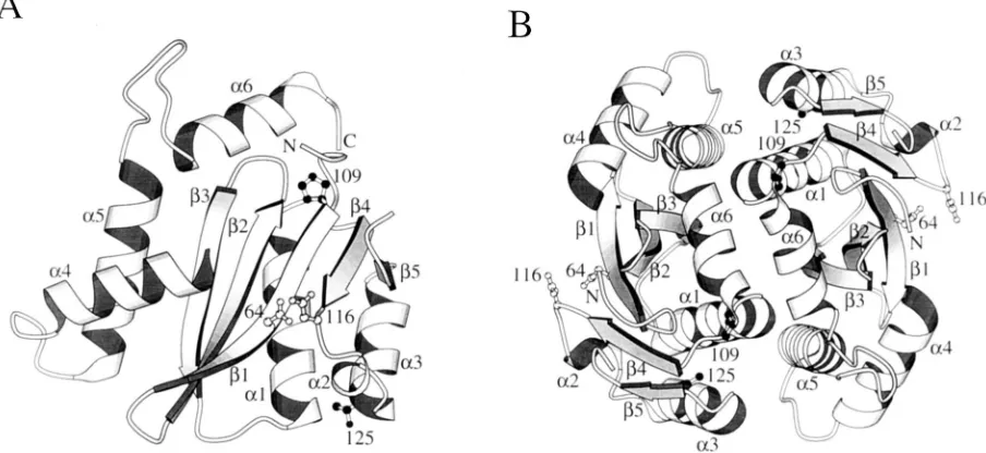
How does T125A restore function to a defective P109S
inte-grase?
Pro-109 and Thr-125 are not in close proximity to each
other in the crystal structure of the catalytic domain of HIV-1
integrase (Fig. 5A). Thus, it is not immediately obvious how
the T125A substitution might rescue the P109S defect(s). Since
Pro-109 is in the turn between
a
1 and
b
4, it is tempting to
speculate that the Ser substitution at position 109 may extend
a
1 and, in doing so, alter the positions of
b
4 and active-site
residue Asp-116 (Fig. 5A). In this model the T125A
substitu-tion would act intramolecularly to restore the integrity of the
active site. Analysis of the crystal structure in this detail,
how-ever, is complicated by three amino acid differences in the
catalytic domains of the NL4-3 and HXBc2 strains of integrase
(7, 46). The core domain of NL4-3 integrase was crystallized
(17), whereas HXBc2 viruses and mutant integrase proteins
were studied in this work. Two of the strain differences lie
between Pro-109 and Thr-125: the crystal contains Val-113 and
Thr-124 where HXBc2 integrase contains Il3-113 and Ala-124.
Both the catalytic (8, 19, 33, 45, 65) and carboxyl-terminal
(30) domains of HIV-1 integrase are important for
multimer-ization, and multimerization is required for 3
9
processing and
DNA strand transfer activities in vitro (19, 65). Pro-109 is
located near the crystallographic dimer interface, and Thr-125
is part of
a
3, which also forms part of this interface (Fig. 5B)
(17). Since the crystal structure of the avian sarcoma virus
integrase catalytic domain shows a similar dimer (3), it seems
likely that the dimer in the HIV-1 structure is important for 3
9
processing and DNA strand transfer activities. We therefore
wondered if the primary effect of the P109S substitution might
be the disruption of the dimer interface and, hence, integrase
activity. In this model T125A would restore function by
stabi-lizing the dimer interface. P109S, P109S T125A, T125A, and
wild-type integrases were analyzed by gel filtration
chromatog-raphy to assess the multimeric states of the proteins.
The results of these analyses showed that P109S integrase
was aggregated. Aggregation may be related to the loss of
activity, but the P109S T125A protein, which displayed
appre-ciable levels of 3
9
processing and DNA strand transfer
[image:6.612.81.533.75.283.2]activi-ties, contained both aggregates and monomers (Fig. 3).
Cer-tain substitutions of other residues in the HIV-1 catalytic
domain, including 81 (42, 64), Cys-65, Phe-121, and
Ser-123 (64), have previously been shown to lower the solubility of
FIG. 5. Locations of Pro-109 and Thr-125 in the crystal structure of the catalytic domain of NL4-3 integrase. The structures were drawn with the program MOLSCRIPT v1.1 (38). (A) Structure of the integrase monomer. Pro-109 and Thr-125 are shown as filled-in ball-and-stick models, and active-site residues Asp-64 and Asp-116 are shown as empty ball-and-stick models. Secondary structural elements are labeled as in reference 17. The amino and carboxyl termini are labeled N and C, respectively. (B) Dimer in the crystal, showing parts ofa1,a3,a5,a6, andb3, which form the dimer interface (17). In this view, the carboxyl terminus lies over Pro-109, and Thr-125 is partially hidden in back ofa3. Other labeling is as in panel A.
on November 9, 2019 by guest
http://jvi.asm.org/
recombinant integrase. Both S81A and S81R recombinant
pro-teins display 3
9
processing and DNA strand transfer activities,
showing that marginally soluble integrase proteins can be
ac-tive (42, 64). Purified P109S T125A monomers displayed the
same specific 3
9
processing and DNA strand transfer activities
as the monomer-aggregate mixture, implying that the P109S
T125A aggregates are also active. We speculate that
aggrega-tion per se is not the reason for the lack of P109S integrase
activity.
Crystal structures of P109S and P109S T125A integrase
pro-teins would help decipher the mechanism of second-site
rever-sion. These changes would have to be introduced into the
catalytic domain (amino acid residues 50 to 212) of integrase
along with the F185K amino acid substitution, which was
nec-essary for crystallization (17, 31). We feel that the tendencies
for both the P109S and P109S T125A full-length HXBc2
pro-teins to aggregate would make the crystallization of the
50-212/F185K core domains extremely difficult, if not impossible.
We therefore probed the P109S integrase defect by using an
in vitro transcomplementation assay (19, 65). Premixing P109S
integrase with the amino-terminal deletion mutant 50-288
yielded 10 to 20% of wild-type levels of 3
9
processing and DNA
strand transfer activities (Fig. 4). How might P109S integrase,
which behaved as a large aggregate on a sizing column, form
functional heteromultimers with the 50-288 protein? In vitro,
3
9
processing and DNA strand transfer activities are catalyzed
by large insoluble aggregates of integrase and DNA (63). It is
therefore not unreasonable that an aggregated protein like the
P109S integrase would form functional mixed multimers in
vitro. The carboxyl-terminal domain, which is a dimer in
solu-tion (18, 43), also contributes to the multimerizasolu-tion of HIV-1
integrase (30). P109S and wild-type integrases displayed equal
metal-independent DNA binding to the 30-bp U5 substrate as
detected by UV cross-linking (data not shown). The
carboxyl-terminal domain of P109S integrase is most likely intact, as this
region is responsible for the metal-independent binding of
HIV-1 integrase to DNA (23, 28, 51, 67, 70). It is therefore
likely that the carboxyl-terminal domains of the 50-288 and
P109S protomers help form and/or stabilize the functional
heteromultimers in the in vitro complementation reaction
mix-ture. The reaction mixture containing the D116N and P109S
mutant proteins also displayed detectable 3
9
processing and
DNA strand transfer activities (Fig. 4), although these levels
were only
#
5% of the levels of wild-type activities. Thus, the
P109S protomer can donate the active site for 3
9
processing
and DNA strand transfer activities under these reaction
con-ditions.
At present we are unable to form a definitive model as to
how the T125A second-site amino acid substitution restores
function to the defective P109S integrase. We suggest that the
T125A substitution stabilizes a structural alteration(s) induced
by the P109S mutation. Both intermolecular (48) and
intramo-lecular (71) models of second-site reversion have been
pro-posed for other systems in which the participating amino acid
residues are not in close proximity to each other. It is
impor-tant to note that the amino- and carboxyl-terminal domains,
which are necessary for the 3
9
processing and DNA strand
transfer activities of recombinant HIV-1 integrase, are absent
in the crystal structure. The T125A second-site substitution
may correct the P109S defect primarily through intramolecular
contacts in the integrase monomer or through intermolecular
contacts involving the catalytic, amino-, and/or
carboxyl-termi-nal domains. Characterization of additiocarboxyl-termi-nal
integrase-defec-tive viruses, together with potentially rescuing second-site
sup-pressor mutations, is expected to reveal more information
about the structure and function of HIV-1 integrase.
ACKNOWLEDGMENTS
We thank Joseph Sodroski for critical review of the manuscript,
Stefano Fiore for criticisms and very helpful discussion, Angela Lippa
and Angela Fresolone for secretarial assistance, and M. Farzan for
help with MOLSCRIPT v1.1.
This work was supported by grants from AIDS Projects of the
Ministry of Health, Rome, Italy (to B.T., F.C. and P.V.), and from the
G. Harold and Leila Y. Mathers Foundation (to A.E.).
REFERENCES
1. Ansari-Lari, M. L., L. A. Donehower, and R. A. Gibbs. 1995. Analysis of human immunodeficiency virus type 1 integrase mutants. Virology 211:332– 335.
2. Brown, P. O., B. Bowerman, H. E. Varmus, and J. M. Bishop. 1989. Retro-viral integration: structure of the initial covalent product and its precursor, and a role for the viral IN protein. Proc. Natl. Acad. Sci. USA 86:2525–2529. 3. Bujacz, G., M. Jaskolski, J. Alexandratos, A. Wlodower, G. Merkel, R. A. Katz, and A. M. Skalka.1995. High-resolution structure of the catalytic domain of avian sarcoma virus integrase. J. Mol. Biol. 253:336–346. 4. Bushman, F. D., and R. Craigie. 1991. Activities of human immunodeficiency
virus (HIV) integration protein in vitro: specific cleavage and integration of HIV DNA. Proc. Natl. Acad. Sci. USA 88:1339–1343.
5. Bushman, F. D., A. Engelman, I. Palmer, P. Wingfield, and R. Craigie. 1993. Domains of the integrase protein of human immunodeficiency virus type 1 responsible for polynucleotidyl transfer and zinc binding. Proc. Natl. Acad. Sci. USA 90:3428–3432.
6. Cannon, P. M., W. Wilson, E. Byles, S. M. Kingsman, and A. J. Kingsman. 1994. Human immunodeficiency virus type 1 integrase: effect on viral repli-cation of mutations at highly conserved residues. J. Virol. 68:4768–4775. 7. Carlini, F., and B. Taddeo. Unpublished observation.
8. Chow, S. A., and P. O. Brown. 1994. Juxtaposition of two viral ends in a bimolecular disintegration reaction mediated by multimers of human immu-nodeficiency virus type 1 or murine leukemia virus integrase. J. Virol. 68: 7869–7878.
9. Chow, S. A., K. A. Vincent, V. Ellison, and P. O. Brown. 1992. Reversal of integration and DNA splicing mediated by integrase of human immunode-ficiency virus. Science 255:723–726.
10. Colicelli, J., and S. P. Goff. 1988. Sequence and spacing requirements of a retrovirus integration site. J. Mol. Biol. 199:47–59.
11. Craigie, R., T. Fujiwara, and F. Bushman. 1990. The IN protein of Moloney murine leukemia virus processes the viral DNA ends and accomplishes their integration in vitro. Cell 62:829–837.
12. Craigie, R., A. B. Hickman, and A. Engelman. 1995. Integrase, p. 53–71. In J. Karn (ed.), HIV, vol. II. A practical approach. Oxford University Press, Oxford.
13. Dayton, A. I., E. F. Terwilliger, J. Potz, M. Kowalski, J. G. Sodroski, and W. A. Haseltine.1988. cis-acting sequences responsive to the rev gene prod-uct of the human immunodeficiency virus. J. Acquired Immune Defic. Syndr. 1:441–452.
14. Donehower, L. A., and H. E. Varmus. 1984. A mutant murine leukemia virus with a single missense codon in pol is defective in a function affecting integration. Proc. Natl. Acad. Sci. USA 81:6461–6465.
15. Drelich, M., M. Haenggi, and J. Mous. 1993. Conserved residues Pro-109 and Asp-116 are required for interaction of the human immunodeficiency virus type 1 integrase protein with its viral DNA substrate. J. Virol. 67:5041– 5044.
16. Drelich, M., R. Wilhelm, and J. Mous. 1992. Identification of amino acid residues critical for endonuclease and integration activities of HIV-1 IN protein in vitro. Virology 188:459–468.
17. Dyda, F., A. B. Hickman, T. M. Jenkins, A. Engelman, R. Craigie, and D. R. Davies.1994. Crystal structure of the catalytic domain of HIV-1 integrase: similarity to other polynucleotidyl transferases. Science 266:1981–1986. 18. Eijkelenboom, A. P. A. M., R. A. P. Lutzke, R. Boelens, R. H. A. Plasterk, R.
Kaptein, and K. Hard.1995. The DNA binding domain of HIV-1 integrase has an SH3-like fold. Nature (London) Struct. Biol. 2:807–810.
19. Engelman, A., F. D. Bushman, and R. Craigie. 1993. Identification of dis-crete functional domains of HIV-1 integrase and their organization within an active multimeric complex. EMBO J. 12:3269–3275.
20. Engelman, A., and R. Craigie. 1992. Identification of conserved amino acid residues critical for human immunodeficiency virus type 1 integrase function in vitro. J. Virol. 66:6361–6369.
21. Engelman, A., and R. Craigie. 1995. Efficient magnesium-dependent human immunodeficiency virus type 1 integrase activity. J. Virol. 69:5908–5911. 22. Engelman, A., G. Englund, J. M. Orenstein, M. A. Martin, and R. Craigie.
1995. Multiple effects of mutations in human immunodeficiency virus type 1 integrase on viral replication. J. Virol. 69:2729–2736.
23. Engelman, A., A. B. Hickman, and R. Craigie. 1994. The core and carboxyl terminal domains of the integrase protein of human immunodeficiency virus type 1 each contribute to nonspecific DNA binding. J. Virol. 68:5911–5917. 24. Englund, G., T. S. Theodore, E. Freed, A. Engelman, and M. A. Martin. 1995.
on November 9, 2019 by guest
http://jvi.asm.org/
Integration is required for productive infection of monocyte-derived macro-phages by human immunodeficiency virus type 1. J. Virol. 69:3216–3219. 25. Fujiwara, T., and K. Mizuuchi. 1988. Retroviral DNA integration: structure
of an integration intermediate. Cell 54:497–504.
26. Goulaouic, H., and S. A. Chow. 1996. Directed integration of viral DNA mediated by fusion proteins consisting of human immunodeficiency virus type 1 integrase and Escherichia coli LexA protein. J. Virol. 70:37–46. 27. Grandgenett, D. P., and G. Goodarzi. 1994. Folding of the multidomain
human immunodeficiency virus type 1 integrase. Protein Sci. 3:888–897. 28. Hazuda, D. J., A. L. Wolfe, J. C. Hastings, H. L. Robbins, P. L. Graham,
R. L. LaFemina, and E. A. Emini.1994. Viral long terminal repeat substrate binding characteristics of human immunodeficiency virus type 1 integrase. J. Biol. Chem. 269:3999–4004.
29. Higuchi, R., B. Krummel, and R. K. Saiki. 1988. A general method of in vitro preparation and specific mutagenesis of DNA fragments: study of protein and DNA interactions. Nucleic Acids Res. 16:7351–7367.
30. Jenkins, T. M., A. Engelman, R. Ghirlando, and R. Craigie. 1996. A soluble active mutant of HIV-1 integrase: involvement of both the core and C-terminal domains in multimerization. J. Biol. Chem. 271:7712–7718. 31. Jenkins, T. M., A. B. Hickman, F. Dyda, R. Ghirlando, D. R. Davies, and R.
Craigie.1995. Catalytic domain of human immunodeficiency virus type 1 integrase: identification of a soluble mutant by systematic replacement of hydrophobic residues. Proc. Natl. Acad. Sci. USA 92:6057–6061. 32. Johnson, M. S., M. A. McClure, D.-F. Feng, J. Gray, and R. F. Doolittle.
1986. Computer analysis of retroviral pol genes: assignment of enzymatic functions to specific sequences and homologies with non viral enzymes. Proc. Natl. Acad. Sci. USA 83:7648–7652.
33. Kalpana, G. V., and S. P. Goff. 1993. Genetic analysis of homomeric inter-actions of human immunodeficiency virus type 1 integrase using the yeast two-hybrid system. Proc. Natl. Acad. Sci. USA 90:10593–10597.
34. Katz, R. A., G. Merkel, J. Kulkosky, J. Leis, and A. M. Skalka. 1990. The avian retroviral IN protein is both necessary and sufficient for integrative recombination in vitro. Cell 63:87–95.
35. Katz, R. A., and A. M. Skalka. 1994. The retroviral enzymes. Annu. Rev. Biochem. 63:133–173.
36. Katzman, M., R. A. Katz, A. M. Skalka, and J. Leis. 1989. The avian retroviral integration protein cleaves the terminal sequences of linear viral DNA at the in vivo sites of integration. J. Virol. 63:5319–5327.
37. Khan, E., J. P. G. Mack, R. A. Katz, J. Kulkosky, and A. M. Skalka. 1991. Retroviral integrase domains: DNA binding and the recognition of LTR sequences. Nucleic Acids Res. 19:851–860.
38. Kraulis, P. J. 1991. MOLSCRIPT: a program to produce both detailed and schematic plots of protein structures. J. Appl. Cryst. 24:946–950. 39. Kulkosky, J., K. S. Jones, R. A. Katz, J. P. G. Mack, and A. M. Skalka. 1992.
Residues critical for retroviral integrative recombination in a region that is highly conserved among retroviral/retrotransposon integrases and bacterial insertion sequence transposases. Mol. Cell. Biol. 12:2331–2338.
40. LaFemina, R. L., C. L. Schneider, H. L. Robbins, P. L. Callahan, K. LeGrow, E. Roth, W. A. Schleif, and E. A. Emini.1992. Requirement of active human immunodeficiency virus type 1 integrase enzyme for productive infection of human T-lymphoid cells. J. Virol. 66:7414–7419.
41. Leavitt, A. D., G. Robles, N. Alesandro, and H. E. Varmus. 1996. Human immunodeficiency virus type 1 integrase mutants retain in vitro integrase activity yet fail to integrate DNA efficiently during infection. J. Virol. 70: 721–728.
42. Leavitt, A. D., L. Shiue, and H. E. Varmus. 1993. Site-directed mutagenesis of HIV-1 integrase demonstrates differential effects on integrase function in vitro. J. Biol. Chem. 268:2113–2119.
43. Lodi, P. J., J. A. Ernst, J. Kuszewski, A. B. Hickman, A. Engelman, R. Craigie, G. M. Clore, and A. M. Gronenborn.1995. Solution structure of the DNA binding domain of HIV-1 integrase. Biochemistry 34:9826–9833. 44. Masuda, T., V. Planelles, P. Krogstad, and I. S. Y. Chen. 1995. Genetic
analysis of human immunodeficiency virus type 1 integrase and the U3 att site: unusual phenotype of mutants in the zinc finger-like domain. J. Virol. 69:6687–6696.
45. Mazumder, A., A. Engelman, R. Craigie, M. Fresen, and Y. Pommier. 1994. Intermolecular disintegration and intramolecular strand transfer activities of wild-type and mutant HIV-1 integrase. Nucleic Acids Res. 22:1037–1043. 46. Meyers, G., S. Wain-Hobson, B. Korber, R. F. Smith, and G. N. Pavlakis.
1993. Human retroviruses and AIDS. A compilation and analysis of nucleic acid and amino acid sequences. Theoretical biology and biophysics group T-10. Los Alamos National Laboratory, Los Alamos, N.Mex.
47. Murphy, J. E., and S. P. Goff. 1992. A mutation at one end of Moloney murine leukemia virus DNA blocks cleavage at both ends by the viral inte-grase in vivo. J. Virol. 66:5092–5095.
48. Nakamoto, R. K., M. K. Alshawi, and M. Futai. 1995. The ATP synthetase gamma subunit-suppressor mutagenesis reveals three helical regions in-volved in energy coupling. J. Biol. Chem. 270:14042–14046.
49. Panganiban, A. T., and H. M. Temin. 1983. The terminal nucleotides of retroviral DNA are required for integration but not virus production. Nature (London) 306:155–160.
50. Panganiban, A. T., and H. M. Temin. 1984. The retrovirus pol gene encodes a product required for DNA integration: identification of a retrovirus int locus. Proc. Natl. Acad. Sci. USA 81:7885–7889.
51. Puras-Lutzke, R. A., C. Vink, and R. H. A. Plasterk. 1994. Characterization of the minimal DNA-binding domain of the HIV integrase protein. Nucleic Acids Res. 22:4125–4131.
52. Quinn, T. P., and D. P. Grandgenett. 1988. Genetic evidence that the avian retrovirus DNA endonuclease domain of Pol is necessary for viral integra-tion. J. Virol. 62:2307–2312.
53. Rice, P. A., and K. Mizuuchi. 1995. Structure of the bacteriophage Mu transposase core: a common structural motif for DNA transposition and retroviral integration. Cell 82:209–220.
54. Roth, M. J., P. L. Schwartzberg, and S. P. Goff. 1989. Structure of the termini of DNA intermediates in the integration of retroviral DNA: dependence on IN function and terminal DNA sequence. Cell 58:47–54.
55. Sambrook, J., E. F. Fritsch, and T. Maniatis. 1989. Molecular cloning: a laboratory manual. Cold Spring Harbor Laboratory, Cold Spring Harbor, N.Y.
56. Sanger, F., S. Nicklen, and A. R. Coulson. 1977. DNA sequencing with chain-terminating inhibitors. Proc. Natl. Acad. Sci. USA 74:5463–5467. 57. Schwartzberg, P., J. Colicelli, and S. P. Goff. 1984. Construction and analysis
of deletion mutants in the pol gene of Moloney murine leukemia virus: a new viral function required for productive infection. Cell 37:1043–1052. 58. Sherman, P. A., and J. A. Fyfe. 1990. Human immunodeficiency virus
inte-gration protein expressed in Escherichia coli possesses selective DNA cleav-ing activity. Proc. Natl. Acad. Sci. USA 87:5119–5123.
59. Shin, C., B. Taddeo, W. A. Haseltine, and C. M. Farnet. 1994. Genetic analysis of the human immunodeficiency virus type 1 integrase protein. J. Virol. 68:1633–1642.
60. Studier, F. W., and B. A. Moffatt. 1986. Use of bacteriophage T7 RNA polymerase to direct selective high-level expression of cloned genes. J. Mol. Biol. 189:113–130.
61. Taddeo, B., W. A. Haseltine, and C. M. Farnet. 1994. Integrase mutants of human immunodeficiency virus type 1 with a specific defect in integration. J. Virol. 68:8401–8405.
62. van Dyke, M. W., M. Sirito, and M. Sawadogo. 1992. Single-step purification of bacterially expressed polypeptides containing an oligo-histidine domain. Gene 111:99–104.
63. van Gent, D. C., Y. Elgersma, M. W. J. Bolk, C. Vink, and R. H. A. Plasterk. 1991. DNA binding of the integrase proteins of human immunodeficiency viruses types 1 and 2. Nucleic Acids Res. 19:3821–3827.
64. van Gent, D. C., A. A. M. Oude Groeneger, and R. H. A. Plasterk. 1992. Mutational analysis of the integrase protein of human immunodeficiency virus type 2. Proc. Natl. Acad. Sci. USA 89:9598–9602.
65. van Gent, D. C., C. Vink, A. A. M. Oude Groeneger, and R. H. A. Plasterk. 1993. Complementation between HIV integrase proteins mutated in differ-ent domains. EMBO J. 12:3261–3267.
66. Vincent, K. A., V. Ellison, S. A. Chow, and P. O. Brown. 1993. Character-ization of human immunodeficiency virus type 1 integrase expressed in Escherichia coli and analysis of variants with amino-terminal mutations. J. Virol. 67:425–437.
67. Vink, C., A. A. M. Oude Groeneger, and R. H. A. Plasterk. 1993. Identifica-tion of the catalytic and DNA-binding region of human immunodeficiency virus type 1 integrase protein. Nucleic Acids Res. 21:1419–1425. 68. Vora, A. C., M. L. Fitzgerald, and D. P. Grandgenett. 1990. Removal of
39-OH-terminal nucleotides from blunt-ended long terminal repeat termini by the avian retrovirus integration protein. J. Virol. 64:5656–5659. 69. Wiskerchen, M., and M. A. Muesing. 1995. Human immunodeficiency virus
type 1 integrase: effects of mutations on viral ability to integrate, direct gene expression from unintegrated viral DNA templates, and sustain propagation in primary cells. J. Virol. 69:376–386.
70. Woerner, A. M., M. Klutch, J. G. Levin, and C. J. Marcus-Sekura. 1992. Localization of DNA binding activity of HIV-1 integrase to the C-terminal half of the protein. AIDS Res. Hum. Retroviruses 8:297–304.
71. Yamaguchi, A., Y. Inagaki, and T. Sawai. 1995. Second site suppressor mutations for the Asp-663Cys mutant of the transposon Tn10-encoded metal-tetracycline/H1antiporter of Escherichia coli. Biochemistry 34:11800– 11806.