JOURNAL OFVIROLOGY, July 1996, p. 4502–4508 Vol. 70, No. 7 0022-538X/96/$04.0010
Copyrightq1996, American Society for Microbiology
Adenovirus Interaction with Distinct Integrins Mediates
Separate Events in Cell Entry and Gene Delivery to
Hematopoietic Cells
SHUANG HUANG,
1TETSUJI KAMATA,
2YOSHIKAZU TAKADA,
2ZAVERIO M. RUGGERI,
2AND
GLEN R. NEMEROW
1*
Departments of Immunology
1and Vascular Biology,
2The Scripps Research Institute, La Jolla, California 92037
Received 27 December 1995/Accepted 5 April 1996
A major impediment to the effective use of adenovirus vectors for gene therapy is a lack of knowledge of how
these vectors interact with diverse cell types in vivo. Adenovirus attachment to most human cell types is
mediated by the fiber protein, which binds to an as yet unidentified cell receptor. In contrast to this, we report
that adenovirus type 2 (Ad2) attachment to hematopoietic cells is facilitated by interaction of the penton base
protein with members of the
b
2 integrin family. Adenovirus particles were capable of binding to human
monocytic cells, which lack fiber receptors, and virus binding could be blocked by a soluble penton base or by
a function-blocking monoclonal antibody to integrin
a
Mb
2. To confirm the role of
a
Mb
2 integrins in Ad2
binding to hematopoietic cells, we analyzed virus attachment and gene delivery to CHO cells expressing
recombinant
b
2 integrins.
a
Mb
2-expressing CHO cells supported 3- to 5-fold-higher levels of Ad2 binding and
5- to 10-fold-larger amounts of gene delivery than did nontransfected CHO cells, indicating that
a
Mb
2
facilitates adenovirus attachment to and infection of hematopoietic cells. While
b
2 integrins promote Ad2
attachment to hematopoietic cells, further studies demonstrated that
a
v integrins were required for the next
step in infection, virus internalization into cell endosomes. These studies reveal a novel pathway of Ad2
infection of hematopoietic cells mediated by distinct integrins which facilitate separate events in virus entry.
They also suggest a possible strategy for selective adenovirus-mediated gene delivery to hematopoietic cells.
Cells of hematopoietic origin are thought to play an
impor-tant role in adenovirus (Ad) pathogenesis, since they
fre-quently infiltrate the primary site of virus infection, a process
associated with immune system-mediated inflammatory
re-sponses (10). Although Ad does not usually replicate in
hema-topoietic cells (2, 6), latent (persistent) infection of these cell
types may promote virus dissemination in the host.
Reactiva-tion of Ad in latently infected cells may also lead to fatal
disseminated disease in immunocompromised individuals (12).
Lymphocytes and/or monocytes are also believed to play a
major role in reducing the efficiency of Ad-mediated gene
therapy by eliminating transduced cells expressing foreign gene
products (7, 8).
Despite the involvement of hematopoietic cells in Ad
patho-genesis and gene delivery, little is known of the specific
inter-actions of Ad with these cell types. Previous studies have
dem-onstrated that the fiber capsid protein is responsible for Ad
attachment to the majority of human cell types (19), although
the cell receptor that mediates the fiber binding has not yet
been identified. It is also unknown as to whether fiber
recep-tors are expressed on all cell types including hematopoietic
cells. Ad entry into cells following virus attachment is mediated
by the penton base (4, 27), which binds to cell integrins
avb
3
and
avb
5. Penton base is composed of five identical subunits,
each containing an RGD peptide sequence (17) that is also
found in a number of cell matrix and adhesion proteins, such as
fibronectin and vitronectin, which bind to a family of cell
sur-face receptors termed integrins (21). Integrins are
het-erodimers composed of noncovalently associated
a
and
b
sub-units (14). There are 14 known
a
and 8 known
b
subunits,
forming at least 21 different heterodimers, which recognize
distinct ligands (14). Integrins that recognize the RGD
se-quence include
a5b
1,
aIIbb
3, and the
av
integrins. Several lines
of evidence suggest that
av
integrins are important secondary
receptors for Ad infection in vitro and in vivo. Recombinant
Ads containing a mutation in the penton base RGD sequence
have decreased infectivity (4). Human monocytes (13) and
fully differentiated airway epithelial cells (11), which do not
express
av
integrins, are also relatively resistant to
Ad-medi-ated gene delivery. In contrast, upregulation of
av
integrins on
monocytes/macrophages (13) and expression of
avb
5 on less
differentiated airway epithelial cells promotes Ad-mediated
gene delivery (11). Penton base interaction with integrin
avb
5
is also involved in Ad-mediated cell membrane
permeabiliza-tion (26).
While there is substantial knowledge about the virus
struc-tural proteins and host cell receptors that mediate Ad
inter-nalization, relatively little is known of the receptor(s) involved
in virus attachment to hematopoietic cells. In previous studies,
we reported that growth factor-stimulated monocytes
ex-pressed decreased levels of the fiber receptor even though they
had shown increased susceptibility to Ad infection (13). This
finding suggested that Ad entry into monocytic cells involved a
pathway distinct from that of virus entry into epithelial cells. In
the studies reported here, we examined Ad attachment and
entry into fully differentiated monocytes (macrophages) as well
as a monocytic cell line, THP-1 cells. These studies revealed a
novel pathway of virus entry that is mediated entirely by the
interaction of the penton base with distinct cell integrins.
MATERIALS AND METHODS
Ads, recombinant proteins, and MAbs.Ad type 2 (Ad2) was purchased from the American Type Culture Collection, Rockville, Md. Ad2 containing a deletion
* Corresponding author. Mailing address: Department of Immunol-ogy, IMM-19, The Scripps Research Institute, 10666 N. Torrey Pines Rd., La Jolla, CA 92037. Phone: (619) 554-8072. Fax: (619) 554-6881. Electronic mail address: gnemerow@scripps.edu.
4502
on November 9, 2019 by guest
http://jvi.asm.org/
in the fiber gene, H2dl807 (5), was kindly provided by Gary Ketner, Johns Hopkins University. Ad.RSVbgal, a replication-defective Ad type 5 (Ad5), was provided by Michel Perricaudet, Institute Gustave Roussy. Ad2 was purified by centrifugation on 16 to 40% cesium chloride gradients as previously described (27). H2dl807 virus was isolated free (,95%) of contaminating helper virus by three cycles of ultracentrifugation on 20 to 35% cesium chloride gradients. For cell-binding studies, purified virions were radiolabeled with Na125I (Iodo-beads;
Pierce). Recombinant Ad2 fiber and penton base proteins were produced in
Trichoplusia ni insect cells as previously described (27). A function-blocking
murine monoclonal antibody (MAb) directed against the penton base RGD sequence (DAV-1) (unpublished data) was purified (Hi-Trap G; Pharmacia) from ascites fluids. Fab fragments of the DAV-1 antibody were generated by papain digestion at pH 8.0 and then purified on a Resource Q (Pharmacia) fast protein liquid chromatography FPLC column. Purified function-blocking MAbs to integrinsavb3 (LM609),avb5 (P1F6), andb1 (P4C10) were kindly provided
by David Cheresh, The Scripps Research Institute. The 69-6-5 function-blocking MAb toavwas generously provided by Maxime Lehmann, Universite´
d’Aix-Marseille. The M1/70 MAb directed against theaMsubunit of Mac-1) (23) was
provided by Ted Fan, The Scripps Research Institute. A hybridoma cell line producing the OKM1 MAb to integrinaMb2 (CD18b/CD11c; Mac-1) was
pur-chased from the American Type Culture Collection. The CP3 MAb was origi-nally raised against integrinaIIbb3 (18); however, sequential
immunoprecipita-tion experiments indicated that it also recognizes integrinaMb2 (unpublished
observation), and a similar observation has been reported by others (9). Cell lines, peripheral blood mononuclear cells, and flow cytometry.The hu-man epithelial cell lines SW480 and A549 and the monocytic cell line THP-1 (American Type Culture Collection) were maintained in Dulbecco modified Eagle medium with 10% fetal calf serum. Chinese hamster ovary (CHO) cells
expressing wild-typeaMb2, mutant D242A (Asp-242 to Ala inaMcDNA), or aLb2 (15) were generated as described previously (15). Briefly, wild-type or
mutantaMoraLcDNA in the pBJ expression vector were used to transfect CHO
cells, together withb2 cDNA. Cells stably expressing wild-type or mutantaMb2
oraLb2 were cloned by cell sorting to obtain high expressors. The transfected
cells were maintained in complete Dulbecco modified Eagle medium containing 350mg of G418 (Geneticin; GIBCO BRL) per ml. Human peripheral blood mononuclear cells were isolated from the blood of healthy adult donors by Ficoll-Hypaque centrifugation as previously described (13). The adherent-cell population, consisting of greater than 80% monocytes, was cultured for 14 to 18 days in RPMI 1640 supplemented with 20% fetal calf serum to promote terminal differentiation of monocytes to macrophages. For analysis of integrin expression, different cell types were incubated in the presence of 10 to 20mg of the anti-av
or anti-b2 integrin MAb per ml for 60 min at 48C. After being washed, the cells were incubated for 30 min at 48C with goat anti-mouse immunoglobulin G (IgG) coupled to fluorescein isothiocyanate (FITC) (FITC-IgG; KPL Laboratories, Gaithersburg, Md.). Following additional washes in phosphate-buffered saline (PBS), the cells were resuspended in 500ml and analyzed by flow cytometry (FACScan; Becton Dickinson) with the Lysis II program.
Ad, fiber, and penton base attachment experiments and gene delivery assays. Binding of recombinant Ad2 fiber protein to cells was assayed by flow cytometry. Epithelial or monocytic cells at 106
ml were incubated for 60 min at 48C with 50 mg of recombinant Ad2 fiber per ml in PBS containing 0.5% bovine serum albumin (BSA). After being washed three times in PBS-BSA, the cells were incubated with a 1:4,000 dilution of a rabbit polyclonal antibody to the fiber protein for 45 min at 48C and then with a 1:1,000 dilution of goat FITC-anti-rabbit IgG (KPL). After final washes, the cells were subjected to flow-cytometric analysis as described above. To measure binding to cells,125
I-labeled Ad2 or FIG. 1. (A) Flow-cytometric analysis of Ad2 fiber binding to cells. SW480 epithelial cells, THP-1 monocytic cells, and terminally differentiated monocytes/ macrophages were incubated with saturating amounts of recombinant fiber, then with an anti-fiber polyclonal antibody, and then with FITC-IgG. Control cell samples were incubated with FITC-IgG alone prior to flow-cytometric analysis. The solid and dashed curves represent cells incubated with and without fiber, respectively. (B) Ad attachment to SW480 and THP-1 cells. Binding of125I-labeled Ad2 (stippled bars) or a fiberless mutant Ad2 (solid bars) was assayed in the presence or absence
of recombinant Ad2 fiber. Nonspecific binding was determined by incubating the cells in the presence of an excess of unlabeled Ad2. The data represent the mean and standard deviation of duplicate samples.
on November 9, 2019 by guest
http://jvi.asm.org/
[image:2.612.63.548.79.426.2]penton base was incubated for 2 h at 48C with 107cells per ml in serum-free
Dulbecco modified Eagle medium containing 0.5% BSA and 20 mM N-2-hy-droxyethylpiperazine-N9-2-ethanesulfonic acid (HEPES). Unattached virus par-ticles or penton base was removed by centrifugation at 13,0003g on a cushion
of 86% silicon oil–14% mineral oil. Nonspecific binding was determined by incubating the cells in the presence of a 50-fold excess of unlabeled virions or a 200-fold excess of penton base. For competition studies, cells were preincubated with 200 mg of recombinant fiber protein or function-blocking anti-integrin MAbs per ml for 30 min at 48C prior to the addition of radiolabeled virus. In certain experiments, labeled penton base or Ad2 was preincubated with 200mg of Fab fragments of the DAV-1 MAb per ml prior to addition to epithelial or monocytic cells. Ad-mediated gene delivery to different cell types was measured as previously described (13) with a recombinant Ad containing the lacZ gene (Ad.RSVbgal) (24).b-Galactosidase activity was quantitated 48 h postinfection by incubating the cells for 60 min at 378C with a buffer containing 0.5% Nonidet P-40 and 3.5 mM o-nitrophenyl-b-D-galactopyranoside (ONPG) as a chroma-genic substrate. The A415, which was linear from 15 to 90 min at 378C, was
measured in a multiwell plate reader (Titertek; Flow Labs, MacLean, Va.). Affinity purification of integrinaMb2 on a penton base column.THP-1 cells (108
) were washed three times in PBS and then solubilized at 48C with 200 mM b-D-octylglucoside in 10 mM Tris-HCl (pH 8.0) containing 150 mM NaCl, 1% BSA, 2 mM MgCl2, 2mg of aprotinin (Sigma Chemical Co., St. Louis, Mo.) per
ml, 20mg of leupeptin per ml, and 5 mM phenylmethylsulfonyl fluoride. Nuclei and cell debris were removed by centrifugation for 15 min at 10,0003g, and the
soluble fraction was applied to a 2-ml column of Affi-Gel-15 (Bio-Rad, Rich-mond, Calif.) containing 2.8 mg of immobilized penton base. In a separate experiment, an equivalent amount of cell lysate was applied to a penton base affinity column in the presence of 20 mM EDTA. The penton base columns were then washed extensively with solubilization buffer, and the bound proteins were eluted with 50 mM diethylamine (pH 11). After neutralization with 1.0 M glycine-HCl (pH 2.0), the column fractions were pooled and concentrated 10-fold (Centricon 10; Millipore Corp., Bedford, Mass.). Samples were then elec-trophoresed on a sodium dodecyl sulfate–7% polyacrylamide gel under reducing conditions and transferred to a polyvinylidene difluoride membrane (Immobilon P; Millipore) by Western blotting (immunoblotting). The protein blot was incu-bated with 5.0% nonfat dry milk (Blotto) to block nonspecific binding sites and then incubated with 10mg of the M1/70 anti-aMMAb per ml in PBS-Blotto.
After further washes, the blot was incubated with anti-mouse IgG conjugated to alkaline phosphatase, washed, and then incubated with a substrate solution as recommended by the manufacturer (ECL Detection System; Amersham Corp.).
RESULTS
Ad attachment and gene delivery to monocytic cells are
independent of the fiber receptor.
To analyze Ad2 entry into
cells of the myeloid lineage, we initially tested whether
mono-cytes/macrophages expressed Ad2 fiber receptors. A human
monocytic cell line, THP-1, as well as cultured human
macro-phages, failed to bind the fiber protein, whereas SW480
epi-thelial cells, as expected, supported fiber binding (Fig. 1A).
Despite lacking fiber receptors, THP-1 cells bound Ad2
parti-cles as well as mutant Ad2 virions (Fib
2; H2dl807) which lack
the fiber protein (5) (Fig. 1B). Monocytic cells exhibited a
lower level of virus binding than did SW480 cells; however, this
difference is probably due to the three- to fourfold-greater size
of epithelial cells. Preincubation of SW480 cells with soluble
fiber abrogated Ad2 attachment, and these cells also bound
much smaller amounts of the fib
2Ad2. In contrast,
preincu-bation of THP-1 cells with the fiber protein did not inhibit the
binding of either the wild-type or mutant virus.
Further studies were performed to compare the
susceptibil-ity of different cell types to Ad-mediated gene delivery (Fig. 2).
Monocytic cells and epithelial cells were both susceptible to
Ad-mediated gene delivery; however, Ad-mediated gene
de-livery to SW480 cells was blocked by preincubation with the
fiber protein, whereas this treatment had no effect on gene
delivery to monocytic cells, suggesting that Ad-mediated gene
delivery to epithelial SW480 cells but not to monocytic cells is
dependent on receptor interaction via the fiber. Incubation of
SW480 cells with the penton base prevented Ad-mediated
gene delivery. This is clearly due to inhibition of
av
integrin-mediated Ad internalization, since function-blocking MAb to
av
integrins abrogated Ad-mediated gene delivery (data not
shown). Incubation of monocytic cells with penton base also
prevented Ad-mediated gene delivery to these cell types.
These studies indicated that both Ad attachment and infection
of monocytic cells are independent of the fiber receptor and
that the penton base protein plays the major role in virus
interactions with these cells.
b
2 integrins promote Ad attachment to and infection of
monocytic cells.
We next investigated whether the penton base
was responsible for Ad binding to THP-1 cells. Virus
attach-ment to THP-1 cells was blocked by chelation of divalent metal
cations with EDTA, by penton base, and also by a
function-blocking MAb (DAV-1) directed against the penton base
RGD sequence (Fig. 3). These studies suggested that the
pen-ton base mediates virus attachment to monocytic cells via
in-FIG. 2. Ad-mediated gene delivery to SW480 epithelial cells and to mono-cytic cells. Cells were incubated with various amounts of Ad.RSVbgal, and b-galactosidase activity was measured 48 h postinfection. Expression of the Ad-transduced lacZ gene in SW480 epithelial cells, THP-1 cells, and macro-phages was quantitated as described in Materials and Methods. In competition experiments, cells were preincubated with recombinant penton base (open squares) or fiber (solid circles) or medium (open circles) prior to the addition of various amounts of Ad.RSVbgal. The data represent the mean of three exper-iments, with standard deviation equivalent to 5 to 10% of the mean.
4504 HUANG ET AL. J. VIROL.
on November 9, 2019 by guest
http://jvi.asm.org/
teraction of its RGD sequence with a cell integrin. To identify
the integrin involved in Ad2 attachment to THP-1 cells, we
examined a panel of function-blocking MAbs to different
in-tegrins for their ability to inhibit penton base binding to THP-1
cells (Fig. 4). Somewhat surprisingly, function-blocking MAbs
to integrins
avb
3 and
avb
5 had only minor effects on penton
base binding. In contrast, the CP3 MAb (18), which recognizes
integrin
aMb
2, completely blocked penton base binding to
THP-1 cells (Fig. 4). This result suggested that Ad interaction
with monocytic cells is facilitated by the interaction of the
penton base with integrin
aMb
2.
To ascertain whether the penton base protein binds directly
to integrin
aMb
2, THP-1 cell membrane proteins were affinity
purified on a penton base column in the presence or absence of
divalent metal cations. Specifically bound proteins were eluted
from the column with low-pH buffer and then analyzed on an
immunoblot for the presence of integrin
aMb
2 (Fig. 5). A
protein band of approximately 160 kDa, consistent with the
size of the
aM
subunit of
aMb
2, was detected by a
non-func-tion-blocking MAb to
aM
(lane 1). This protein was not
de-tected in membrane fractions that had been affinity purified in
the presence of 20 mM EDTA (lane 2). These results indicated
that the interaction of integrin
aMb
2 with penton base requires
divalent metal cations, a property of most if not all
integrin-ligand interactions.
Ad interaction with CHO cells lacking or expressing
recom-binant
b
2 integrins.
To confirm that
b
2 integrins facilitate Ad
attachment to cells, we first established a CHO cell line stably
expressing
aMb
2. When analyzed by flow cytometry, CHO/
aMb
2 cells exhibited significant reactivity with the anti-
aM
CP3
and OKM1 MAb but CHO cells did not (Fig. 6A). We then
compared virus binding to nontransfected CHO cells which
lack
b
2 integrins or to
b
2-integrin-expressing CHO cells (Fig.
6B). Since CHO cells also do not express Ad2 fiber receptors
(data not shown), this allowed us to analyze Ad attachment
independent of the fiber receptor.
aMb
2-expressing CHO cells
bound approximately fivefold-higher levels of Ad2 than did
nontransfected CHO cells. The majority of Ad2 attachment to
aMb
2-expressing CHO cells was mediated by the penton base
protein interaction with integrin
aMb
2, since it could be
inhib-ited by preincubating cells with the CP3 MAb or with the
penton base (Fig. 7). In contrast, Ad2 fiber or
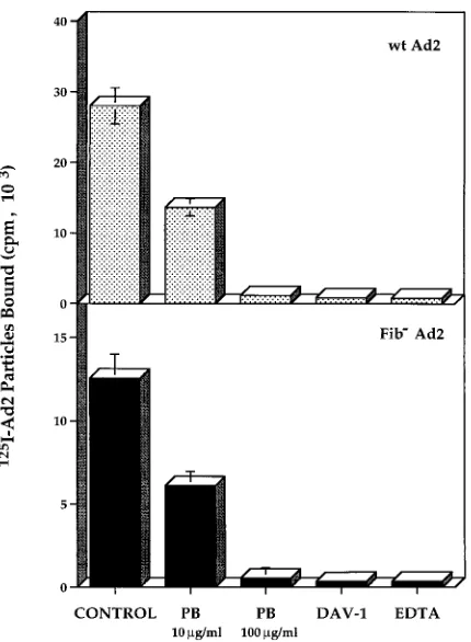
function-block-FIG. 3. The penton base facilitates Ad2 attachment to THP-1 monocytic cells. Binding of125
I-labeled wild-type (wt) Ad2 or a fiberless mutant Ad2 to THP-1 cells was measured in the presence of saturating amounts of the penton base (PB) protein, 20 mM EDTA, or Fab fragments of the DAV-1 anti-penton base MAb. The data represent the mean and standard deviation of duplicate experiments.
FIG. 4. Effect of anti-integrin MAbs on penton base binding to monocytic cells. Binding of125
I-labeled penton base to THP-1 cells was measured in the presence of 200mg of function-blocking MAbs to integrinsaMb2 (CP3),avb3
(LM609),av(69-6-5), andb1 (P4C10) per ml. The data represent the mean and
[image:4.612.71.286.69.362.2]standard deviation of three experiments.
FIG. 5. Detection of penton base protein interaction with integrinaMb2 by
immunoblotting. Detergent-solubilized THP-1 cell membrane proteins were af-finity purified in the absence (lane 1) or presence (lane 2) of 20 mM EDTA on a Affi-Gel-15 column containing immobilized penton base. Bound proteins were eluted with low-pH buffer and then neutralized and concentrated. Fractions were then analyzed on an immunoblot for the presence of integrinaMb2 by using the
M1/70 MAb as described in Materials and Methods.
on November 9, 2019 by guest
http://jvi.asm.org/
ing MAbs directed against integrins
avb
5 and
b
1 had no effect
on virus attachment.
aMb
2-expressing CHO cells were also
approximately 5- to 10-fold more susceptible to Ad-mediated
gene delivery than were nontransfected CHO cells (Fig. 8),
indicating that integrin
aMb
2 promotes not only Ad2
attach-ment but also virus infection. Interestingly, CHO cells with a
point mutation (Asp-242 to Ala) in the A domain of the
aM
subunit of
aMb
2 also showed enhanced susceptibility to
Ad-mediated gene delivery (Fig. 8). Since this mutation prevents
binding of the complement fragment of iC3b to
aMb
2 (16),
these results suggest that Ad2 penton base interacts with a
region in
aMb
2 distinct from that used for binding to iC3b. In
further studies, we found that CHO cells expressing another
member of the
b
2 integrin family,
aLb
2 (LFA-1), a receptor
found on human lymphocytes (3), also enhanced (threefold)
Ad2 attachment, as well as Ad-mediated gene delivery
(five-fold) (data not shown). Thus, these studies demonstrate that
two distinct members of the
b
2 integrin family promote Ad
interaction with CHO cells.
Ad internalization into hematopoietic cells is mediated by
a
vintegrins.
While the studies reported here demonstrated
that
b
2 integrins promote Ad attachment to hematopoietic
cells, we hypothesized that
av
integrins were, nonetheless,
re-quired for virus internalization. In support of this hypothesis,
we found that Ad2 uptake into both THP-1 monocytic cells
and
aMb
2-expressing CHO cells could be inhibited by
func-tion-blocking MAbs to
av
integrins (Fig. 9), although these
antibodies had no effect on virus attachment (Fig. 4). These
results indicate that
av
integrins rather than
b
2 integrins
me-diate Ad2 internalization into hematopoietic cells. Thus, Ad
interaction with distinct integrins is required for virus
attach-ment and entry into hematopoietic cells.
DISCUSSION
The identity of cell receptors that mediate Ad2 attachment
to different cell types in vivo has yet to be firmly established.
Previous studies had indicated that the cell receptor for the
Ad2 fiber protein was probably responsible for virus
attach-ment to the majority of different cells (19); however, there is
relatively little information on whether this receptor is present
on hematopoietic cells. The studies reported here demonstrate
that cells of the myeloid lineage do not express Ad2 fiber
receptors, although they permit virus attachment via
interac-tion of the penton base protein with integrin
aMb
2. A
Scat-chard analysis (unpublished observation) indicates that
mono-cytic cells, which express both
aMb
2 and
av
integrins, possess
both high- and low-affinity binding sites for the penton base
protein, whereas only low-affinity penton base-binding sites
have been observed on epithelial cells, which express
av
but not
b
2 integrins (27). This finding is consistent with the current
studies that demonstrate that
b2
integrins serve as alternative
Ad2 attachment receptors on certain hematopoietic cell types
which lack fiber receptors.
While
b
2 integrins were shown to promote Ad attachment to
monocytic cells, the precise sequence in the penton base that
governs virus interaction with
b
2 integrins remains to be
de-termined. Penton base and Ad2 binding to monocytic cells is
inhibited by an GRGDSP synthetic peptide (results not shown)
and by a MAb that recognizes the penton base RGD sequence
FIG. 6. (A) Flow-cytometric analysis of integrinaMb2 expression on CHO
cells. Nontransfected CHO cells or CHO cells transfected with cDNAs encoding aMb2 (15) were evaluated foraMb2 expression by flow cytometry with the CP3
MAb (dashed line) or another MAb toaMb2, OKM1 (solid line), followed by
incubation with FITC-IgG. Control samples were incubated with FITC-IgG alone prior to analysis by flow cytometry. (B) Ad attachment to CHO cells lacking integrinaMb2 or to CHO cells expressing recombinantaMb2. Binding of
increasing amounts of125I-labeled Ad2 was measured in the presence or absence
of unlabeled virus to determine the level of nonspecific binding. The data rep-resent the mean of three experiments, with the standard deviation being equiv-alent to 5 to 10% of the mean.
4506 HUANG ET AL. J. VIROL.
on November 9, 2019 by guest
http://jvi.asm.org/
[image:5.612.65.446.74.452.2](Fig. 3), suggesting that the penton base RGD motif is involved
in interactions with
b
2 integrins. Interestingly, the CP3 MAb,
which blocks Ad and penton base interaction with
b
2 integrins
(Fig. 7), contains an RYD sequence in its antigen-combining
site (25). Since the RYD sequence in this MAb is believed to
mimic the RGD motif involved in integrin binding, this finding
further supports the notion that the penton base RGD
se-quence mediates interaction with
b
2 integrins. Further penton
base mutagenesis studies, however, are necessary to confirm
this possibility. The fact that two different
b
2 integrins which
have different
a
subunits facilitated Ad2 attachment and gene
delivery also suggests that the
b
2 subunit may play the major
role in penton base interactions.
Although
b
2 integrins promote Ad attachment, these
recep-tors do not appear to play a direct role in Ad2 internalization
(Fig. 9). Consistent with the present studies, freshly isolated
monocytes express integrin
aMb
2 and also support virus
at-tachment; however, they are not susceptible to Ad infection
and gene delivery (13). In contrast, upregulation of
av
integrin
expression on terminally differentiated
monocytes/macro-phages or expression of recombinant
b
2 integrins in CHO cells
that also have endogenous
av
integrins (Fig. 9) renders these
cells competent for virus internalization and Ad-mediated
gene delivery. Thus, penton base interaction with two distinct
integrins is required for efficient Ad entry into these cells.
Further studies are required to evaluate the overall
contribu-tion of this fiber-independent pathway to Ad infeccontribu-tion of other
hematopoietic cell types, including B and T lymphocytes and
NK cells. The interaction of other microorganisms such as
Bordetella pertussis, a bacterial pathogen, with different cell
integrins has been reported previously (20); however, these
interactions are thought to facilitate the same stage
(attach-ment) in infection.
The findings reported here also suggest that a modified Ad
vector lacking the fiber protein could be used to selectively
deliver genes to hematopoietic cells in vivo because of its
limited capacity to interact with the majority of other cell types
that express the fiber receptor. Host immune responses
di-FIG. 7. Effect of anti-integrin MAbs on Ad attachment toaMb2-expressing
CHO cells.aMb2-expressing CHO cells were incubated with recombinant fiber,
penton base (PB), or function-blocking MAbs toaMb2 (CP3),avb5 (P1F6), or b1 (P4C10), and then the binding of125I-labeled Ad2 was measured.
FIG. 8. Ad-mediated gene delivery to CHO cells lacking or expressing inte-grinaMb2. Expression of the Ad-transduced lacZ gene in nontransfected CHO
cells lackingaMb2, CHO cells expressing wild-type (WT)aMb2, and in CHO
[image:6.612.60.296.492.669.2]cells expressing a point mutation (D242A) in theaMsubunit ofaMb2 is shown. b-Galactosidase expression was assayed as described in Materials and Methods. The data represent the mean of three experiments, with the standard deviation being equivalent to 5 to 10% of the mean.
FIG. 9. Ad internalization is mediated by avintegrins. Internalization of 125I-labeled Ad2 into THP-1 cells (A) ora
Mb2-expressing CHO cells (B) was
assayed at various times after incubation at 378C by resistance to trypsin diges-tion (27) (open circles). In parallel studies, cells were preincubated with a function-blocking MAb toav(69-6-5) (A) oravb5 (P1F6) (B) (solid circles)
before measurement of virus internalization. The data represent the mean of three experiments, with the standard deviation being 5 to 10% of the mean.
on November 9, 2019 by guest
http://jvi.asm.org/
rected against Ad structural proteins limit the usefulness of Ad
vectors (8, 28); therefore, a vector lacking a major structural
protein (fiber) might also be less immunogenic, thereby
pro-viding longer-term expression of a therapeutic gene. Previous
studies have demonstrated that fiberless Ad particles can be
properly assembled (5); however, further development of
packaging cell lines is needed to produce significant amounts
of such particles in the absence of contaminating helper virus.
Since monocytes/macrophages represent a site of persistent or
latent virus infection by a number of important human
patho-gens including cytomegalovirus and human immunodeficiency
virus, the ability to selectively deliver antiviral agents (e.g.,
ribozymes) into these cells via Ad vectors may have therapeutic
value. Ad-mediated targeting of cytokine genes to monocytes/
macrophages may also prove beneficial for anticancer therapy,
given the well-established role of these cells in tumor cell
killing (1, 22). In addition to potential practical applications,
the current studies provide fundamental knowledge of
virus-host cell interactions and also contribute new insights into the
function of cell integrins.
ACKNOWLEDGMENTS
We express our gratitude to Michel Perricaudet for providing Ad.RSVbgal virus, Gary Ketner for the H2dl807 virus, and David Cheresh for helpful advice during the course of these studies. We also thank Patricia Mathias for her technical expertise and Catalina Hope and Joan Gausepohl for preparation of the manuscript.
This work was supported in part by U.S. Public Service grants HL54352 and GM49899 and GCRC grant 2MO1 RR00833.
REFERENCES
1. Addison, C. L., T. Braciak, R. Ralston, W. J. Muller, J. Gauldie, and F. L. Graham.1995. Intratumoral injection of an adenovirus expressing interleu-kin 2 induces regression and immunity in a murine breast cancer model. Proc. Natl. Acad. Sci. USA 69:8522–8526.
2. Andiman, W. A., and G. Miller. 1982. Persistent infection with adenovirus types 5 and 6 in lymphoid cells from humans and woolly monkeys. J. Infect. Dis. 145:83–88.
3. Arnaout, M. A. 1990. Structure and function of the leukocyte adhesion molecules CD11/CD18. Blood 75:1037–1050.
4. Bai, M., B. Harfe, and P. Freimuth. 1993. Mutations that alter an Arg-Gly-Asp (RGD) sequence in the adenovirus type 2 penton base protein abolish its cell-rounding activity and delay virus reproduction in flat cells. J. Virol. 67:5198–5205.
5. Challberg, S. S., and G. Ketner. 1981. Deletion mutants of adenovirus 2: isolation and initial characterization of virus carrying mutations near the right end of the viral genome. Virology 114:196–209.
6. Chu, Y., K. Sperber, L. Mayer, and M.-T. Hsu. 1992. Persistent infection human adenovirus type 5 in human monocyte cell lines. Virology 188:793– 800.
7. Crystal, R. G. 1995. Transfer of genes to humans: early lessons and obstacles to success. Science 270:404–410.
8. Dai, Y., E. M. Schwarz, D. Gu, W.-W. Zhang, N. Sarvetnick, and I. M. Verma.1995. Cellular and humoral immune responses to adenoviral vectors
containing factor IX gene: tolerization of factor IX and vector antigens allows for long-term expression. Proc. Natl. Acad. Sci. USA 92:1401–1405. 9. De Nichilo, M. O., D. R. Shafren, W. M. Carter, M. C. Berndt, G. F. Burns, and A. W. Boyd.1996. A common epitope on platelet integrinaIIbb3
(gly-coprotein IIbIIIIa; CD41b/CD61) andaMb2 (Mac-1; CDIIb/CD18) detected
by a monoclonal antibody. J. Immunol. 156:284–288.
10. Ginsberg, H. S., L. L. Moldawer, P. B. Sehgal, M. Redington, P. L. Kilian, R. M. Chanock, and G. A. Prince.1991. A mouse model for investigating the molecular pathogenesis of adenovirus pneumonia. Proc. Natl. Acad. Sci. USA 88:1651–1655.
11. Goldman, M. J., and J. M. Wilson. 1995. Expression ofavb5 integrin is necessary for efficient adenovirus-mediated gene transfer in the human air-way. J. Virol. 69:5951–5958.
12. Hierholzer, J. C. 1992. Adenoviruses in the immunocompromised host. Clin. Microbiol. Rev. 5:262–274.
13. Huang, S., R. I. Endo, and G. R. Nemerow. 1995. Upregulation of integrins avb3 andavb5 on human monocytes and T lymphocytes facilitates adeno-virus-mediated gene delivery. J. Virol. 69:2257–2263.
14. Hynes, R. O. 1992. Integrins: versatility, modulation, and signaling in cell adhesion. Cell 69:11–25.
15. Kamata, T., R. Wright, and Y. Takada. 1995. Critical threonine and aspartic acid residues within the I domains ofb2 integrins for interactions with intercellular adhesion molecule 1 (ICAM-1) and C3bi. J. Biol. Chem. 270: 12531–12535.
16. Michishita, M., V. Videm, and M. A. Arnaout. 1993. A novel divalent cation-binding site in the A domain of theb2 integrin CR3 (CD11b/CD18) is essential for ligand binding. Cell 72:857–867.
17. Neumann, R., J. Chroboczek, and B. Jacrot. 1988. Determination of the nucleotide sequence for the penton-base gene of human adenovirus type 5. Gene 69:153–157.
18. Niiya, K., E. Hodson, R. Bader, V. Byers-Ward, A. A. Koziol, E. F. Plow, and Z. M. Ruggeri.1987. Increased surface expression of the membrane glyco-protein IIb/IIIa complex induced by platelet activation. Relationship to the binding of fibrinogen and platelet aggregation. Blood 70:475–483. 19. Philipson, L., K. Lonberg-Holm, and U. Petterson. 1968. Virus-receptor
interaction in an adenovirus system. J. Virol. 2:1064–1075.
20. Relman, D., E. Tuomanen, S. Falkow, D. T. Golenbock, K. Saukkonen, and S. D. Wright.1990. Recognition of a bacterial adhesin by an integrin: mac-rophage CR3 (aMb2, CD11b/CD18) binds filamentous hemagglutinin of Bordetella pertussis. Cell 61:1375–1382.
21. Ruoslahti, E. 1991. Integrins. J. Clin. Invest. 87:1–5.
22. Schmidt-Wolf, G. D., and I. G. H. Schmidt-Wolf. 1995. Cytokines and gene therapy. Immunol. Today 16:173–175.
23. Springer, T., G. Galfre, D. S. Secher, and C. Milstein. 1979. Mac-1: a macrophage differentiation antigen identified by monoclonal antibody. Eur. J. Immunol. 9:301–306.
24. Stratford-Perricaudet, L. D., I. Makeh, M. Perricaudet, and P. Briand. 1992. Widespread long-term gene transfer to mouse skeletal muscles and heart. J. Clin. Invest. 90:626–630.
25. Tomiyama, Y., E. Brojer, Z. Ruggeri, S. J. Shattil, J. Smiltneck, J. Gorski, A. Kumar, T. Kieber-Emmons, and T. J. Kunicki.1992. A molecular model of RGD ligands. J. Biol. Chem. 267:18085–18092.
26. Wickham, T. J., E. J. Filardo, D. A. Cheresh, and G. R. Nemerow. 1994. Integrinavb5 selectively promotes adenovirus mediated cell membrane per-meabilization. J. Cell Biol. 127:257–264.
27. Wickham, T. J., P. Mathias, D. A. Cheresh, and G. R. Nemerow. 1993. Integrinsavb3andavb5promote adenovirus internalization but not virus
attachment. Cell 73:309–319.
28. Yang, Y., F. Nunes, K. Berencsi, E. E. Furth, E. Go¨nczo¨l, and J. M. Wilson. 1994. Cellular immunity to viral antigens limits E1-deleted adenoviruses for gene therapy. Proc. Natl. Acad. Sci. USA 91:4407–4411.
4508 HUANG ET AL. J. VIROL.