C
H A R A C T E R I ZA T I O N O F T H E HU M A NI
M M U N E RE S P O N S E T OCR Y P T O S P O R I D I U M SP P.
I NSO U T H E R N I
N D I ATHESIS SUBMITTED TO
THE TAMIL NADU DR M.G.R. MEDICAL UNIVERSITY, CHENNAI FOR THE DEGREE OF
DOCTOR OF PHILOSOPHY
BY
A.
SI T A RA SW A R NA RA OTHE WELLCOME TRUST RESEARCH UNIT DEPARTMENT OF GASTROINTESTINAL SCIENCES
CHRISTIAN MEDICAL COLLEGE, VELLORE 632004, TAMIL NADU, INDIA
•
TA B L E O F
CO N T E N T S
Chapter Page
number
1 Introduction 1
2 Aims and Objectives 4
3 Review of Literature 6
4 Scope and Plan of the study 28
5 Materials and Methods 30
6 Molecular and Spatial Epidemiology of Cryptosporidial Diarrhea in Children– results from a birth cohort in a semi urban slum
community
40
7 Effects of Multiple Symptomatic and Asymptomatic Cryptosporidial Infections in Children in the Community
52
8 Molecular Characterization of Cryptosporidial Isolates in
Hospitalized Children with Diarrhea and Quantitation of Oocyst Burden with Real Time PCR
61
9 Serum IgG responses to Cryptosporidium Antigens in a Birth Cohort of Children in a Semiurban Slum Community in South India
71
10 Molecular Epidemiology and Immune response to Cryptosporidial Infection in HIV Infected Adults and Quantitation of Oocysts in Patients with and without Diarrhea
85
11 Summary and Conclusions 96
Recommendations 101
•
A
C K N O W L E D G E M E N T SI would like to thank my guide, Prof Gagandeep Kang or Cherry as she is more fondly
known for first giving me a job and then encouraging me to do a PhD. I also thank Prof
Honorine Ward, Professor at Tufts Medical Center, Boston, our collaborator in research,
for being a mentor and providing training at her laboratory.
I would also like to thank Prof Deva Prasanna Rajan, my co-guide for his support, Prof
Jayaprakash Muliyil at the Dept of Community Health and Dr Elena Naumova, Tufts
Medical Center, Boston who provided a lot of insight during data analysis. Thanks to
Geneve Allison, Roberta O’Conner, Anne Kane and Brett Leav at the Tufts Medical
Center, Boston who provided me with the resources to transfer several of the assays to
Vellore.
I would be remiss if I didn’t especially thank Arun Kannan, Farzana Begum Liakath and
Gunashekaran Chandrabose, who work really long hours with me currently and Charles
Livingston, Premi Shankaran, Priya Rajendran and Kalyan Banda who have worked with
me previously on various projects.
I thank Rajiv Sarkar and Beryl Gladstone, the epidemiologists without whom all the data
collected would have meant very little.
I would also like to thank various members of the labs in Boston and Vellore including
Rekha Pai and Sheela Roy and our department staff, Thirumani and Kumari for all their
help. I would also like to thank all the clinical research officers, laboratory technicians
and field workers who were responsible for collecting clinical data and samples.
I would like to thank friends everywhere who have always made life interesting and
finally and most importantly, my fantastic partner in life, Mahesh who has helped me set
•
LI S T O F
AB B R E V I A T I O N S
bp - base pair µ - micron µl - micro litre
AIDS - acquired immunodeficiency syndrome ART- antiretroviral therapy
BMT - bone marrow transplant BSA - bovine serum albumin CD - cluster of differentiation CDC- Centers for Disease Control CMC- Christian Medical College
COWP – Cryptosporidium oocyst wall protein Cp – Cryptosporidium parvum
DHFR – Dihydrofolate reductase DNA- Deoxyribonucleic acid
EDTA –Ethylenediamine tetra acetic acid ELISA –Enzyme linked immunosorbent assay EST – expressed sequence tag
ETEC – Enterotoxigenic E. coli GIS – geographic information systems gp – glycoprotein
GPI –Glycosylphosphatidylinositol
HAART – Highly active antiretroviral therapy HAZ – Height for age Z score
HIV – human immunodeficiency virus IFN – interferon
Ig - Immunoglobulin IL – interleukin
IPTG Isopropyl β-D-1-thiogalactopyranoside IQR Interquartile range
ITS – internal transcribed spacer kDa Kilodalton
Mabs Monoclonal antibodies Mb- mega base pair
Mg - milligram
MPN- most probable number NFkB – Nuclear Factor k B
N-WASP – Neural Wiskott–Aldrich syndrome protein OD – Optical density
OI – Opportunistic infection PBS - Phosphate buffered saline PCR - Polymerase chain reaction PV – Parasitophorous vacuole
RFLP – Restriction fragment length polymorphism rRNA - ribosomal RNA
SD – Standard deviation TCR – T - cell receptor TNF- tumor necrosis factor
TRAP - Thrombospondin-related adhesive protein WAZ- Weight for age Z score
WHO -World Health Organization
•
C
H A P T E R1
Cryptosporidiumspp. are apicomplexan parasites that are an important cause of diarrhea,
both in children in developing countries, where it is endemic and in
immunocompromised individuals where infection may result in protracted and life
threatening diarrhea in HIV infected patients. In the west, cryptosporidiosis is causally
associated with sporadic infections as well as waterborne outbreaks. In cryptosporidial
infection, the immune status of the host plays an important role in determining disease.
Immunocompetent persons develop a self-limited diarrheal illness but severe and
persistent infections occur in subjects with compromised immune function.
Cryptosporidiosis in humans is mainly attributable to 2 species - Cryptosporidium
hominis and Cryptosporidiumparvum.
Transmission occurs by multiple routes including direct person-to-person spread,
ingestion of contaminated food or water or contact with infected animals. The spherical
thick walled, environmentally hardy oocysts, shed in the fecal material of the infected
host, are immediately infectious, unlike other coccidian parasites, and have a low
infectious dose (42). The potential to cause explosive and massive waterborne outbreaks
has also been well documented in developed countries. As there is a distinct potential for
intentional contamination of water supplies, the CDC has included this parasite as a
Approximately one in five deaths in children aged less than 5 years in India is attributable
to diarrhea and Cryptosporidiumspp. is a leading cause of infectious diarrhea in children
in India. Infection in early childhood has been associated with subsequent impairment in
growth, physical fitness and cognitive function in children in developing countries (25,
86). In these countries, malnourished children may be at greater risk of developing
cryptosporidiosis, and in addition, those who do so are at risk of further nutritional
compromise during the course of infection. This protozoan parasite accounts for around
12% of diarrhea in children below? 5 years in developing countries with a peak incidence
in children less than 2 years of age. A number of studies in India have reported
Cryptosporidium spp. in diarrheal stool samples of children by microscopy with
positivity rates ranging from 1.1% to 18.9%. There have however, been very few studies
examining the molecular epidemiology of cryptosporidiosis in children and none
examining immunity to cryptosporidiosis in Indian children.
Cryptosporidiosis is also a substantial threat to HIV-infected individuals and affects up to
50% of patients with AIDS in the developing world, (64). Patients can have chronic
watery diarrhea that can last for more than 2 months, resulting in severe dehydration,
weight loss, malnutrition, extended hospitalizations, and mortality. AIDS patients with
cryptosporidiosis also have a significantly shorter duration of survival from the time of
diagnosis (138). Cryptosporidiosis remains a major risk to immunocompromised because
of the lack of effective specific therapy. Antiretroviral therapy greatly influences the
resulting in a sustained therapeutic effect on follow-up. However, despite the use of
HAART, HIV-infected patients still present with coccidian diarrhea, possibly due to
non-compliance with medications, viral drug resistance to the drugs or decreased drug
bioavailability. Reports from the mid 1990s on the prevalence of symptomatic
cryptosporidiosis in HIV infected adults from different parts of India, range from as low
as 0.7% to 81%. Some of these studies that documented mean CD counts in patients,
showed that that symptomatic cases had CD counts
4
4 < 200 cell/mm3 reinforcing the
importance of CD4 T cells in mediating resistance to this pathogen.
Due to its sequestered location within the host cell, ability to set up an auto-infective
cycle in the host, innate antimicrobial resistance and ability to affect several host species,
this parasite has a large survival advantage over other enteric pathogens. Till date, there
is no effective treatment or vaccine for cryptosporidiosis. Nitazoxanide was licensed for
use in immunocompetent patients, but a recent meta-analysis found it to be ineffective in
HIV-infected individuals (3). The urgent need for development of tools for early
detection, treatment and prevention of cryptosporidiosis especially in developing
countries is highlighted by its inclusion in the ‘Neglected Diseases Initiative’ in
September 2004 (203). This study attempts to present the much needed information on
epidemiology, transmission and immune response to Cryptosporidium in 2 vulnerable
•
CH A P T E R
2
The aim of this study was to document cryptosporidial infections and immune response
in children under the age of 5 years and HIV infected adults – the two populations most
vulnerable to infection in India. Children in a longitudinal birth cohort study were
included to study the commonly circulating species and subgenotypes in the community,
transmission patterns, effect of multiple infections and longitudinal antibody responses.
Hospitalized children were included to study severity of diarrhea and quantitate oocyst
burdens and contrast species and subgenotypes obtained with that in the community.
HIV infected adults recruited from the infectious diseases clinic in the hospital were
included to study infecting species and subgenotypes, immune response and oocyst
burdens.
The specific aims were:
1. To study the molecular epidemiology of Cryptosporidiosis
a) To study the molecular epidemiology of cryptosporidial diarrhea in a birth
cohort of children in the community
b) To identify the effects of multiple symptomatic and asymptomatic
cryptosporidial infections in early childhood
c) To characterize cryptosporidial isolates in hospitalized children with diarrhea
and quantitate oocyst burden with real time PCR
d) To characterize cryptosporidial isolates associated with symptomatic and
asymptomatic infections in HIV infected adults in the hospital and to determine
2. To study the immune response to cryptosporidial infections
a) To determine maternal and longitudinal serum antibody levels to recombinant cryptosporidial antigens in children in the birth cohort
b) To evaluate subtype-specific antibody responses in children before and after
cryptosporidial diarrhea
c) To determine serum antibody responses to the immunodominant
•
CH A P T E R
3
Historical background – The parasite and its life cycle was first described in seminal studies in mice by a medical parasitologist, Ernest Edward Tyzzer, at Harvard University
in an observation in 1907, and subsequent publications in 1910 and 1912 (231). He
named this genus “Cryptosporidium” or “hidden sporocysts” due to the absence of
sporocysts, which are seen in most other coccidian parasites. This protozoan then
remained largely unrecognized as a human pathogen until the first cases reports in
humans emerged in the late 1970s with the advent of the AIDS epidemic. Although the
first human case report in 1976 was a child with a self limiting enterocolitis that lived on
a farm (167), a review of the first 7 reported cases in 1980 showed that this was a disease
of the immunocompromised (231).
The potential to cause waterborne outbreaks, due to the ability of the oocyst to survive in
the environment, was documented in Milwaukee where the largest outbreak of water
borne disease ever took place in 1993, affecting an estimated 403,000 persons (135). This
and the inclusion of the parasite as a Category B pathogen for biodefense as a water
safety threat by the Centers for Disease Control and Prevention, Atlanta (CDC)
(http://www.bt.cdc.gov/agent/agentlist-category.asp) due to the potential for intentional
contamination of water supplies, has resulted in a new interest in research on this
Since the 1980s, the life cycle and invasive process of Cryptosporidium in the intestinal
epithelial cell has been studied (139), the taxonomy and host specificity revised (247), the
number of chromosomes established as 8, expressed sequence tag (EST) data made
available (222) and in vitro culture attempted with partial success (15). In 2004, the 9 Mb
genome sequence of C. parvum and C. hominis was released (2). This has resulted in a
clearer understanding of the biology and pathogenesis of this protozoan parasite.
However, in spite of these advances, till date there is no effective therapy for
cryptosporidiosis in HIV and no vaccine for prevention of disease in susceptible
populations.
Taxonomy and Classification – Cryptosporidium spp. belongs to the phylum Apicomplexa, group Alveolata. They are characterized by the presence of an apical
complex containing apical rings, a conoid, micronemes, rhoptries, and dense granules
which play an important role in host cell invasion. Although long believed to be related
to the other coccidian parasites like Sarcocystis and Eimeria with whom this parasite
shares an ecological niche, there is now phylogenetic evidence to show that
Cryptosporidium is more closely related to another group of apicomplexan protists called
gregarines (23). This new classification may also help explain the several differences
between Cryptosporidium and other intestinal coccidians including its extracytoplasmic
location in the host cell and absence of sporocyst as well as the unique attachment
Cryptosporidium was also found to lack the apicoplast and mitochondrial genome usually
found in other apicomplexan parasites.
Early taxonomical classifications of species in this genus were based on oocyst
morphology and size, infectivity and host species resulting in several erroneously named
species. Recent advances in molecular characterization based on the 18S rRNA gene
sequence have now helped validate the presence of at least 16 species (31) and an
additional ~ 40 isolates referred to as genotypes. Of importance is the emergence of the
species ‘C. hominis’ to denote what was previously described as C. parvum (human
genotype) thereby clearly differentiating the zoonotic species C. parvum (previously
known as bovine genotype) from isolates that were found in humans. This was based on
multi-locus analysis and infectivity studies (150) although the 2 species share an average
nucleotide similarity of 96.7%. These 2 species are the most common cause of disease in
humans although infections with C. felis, C. canis, C. muris, C. suis and C. meleagridis
have also been reported.
Life cycle, Excystation and Invasion – Several factors have made cryptosporidial research extremely difficult, including the inability to maintain this organism in culture
instead requiring passage in larger animals like calves or pigs and the species that affects
humans, C. hominis is even more difficult to obtain. Therefore C. parvum has been used
The life cycle of Cryptosporidium spp. is completed within the small intestine and colon
of the host, with the developing stages associated with the luminal surface of the
intestinal epithelial cells, where it remains intracellular but extracytoplasmic (95).
Infection in a new host occurs following the ingestion of as few as 10 oocysts (ID50 132
oocysts), as evidenced in healthy adult volunteer studies (42). A clear dose response
relationship in C. hominis was also found with diarrhea in 40% of cases when infected
with 10 oocysts increasing to 75% with 500 oocysts.
The oocyst wall is a trilaminar structure and is interrupted at one pole by a single seam or
suture that extends one-third to one-half the way around the periphery. Sporozoites exit
the oocyst via this suture during excystation. The outer wall is thought to be a
glycoprotein while the central layer of the wall is thought to be a glycolipid/lipoprotein
responsible for the acid-fast staining property of the oocyst. Thin-walled oocysts differ in
that the wall does not contain a thick inner layer. The release of 4 motile sporozoites from
each oocyst is triggered by temperature, exposure to acidic pH followed by bile salts and
trypsin along with proteases released by the parasite (215). Prior to invasion, the
adherence of oocyst wall and sporozoites to the intestinal mucous layer is thought to be
mediated by surface Gal/GalNAc lectins (215). Using a process of helical or gliding
motility, based on the parasites intracellular actin-myosin like mechanism called the
actin-aldolase-TRAP complex, the sporozoites move over the surface of cells and deposit
Table 3.1: Cryptosporidial Proteins Associated with Host – Parasite Interactions
Protein Size and type Location and Function
CSL 1300 kDa glycoprotein Seen in the apical region of sporozoites and released in trails, binds to host cells via 85 kDa receptor gp900 ~ 900 kDa mucin like
glycoprotein
Seen on the surface of sporozoites and shed in trails – associated with adherence to mucin and gliding motility
gp40/15 40 and 15 kDa (Cp17) mucin like glycoproteins
gp40 seen on apical region and associated with adherence gp15 over the entire surface, shed in trails, gliding motility
Cp47 47kDa glycoprotein Apical region, adherence
P23 23/27 kDa glycoprotein Seen over entire surface, shed in trails, associated with adherence and gliding motility
Cpa 135 135 kDa glycoprotein Apical region, Co-localizes with gp900
P30 31 kDa protein Gal/GalNAc lectin, adherence to intestinal mucin layer and epithelial cell epitopes
Cp2 82 kDa, glycoprotein Associated with PV membrane, sexual development TRAP C1 76 kDa protein Apical region, Adhesion and gliding motility
CPS 500 Glycolipid Gliding motility
Cp12 Oocyst and sporozoites surface adherence
CpMuc 1-7 Family of mucins, 28-55 kDa
ligand (CSL), gp900, gp40/15, p23 and Cpa 135 (38, 215). Other proteins described to
be involved in host cell adhesion include Cp47, Cp12 and Cp2 (38, 215) (Table 3.1).
The attachment and invasion process is dependent on temperature, intracellular calcium,
and the host cell cytoskeleton and is an extremely rapid process with invasion occurring
in less than 7 minutes (48, 215), with release of proteins by the apical complex.
Activation of cell signaling pathways that trigger cdc42, a key regulator of actin
remodeling, induces host cell actin remodeling at the attachment site with accumulation
of actin and related proteins like tropomyosin 5, N-WASP and Arp 2/3 (46, 48). This
results in the formation of a parasitophorous vacuole (PV) (95), a unique sequestered
extracytoplasmic niche. A feeder organelle on the parasitophorous vacuole membrane
comprised of a tunnel-like structure is believed to function as a portal to allow nutrients
to pass from the host cell to the parasite (215). Within the PV, the sporozoites develops
into a transitional trophozoite stage which give rise to meronts, which replicate by an
asexual cycle (merogony) to form merozoites that infect adjacent epithelial cells (within
3-5 days post infection) and by a sexual cycle resulting in the production of micro- and
macro-gamonts (within a week of infection) which fuse to form zygotes. How
microgametes detect cells containing the macrogamonts is not known, but once it does,
the microgamete and macrogamont fuse (139). The resultant zygotes then mature to form
sporulated thin-walled and thick-walled oocysts. The parasite also affects host cell
oocysts can excyst endogenously, resulting in autoinfection, which, along with repeated
first-generation merogony, helps to explain persistent infections in AIDS patients. The
spherical thick-walled, environmentally resistant oocysts (3–6 µm in diameter), shed in
the fecal material of the infected host, are immediately infectious, unlike other coccidian
parasites, and hence can be transmitted from person to person. The more common routes
of transmission are by the fecal-oral route with ingestion of contaminated food or water
or contact with infected animals.
Pathophysiology of cryptosporidial diarrhea - Cryptosporidial diarrhea has a pre-patent period ranging from 4 to 22 days for humans as shown in volunteer studies (60).
The median duration of diarrhea in these studies was 6 days with oocyst shedding
recorded up to 30 days after challenge. The alterations in the intestinal structure and
physiology that lead to the pathogenesis of cryptosporidiosis include rapid loss of the
microvillus border, shortening and fusion of the villi and lengthening of the crypts which
occurs in a dose dependent manner, resulting in malabsorption due to loss of
membrane-bound digestive enzymes, decreased absorption, reduced glucose-NaCl absorption and
increased chloride anion secretion. (173, 230).
Cryptosporidium infection stimulates the production of proinflammatory cytokines,
IFN-γ, IL-8 and TNF-α. While IFNγ is important for the resolution of symptoms, TNFα
prostaglandins, neural peptides and reactive nitrogen intermediates (128). Neutrophils
and mononuclear cells infiltrate the lamina propria. Substance P has also been found to
be up regulated in cryptosporidiosis and could also cause elevation of pro-inflammatory
cytokines (197). A marker of inflammation, fecal lactoferrin has been found to be
elevated in diarrheal stool of children with cryptosporidial diarrhea (119).
Cryptosporidium has also been found to cause disruption of the epithelial tight junction
leading to a leaky and dysfunctional epithelium and alteration of solute transport (230). In
a suckling rat model cryptosporidial infection was found to impair nutrient intake, growth
and muscular protein synthesis and this persists beyond the duration of infection (227).
Most infections present with a self-limited watery diarrhea although people with
pre-existing antibodies are found to be less susceptible or may have asymptomatic infections.
Associated symptoms like vomiting, malaise, loss of appetite and fever may also occur.
The disease is usually self limited in the immunocompetent but can result in a prolonged,
protracted infection in HIV infected adults.
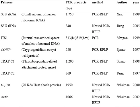
Molecular typing of cryptosporidial isolates – PCR based identification of
Cryptosporidium and differentiation of species based on polymorphisms has been carried
out using several loci. These include β-tubulin, actin, TRAP-C1, TRAP-C2, ITS1,
polythreonine repeat (Poly-T), dihydrofolate reductase (DHFR) and heat shock proteins
(Hsp70) (105) (Table 3.2). Currently however, most workers carry out a nested PCR and
More heterogenous loci used for subgenotyping of cryptosporidial isolates in
epidemiological and population genetics studies include micro satellite 1 and 2, mini
satellites, ITS-2 and gp60 or Cpgp40/15.
Extensive polymorphisms among Cryptosporidium isolatesat the Cpgp40/15 locus has
also been used as a epidemiological tool for subgenotyping in several studies (130). This
gene encodes a precursor glycoprotein which is proteolytically cleaved into two
glycoproteins, gp40 and gp15 (Table 3.1) which are implicated in attachment and
invasion (170). A striking feature of the Cpgp40/15 gene (also referred to as GP60) is the
high degree of polymorphism. Numerous single nucleotide and amino acid
polymorphisms in the Cpgp40/15 locus, define at least 8 allelic subtypes that can be
reliably identified by PCR-RFLP. The identifying polymorphisms are predominantly
clustered in the variant domain of the gp40 portion of the molecule, located directly
C-terminal of the polyserine domain. Currently, Cpgp40/15 subtype analysis is commonly
used as a tool to investigate transmission patterns, waterborne outbreaks and clinical
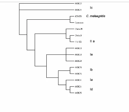
correlates of cryptosporidiosis. The common subgenotypes of C. hominis are Ia, Ib, Id, Ie
and If and that of C. parvum are IIa, IId and the anthroponotic IIc (248). Additional
Table 3.2: Tools for Molecular Typing of Cryptosporidial Species
Primers PCR products
(bp)
method Author year
SSU rRNA (Small subunit of nuclear ribosomal RNA)
1,750 PCR-RFLP Xiao 1999
SSU rRNA 840 Nested
PCR-RFLP
Jiang 2005
ITS1 (Internal transcribed spacer
of nuclear ribosomal DNA)
513(hu)519(bov) PCR Morgan 1999
COWP (Cryptosporidium oocyst
wall protein)
550 PCR-RFLP Spano 1997
TRAP-C1 (Thrombospondin related
attachment protein gene)
1,200 PCR-RFLP Spano 1998
TRAP-C2 369 PCR-RFLP Peng 1997
Hsp70 (70 Kda Heat shock protein) 1950 Nested
PCR-RFLP
Sulaiman 2000
Actin 1066 Nested
PCR-RFLP
Molecular epidemiology – Among the 9 species of Cryptosporidium that cause human disease, C. hominis and C. parvum alone account for greater than 90% of cases. In
developed countries, there are geographic differences between the 2 species with a
slightly greater burden of C. parvum than C. hominis in the United Kingdom, New
Zealand and some parts of Europe (248) but in North America and Australia, C. hominis
is more common. This difference could be attributed to different sources of infection and
route of transmission with C. parvum associated with farming and rural areas. Also, in
these countries, the Cpgp40/15 subgenotype of C. parvum is usually IIa or IId indicating
a zoonotic transmission. In these countries, cryptosporidiosis is a sporadic disease that
occasionally causes outbreaks due to contamination of drinking or recreational water.
In developing countries, the majority of infections are due to C. hominis not C. parvum.
A higher prevalence of more exotic species like C. felis, C. canis and C. meleagridis are
also seen. Cryptosporidiosis is endemic in developing countries and mainly affects
susceptible populations like young children, malnourished and immunocompromised.
Transmission of cryptosporidiosis is anthroponotic in these countries and is reflected in
the fact that most C. parvum isolates belonged to the IIc subgenotype (129, 248). In this
setting, transmission is both endemic involving person to person routes, contamination of
food and from animals as well as epidemic involving the water supply. Seasonality has
also been demonstrated in several developing countries and usually is associated with
The importance of differentiating between species using molecular tools is highlighted by
several recent findings. These include a report of longer periods of oocyst shedding in C.
hominis compared to C. parvum-infected patients in Peru (245), significantly higher
oocysts shedding and greater growth shortfalls at 6 months post infection in children with
C. hominis in Brazil (29) and a longer duration of symptoms, higher rate of asymptomatic
infection and lower CD4+ cell count occurred in HIV-infected patients with C. hominis
infection compared to C. parvum infection in Tanzania (94). A recent study showed that
infection with C. hominis, but not C. parvum was associated with an increased risk of
non-intestinal sequelae in immunocompetent adults and children (97). Another recent
study in Peru showed that zoonotic species like C. canis, C. felis, and subgenotype Id of
C. hominis were associated with diarrhea, and infection with C. parvum was associated
with chronic diarrhea and vomiting in the HIV infected (35).
Some association between Cpgp40/15 subgenotypes and transmission and disease has
also been described. Studies on waterborne outbreaks showed an association with
C.hominis subgenotype Ib indicating that it was potentially more infectious or virulent
(51). Therefore identifying the species and subgenotypes to differentiate between
anthroponotic and zoonotic strains and documenting the molecular epidemiology of
cryptosporidiosis will help plan interventional measures to prevent and treat
Cryptosporidium in children in developing countries - Diarrhea is one of the most
common childhood illnesses with every child aged below 5 years in the developing world
experiencing at least 3 episodes of diarrhea per year (121). In addition to the mortality
and morbidity associated with diarrhea due to dehydration, chronic and recurrent diarrhea
leads to poor absorption of nutrients resulting in stunting and developmental delay (25).
Amongst the more than 20 viral, bacterial and parasitic causes of diarrhea identified till
date, the most frequently reported etiological agents in children in the developing world
include rotavirus, diarrheagenic Escherichia coli, Campylobacter jejuni, Shigella spp,
non-typhoidal Salmonella, Giardia lamblia, Cryptosporidium spp. and Entamoeba
histolytica (172). Astrovirus, enteric adenovirus and calicivirus have also been reported,
but previous studies have indicated a lower relative contribution to the total number of
cases due to higher rates of bacterial and parasitic infections in these countries (172).
In developing countries, cryptosporidiosis, is seen more frequently in malnourished than
well-nourished children and the consequences are more severe in the former than the
latter, possibly because of impaired T cell responses. Studies of Haitian children with
cryptosporidiosis reported that malnourished children have increased levels of systemic
and fecal proinflammatory cytokines (119). Watery diarrhea, vomiting, anorexia and
weight loss are the commonest symptoms of cryptosporidiosis, with persistent diarrhea
frequently reported from developing countries. Studies done in Brazil suggest that even a
single episode of cryptosporidiosis predicts a subsequent increased risk of diarrheal
Diarrhoea is the cause of death in approximately 23% of Indian children who die before
the age of 5 years (107). Cryptosporidiumspp. are a leading cause of infectious diarrhea
in children in India. A number of studies have reported Cryptosporidium spp. in
diarrheal stool samples of children by microscopy with positivity rates ranging from
1.1% to 18.9. Many studies also examined asymptomatic or “control” children (i.e.
without diarrhea) and reported positivity rates between 0% and 9.8% (Table 3.3). These
studies differed in their methods of patient recruitment and stool examination, but only 3
studies conducted till date have used molecular techniques for identification and typing
indicating that actual infection rates may be significantly higher. In a hospital based study
from Calcutta, of 40 microscopy positive samples identified over 5 years, 35 were
positive for C. hominis, 4 were positive for C. parvum, and 1 was positive for C. felis
(57). A recent hospital based study from Secunderabad on adults and children also found
that C. hominis (69%) was the most common genotype (158). A study from Vellore
revealed that Cryptosporidium along with rotavirus, enteric adenovirus and Group B
Salmonella, was a common cause of nosocomial diarrhea in children aged < 3 years
(109). In more recent studies in Vellore, this parasite was also the commonest cause of
parasitic diarrhea among children attending the hospital and also had a high
pathogenecity index. When conventional and molecular methods were compared for the
detection of Cryptosporidiumspp. among the children with diarrhea, it was found that the
detection rates were significantly higher when a combination of molecular and
conventional methods were applied as compared to the conventional method of
Table 3.3: Cryptosporidiosis in Children in India
Place Year Reference Age
(years) %
symptomatic %
asymptomatic
Vellore 1985 Mathan (140) <3 13.1 9.8
Chandigarh 1987 Malla et al(137) <12 1.3
Calcutta* 1987 Das et al (56) <12 5.9
Calcutta 1988 Sengupta et al(205) <5
6.1 1
Varnasi 1988 Singh et al(213) 0.5-3 3 0
Mumbai 1989 Pherwani (184) <5 4.4 0
Bhubaneswar 1989 Subramanyam et al(224). <8 13 0
Calcutta 1989 Pal et al(175). <5 5.6 1.2
Idukki 1989 Reinthaler et al(193). <10 6 3
Delhi 1991 Kaur(114) <2 5 0
New Delhi 1991 Uppal(234) <10 4.9
Calcutta 1993 Das et al(58) <12 5.5 1.1
Varanasi 1993 Nath et al(160). <5 3.8 1.8
Amritsar 1995 Jindal (106) <3 1.3
Manipal 1995 Shetty(210) <5 1.8
Chandigarh 1999 Sethi et al.(206) <12 1.4 -
Secunderabad 2001 Nagamani et al(159) 0.25 - 3 6
Vellore* 2001 Kamalaratnam(109) <3 7.2
Manipal 2002 Ballal (19) <5 15.6 3
Delhi 2002 Kaur et al(115). <5 18.9 -
Calcutta 2005 Palit(176) <12 - 2.3
Calcutta 2006 Das et al(57) <5 4.6 1.2
Secunderabad 2007 Nagamani et al (158) <12 7.6
Although diarrhea in itself is a leading cause of morbidity and mortality among children
in developing countries, the actual effects of repeated and chronic diarrhea on child
development and future productivity as adults is grossly under-estimated (87). Recurrent
and persistent diarrhea has been consistently associated with stunting in children (194). In
studies on children living in slums in Brazil and Peru, early childhood diarrhea (defined
as episodes of diarrhea in the first 2 years of life (86)) negatively correlated with tests of
cognitive function, verbal fluency and physical fitness and resulted in long term growth
faltering (86, 134, 166, 178).
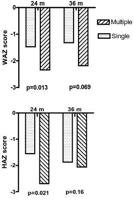
Children who develop cryptosporidiosis are at risk of further nutritional compromise
during the course of the infection. Studies from Peru suggest that both symptomatic and
asymptomatic cryptosporidiosis in children are associated with growth faltering in the
month after infection and recovery is slower in children with symptomatic infection (25).
In these studies, diarrhea due to Cryptosporidium spp. were frequently found to be
associated with lower cognitive function scores and stunting, and the risk increased with
the number of episodes per year (25, 86). Studies in other countries like Malaysia (8),
Turkey (37) and Bangladesh (225) have also implicated diarrhea with Cryptosporidium
spp (44, 45, 86, 166) as an independent predictor of stunting. The role of cryptosporidial
infections on cognitive function and growth in Indian children has not been assessed.
around 2.47 million people living with HIV/AIDS (PLHA) and the four states of Andhra
Pradesh, Maharashtra, Tamil nadu and Karnataka contributed to 63% of all reported cases
(157). The common enteric opportunistic pathogens described are Cytomegalovirus,
Cryptosporidium spp., Isospora belli, Microsporidium spp., Mycobacterium avium
intracellulare and more recently, Clostridium difficile. Cryptosporidium spp. and I. belli
have been reported as the most common causes of diarrhea, however, most studies have
focused only on protozoan etiology (17, 22). Patients can have chronic watery diarrhea
that can last for more than 2 months and shed oocysts in stool during the entire period,
resulting in severe dehydration, weight loss and malnutrition, extended hospitalizations,
and mortality(3, 98). Other symptoms are abdominal cramps, anorexia, nausea, vomiting,
fatigue and low-grade fever. AIDS patients with cryptosporidiosis also have a
significantly shorter duration of survival from the time of diagnosis (138). In the
immunocompromised host, Cryptosporidium spp is also the most commonly isolated
pathogen in the biliary tract in patients with AIDS-cholangiopathy (47). There have been
also been a few reports on other sites of infection, involving the pancreas and lungs
(133).
In India, there have been reports from the mid 1990s on the prevalence of symptomatic
cryptosporidiosis in HIV infected adults from different parts of the country ranging from
as low as 0.7% to 81% (Table 2.4). A high prevalence of around 80% was reported from
a study in Imphal (5) and another in Maharashtra (79) but both had very small sample
modified acid fast staining of concentrated stool samples. The data on prevalence is
highly varied and could reflect geographical differences, as well as differences in the
populations being studied especially with respect to socioeconomic status and access to
potable water. A few recent studies that also carried out ELISA and PCR for detection
(102, 116) found a higher sensitivity for PCR and lower for antigen detection ELISA,
compared to microscopy also contributing to variability in prevalence rates. In children
with HIV in India, chronic diarrhea has been reported in 6.8 to 42 % of cases but very
few studies examined the specific etiology of diarrhea (207).
Several studies in India have also documented mean CD4 counts in HIV infected patients
with most studies showing that symptomatic cases had CD4 counts < 200 cell/mm3 and
asymptomatic cases had CD counts > 300 cell/mm3 reinforcing the importance of CD4 T
cells in mediating resistance to this pathogen (17, 61, 80, 156). A study from Delhi that
stratified patients based on their CD4 counts showed that Cryptosporidium was the
commonest parasite seen in 46% patients with counts < 200 cell/mm3. The prevalence of
cryptosporidial diarrhea (56%) was also significantly higher when CD4 counts were
Table 3.4: Cryptosporidiosis in HIV Infected Patients in India
*data based on analysis of results in the publication
Place Year Reference Cases %
symptomatic %
asymptomatic
Chennai 1995 Kumarasamy et al (126) Adults 16 -
Northern India*
1995 Giri et al(80) Adults and children 4 -
Manipur 1996 Anand et al(10) Adults (iv drug users) 46.6 -
Karad, South Maharashtra
1996 Ghorpade et al(79) - 83.3 † -
Mumbai* 1996 Lanjewar et al (127) Adults 31 -
Manipur* 1997 Anand et al(11) Adults
(asymptomatic iv drug users)
- 57.8
Chennai 1997 Ananthasubramaniam et al (12)
- 6.5 0
Imphal 1998 Agarwal(5) Adults 81.8† -
Vellore 1999 Mukhopadhya et al (155) Adults 9.8 6
Lucknow 2000 Prasad (185)et al Adults, chronic
diarrhea
11.5 -
Chandigarh* 2002 Mohandas(144) et al Mostly adults 22.6 1.49
Mumbai 2002 Joshi(108) et al - 8.5 -
Chennai 2002 Kumar et al(124) Adults 13.7 8
New delhi 2003 Vajpayee et al(235) Adults 18.6 -
Mangalore 2003 Shenoy et al(209) - 17.5 -
Manipal 2003 Singh et al(211) Adults - 43
New Delhi* 2004 Sharma et al(208) Adults 9.3 -
Vellore 2005 Banerjee et al(22) - 15.5 -
Manipal 2005 Ballal et al(18) - 18.57 -
Delhi 2005 Sadraei et al(199) Adults 42.1 38.1
Nepal 2006 Adhikari et al(4) Adults 5.2
Vellore 2006 Muthusamy et al(156) Adults 25.2 4.7
Varanasi 2006 Attili et al(16) - 5.71 -
Bangalore 2007 Becker et al(24) Adults 0.7 2
Delhi 2007 Dwivedi ey al(61) Adults 66.6 8
Although cryptosporidiosis is endemic in tropical countries, only a limited number of
isolates have been typed from developingcountries, especially from HIV-infectedpeople.
Data on genotypes and species in HIV infected patients in India is limited to a single
study from Vellore which showed a prevalence of 25% among HIV seropositive
individuals with diarrhea and 4% in those without diarrhea. Although C. hominis was the
most common species identified comprising 64% of positive cases, a strikingly high prevalence of potentially zoonotic species was seen including, C. parvum, C. meleagridis,
C. felis and C. muris in symptomatic as well as asymptomatic HIV-infected adult
patients, but no significant animal contact was found in the cases with potentially
zoonotic infections.
In developed countries, zoonotic infections are usually due to contact with domestic pets
but studies from endemic countries like Thailand and India have shown a high prevalence
of zoonotic species of up to 50% with no significant contact with pets in these cases (75,
156). A recent study from Delhi has documented history of contact with animals in 87%
of patients with diarrhea and in 32% of cases with cryptosporidiosis, however,
genotyping to identify zoonotic cryptosporidial species was not carried out (61). Studies
on bovine cryptosporidiosis in Calcutta and Punjab have found prevalence rates ranging
from 26% to 50% in diarrheic and 8.5% to 25.7% in non diarrheic calves with the
predominant species being C. parvum (179, 201) with a high mortality rate of 35 %
Table 3.5: CD4 counts Associated with Symptomatic and Asymptomatic
Cryptosporidiosis in Indian patients
CD4 counts (cells/mm3)
Place Year Author
diarrhea no diarrhea
Northern India 1995 Giri et al(80) 56 -
New Delhi 2003 Vajpayee et al(235) 227 -
New Delhi 2004 Sharma(208) 61 -
Vellore 2006 Muthusamy et
al(156)
145 312
Varanasi 2006 Attili et al(16) 255.2 366.9
and other animals, the high prevalence and asymptomatic cryptosporidial infections in
these animals could serve as a reservoir of infection to susceptible human hosts in India.
Antiretroviral therapy also greatly influences the outcome of cryptosporidiosis both
indirectly by immune restitution and increase in CD4 counts (204) and also by the direct
effect of protease inhibitors on oocyst shedding (142) resulting in a sustained therapeutic
effect after follow-up. However, despite the use of HAART, HIV-infected patients can
still present with coccidian diarrhea, possibly due to noncompliance with medications,
viral drug resistance or decreased drug bioavailability (149). Relapses after
discontinuation of HAART have also been documented (251). In India, recently HAART
has become available at an affordable cost or through the government’s ART roll out
programme in 2004 and a recent study from Chennai on the current NNRTI based
therapy initiated when CD4 counts were < 250 cells/mm3 showed a substantial increase in
life expectancy (67). Another study examining the effect of HAART on the incidence of
OIs in India has found that there has been a decrease in both OIs and tuberculosis in
patients on therapy. Hence, increased access to HAART has impacted the natural history
of HIV infection (125). Although western data indicates a shift in etiology of diarrhea in
HIV infected patients (33) and there seems to be a decrease in OIs in a single study from
India, the effect of HAART on the occurrence of cryptosporidiosis and etiology of
Among the other coccidian parasites, Isospora belli has also been reported frequently
with prevalence rates ranging from 2.5 to 60% in patients with diarrhea. Most recent
studies in India show lower prevalence rates than Cryptosporidium (probably due to
prophylactic treatment with trimethoprim-suphamethoxazole)(17, 61, 116, 156, 191) but
a few have recorded higher prevalence rates (89, 237). Cyclospora, on the other hand has
been isolated very infrequently from HIV infected patients in India with low prevalence
rates ranging from 0.98 to 6.5% in symptomatic cases (144, 155, 156, 208).
Other susceptible populations in India- There have been few studies documenting cryptosporidiosis in transplant patients in India. A previous study on patients undergoing
allogeneic bone marrow transplantation in Vellore identified Cryptosporidium spp. in 7
of 65 cases, both in the pre- and post-transplantation period and also found a higher
mortality in patients with enteric pathogens than those without. The other enteric
pathogens identified were rotavirus, adenoviruses, Clostridium difficile and diarrheagenic
E. coli (112). In more recent studies from the same center, Cryptosporidium was
identified in 2.9% of all allogeneic bone marrow transplant recipients and 1.7 % of
pediatric allogeneic BMTs (77, 78). Rates of infections in these patients were similar to
those in the West, possibly attributable to good infection control strategies. A study on
renal transplant recipients in North India identified cryptosporidial diarrhea in 16.6% of
cases (232). Only one study from Varanasi, evaluating risk factors for cryptosporidiosis
in the geriatric age group has been carried out, in which 18.3% of cases had
Immune response - Immune responses involved in mediating resistance to infection with this parasite are not well understood, but evidence obtained from immunodeficient
animal models have shown that a Th1 response involving primarily TCR αβ+ CD4+
lymphocytes, IFN-γ and IL-12 play a major role in the control of C. parvum infection
(62). The susceptibility of HIV/AIDS patients to this pathogen and resolution of
cryptosporidiosis following immune restitution underscores the importance of CD4 T
cells (204). In addition, a number of studies have shown that the cytokine IFN-γ is
critical in protection from and clearance of the infection (195). In human volunteer
studies, IFN-γ expression could be detected in jejunal biopsies of Cryptosporidium
-infected adults and was associated with the presence of anti-Cryptosporidium serum
antibodies and absence of oocyst shedding (240). Peripheral blood mononuclear cells
(PBMCs) from infected humans proliferate in response to recombinant and crude
preparations of C. parvum antigens (83) The cytokines IL-4, IL-10, IL-13 and IL-15
have also been shown to be important (128).
Increasing evidence for the protective effect of an initial innate response has also been
documented. Decreased levels of mannose binding lectin in Haitian children and HIV
infected adults correlated with susceptibility to cryptosporidiosis (120). Toll like
receptors 2 and 4 mediated response to infection via MyD88 and NFκB in both mice
models and in epithelial cell lines (49). NFκB then results in the expression of
The role of antibodies in resistance to infection and protective immunity is less clear.
Humoral immune response to oocyst lysates, as well as to specific glycoprotein antigens,
have been characterized in volunteer studies and in seroepidemiological studies (195).
Many of these have been seroprevalence studies which have reported a wide range of
seropositivity depending on age, geographic location, living and environmental
conditions. In general, IgA, IgM and IgG rise during an infection but IgG levels persist.
In adult human volunteer studies, serological response to cryptosporidial antigens was
coincident with resolution of symptoms and preexisting antibodies associated with
decreased severity and duration of infection, but these antibody responses may only be
markers of other cell mediated protective responses (195). Cryptosporidium-specific fecal
IgA antibody responses in human volunteer studies correlated significantly with the
presence of active or recent infection (54). In children in developing countries, antibody
titers increase with age (53) but children with persistent diarrhea in Bangladesh showed a
decrease in IgA and IgM titers (118).
Immunodominant antigens - Immunoblot analyses of sera from infected humans have identified antibody responses to several antigens, most consistently to groups of 15-17
kDa (also called gp15 or Cp17) and 23-27 kDa proteins (also called Cp23). Both gp15
and Cp23 are largely conserved among C. hominis and C. parvum isolates. The most
immunodominant is the 15-17 kDa antigen which has been consistently identified and
used to assess antibody response in a number of studies (195). A study in human
with protection from diarrhea. The study in children in Peru also reported higher IgG
levels against the 15 kDa antigen in children with asymptomatic infection, also possibly
indicating protection. In a recent study from a birth cohort of children in Peru, serum IgG
to partially purified native 15 kDa and recombinant 27kDa antigen were used to identify
cryptosporidial infection in children and found an increase in serum IgG levels with age.
Children with asymptomatic infections had higher antibody levels than those with
symptomatic infection (187). A recent study reported IFN-γ production in response to C.
hominis gp15 in immunocompetent adults with prior cryptosporidial infection (186).
Cp23 plays an important role in cell mediated immunity. The antigen stimulated specific
proliferative response by splenocytes and mesenteric lymph node cells from infected
IFN-γ knockout BALB/c mice, and also induced TNF-α, IL-2, and IL-5 mRNA
production by spleen cells from infected animals (28). A study on Haitians showed that
IgG responses were greater among persons who exhibited T cell responses to Cp23 (216).
The presence of serum antibodies to Cp23 was associated with protection from diarrhea
in HIV-infected subjects (18). Additionally, studies have shown that Cp23 induces a
proliferative T cell response in immunocompetent adults (8, 38). Further, DNA encoding
Cp23 was shown to elicit protective immune responses in mice (14).
that were injected with recombinant Cp23 did not develop diarrhea when exposed and
also had a significant decrease in oocyst production. A recent study using DNA vaccine
vector expressing Cp23 showed clearance of infection and antibody response to the
antigen in C57BL/IL-18 KO mice (244). Similar animal studies using gp15 also showed
high serum and colostrum antibody responses in gp15 plasmid injected sheep (103).
Nasal immunization of pregnant goats also resulted in less oocyst shedding in the
suckling kids (200). Another group recently tested a recombinant plasmid containing both
Diagnosis – Microscopy has remained the ‘gold standard’ for diagnosis of intestinal
cryptosporidiosis in humans. The most commonly used method is the modified acid fast stain which is carried out on concentrated stool specimens, collected on 3 consecutive days. Cryptosporidial oocysts appear acid fast and irregularly stained and in the size range of 4-6µ (Figure 3.1). Other non-morphology based tests include IFA, ELISA and immunochromatography for detection of oocysts or antigen in stool (203). Flow cytometry based tests using standardized methods are used to detect oocysts in large volume samples such as water or sewage, but are available only in developed countries. PCR based tests are also used in surveillance and environmental samples (129).
Treatment - Therapeutic approaches for cryptosporidiosis in the past have included
macrolide antibiotics, paramomycin, rifaximin, octreotide and immunotherapy, among
others. Although nitazoxanide has been recently licensed for treatment of
cryptosporidiosis in children (9) and immunocompetent individuals in most countries
including India, a recent meta-analysis found it to be ineffective in HIV patients (3).
Unlike other coccidian causes of diarrhea like Isospora spp. and Cyclospora spp., which
can be treated with co-trimoxazole, there is still no effective treatment for
•
C
H A P T E R 4A vaccine for prevention of cryptosporidiosis in children and HIV infected adults in
developing countries is necessary in the light of the fact that there is still no efficient
antimicrobial agent to treat this infection. The characterization of immune responses to
Cryptosporidium spp. in a developing country setting where diarrhea is known to be an
important cause of morbidity and mortality is vital to facilitating development of
interventions appropriate to the communities in which cryptosporidiosis is common.
There have been few studies on the molecular epidemiology of cryptosporidiosis in
children in India (73), all hospital based and two previous studies on HIV infected adults
(117), including one from our center (156). Previous birth cohort studies in the
community on cryptosporidiosis have been in South America (26, 29) and Africa (181,
229) but none from the Indian subcontinent. This study will provide detailed information
on the circulating species and subgenotypes in the country in these 2 susceptible
populations. Cryptosporidiosis in children will be studied in two settings - the community
in Vellore and among hospitalized children in Delhi, Vellore and Trichy.
Cryptosporidiosis among HIV-infected adults attending the hospital in Vellore will also
be studied. These data will help elucidate the transmission pattern and epidemiology of
cryptosporidiosis in the country. It will help delineate whether this is truly an
anthroponotic disease or whether a significant zoonotic component exists. Factors like
sociodemographic status, housing, number of siblings, water supply, contact with animals
etc will also be studied to identify risks for acquiring cryptosporidiosis. In addition, any
seasonal effect on incidence of cryptosporidiosis will also be studied. This information is
Humoral immune response to cryptosporidial antigens will be studied in children and
HIV infected adults. Previous studies on immune response to cryptosporidiosis in India
are limited to one study from Chandigarh (117). However, this study used a crude antigen
preparation to document the cytokine profile. In this study, we plan to use 3 well
characterized immunodominant antigens, gp15, gp40 and Cp23 to study immune
response. Studies on the immune response and genotyping will lead to identification of
the correlates of protective immunity and form the basis for future studies on vaccine
•
C
H A P T E R 5Study Populations
This study recruited participants from three different settings – children in the community
in Vellore who were followed up from birth, children with diarrhea who were
hospitalized at Vellore, Delhi and Trichy and HIV infected adults attending a tertiary care
hospital at Vellore.
The birth cohort - The subjects in this study were part of a community based birth cohort of 452 children recruited to study rotaviral diarrhea in the semi-urban slum areas
of Ramnaikapalayam, Chinnallapuram and Kaspa in Vellore, South India between April
2002 and July 2003, who were followed up for 3 years (20) (Figure 5.1). The study area
had a population of approximately 33,390 and all households in the area were mapped
using Geographic Information Systems (GIS) (Figure 5.3). Garmin GPS V (GARMIN
International Inc., Kansas, USA) was used for collecting waypoints and trackpoints
(latitude, longitude). These were then downloaded using GPS Utility 4.10.4 (GPS Utility
Ltd., Southampton, England) and mapped using ArcView GIS 3.3 software
(Environmental Systems Research Institute Inc., California, USA). Data on
sociodemographic, environmental, and clinical characteristics were collected during the
study and included information on the composition of the household in terms of residents,
family structure, occupation and socio-economic status. Data on housing structure,
arrangements for eating, sleeping, water supply and toilet facilities were also recorded.
Each child in the cohort was visited at home twice a week to record any morbidity or
Figure 5.2: Twice Weekly Follow Up Visits of Children in the Birth Cohort
Diarrhea was defined as the passage of three watery stools in a 24 hour period. In
children less than 6 months of age, a change in number or consistency of stools reported
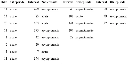
by the mother was considered indicative of diarrhea. An episode was defined as at least 1
day of diarrhea, preceded and followed by 2 or more days without diarrhea. The episode
was considered to have ended on the day bowel movements returned to normal (152).
Any child who had diarrhea was assessed clinically and treated appropriately. Fecal
samples were collected from the child and the child was followed up daily until cessation
of diarrhea. Laboratory testing was done on the diarrheal stool sample to identify
bacterial, parasitic or viral agents of gastroenteritis. Serum and fecal samples were
collected in the neonatal period. A serum sample was also collected from the mother at
the time of delivery. Serum samples were then collected every 3 months during the first 2
years of life and every six months during the third year. Diarrheal samples were screened
for Cryptosporidium spp. by microscopic examination of fecal smears stained by
modified acid fast stain. Children with no cryptosporidial diarrhea were defined as
children who did not have Cryptosporidiumspp. detected by microscopy or PCR in any
diarrheal stool sample up to 2 years of age. For this study, maternal sera collected at
delivery, sera from children with cryptosporidial diarrhea and without cryptosporidial
diarrhea at 3.5, 9 and 24 months of age as well as sera from cases collected 2 to 12 weeks
before (pre-infection) and after (post-infection) the first episode of cryptosporidial
diarrhea were tested. Informed consent was obtained from the parent prior to enrollment
Hospital based study on children with diarrhea- This study was carried out with stool samples originally collected for a multi-center rotavirus surveillance programme called
the Indian Rotavirus Strain Surveillance Network. Samples from 3 centers – Christian
Medical College, Vellore, St. Stephen's Hospital, Delhi and Child Jesus Hospital, Trichy
were available for this study representing both south and north India. Children aged less
than 5 years presenting to one of the three study hospitals with acute gastroenteritis and
requiring hospitalization for rehydration for at least 6 hours were enrolled in the study. A
detailed clinical evaluation of the episode of diarrhea including duration and frequency of
diarrhea and vomiting, fever and degree of dehydration was recorded and severity of the
diarrheal episode was assessed. Informed consent was obtained from the parent/guardian
before sample collection and the study was approved by the Institutional Review Board
of Christian Medical College, Vellore. All stool samples collected at CMC, Vellore were
transported within two hours to the laboratory and stored at 4ºC till testing. Samples from
St.Stephen's Hospital, Delhi and Child Jesus Hospital, Trichy were stored at 4ºC upon
collection and were transported in boxes with ice-packs at 15 day intervals to CMC. All
samples were processed on the same day of receipt and aliquots of samples were stored at
-70ºC for further characterization studies. Stool specimens collected from each child
enrolled in the study were tested for Cryptosporidium spp. by modified acid fast staining
and microscopy. In addition, a few children found to have cryptosporidial diarrhea at the
CMC, Vellore center were followed up and stool collected for up to 3 weeks to determine
HIV infected adults – HIV-infected adults who presented to the outpatient unit or who were admitted to the Department of Medicine Unit – I at Christian Medical Hospital,
Vellore were enrolled in this study after obtaining informed consent. A stool sample and
5 ml of blood was collected at enrollment. CD4 T cells were counted for most patients if
no CD4 count was available within 3 months prior to enrollment. CD4 counts were carried
out by flow cytometry using the Guava Technologies PCA instrument with Easy CD4
software in the Department of Clinical Virology. Details on the current diarrheal
episodes as well as any other co-infection, history of diarrhea and anti retroviral treatment
were collected. Data on concurrent opportunistic infections was also collected by
reviewing case records. For patients with diarrhea, detailed clinical information on
frequency, duration, and associated symptoms such as weight loss was collected. In this
study, all stool samples were screened for Cryptosporidium by PCR and serum samples
were tested for anti cryptosporidial antibodies. Diarrhea was defined as three or more
stools per day for at least 72 hours. Acute diarrhea was defined as diarrhea of less than
14 days duration. Persistent diarrhea was defined as diarrhea for 14 or more days. The
study protocol, questionnaires and consent forms were approved by the institutional
review board.
Laboratory Methods
Microscopy - Stool samples were concentrated by formalin-ether sedimentation method and smears prepared from the deposit. The dried, fixed smears were then stained by the
modified Ziehl-Neelsen staining technique which involves staining with strong
blue (72). Cryptosporidium oocysts appear acid-fast, round or slightly ovoid and 4.5-6.0
µm in diameter with variable degrees of staining.
DNA extraction– DNA was extracted from stool samples by the QIAamp DNA stool mini kit for DNA (Qiagen Inc, Valencia, CA). In this protocol, around 180-220 mg or, if
liquid, 200 µL stool was weighed in a 2 ml microcentrifuge tube and placed on ice.
Around 1.4 ml of buffer ASL was added and the suspension thoroughly vortexed and
heated for 5 min at 95°C and stool particles was pelleted by centrifugation. The
supernatant was pipetted into a new microcentrifuge tube and the pellet discarded. The
InhibitEX (Qiagen Inc, Valencia, CA) tablet was added to each sample, vortexed until the
tablet was completely suspended and then incubated for 1 minute at room temperature to
allow inhibitors in the sample to adsorb to the InhibitEX matrix. The samples were then
spun down twice to pellet the inhibitors. Around 200 µL of the supernatant was pipetted
into a new microcentrifuge tube containing 15 µL Proteinase K to which 200 µL of
Buffer AL was added and vortexed to form a homogeneous solution. This was incubated
at 70°C for 10 min following which 200 µL of ethanol (96-100%) was added to the lysate
and mixed. This was added to a QIAmp spin column which was then centrifuged at full
speed for 1 minute and placed in a new collection tube and the filtrate discarded. 500 µl
of Buffer AW1 and AW2 were added in succession to the spin column and centrifuged.
Finally, the spin column was placed into a new, labeled 1.5 mL microcentrifuge tube and
200 µL of Buffer AE pipetted directly onto the membrane. This was incubated for 1
PCR for screening and typing of cryptosporidial DNA - DNA from microscopy positive and negative samples were screened for Cryptosporidium spp. by a nested SSU
rRNA (Small subunit RNA) PCR (105) (Figure 5.4). Samples that were microscopy
positive but SSU rRNA PCR negative were also screened by COWP (Cryptosporidium
oocyst wall protein) and TRAP C1 (thrombospondin related adhesive protein) PCR (105).
The primers and cycling conditions used for each of these PCR reactions is summarized
in table 5.1. Samples that were PCR positive were then speciated by SSU rRNA PCR
RFLP and subgenotyped by Cpgp40/15 PCR RFLP.
RFLP – PCR amplicons from the SSU rRNA PCR (Figure 4.4) were subjected to a restriction digestion using the enzymes SspI (New England Biolabs) and VspI (Promega)
for species determination (Table 4.2) (Figure 5.5) (105). In this protocol, 20-30 µl of the
second round PCR product was incubated with 2 µl enzyme and 5 µl of the 1X RE
buffer. The volume was made up to 50 µl with sterile water and incubated in a 37°C
water bath for 1 hour. PCR amplicons from the Cpgp40/15 PCR (Figure 5.6) were
subjected to a restriction digestion using the enzymes AluI and RsaI (New England
Biolabs) for subgenotyping (Figure 5.7) (Table 5.2) (130). Ten µl of the second round
PCR product was incubated with 1µl enzyme and 2 µl of the 1X RE buffer and the
volume was made up to 20 µl with sterile water and incubated in a 37°C water bath for 1
hour.
Locus Primers and primer sequence Cycling conditions Amplicon size SSU rRNA
(nested)
SSU 3: 5’ TTC TAG AGC TAA TAC ATG CG 3’ SSU 4: 5’ CCC TAA TCC TTC GAA ACA GGA 3’
SSU 5: 5’ GGA AGG GTT GTA TTT ATT AGA TAA AG 3’ SSU 6: 5’ AAG GAG TAA GGA ACA ACC TCC A 3’
95°C 2 min, 72°C 1 min 94°C 45 sec
55°C 45 sec 35 cycles 72°C 1 min
72°C 10 min
820 bp
COWP C0WP 1: 5’- GTAGATAATGGAAGAGATTGTG -3’
COWP 2:5’- GGACTGAAATACAGGCATTATCTTG -3’
95°C 2 min, 72°C 1 min 94°C 60 sec
55°C 30 sec 34 cycles 72° C 50 sec
72°C 10 min
600 bp
TRAP TRAP1: 5– GGATGGGTATCAGGTAATAAGAA -3
TRAP2: 5- CCATTCTCTCCCTTTACTTC -3
Similar to COWP PCR 500 bp
Cpgp 40/15
(nested)
7 - 5’-ATGCAAAAATACGTGGACTGGG-3’ 8 - 5’-TCGCACGAAAGATTTCCATTG-3’ 9 - 5’-TTACTCTCCGTTATAGTCTCCGCTG-3’
95°C 15min
94°C 40 sec
55°C 50 sec 40 cycles
~ 1kbp