STUDY OF SERUM MAGNESIUM LEVEL
IN TYPE 2 DIABETES MELLITUS
Dissertation submitted in partial fulfilment of requirements for
M.D.DEGREE IN GENERAL MEDICINE BRANCH I
Of
THE TAMILNADU Dr. M.G.R. MEDICAL UNIVERSITY, CHENNAI, INDIA.
THANJAVUR MEDICAL COLLEGE , THANJAVUR - 613004.
THE TAMILNADU Dr. M.G.R. MEDICAL UNIVERSITY, CHENNAI-600032.
CERTIFICATE
This is to certify that the dissertation entitled “STUDY OF SERUM
MAGNESIUM LEVEL IN TYPE 2 DIABETES MELLITUS” is a bonafide
work done by Dr.SUBASH CHANDRABOSE G. at Thanjavur Medical
College,Thanjavur in partial fulfilment of the university rules and regulations
for award of M.D., Degree in General Medicine (Branch-I) under my guidance
and supervision during the academic year 2010-2013.
Prof.Dr.C.GUNASEKARAN M.D.D.C.H,
The Dean I/C,
Thanjavur Medical College, Thanjavur–613004.
Prof.Dr.S.MUTHUKUMARAN,M.D.,
Head of the Department,
Department of Internal Medicine, Thanjavur Medical College, Thanjavur–613004.
Prof.Dr.K.NAGARAJAN,M.D.,
Unit Chief M-III,
DECLARATION
I solemnly declare that the dissertation titled “STUDY OF SERUM
MAGNESIUM LEVEL IN TYPE 2 DIABETES MELLITUS” is done by
me at Thanjavur Medical College, Thanjavur during 2010-2013 under the
guidance and supervision of Prof. Dr.K.NAGARAJAN,M.D., The dissertation
is submitted to The Tamilnadu Dr.M.G.R. Medical University towards the
partial fulfilment of requirements for the award of M.D. degree in General
Medicine (Branch Ι).
Place: Thanjavur Signature of candidate
ACKNOWLEDGEMENT
At the outset, I thank Prof. Dr.C.GUNASEKARAN M.D.D.C.H,
The Dean I/C, Thanjavur Medical College and Hospital, Thanjavur for granting
me permission to do my dissertation and for allowing me to use institutional
facilities.
I am grateful to Prof. Dr.S.MUTHUKUMARAN M.D., Professor and
Head, Department of Internal Medicine, Thanjavur medical college and
Hospital for his constant support and guidance throughout my Post graduate
period and during the course of this study.
I am indebted to Prof. Dr.K.NAGARAJAN M.D., Professor of
Medicine and my unit chief, who constantly helped me and guided me in this
study and during my post-graduate period.
I profoundly express my heartiest thanks to my respected Professors
Prof. Dr.P.G.SANKARANARAYANAN M.D, Prof. Dr.S.MANOHARAN
M.D, Prof. Dr C.GANESAN M.D, and Prof. Dr.D.NEHRU M.D. D.M.R.D,
for their advice, guidance and valuable criticism which enabled me to do this
work effectively.
I would also like to thank Prof. Dr.M.THANGARAJ M.D.D.M, Head
of Department of Neurology, Department of Neurology, Thanjavur Medical
College and Hospital, Thanjavur who supported and helped me during this
I would also like to thank Assistant Professors Dr.S.ELANGOVAN
M.D.D.M, Dr. K.BALAMURALI M.D.D.M, Department of Neurology,
Thanjavur Medical College and Hospital, Thanjavur for their encouragement
during this study.
I would also like to thank Dr. C. PARANTHAGAN M.D. Registrar,
Department of Internal Medicine for his support and guidance.
I would also like to thank Dr.J.VIJAYBABU M.D.D.M.,.
Dr.S.VETRIVEL M.D., and Dr. THIRUMURUGAN M.D., Assistant
Professors of my unit, Department of Internal Medicine for their constant help,
encouragement and support throughout this study and during my post graduate
period.
I would also like to thank J.S.JESUS RAJA M.E, for his excellent
support in statistical analysis.
I express my sincere gratitude to all the patients who participated in the
study.
Lastly, I thank all my professional colleagues for their support and
CONTENTS
SL.NO TITLE PAGE NO
1 INTRODUCTION 1
2 AIMS AND OBJECTIVES 3
3 REVIEW OF LITERATURE 4
4 METHODOLOGY 53
5 RESULTS 58
6 DISCUSSION 80
7 CONCLUSION 86
8 ANNEXURES
BIBLIOGRAPHY
ANNEXURE I -ABBREVIATIONS
ANNEXURE II- PROFORMA
ANNEXURE III-MASTER CHART
ANNEXURE IV- KEYS TO MASTER CHART
STUDY OF SERUM MAGNESIUM LEVEL IN TYPE 2
DIABETES MELLITUS
ABSTRACT
BACKGROUND
A high prevalence of magnesium deficiency is reported in diabetics. Magnesium depletion has a negative impact on glucose homeostasis and insulin
sensitivity in type 2 diabetic patients as well as on the evolution of complications such as retinopathy, nephropathy, neuropathy and arterial atherosclerosis. The aim of this study is to estimate the prevalence of hypomagnesemia in patients with type 2 diabetes mellitus and its correlations
with microvascular complications of diabetes like retinopathy, nephropathy, neuropathy.
MATERIALS AND METHODS:
Patients with type 2 diabetes admitted in Thanjavur Medical College and
Hospital over a period of one year between October 2011 to October 2012 formed the study population. The sample size was 100 patients. Serum magnesium concentration was measured by calmagite dye method.
RESULTS:
serum magnesium. Significant association was found between hypomagnesemia and diabetic retinopathy, nephropathy, neuropathy. Significant correlations were not found with co morbidities such as ischemic heart disease and hypertension.
Low serum magnesium concentrations are common in type 2 diabetics. Magnesium deficiency is conclusively associated with diabetic retinopathy, nephropathy, neuropathy.
KEY WORDS:
1
INTRODUCTION
India is frequently referred to as the diabetic capital of the world as it has the highest number of cases in the world.
In worldwide the last 2 decades, incidence is suddenly increased from 30
million cases in 1985 to 171 million in 2000. Recent data suggests that prevalence of DM by the year 2030 could be 360 million. DM is worldwide in distribution and the incidence of both types is rising.1, 2
The distribution of both T1 DM and T2DM varies worldwide, due to
relative difference in genetic and environmental factors in different parts of the world. Recent data shows it is associated with 10-30% reduction of life expectancy, most common cause of blindness in the age group of 20 to 65 years,
25 fold increased risk of non traumatic lower limb amputations and increase incidence of end stage renal disease approximately 1000 patients per year2.
India had around 31.7 million cases in year 2000 which is expected to
rise alarmingly to around 79.4 million in 2030 by which time every fifth diabetic subject in the world would be an Indian3.
In Tamilnadu the prevalence in 2008 is 18.6% in urban areas and 9.1% in rural areas3.
2
that play important role in glucose metabolism. Animal studies found that low magnesium has a negative effect on post receptor signalling of insulin. Some studies have also found that magnesium supplementation improves insulin
action and glucose metabolismin diabetics4.
Magnesium is involved in multiple levels of insulin secretion. Magnesium deficiency can modify the Na+ K+ ATPase channel that maintains
sodium, potassium and glucose transport5.
In DM there is a direct correlation between serum magnesium concentration in blood and cellular glucose disposal that is independent of insulin secretion.6
Low serum magnesium level has direct correlation with microvascular
complications (retinopathy7, neuropathy. nephropathy) and macrovascular
complications (IHD and cerebrovascular disease)8.
In elderly type2 DM Paolisso hypothesized that oral magnesium supplementation for 4 weeks results in decreased fasting blood sugar level
increased plasma and RBC magnesium level.9
3
OBJECTIVES
The aims of this study are
1. Measurement of serum magnesium concentration in diabetes mellitus .
2. To estimate the prevalence of hypomagnesemia in patients with type 2 diabetes mellitus
3. Its correlation with microvascular complications like retinopathy,
4
REVIEW OF LITERATURE
DIABETES MELLITUS
Diabetes Mellitus is a clinical syndrome characterized by high blood
sugar level (hyperglycemia) and glycosuria due to relative or absolute deficiency of insulin secretion or its action, or insulin resistance that leads to disturbances in carbohydrate ,protein, fat metabolism , water and electrolyte
homeostasis.
THE HISTORICAL ASPECTS
Its history has been characterized by numerous cycles of discovery, neglect and rediscovery. Its history may be divided into four major periods. The „ANCIENT‟ period showed the first clinical features of DM and its
complications. The 16 th to 18th centuries have been termed the „DIAGNOSTIC‟
period, as DM was then identified as a separate disease entity. The mid to late
19th centuries may be consider as the first „EXPERIMENTAL‟ period. During
this period role of the pancreas became clear and the molecular level of diabetes
were initially identified10. Finally, in the 20th century knowledge about diabetes
5
The word Diabetes in Greek means – “I run through Siphon”. Indian
name for Diabetes is Madhumeha – Honey in rain. In 16th century, Susrutha in
the Sanskirit book of surgery, and Charaka in the Sanskirit book of medicine
have mentioned about Diabetes. The first person -Vaidys – tested the urine of diabetic patients.
SPECTRUM OF DIABETES MELLITUS & GLUCOSE HOMEOSTASIS
Types Normal glucose tolerance (mg/dl) HYPERGLYCEMIA PREDIABETES IFG & IGT (mg/dl) DIABETES MELLITUS NOT INSULIN REQUIRED INSULIN REQUIRED FOR CONTROL INSULIN REQUIRED FOR SURVIVAL T1DM T2DM
Other types GDM Time (years) FBG (mg/dl) 2-h PPBG (mg/dl)
< 110 < 140
110-125 140 – 199
≥ 126 ≥ 200
ETIOLOGICAL CLASSIFICATION OF DIABETES 1. Type 1 Diabetes
2. Type 2 Diabetes 3. Specific types
6
SPECIFIC TYPES OF DIABETES
A. Genetic mutation in the β-cell function: 1. MODY - maturity onset diabetes of the young Type1- Hepatocyte nuclear transcription factor 4α.
Type2 -Glucokinase. Type 3 -HNF 1α .
Type 4- Insulin promoter factor 1. Type5- HNF 1β.
Type 6- Neuro D1. 2. Mitochondrial DNA
3. Proinsulin or insulin conversion
B. Genetic mutations in insulin action: 1. Lipodystrophy syndromes
7
C. Diseases of the pancreas:
It includes pancreatitis, pancreatectomy, pancreatic malignancy, cystic fibrosis, etc.
D. Endocrine disorders:
Pheochromocytoma, somatostatinoma, aldosteronoma, etc. E. Drugs:
Glucocorticoids, diazoxide, beta agonists, thiazide diuretics. F. Infectious causes:
Rubella, cytomegalovirus, coxsackie virus. G. Immune-mediated Diabetes:
Stiff-man syndrome and anti-insulin receptor antibodies. H. Genetic syndromes
Myotonic dystrophy, porphyria, Prader-Willi syndrome. , Klinefelter‟s, Turner‟s
and Down‟s syndrome.
GESTATIONAL DIABETES MELLITUS (GDM)11
It is the occurrence of reduced glucose tolerance during gestation.
Incidence: 7% (range 2–10%) of pregnancies.
8
Effects of diabetes on pregnancy:
Hydraminos
Toxaemia of pregnancy
Maternal infections
Difficult labour
Recurrent abortions
Postpartum haemorrhage
Puerperal sepsis.
Effects of diabetes on the foetus:
Prematurity
Still birth
Macrosomia
Postpartum hypoglycaemia
Respiratory distress syndrome
Hyperbilirubinemia
Congenital heart disease
9
TYPE 1 DIABETES MELLITUS
Pathogenesis Of Type 1 Diabetes Mellitus:
Normal islet
?Viral infection in pancreatic beta cell
Secretion of IFN-α by pancreatic beta cell
Hyperexpression of MHC class1 antigen within beta cell
Insulitis
Insulin deficient islets
Immune Mediated Diabetes (Type 1A)
Its incidence is around 5-10% all diabetes. It occurs due to immune mediated destruction of pancreas.
Pathogenesis:
1. Genetic predisposition: Diabetogenic genes are located in short arm of chromosome 6
2. Environmental triggers
a) Viral infections like rubella virus, CMV, mumps virus, etc.
10
Viruses and above agents directly act on beta cells and initiate autoimmune processes against these cells.
3. Immune mechanism
DM can occur with other autoimmune disorders. It is due to presence of
diabetogenic peptide which triggers the immune system. .Antibodies against
β-cells include
1. Islet cell auto antibodies, 2. Autoantibodies to insulin,
3. Autoantibodies to glutamic acid decarboxylase (GAD65) and 4. Autoantibodies to tyrosine phosphatases IA-2 and IA-2B.
Anyone of above auto antibodies present in 85-90% of patients.
Age of onset is usually less than 30 years. Usually associated with prominent muscle wasting. It has an abrupt onset with rapid progression course.
Classical symptoms of diabetes like polyuria, polyphagia, polydipsia are present. Family history of diabetes mellitus is usually absent.
HLA DR3 or DR4 seen in>90% of patients. Pancreatic islet cells (of
11
Idiopathic Diabetes (Type 1B)
These persons have absolute deficiency of insulin and are at risk of
12
TYPE2 DIABETES MELLITUS
90-95% of diabetics are Type 2 DM. Patients have insulin resistance and have relative deficiency of insulin. Specific aetiologies are not known. It usually
starts after the age of 30 years. It has insidious onset, with gradual progressive course. Polyuria, polyphagia, polydipsia are not so classically seen as in type 1 DM. Family history of diabetes mellitus is usually present. No HLA links are
seen. 50% concordance is seen in identical twins. Pancreatic islet cells are not totally destroyed .Plasma insulin in serum normal to high. Complications like hyperosmolar hyperglycaemic non ketotic coma are very often seen.
Risk of developing T2DM:
Age of onset of T2DM in proband
(years)
Age corrected risk of T2DM for
Siblings(%)
25-44 53
45-54 37
55-64 38
65-80 31
Pathogenesis of Type 2 DM:
1) Insulin Resistance
13
Insulin resistance:
Exact cause of insulin resistance is unknown. Possible mechanism is increase release of free fatty acids from adipose tissue that induce insulin
resistance. In addition there is increased release of adipokines which act on receptors of insulin and dampen the action of insulin. There is a higher prevalence of insulin resistance is seen in metabolic syndrome.
Pancreatic Beta cell failure:
In the earlier stage of diabetes mellitus only moderate reduction of insulin secreting beta cells lost. At the time of diagnosis of type 2 DM more than 50% reduction of beta cell is seen. Most important pathological feature is the
14
Differences between Type 1 and Type 2 Diabetes Mellitus:
T1DM T2DM
Age of onset(years) Less than 40 More than 50
Duration of symptoms Usually weeks Months to years
weight Normal or decrease Obese
Ketone bodies in urine Present Absent
Insulin requirement for survival
Needed Not needed
Auto antibodies Present Not present
Diabetic complications at
the time of presentation
Not present Present
Family history Uncommon Seen in 25%
Other autoimmune
disorders
common Uncommon
Clinical features of DM
Polyuria
Increased thirst
Dry mouth
Nocturia
Tiredness, lethargy, fatigability
15
Excessive weight loss
Nausea, vomiting, headache
Polyphagia
Genital candidiasis
Mood changes, irritability
Diagnostic criteria for Diabetes mellitus
It includes
A. Symptoms of diabetes plus random blood sugar ≥ 200 mg/dl B. Fasting plasma sugar ≥ 126 mg/dl
C.2 hour postload plasma sugar ≥ 200 mg/dl during an oral GTT
D.HbA1C >6.5%
The new Diagnostic criteria for pre-diabetes and diabetes mellitus FBS > 100 mg/dl = normal fasting glucose
FBS ≥100 mg/dl and <126 mg/dl =impaired fasting glucose (IFG)
FBS ≥ 126 mg/dl = provisional diagnosis of diabetes (on more than one occasion)
The corresponding categories when the oral GTT is used ,are as follows
2 h BS < 140 mg/dl = normal glucose tolerance
2 h BS ≥ 140 mg/dl and < 200 mg/dl = impaired glucose tolerance (IGT)
2 h BS ≥ 200 mg/dl = provisional diagnosis of diabetes (must be confirmed on
16
FBS-fasting blood sugar, 2 h BS- 2 hour post load blood sugar.
The prognostic significance and outcome are same whether it is the FBS >126 mg/dl or 2 hour postprandial blood sugar >200 mg/dl(in diabetes).The
FBG test is now mostly performed because of ease of administration , convenience, acceptability to patients and its lower cost.
2-hour post load glucose is done by taking 75 g of glucose dissolved in
300 ml of water.
Fasting is defined as no food intake (i.e. overnight) for at least 8 hours. Random is defined as any time of day without regard to time since the last meal. Screening for diabetes:
Diabetes is one of the diseases diagnosed late when multiple
complications have appeared. Nearly 1/3rd of group remain undiagnosed. But no
studies have supported screening of asymptomatic persons. But use of fasting
blood sugar as a screening test for Type 2 DM is justified in individuals at high risk group.
Glycosylated haemoglobin C is also recommended for screening.
Adverse factors for Type 2 Diabetes Mellitus 1. Obesity
2. Family history of DM
3. History of diabetes mellitus during pregnancy (GDM) or Birth of baby
weight > 4kg
17
5. Race/ethnicity 6. Type A personality
7. Evidence of vascular disease features
Standards of medical care in DM
It is a chronic progressive disease that required frequent medical care for to decrease acute and chronic complications.
Initial Evaluation includes
Detailed Medical history
Previous HbA1C reports.
Detailed history of dietary pattern, and weight history and any
developmental delay in earlier age.
Detailed history of prior treatment and present treatment of DM.
Exercise history.
Previous or present infections, particularly skin, foot, oral GIT and
urinary tract infections. 2) Physical examination:
Height, weight and body mass index
Sexual maturation staging.
BP measurement, including orthostatic measurements
Examination of pulses
18
Thyroid gland examination.
Cardiac evaluation.
Eye-fundoscopic examination
Abdominal examination
Hand examination.
Foot examination.
Dermatological examination (for tinea, acanthosis nigricans and
insulin-injection sites).
CNS examination.
Examination for secondary causes of diabetes
(e.g.hemochromatosis,pancreatic disease).
3) Laboratory evaluation
HbA1C measurement
Fasting serum lipid profile levels, which includes total cholesterol, HDL,
Triglycerides, LDL, VLDL.
Examination of micro albumin in urine in T1DM who have had duration
of > 5 years and in all cases with T2 DM .
Serum creatinine.
Thyroid stimulating hormone (TSH) level in all T1DM, in T2DM with
clinical features.
19
Examination of urine for ketone bodies, protein and sediments.
4)Referrals:
• Ocular examination.
• Behavioural specialist. • Podiatrist.
Goals to be achieved in Diabetes mellitus: HbA1C < 7.0%
Fasting plasma sugar: 90 – 130 mg/dl
Post prandial plasma sugar: < 180 mg/dl Blood Pressure :< 130/80 mmHg
LDL < 100 mg/dl
Triglycerides < 150 mg/dl HDL> 40 mg/dl
Blood Sugar Control Goals- An Approach : •Goals vary from person to person.
•Following persons (Children, Women who have conceived and older age) need
special care.
•Less severe control is required in patients with recurrent episodes of
hypoglycaemia.
•More adequate control of blood glucose (i.e. a normal HbA1c < 6%) may
20
•Postprandial sugar may be targeted if glycosylated haemoglobin C levels are
not met despite reaching fasting sugar level. Complications of Diabetes Mellitus:
Diabetes has both acute and chronic complications. Acute complications:
It includes
1.Diabetic ketoacidosis (DKA):
It is an acute medical emergency, characterised by
Increase blood sugar
Ketone bodies in serum and urine
Metabolic acidosis
Complications:
Cerebral edema
Acute respiratory distress syndrome
Disseminated intra vascular coagulation
Thromboembolism
Circulatory failure
2. Hyperglycemic Hyperosmolar state (HHS): It is characterised by
hyperglycemia without metabolic acidosis or ketone bodies
21
3. Hypoglycemia:
It is more common in insulin treated patients.
4. Lactic acidosis
Chronic Complications:
1.Diabetic Retinopathy 2.Diabetic Neuropathy 3.Diabetic Nephropathy
4.IHD
5. Cerebrovascular accident
Transient ischemic attack, stroke
6.Peripheral vascular disease claudication, ischaemia 7. HT
8.Infection like Tuberculosis, Candidaisis, Mucormycosis, Necrotising fasciitis,
Periodontitis
9.Dupuytren's contracture
22
DIABETIC RETINOPATHY12
It is one of the commonest cause of blindness in adults in the age group30 to 65 years in developed countries. During the 1st two decades of disease, nearly
all patients with T1DM and >60% with T2DM have retinopathy. 21 percent of
T2DM patients have presented with retinopathy at first visit.
Classification:
Nonproliferative Diabetic Retinopathy (NPDR): 1. Mild type:
Presence of 1 micro aneurysm with one or more of the following :
Retinal haemorrhage,
Hard and soft exudates.
2. Moderate type:
Presence of Haemorrhage/ micro aneurysms or
Presence of both in at least one quadrant with one or more of the
following:
Soft exudates, venous beading and intra retinal microvascular
abnormalities. 3. Severe type:
Haemorrhage or micro aneurysms or
23
Venous beading in two or more quadrants , intra retinal microvascular
abnormalities in at least one quadrant. Proliferative Diabetic Retinopathy (PDR): 1. Early: One or more of the following: a) NVE
b) NVD
c) Vitreous or preretinal haemorrhage d) NVE< ½ disc area.
2. High risk: One or more of the following.
a) NVD > ¼- 1/
3 disc area
b) NVD with vitreous or preretinal haemorrhage
c) NVE > ½ disc area. Preretinal or vitreous haemorrhage. 3. Advanced PDR:
High risk PDR, traction retinal detachment involving macula or
Vitreous haemorrhage obscuring ability to grade NVD or NVE.
IRMA – Intraretinal micro vascular abnormalities. NVE – Neovascularisation elsewhere.
NVD – Neovascularisation over the disc
Clinical features of Retinopathy
Micro aneurysms
24
Exudates
Cotton wool spots
Neovascularisation of retina and iris
Subhyaloid haemorrhage
Vitreous haemorrhage and fibrosis
Macular edema can occur at any stage of diabetic retinopathy. Non
proliferative diabetic retinopathy usually appear at end of first decade or early second decade in cases of type2 diabetes mellitus. Proliferative diabetic retinopathy usually appears within 5 years of non proliferative diabetic retinopathy. Pregnancy, uncontrolled diabetes mellitus, uncontrolled HT can
accelerate these changes.
UKPDS study showed that strict sugar control(i.e. for every percentage of
reduction of HbA1C) associated with a 35% reduction in risk of retinopathy13,
25
Pathophysiology of microvascular complications: Increase glucose level
Activation of protein kinase c, Endothelial nitric oxide synthase uncoupling, Increase production of advanced glycation products, Activation of polyol
pathway
Activation of reactive oxygen species
Increase oxidative stress
1.Altered gene expression
2.Decrease nitric oxide synthesis
3.Activation of protein kinase C
4.Increased formation of advanced glycation products
5.Induction of DNA damage
26
Other ocular complications :
Cataract
Glaucoma
Retinal detachment
Macular edema
Investigations for retinopathy:
Visual acuity
Fundus examination
Fundus fluorescein angiography
Slit lamp examination
Treatment options for retinopathy:
Laser photocoagulation
Injection of steroids
27
DIABETIC NEPHROPATHY
This is most common cause of stage five chronic kidney disease in world wide. Compared to type2 diabetes progression to chronic kidney disease is
higher in type1 DM.
The diabetic nephropathy progresses from stage of microalbuminuria to stage of macroalbuminuria / clinical albuminuria to end stage renal disease.
Progression from micro to macroalbuminuria usually taken upto 10-15 years. ESRD develops in half of T1DM patients with clinical nephropathy within ten years and 3/4of patients by twenty years. But in type 2 DM, even after twenty years of overt nephropathy only 20% progress to ESRD.
Screening for Microalbuminuria:
A screening test should be done for urine microalbumin at the time of diagnosis in persons with T2 DM at the time of first visit , repeated after five
years of disease duration. For patients with T1DM, test should be repeated yearly.
Screening for microalbuminuria can be performed by 3 ways.
1. Calculation of the albumin to creatinine ratio in a spot urine sample. 2. 24 hour Urine sample and measurement of albumin excretion.
28
CATEGORY
Spot collection
(μg/mg creatinine)
24 Hr collection (mg/24 hrs)
Timed collection
(μg/min)
Normal <30 <30 <20
Microalbuminuria 30 – 299 30-299 20-199
Macroalbuminuria ≥300 ≥300 ≥200
29
DIABETIC NEUROPATHY
It occurs in almost half of cases with long duration of diabetes. The
development of neuropathy is directly in correlation with the duration and degree of glycemic control2,15.
Classification:
Somatic
Visceral or autonomic
Somatic:
1) Polyneuropathy
Symmetrical mainly sensory and motor
Asymmetrical mainly motor and proximal including amyotrophy
2) Mononeuropathy including mononeuritis multiflex
Visceral:
Cardiovascular
GIT
Pupillary
Genitourinary
Sudomotor
Vasomotor
Possible causes of neuropathy in DM includes
30
Formation of advanced glycation end products
Increased level of protein kinase c
Activation of polyol pathway leads to accumulation of sorbitol
Histopathology:
Axonal degeneration of both myelinated and unmyelinated fibres
Schwann cell basal lamina hypertrophy
Patchy, segmental demyelination
Basement membrane thickening and presence of microthrombi in neural
vessels
The most common type reported is distal symmetrical sensory poly neuropathy. Most cases frequently presented with distal sensory loss,
hyperesthesia, paresthesia and dysesthesia.
Painful neuropathies may also occur. It can be an acute (lasting < 12 months) and a chronic (lasting > 12 months).
Individuals with long standing T1 or T2 DM are prone to develop autonomic diabetic neuropathy.
Clinical features of Autonomic Neuropathy: Cardiovascular features
Orthostatic hypotension
31
Resting tachycardia
Gastrointestinal features
Dysphagia
Nausea, vomiting, abdominal fullness
Constipation
Nocturnal diarrhoea
Genitourinary features
Erectile dysfunction
Urinary incontinence
Recurrent urinary tract infections
Retrograde ejaculation
Pupillary features
Small pupil size
Delayed response to light reflex
Sudomotor features
Gustatory sweating
Increased sweating in the night
Anhidrosis and fissures in the feet
Vasomotor features
Dependent edema
Bullous formation
32
Tests for cardiovascular autonomic functions
1) Simple reflex test
A) Heart rate responses
to valsalva manoeuvre ( fifteen seconds): ratio of longest to shortest R- R
interval Normal ≥1.21 ,abnormal≤1.20
to deep breathing :6 breaths over 1 minute Normal ≥15 abnormal≤10
to standing after lying :Ratio of R- R interval of 30th to 15th beats Normal
≥1.04 ,abnormal≤1
B) Blood pressure response to standing -Systolic BP fall Normal ≤10
abnormal≥30
C) Special Tests
heart rate and BP in response to handgrip
heart rate and BP variability using domain analysis of ambulatory
monitoring
MIBG scan of heart
heart rate and BP variability using power spectral analysis of ECG
monitoring Diagnosis:
Examination of the feet
Look for ulceration,
33
Tuning fork using 128-Hz shows decreased vibration perception
Semmes-Weinstein monofilament for pressure sensation
Normal results on vibration testing (Likelihood ratio range, 0.33–0.51) or
monofilament (Likelihood ratio range, 0.09–0.54) make large fiber peripheral neuropathy from diabetes less likely.
Nerve conduction studies may show decreased conduction of the
34
MAGNESIUM
Magnesium is the 4th most abundant cation in the body. It is also the 2nd
largest amount cation intracellularly, next to potassium. A normal adult has 21-28gms (approximately 2000mEq) of magnesium. Nearly 60% of total body magnesium is present in bone, 38% is in soft tissues with slightly higher
concentrations in liver and skeletal muscle. (15-20 mEq/Kg). and less than 2% is present in extracellular compartment16.
Serum concentration of magnesium varies from 1.7 to 2.4 mg/dL. The
plasma concentration in healthy adults remain remarkably constant with very less fluctuations due to sensitive control mechanisms that are not fully understood
The mean daily oral intake of magnesium is roughly 25 mEq (140-360
mg/day). About 40% of the dietary magnesium is absorbed in the small intestine mainly the ileum. Elimination is mainly through kidneys and is about 100mg/day and it is well regulated. So when blood levels rise more than 2.4
mg/dl, magnesium excretion increases many folds. In event of magnesium loss absorption is increased dramatically, the main site being the thick ascending loop of Henle. Many factors inhibit reabsorption, like increased ECF volume,
35
Biochemical importance of Magnesium:
Magnesium is an activator of critical enzyme systems that maintain cellular metabolism. The most important being the activation of enzymes that
hydrolyze and transfer phosphate groups in the processes involving adenosine triphosphate (ATP). This ATP is essential for glucose catabolism as well as , lipid, amino acids, etc .
Magnesium is a cofactor for oxidative phosphorylation inside the mitochondria.
The macromolecular structure of DNA, RNA and ribosomes is
maintained by magnesium.18,19 It is also involved in protein synthesis by
regulating the attachment of mRNA to the 70s ribosome.20
Regulation of Serum Magnesium
A)Renal regulation:
Renal regulation of serum magnesium concentration is mainly by altering its reabsorption at the thick ascending loop of Henle. Magnesium reabsorption is increased by parathyroid hormone and is reduced by hypercalcemia and
36
B) Intestinal absorption :
Intestinal magnesium absorption mainly at jejunum and ileum is increased by 1,25 (OH)
2 Vitamin D
C) Hormonal factors23
Increasing serum magnesium,
Parathyroid hormone
Glucagon
1, 25(OH)
2 Calcitriol. Decreasing serum magnesium,
Aldosterone
Vasopressin (ADH)
Thyroxine
Calcitonin
Selected Food Sources of Magnesium22-Magnesium content in mg/100gm)
Nuts
Almonds – 315
Cashews – 260
37
Legumes
Split Beans – 50
Soyabean – 86
Fruits
Dates – 35
Banana – 30
Oranges – 10
Apple – 5
Dairy Products
Milk – 24
Butter – 20
Yoghurt – 12
Cereals
Shredded Wheat -110
Rice – 40
Meat and fish
Pork – 22
38
Beef – 18
Fish – 22
Hypermagnesemia :
Hypermagnesemia is very uncommon in the absence of renal insufficiency, kidneys can excrete huge quantities of magnesium when needed
(up to 250 mmol/d).24
Causes of hypermagnesemia A) Impaired magnesium excretion
Renal failure
Familial hypocalciuric hypercalcemia
B) Excessive magnesium intake
Cathartics
Antacid preparations
Parentral magnesium administration (eg.magnesium sulfate in PIH)
C) Rapid magnesium mobilisation from soft tissues
Trauma
Extensive burns
Shock, sepsis
39
D) Other disorders
Adrenal insufficiency
Hypothyroidism
Hypothermia
Clinical features:
The most important clinical presentation of hypermagnesemia are
vasodilation and neuromuscular blockade, occurring at concentrations > 4.8 mg/dL (>2mmol/L). Hypotension usually does not respond to intravenous fluids and vasopressors.
Lethargy and weakness may progress to respiratory failure, paralysis and coma with depressed deep tendon reflexes occurs at serum magnesium levels > 4 mmol/L). Paralytic ileus may occur.
Prolongation of PR, QRS intervals, heart blocks and asystole occurs at
serum magnesium levels around 10 mmol/L.
Treatment of Hypermagnesemia:
Generally involves recognising and removing the magnesium source
40
Hypomagnesemia :
Hypomagnesemia means a significant decrease in body magnesium stores (0.5 to 1 mmol/Kg). . Dietary magnesium deficiency is uncommon except in
persons consuming alcohol.
Causes of Hypomagensemia24
I.Impaired intestinal absorption
A.Primary infantile hypomagnesemia B.Malabsorption syndromes
C.Vitamin D deficiency. II.Increased intestinal losses
A.Protracted vomiting / diarrhoea
B.Intestinal drainage, fistulae III.Impaired renal tubular reabsorption
A.Genetic magnesium wasting syndromes. 1.Gitelman syndrome
2.Bartter syndrome
3.Na-K ATPase g-subunit mutations B.Acquired renal disease
1.Tubulointerstitial disease
41
C.Drugs 1.Ethanol
2.Diuretics (loop, thiazide and osmotic)
3.Cisplatin, cyclosporine
4.Aminoglycosides, Amphotericin B IV.Metabolic causes
1.Hyperaldosteronism 2.SIADH
3.Diabetes mellitus 4.Metabolic acidosis
5.Hypercalcemia 6.Hyperthyroidism V.Others
1.Pancreatitis
2.Excessive sweating 3.Osteoblastic metastases
Many genetic/hereditary magnesium losing conditions are described , but are exceedingly rare. Prolonged nasogastric aspiration, intravenous fluids, infectious diarrhoea, fat losing enteropathies and inflammatory bowel disease
may produce hypomagnesemia.23 Magnesium deficiency is quite frequent in
42
Incidence :
Hypomagnesemia is a relatively common electrolyte imbalance seen in
around 12% of in- patients.25 .In intensive care settings incidence is very high (60%).Parentral nutrition, diuretic usage, decreased serum Albumin levels , and
aminoglycosides may be responsible for this high frequency26
Risk of incidence:27
2% in common people.
10 - 20% in hospitalized patients.
60-70% in ICU settings.
30 - 80% in Alcoholics.
25% in diabetic patients.
Hypomagnsemia occurs equally in males and females.
Clinical features:28
The clinical features appear only when serum magnesium concentrations are <1.2 mg/dL (0.5 mmol/L). It is presented as irritability, Central nervous
43
History :
History concerned with hypomagensemia are usually nonspecific. They present with weakness, muscular cramps / palpitations. Sometimes abnormal
behaviour in the form of irritability, apathy, psychosis, may be seen in severe cases. In few vertigo, incoordination, depression, or fits may occur.
Physical signs :
Exaggerated deep tendon reflexes.
Trousseau and Chvostek sign
Difficulty in swallowing due to esophageal hypomotility
Altered mental status
Ataxia, nystagmus or seizures (at levels <0.8 mg/dl)
Pulses may be irregular due to VPCs
ECG :
Hpomagnesemia can produce non specific alterations in the electrocardiogram. Mild to moderate hypomagnesemia (1.2 to 1.7 mg/dl)
producing widened QRS complexes with tall T-waves. Severe
Hypomagnesemia (<1.2 mg/dl) produces increased PR interval, broad QRS
44
Cardiac arrhythmias including sinus tachycardia, other supraventricular tachycardia and ventricular tachyarrhythmia also can occur.
Investigations:
Serum magnesium levels are estimated by several methods.
Neutron activation analysis
Atomic absorption spectrometry
Ion selective electrodes (ISE)
Equilibrium dialysis
Calmagite dye method.
Calcium, potassium and phosphorous levels must be assessed. BUN and creatinine levels And Blood glucose levels should also be measured.
Treatment of Hypomagnesemia:
The route of magnesium replacement is according to severity of the
clinical manifestations. Moderate to severe manifestations like patients with tetany or ventricular arrhythmias require 50 mEq of intravenous route slowly over 8 to 24 hours and repeated as and when needed to maintain concentration
45
Oral replacement ( magnesium chloride & Magensium lactate) should be given in patients with milder presentations. The background cause should be corrected.
Hypomagnesemia and other diseases:
A) Magnesium and cardiovascular diseases32
Arrhythmias can be precipitated in hypomagnesemia especially in acute
coronary syndrome, cardiac failure and associated hypokalemia.
Torsade De pointes is a fatal arrhythmia precipitated by drugs that increase QT interval, electrolyte imbalance (low potassium and magnesium), or
a decrease heart rate. Intravenous magnesium is recommended for this fatal condition.
Trials show significant correlation with ischemic heart disease. decreased level of serum magnesium is associated with higher incidence of ischemic heart
disease.39 The underlying mechanism for the increased risk is not fully
understood.
Magnesium supplementation may improve the exercise tolerance, antithrombotic effect with aspirin and thus improves outcome in individuals
46
Acute MI(myocardial infarction) is often associated with significant
reduction in serum magnesium levels.41Hypomagnesemia associated with acute
MI is probably responsible for increase in the frequency of fatal arrhythmias in
the acute period. 33
A positive correlation is seen in patients with hypomagnesaemia and CCF .It occurs due to use of diuretics. Whether this is associated with increased
mortality is not known.41
B) Hypertension
Magnesium has significant role in controlling BP.35 Hypomagnesemia leads to increased intracellular potassium and calcium levels which leads to vasoconstriction and increased peripheral vascular resistance. Magnesium may
also have some direct effect on vascular smooth muscle.36
Studies have also found that requirement of anti hypertensive dosage is more in patients with low serum magnesium levels.
As per JNC recommendation high magnesium rich diet is necessary for
47
C) Stroke
Studies have also found that increased magnesium intake decreases
incidence of stroke.42
D) Osteoporosis
There is a significant correlation between serum magnesium levels and
bone mineral density.43
E) Asthma:
Normal functioning of lung requires magnesium rich diet. There is a significant correlation between hypomagnesemia and incidence of bronchial asthma.
F) Dyslipidemia
Hypomagnesemia is associated with increased plasma LDL,
48
MAGNESIUM AND DIABETES MELLITUS
Magnesium is a essential element for metabolism of carbohydrate . 25 to 39% diabetes mellitus patients have hypomagnesemia
The clinical implications of magnesium deficiency in diabetes are many. Hypomagnesemia can be a result of hyperglycemia and can produce or increase insulin resistance. Hypomagnemia is implicated in the development of diabetic
retinopathy.most often higher incidence occur if associated with hypertension,38
thrombotic tendency,45 insulin resistance46 and the Reaven – Modan syndrome, a
unique clinical entity that connects diabetes mellitus, hyperinsulinemia, hypertension and increased thrombotic tendency – all producing adverse
cardiovascular outcomes.47
Causes of Hypomagnesemia in Diabetes Mellitus :
Initially, the cause of hypomagnesemia in diabetes was supposed to be due to
(1) Osmotic renal loss from glycosuria.
(2) Impaired absorption from GIT .
(3) Insulin mediated transfer of magnesium to RBC.
49
treatment has been shown to reduce magnesium losses through kidneys.
Garland48 suggested that delay in insulin treatment is less effective in correcting
the renal magnesium losses, probably due to certain irreversible changes.
The role of magnesium in Insulin Action :
Magnesium is a essential cofactor for insulin secretion, and activity.
Hypomagnesemia decreases insulin secretion by the pancreas.49
Multiple studies have shown hypomagnesemia to increase insulin resistance. This insulin resistance is a post receptor defect and may be
associated with calcium mediating the signal for insulin action. 46
Number of studies have shown that tissue response to insulin is more in the presence of magnesium.
In a recent study, the cellular uptake of magnesium, was shown to be
reduced in diabetics.50 there is also some proof that low magnesium levels
directly produces insulin resistance. Nadler et al.56 analysed 16 non diabetic
individuals and found decreased insulin sensitivity after production of magnesium deficiency.
Similarly elderly non diabetic individuals had improved glucose tolerance,
50
obese subjects, insulin resistance was found to be associated with low
magnesium levels, compared to non obese individuals.52
Tonyai, et al.53 suggested that a low erythrocyte magnesium content can
change membrane viscosity, and impair the binding of insulin with its
membrane. Paolisso, et al.51 were able to rectify the altered erythrocyte
microviscosity with long-term magnesium supplementation.
Role of magnesium deficiency in diabetic end organ damage:
Magnesium deficiency is associated with diabetic microvascular disease.
Hypomagnesemia has been shown in patients with diabetic retinopathy, with
still lower magnesium levels in severe diabetic retinopathy. Magnesium
depletion is also related to development of diabetic polyneuropathy. Corsonello, et al have suggested a relationship between diabetic microalbuminuria and
serum ionized magnesium levels. Magnesium depletion also has been associated
with multiple macrovascular complications. 43,50
Grafton, et al57 have suggested on the inositol transport theory for the
development and progression of diabetic complications. According to this theory hyperglycemia leads to excessive stimulation of aldose reductase causing high cellular level of sorbitol. This sorbitol inhibits transport of inositol, thus
51
hypomagnesaemia causes a decrease in the affinity of the inositol transport protein for inositol, leading to a two fold reduction in rate of inositol transport and accelerated development of diabetic complications.
Resnick proposed that the „primary‟ defect is the abnormal cellular ion
handling leading to complications47
There is a significant correlation between magnesium and antioxidant
levels. Weglicki, et al have hypothesized that a fall in magnesium level leads to loss of cellular activity against oxidative substances. Magnesium deficiency has also been shown to lower the efficiency of substances acting againt oxidants
such as glutathione, vitamin c and Tocopherol.
Evidence for efficacy of Magnesium Supplementation in Diabetes Mellitus:
There is strong evidence that repletion of magnesium can reduce the
insulin resistance, platelet reactivity and other cardiovascular risk factors .54
In a study involving 16 diabetics and 30 healthy controls, oral supplementation with magnesium hydroxide (250mg twice daily) resulted in
reduced insulin requirements in the diabetic patients. Paolisso,51 et al
52
In type 2 diabetics, oral supplementation of magnesium decreases platelet reactivity, and decreases the incidence of hypertension and improves lipid
profile.44
Clinical approach to Magensium Supplementation in Diabetes Mellitus
Diabetic patients at risk for hypomagnesemia such as ACS, DKA, chronic
alcohol intake, prolonged duration of parentral therapy, chronic diarrhoea , diuretic therapy should have their magnesium levels measured.
Overt serum hypomagnesaemia should always be corrected. If
hypomagnesemia is clinically suspected, but not supported by serum levels, a further test with erythrocyte or platelet magnesium concentration is mandatory
Magnesium chloride is the preparation of choice. Doses vary from 100 to
600mg/day .54 Diarrhoea is the most important and the dose limiting side effect.
Renal insufficiency with Glomerular filtration rate of less than 30 ml/min is the
53
METHODOLOGY
SOURCE OF DATA
Patients with type 2 diabetes admitted in THANJAVUR MEDICAL COLLEGE & HOSPITAL, THANJAVUR, who satisfied the inclusion criteria
and consented to participate in the study were included.
PERIOD OF STUDY
October 2011-October 2012
TYPE OF STUDY
Cross sectional study
100 patients were randomly selected of which 50 were males and 50 were
females. Cases with renal failure, Acute Coronary Syndromes, patients on diuretics, alcoholics or with malabsorption were excluded. None were taking magnesium supplements or magnesium containing antacids. Informed consent
was obtained.
INCLUSION CRITERIA
All patients both males and females above 13 years of age group with type 2 diabetes mellitus admitted in Internal medicine units of Thanjavur
54
EXCLUSION CRITERIA
1. Patients with chronic renal failure.
2. Acute myocardial infarction in last 6 months.
3. Patients on diuretics.
4. Patients with history of alcohol abuse.
5. Patients receiving magnesium supplements or magnesium containing
antacids.
6. Malabsorption or chronic diarrhea. 7.Age<13years.
DATA COLLECTION
The 100 diabetics (50 Men & 50 Women) were included in the study. Detailed history – including duration of diabetes, treatment mode, symptoms suggestive of diabetic neuropathy, associated diseases such as hypertension and ischemic heart disease was obtained as per the proforma.
Detailed physical and neurological examination was done. Retinopathy was assessed by direct opthalmoscopy.
Samples were collected for estimation of fasting blood glucose and
55
experience neurologist with symptoms & signs suggestive of neuropathy. Blood urea, serum creatinine and 24 hour urinary albumin were estimated. Serum magnesium was estimated by Calmagite dye method. HbA1C measurement
done by a modified calorimetric method.
CALMAGITE DYE METHOD – TEST PRINCIPLE
Under alkaline conditions, magnesium combines with calmagite dye to
form a red colour which is read spectrophotometrically at 530 nm. Formation of colour is depends on magnesium levels. To eliminate the interference of calcium during estimation, EDTA is included in the reagent. Cyanide reduces
heavy metal interference. Surfactant reduces protein interference.
TEST PROCEDURE
Three test tubes labeled Blank, Standard and Test are prepared as in table.
In test tubes Blank Standard Test
Calmagite 1 ml 1 ml 1ml
Standard sample
- 10 ml -
Patient‟s sample
- - 10 ml
Distilled water
56
This test tubes are incubated at room temperature (22-28ºC). The absorbance of Test (A
T), Standard (AS) and Blank (AB) are read at 530nm in spectrophotometer. Magnesium concentration is calculated by the following formula.
Magnesium concentration (mEq/L) = (A
T-AB / AS-AB) x 2
Serum magnesium concentration is expressed in mg/dl by linearity of 1
mEq/L = 1.2 mg/dl.
According to magnesium levels patients were classified into:
1) Normal, 1.7 to 2.4 mg/dl,
2) Low <1.7mg/dl,
3) High >2.4 mg/dl.
Patients were also categorized on the basis of duration of diabetes, presence of ischemic heart disease or hypertension, mode of treatment,
presence/absence of retinopathy, neuropathy and nephropathy, and glycemic control (FBS and HbA1C).
Cases with diabetic retinopathy were further divided into
a) Nonproliferative diabetic retinopathy.
57
Diabetic nephropathy was graded depending on 24 hour urinary excretion of albumin as follows:
No nephropathy, < 30mg/24hour.
Microalbuminuria 30 – 299mg/24hour
Macroalbuminuria (clinical proteinuria) >= 300 mg/24hour.
STATISTICAL ANALYSIS
The statistical analysis was done by SPSS 15 software.MS Word and
Excel were used to generate tables and charts. Following tests were used: 1. Chi square test
2. Student T test
3. Oneway ANOVA test
58
RESULTS
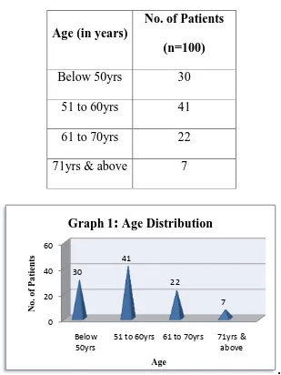
[image:67.595.142.451.206.618.2]100 cases of type 2 DM (50 males, 50 females, mean age 56.87 years) comprised the study group.
Table 1: Age Distribution
Age (in years)
No. of Patients
(n=100)
Below 50yrs 30
51 to 60yrs 41
61 to 70yrs 22
71yrs & above 7
.
Patients were distributed across the age spectrum of 42 to 78 years. Mean age 56.87 years. Most patients (n=41) were present in 51-60 group. Youngest patient was 42 years old.
0 20 40 60 Below 50yrs
51 to 60yrs 61 to 70yrs 71yrs & above 30 41 22 7 No . o f P a tient s Age
59
Table 2: Characteristics of study population
Characteristics
No. of subjects 100
Age (Years) 56.87 (42 – 78)
Men 50
Women 50
Duration of diabetes (years)
8.89(2-23)
Medication
67 Oral hypoglycemics
Insulin and oral
hypoglycemics
33
Diet only 0
Comorbidities
30 Hypertension
Ischemic heart disease 15
Diabetic retinopathy
NPDR
33
PDR 2
Diabetic neuropathy 15 Diabetic nephropathy
Microalbumineria
29
Macroalbumineria 4
60
.
The average duration of diabetes in study population was 8.89 years and range was 2 year to 23 years. 67 patients received only oral hypoglycemic
agents and 33 patients received both. 30 patients had hypertension and 15 patients had ischemic heart disease and 55 patients had no comorbidities. Total 35 patients had diabetic retinopathy. Total of 15 patients had diabetic
neuropathy. 33 patients had Nephropathy.
0 5 10 15 20 25 30 35 40
No
.o
f
P
a
tient
s
Characteristics
61
Table 3: Prevalence of Hypomagnesemia
Sex
Magnesium
Statistical inference Normomagnesemia
(n=65)
Hypomagnesemia
(n=35)
Male 34 (52.3%) 16 (45.7%) X2=.396
Df=1 .529>0.05
Not Significant
Female 31 (47.7%) 19 (54.3%)
Sl.no MG Mean S.D Statistical inference
1 Male (n=50) .4920 .72530 T=-.622 df=98
.536>0.05 Not Significant
62
Hypomagnesemia was found in 35 patients. 65 patients had normomagnesemia No patient had hypermagnesemia. No correlation was found between hypomagnesemia in men and women (45.7% and 54.3%%
respectively).
Graph 3
:
Prevalence of Hypomagnesemia63
Table 4: Prevalence of Hypomagnesemia and duration of diabetes
Oneway ANOVA
Sl.no MG Mean S.D SS Df MS
Statistical
inference
1
Between Groups
32.917 4 8.229
F=13.265 .060<0.05 Not Significant 2 Below 5yrs (n=45)
.0000 .00000
3
6 to 10yrs
(n=25)
.8040 .78977
4
11 to 15yrs (n=15)
1.4333 .40119
5
16 to 20yrs (n=10)
1.2200 .64601
6
21yrs & above (n=5)
.0000 .00000
64 Sl. no Duration(years) Magnesium Statistical inference Normomagnesemia (n=65) Hypomagnesemia (n=35)
1 Below 5yrs 46 (70.8%) 2 (5.7%) X2=4.568
Df=4 .067>0.05
Not
Significant
2 6 to 10yrs 12 (18.5%) 13 (37.1%)
3 11 to 15yrs 1 (1.5%) 11 (31.4%)
4 16 to 20yrs 2 (3.1%) 8 (22.9%)
5 21yrs & above 4 (6.2%) 1 (2.9%)
chi-square (χ2) value is 4.568.So the correlation is insignificant p value. So, the
duration of diabetes not significantly predict serum magnesium concentration. Oneway ANOVA F=13.265 in between groups also shows no significant correlation. The mean duration was 8.89 years (2-23).
0 10 20 30 40 50 Below 5yrs 6 to 10yrs 11 to 15yrs 16 to 20yrs 21yrs & above No .o f P a tient s
Age in Years
Graph 4
:
Prevalance of Hypomagnesemia and Duration of Diabetes65
Table 5: Prevalence of Hypomagnesemia and Mode of Diabetic treatment
Treatment mode Magnesium Statistical inference Normomagnesemia (n=65) Hypomagnesemia (n=35)
OHA 45 (69.2%) 22 (62.9%) X2=.418
Df=1 .518>0.05
Not Significant
OHA+insulin 20 (30.8%) 13 (37.1%)
20 45 13 22 0 5 10 15 20 25 30 35 40 45 50
Insulin treated Non insulin treated
Graph 5
:
Prevalence of Hypomagnesemia66
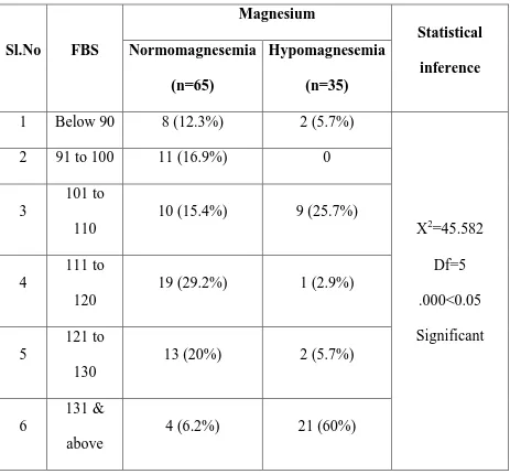
Table 6: Prevalence of Hypomagnesemia and Fasting blood sugar
Sl.No FBS
Magnesium
Statistical
inference Normomagnesemia
(n=65)
Hypomagnesemia
(n=35)
1 Below 90 8 (12.3%) 2 (5.7%)
X2=45.582 Df=5 .000<0.05 Significant
2 91 to 100 11 (16.9%) 0
3
101 to 110
10 (15.4%) 9 (25.7%)
4
111 to
120
19 (29.2%) 1 (2.9%)
5
121 to 130
13 (20%) 2 (5.7%)
6
131 & above
67
.
Prevalence of hypomagnesemia is high when fasting blood sugar >131(60%). The chi-square (χ2) value is 45.582. So the correlation is significant.
So, fasting blood sugar can significantly predict serum magnesium concentration.
0 5 10 15 20 25
Below 90 91 to 100 101 to 110 111 to 120 121 to 130 131 & above
No
.
o
f
pa
tient
s
Treatment
Graph 6
:
Prevalence of Hypomagnesemia and Fasting Blood Sugar68
Table 7: Prevalence of Hypomagnesemia and HbA1c
Sl.no HbA1C
Magnesium
Statistical
inference Normomagnesemia
(n=65)
Hypomagnesemia
(n=35)
1 Below 6 8 (12.3%) 2 (5.7%)
X2=10.408 Df=5 .064>0.05 Not Significant
2 6 to 7 18 (27.7%) 5 (14.3%)
3 7 to 8 15 (23.1%) 5 (14.3%)
4 8 to 9 5 (7.7%) 9 (25.7%)
5 9 to 10 8 (12.3%) 4 (11.4%)
6
10 &
above
11 (16.9%) 10 (28.6%)
Serum magnesium concentration showed no significant association with
HbA1C. Chi square X2=10.408,Df=5 P value .064 . Higher prevalance of
69
.
0 2 4 6 8 10 12 14 16 18
Below 6 6 to 7 7 to 8 8 to 9 9 to 10 10 & above
No
o
f
pa
tient
s
HbA1C
Graph 7
:
Prevalence of Hypomagnesemia and HbA1C70
Table 8: Prevalence of Hypomagnesemia and Diabetic Retinopathy
Particulars
No.of Patients
(n=100)
Percentage
(100%)
No Retinopathy 65 65.0
NPDR 33 33.0
PDR 2 2.0
Sl.no Retinopathy
Magnesium
Statistical
inference Normomagnesemia
(n=18)
hypomagnesemia
(n=17)
1 NPDR 18 (100%) 15 (88.2%) X2=4.746
Df=1 .034<0.05
Significant
71
Observations revealed a definite correlation between hypomagnesemia and diabetic retinopathy. The chi-square (χ2) value is 4.746. Df=1 .p value is
0.034.
0 10 20 30 40 50 60 70
Retinopathy No Retinopathy
No
O
f
P
a
tient
s
Graph 8
:
Prevalence of Hypomagnesemia and Diabetic Retinopathy72
TABLE.9: Prevalence of Hypomagnesemia and Diabetic Neuropathy
Particulars
No. of patients
(n=100)
Percentage
(100%)
No neuropathy 85 85.0
neuropathy 15 15.0
Sl.no Neuropathy
Magnesium
Statistical
inference Normomagnesemia
(n=65)
Hypomagnesemia
(n=35)
1 Negative 60 (92.3%) 25 (71.4%) X2=7.778
Df=1 .005<0.05 Significant
2 Positive 5 (7.7%) 10 (28.6%)
73
0 10 20 30 40 50 60 70 80 90
Neuropathy No Neuropathy
No
o
f
pa
tient
s
Graph 9
:
Prevalence of Hypomagnesemia and Diabetic Neuropathy74
Table 10: Prevalence of Hypomagnesemia and Diabetic Nephropathy
Particulars
No.of persons
(n=100)
Percentage
(100%)
No albumminuria 67 67.0
Macroalbuminuria 4 4.0
Microalbuminuria 29 29.0
Sl.
no
Nephropathy
Magnesium
Statistical
inference Normomagnesemia
(n=65)
Hypomagnesemia
(n=35)
1 No albuminuria 65 (100%) 2 (5.7%) X2=91.471
Df=2 .000<0.05
Significant
2 Macroalbuminuria 0 4 (11.4%)
75
.
Observations revealed a definite correlation between hypomagnesemia
and diabetic nephropathy. The chi-square (χ2) value is 91.471. Df=2 .
0 10 20 30 40 50 60 70
No
.
o
f
pa
tient
s
Graph 10
:
Prevalence of Hypomagnesemia and Diabetic Nephropathy76
Table 11: Prevalence of Hypomagnesemia and Ischemic Heart Disease
Sl.no IHD
Magnesium Statistical inference Normomagnesemia (n=65) Hypomagnesemia (n=35)
1 Absent 54 (83.1%) 31 (88.6%) X2=.539
Df=1
.463>0.05 Not Significant
2 Present 11 (16.9%) 4 (11.4%)
Serum magnesium concentration showed no significant relation with ischemic heart disease, X2=.539,Df=1.P value is insignificant.
. . 0 10 20 30 40 50 60 70 80 90
IHD No IHD
No o f pa tient s
Graph 11
:
Prevalence of Hypomagnesemia and Ischemic Heart Disease77
Table 12: Prevalence of Hypomagnesemia and Hypertension
Sl.no SHT
Magnesium Statistical inference Normomagnesemia (n=65) Hypomagnesemia (n=35)
1 Absent 43 (66.2%) 27 (77.1%) X2=1.308
Df=1 .253>0.05 Not Significant
2 Present 22 (33.8%) 8 (22.9%)
Serum magnesium concentration showed no significant with systemic
hypertension, X2=1.308,Df=1.P value is insignificant
. 0 10 20 30 40 50 60 70
HTN No HTN
No o f pa tient s
Graph 12