Copyright © 2000, American Society for Microbiology. All Rights Reserved.
Liver-Specific Alpha 2 Interferon Gene Expression Results in
Protection from Induced Hepatitis
LUIGI AURISICCHIO, PAOLA DELMASTRO, VALENTINA SALUCCI, ODALYS GONZALEZ PAZ, PATRIZIA ROVERE, GENNARO CILIBERTO, NICOLA LA MONICA,ANDFABIO PALOMBO*
IRBM P. Angeletti, Rome, Italy
Received 22 September 1999/Accepted 4 February 2000
The current therapy for hepatitis B and C is based on systemic administration of recombinant human alpha
interferon (r-hIFN-␣). However, systemic delivery of r-hIFN-␣is associated with severe side effects, but more
importantly, it is effective in only a small percentage of patients. In an effort to maximize IFN-␣ antiviral
efficacy, we have explored the therapeutic potential of murine IFN-␣2 (mIFN␣2) selectively expressed in the
liver. To this end, we have developed a helper-dependent adenovirus vector (HD) containing the mIFN-␣2 gene
under the control of the liver-specific transthyretin promoter (HD-IFN). Comparison with a first-generation
adenovirus carrying the same mIFN-␣2 expression cassette indicates that at certain HD-IFN doses, induction
of antiviral genes can be achieved in the absence of detectable circulating mIFN-␣2. Challenge of injected mice
with mouse hepatitis virus type 3 showed that HD-IFN provides high liver protection. Moreover, liver protec-tion was also observed in acute nonviral liver inflammaprotec-tion hepatitis induced by concanavalin A at 1 month postinfection. These results hold promise for the development of a gene therapy treatment for chronic viral
hepatitis based on liver-restricted expression of IFN-␣2.
Interferon (IFN) was discovered by Isaac and Lindenmann in 1957 (18), and recently the U.S. Food and Drug Adminis-tration has approved recombinant human IFN-␣ (r-hIFN-␣) for the treatment of hepatitis B virus (HBV) and hepatitis C virus (HCV) infections. IFN-␣acts on target cells to confer a state of resistance to viral infectivity at one or more stages of virus entry or replication. These biological effects require bind-ing to the type I IFN receptor complex, which is composed of two subunits, ␣ and  (12). Both subunits undergo rapid li-gand-dependent tyrosine phosphorylation, and the ␣ subunit itself acts as a species-specific transducer for type I IFN action (7). At least 30 genes are known to be transcriptionally induced by type I IFNs, including 2⬘,5⬘-oligoadenylate synthetase (2⬘5⬘OAS), the double-stranded RNA-activated protein ki-nase, and the IFN-I response factor I (8, 10). 2⬘5⬘OAS is important for antiviral response, and its activity is required by cells to activate the endonuclease RNase L, which degrades RNA (31).
The currently available treatment for HCV with r-hIFN-␣
results in clearance of the virus in only 20% of patients. How-ever, recent clinical trials have shown that a combination of r-hIFN-␣and the antiviral drug ribavirin can increase the per-centage of recovery up to 40% (9, 30). Although these results appear to be very promising, systemic injection of r-hIFN-␣is associated with severe side effects, which worsen in combination with ribavirin, causing the withdrawal of 20% of patients from therapy.
It is not clear why r-hIFN-␣treatment is effective in only a minority of patients. One possible explanation has been pos-tulated on the basis of association of specific HCV genotype and lack of sustained response. HCV proteins may block
IFN-␣-induced antiviral polypeptides, thus allowing virus replica-tion to take place (26, 35, 36). However, the inducreplica-tion of a
stronger antiviral response may overcome this blockage, sug-gesting that treatment of these species may require a higher amount of IFN-␣.
These clinical limitations in addition to the high cost of the treatment have prompted research for new delivery systems, either by modifying the recombinant protein (13) or through a gene therapy approach. To this end, the antiviral properties of different IFN-␣types have been explored in mice using naked DNA injection into muscle. A prophylactic injection of plasmid DNA expressing different murine IFN-␣(mIFN-␣) types suc-ceeded in reducing cytomegalovirus replication in the injected mice to a limited extent (42). In the case of HBV, transgenic mice have been developed as a model for viral replication and gene expression. Injection of a first-generation adenovirus (Ad) vector expressing the-galactosidase gene (Ad-gal) led to a transient inhibition of HBV virus replication in association with the advent of inflammatory cytokines induced by adeno-virus injection (5).
IFN-␣exhibits a short half-life in the blood after parenteral protein administration (15, 40), suggesting that the limited performance of IFNs in hepatitis treatment may be caused, at least in part, by insufficient or lack of sustained delivery of the protein to the liver. Because liver-specific gene delivery could overcome these limitations, we have explored the effects of mIFN-␣2 gene delivery mediated by Ad vectors that have been shown to transduce mainly in the liver when injected intrave-nously (17). To further limit mIFN-␣2 expression to the target tissue, the mIFN-␣2 gene was cloned under the control of the liver-specific transthyretin promoter (TTR) (27). The mIFN-␣2 expression cassette was rescued in a first-generation Ad vector, Ad-IFN, and in a helper-dependent (HD) vector, HD-IFN. The HD vectors do not express any viral proteins and result in prolonged transgene expression in immunocompetent mice upon systemic delivery (4, 20, 24, 33). In this study, we show that the combination of an HD vector and liver-specific pro-moter resulted in intrahepatic IFN-␣ expression, which pro-tected the liver in acute hepatitis models.
* Corresponding author. Mailing address: IRBM P. Angeletti, Via Pontina Km 30,600, 00040 Pomezia (Rome), Italy. Phone: 39-06-91093-234. Fax: 39-06-91093-225. E-mail: palombo@irbm.it.
4816
on November 9, 2019 by guest
http://jvi.asm.org/
MATERIALS AND METHODS
Cell lines.293 and 293Cre4 cells (23) were grown in minimal essential medium (MEM) supplemented with 10% heat-inactivated fetal calf serum (FCS). 911 (human embryonic retinoblasts), L-929 (mouse fibroblasts), and HuH-7 (human hepatoma) cells were grown in Dulbecco’s modified Eagle’s medium (DMEM) supplemented with 10% FCS. DBT cells (mouse fibroblasts) were grown in DMEM supplemented with 10% tryptose phosphate broth and 5% fetal calf serum.
Mouse strains.The mice used in this study were immunocompetent, 6- to 8-week-old (at the time of injection), C57/B6 females purchased from Charles River. Groups of four to five mice received injections in the tail vein of Ad vectors diluted in physiologic solution in volumes of 200l. Blood was obtained by retroorbital bleeding, and serum was stored at⫺80°C. At the indicated time, mice were sacrificed, and organs were rapidly frozen in liquid nitrogen and stored at⫺80°C.
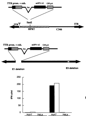
Construction of mIFN-␣2 expression cassette.A synthetic mIFN-␣2 gene was assembled by PCR with 28 oligonucleotides, 40 nucleotides in length, which collectively encoded both strands of the gene, as previously described (34). The PCR product was then cloned in pBluescript II (pBS-mIFN) and sequenced. The bovine growth hormone poly(A) site was amplified by PCR from plasmid pcDNA (Invitrogen), adding anXbaI site at the 5⬘end and aNotI site at the 3⬘end. This fragment was cloned downstream of the mIFN-␣2 cDNA in pBS-mIFN, gener-ating pBS-mIFN-bGH. The liver-specific TTR gene minimal enhancer and pro-moter (27) was excised as aHindIII fragment from pTTR-CAT (kindly provided by R. H. Costa) and inserted upstream of the mIFN cDNA in pBS-mIFN-bGH, generating pTTR-mIFN-bGH. To improve the level of expression, an artificial intron was amplified by PCR from pCAT3basic (Invitrogen) and subcloned into theHindIII andEcoRI sites between the TTR promoter and enhancer and the mIFN cDNA, generating pTTR-intr-mIFN-bGH (Fig. 1).
Ad vectors.To construct pHD-IFN, the mIFN-␣2 expression cassette was excised from pTTR-intr-mIFN-bGH using SalI andNotI and subcloned in pABS4 (Microbix). Subsequently, the mIFN-␣2 cassette and kanamycin resis-tance gene were recovered as aPvuII fragment and subcloned in theSmiI site of pSTK120 (24, 29), generating pHD-IFN. Plasmid pAd1-IFN was generated by homologous recombination inEscherichia colias previously described with slight modifications (6). Briefly, the mIFN-␣2 cassette was subcloned in plasmid p⌬E1sp1B and recombined in vector pHVAd1 by transformation inE. colistrain BJ 5183 (Fig. 1).
To rescue the helper-dependent HD-IFN vector, plasmid pHD-IFN was cleaved withPmeI and transfected into 293Cre4 cells. Subsequently, the cells were infected with the helper Ad AdLC8cluc (25). The titer of the HD vector during amplification passages on 293Cre4 cells was monitored by infecting HuH-7 cells with lysates from each passage and determining the amount of mIFN-␣in cell supernatants at 48 h postinfection by the vesicular stomatitis virus (VSV) inhibition assay. Similarly, pAd-IFN was linearized withPmeI and trans-fected into 293 cells. Multiple virus passages were performed to reach a high titer. Both Ad vectors were purified on a CsCl density gradient, and physical particles were measured by optical density of DNA. The titer of Ad-IFN, mea-sured as plaque-forming units, was determined on 911 cells by standard plaque assay. Ad-gal is a first-generation vector that carries the-galactosidase gene under the transcriptional control of a cytomegalovirus immediate-early promoter (Quantum Biotechnologies).
Cythopathic inhibition assay for IFN.The viral cytopathic inhibition assay with VSV has been described elsewhere (1). mIFN-␣2 activity is expressed in units per milliliter. mIFN-␣2 activity was calibrated against the curve obtained with a standard recombinant mIFN-␣(Calbiochem).
Intrahepatic mIFN-␣2 measurement.Livers were weighed and homogenized in phosphate-buffered saline with a Polytron homogenizer, and lysates were centrifuged for 30 min at 4°C at 14,000 rpm to eliminate cell debris. Since IFN-␣
is acid stable, HCl (0.5 N) was added to achieve pH 2.0, and extracts were incubated overnight at 4°C. Neutral pH was reached by adding NaOH, and the extracts were centrifuged for 30 min at 4°C at 14,000 rpm. Clarified extracts were then analyzed by the VSV inhibition assay.
Northern blot analysis and RNase protection assay.Frozen tissues were me-chanically pulverized, and RNA was isolated from tissues with the Ultraspec RNA reagent (Biotecx Laboratories) according to the manufacturer’s instruc-tions. Total RNA (20g) was used in Northern blot analysis. The intensity of bands was quantified by PhosphorImager analysis. The RNase protection assay for quantification of mRNA was performed with the RiboQuant Multi-Probe RPA assay system (PharMingen) according to the manufacturer’s instructions.
Induction of acute hepatitis.Mouse hepatitis virus type 3 (MHV-3) was am-plified on mouse fibroblast DBT cells, and the titer was measured in a standard plaque assay. A total of 100 PFU was injected intraperitoneally (i.p.) in 100l of physiologic solution. To measure MHV-3 infectious particles present in the liver, fractions were homogenized with a Polytron homogenizer, and the lysate was centrifuged for 30 min. The titer of MHV-3 in the supernatant was determined by plaque assay. Concanavalin A (ConA; Sigma) was suspended in physiologic solution and injected intravenously (i.v.).
Biochemical and histological analysis.The extent of hepatocellular injury induced by MHV-3 or ConA injection was monitored by measuring serum alanine aminotransferase (GPT) activity at the indicated time points. GPT
ac-tivity was measured in a Spotchem model SP-4410 according to the manufactur-er’s instructions. For histological analysis, liver fractions that had been fixed by immersion in 10% Formalin in 0.1 M phosphate buffer (pH 7.4) were dehydrated in graded alcohol and xylene and embedded in paraffin. Sections were cut and stained with Harris hematoxylin and counterstained with eosin Y. Tissue sections were subsequently washed with distilled water, dehydrated in graded alcohol and xylene, and mounted with Permount. Tissue sections were assessed for the presence of necrosis and inflammatory cell infiltration.
RESULTS
Construction of Ad vectors.To obtain liver-specific
expres-sion of mIFN-␣2, we rescued an HD vector carrying the mIFN-␣2 gene under the control of the TTR promoter and enhancer (28) (Fig. 1A). To compare the transduction effi-ciency of HD-IFN to that of a first-generation Ad vector, Ad-IFN was constructed. This vector is an E1- and E3-deleted Ad in which the same expression cassette present in HD-IFN was inserted in the E1 region (Fig. 1B). The infectious titer of HD-IFN was established on the basis of mIFN-␣2 units pro-duced in the supernatant of hepatoma Huh-7 cells infected with different amounts of both virus preparations, and the HD-IFN titer was expressed as plaque-forming unit equiva-lents (PFU-E).
To assess tissue-specific expression, two different cell lines were infected with both viruses (Fig. 1C). Similar amounts of mIFN-␣2 were produced over time by HD-IFN- and Ad-IFN-infected human hepatoma cells (Huh-7). In contrast, mIFN-␣2 was not detected upon infection of nonhepatic cells (HeLa). This result indicates that both Ad vectors direct hepatic ex-pression of the mIFN-␣2 gene.
Assessment of biological response.To verify that mIFN-␣2
gene delivery could elicit a biological response in vivo, C57/B6 mice were injected with increasing doses of HD-IFN: 2⫻108, 9⫻ 108, and 1.8⫻ 109 PFU-E. For comparison, a group of mice were injected with 1.6⫻ 109 PFU of Ad-IFN. Last, a group of mice were injected with 2⫻109PFU of the Ad-gal vector to evaluate induction of antiviral genes mediated by Ad injection in the absence of mIFN-␣2 transgene expression. Mice were sacrificed at day 7 postinjection (p.i.), and serum and livers were collected to examine mIFN-␣2 content and biological effects. Total liver RNA was prepared from a liver fraction, and mIFN-␣2 expression was measured by Northern blot analysis. As shown in Fig. 2A, mIFN-␣2 mRNA was de-tectable only at the highest dose of HD-IFN injected, whereas a faint hybridization signal was detected in livers from mice injected with 9⫻108PFU-E. No mIFN-␣2 transcripts were detected in mice injected with the lowest HD dose, either with Ad-IFN or with Ad-gal. We next examined the presence of mIFN-␣2 in the liver. For this purpose, fractions were homog-enized and incubated overnight at low pH to eliminate the acid-labile IFN-␥that could have been released by the inflam-matory response against Ad particles (5). The acid-stable mIFN-␣2 was measured by the VSV inhibition assay (1). mIFN-␣2 was not detected in Ad-gal-injected mice, and in Ad-IFN-injected mice, an almost undetectable level was ob-served only in one of two mice examined (Fig. 2B). In contrast, injection of HD-IFN resulted in a dose-response correlation, with the dose of 1.8⫻109PFU-E yielding almost 1,000-fold-more hepatic mIFN-␣2 than the corresponding dose of Ad-IFN. More importantly, injection of the lowest HD-IFN dose resulted in detectable levels of mIFN-␣2 in both mice analyzed (in the range of 50 U/g). The presence of mIFN-␣2 in the serum was clearly detected only in mice injected with HD-IFN at the dose of 1.8⫻109PFU-E and almost undetectable at the dose of 9⫻108PFU-E (Fig. 2C).
To ascertain whether genes normally involved in the
on November 9, 2019 by guest
http://jvi.asm.org/
ral response induced by mIFN-␣2 were also activated upon injection of Ad vectors, the same filters were hybridized with the 2⬘5⬘OAS probe (Fig. 2D). A weak hybridization signal was observed in mice injected with 2⫻109PFU of Ad-gal. The induction of 2⬘5⬘OAS may have been triggered by IFN-␥ pro-duction, as a result of an inflammatory response directed against the first-generation Ad vector (5). At the same dosage, Ad-IFN yielded a slightly higher induction of 2⬘5⬘OAS than that mediated by Ad-gal. This induction may be explained by the low level of expression of the mIFN-␣2 gene, as indicated by the presence of mIFN-␣2 protein in the liver (Fig. 2B). As observed for the intrahepatic content of mIFN-␣2, the
induc-tion of 2⬘5⬘OAS correlated with the dose of HD-IFN. The injection of 1.8⫻109 PFU-E induced a 25-fold-greater acti-vation of 2⬘5⬘OAS than the corresponding dose of Ad-gal. Also, injection of 2⫻ 108 PFU-E, which resulted in a small amount of intrahepatic mIFN-␣2, induced a significant biolog-ical response.
[image:3.612.137.473.68.517.2]Lastly, the presence of inflammatory cytokines was exam-ined in an RNase protection assay (Fig. 2E). Tumor necrosis factor alpha (TNF-␣) was induced by all vectors in a dose-dependent manner, with an almost undetectable signal at the lowest HD-IFN dose. In order to establish a more precise comparison between IFN response consequent to local gene
FIG. 1. Structure of HD-IFN and Ad-IFN and expression of mIFN-␣2 in vitro. (A and B) mIFN-␣2 expression cassette used in this study. The mIFN-␣2 gene was cloned downstream of the TTR promoter (prom.) and enhancer (enh.) followed by the polyadenylation signal of bovine growth hormone (GH pA). Int., intron. (A) To generate the HD-IFN vector, the expression cassette was cloned in theSmiI site of the STK120 backbone vector, which contains the following sequence: the left-terminus Ad5 internal terminal repeats (ITR) and packaging signal (); a fragment of the human hypoxanthine guanine phosphoribosyltransferase (HPRT); a human fragment of the C346 cosmid; and the right-terminus Ad5 ITR sequence. (B) To derive the Ad-IFN vector, the expression cassette was recombined in the E1 region of pHVAd1 vector as described in Materials and Methods. (C) The human cell lines Huh-7 and HeLa were transduced with either HD-IFN (open bars) or Ad-IFN (solid bars) at a multiplicity of infection of 10. Secretion of mIFN-␣2 into the cell culture medium was measured by the VSV cythopathic inhibition assay as described in Materials and Methods.
on November 9, 2019 by guest
http://jvi.asm.org/
delivery and liver inflammation induced by Ad vectors, the mRNA expression levels of 2⬘5⬘OAS and TNF-␣were normal-ized, and their ratio was calculated (Fig. 2G). Interestingly, the HD vectors at all doses tested gave a higher value than the first-generation vectors, and the best conditions (high IFN response and least inflammation) were seen with the lowest viral load. Taken together, these results indicate that the bio-logical response induced by HD-IFN is not a consequence of the inflammation induced by Ad per se but is mainly driven by mIFN-␣2 transgene expression.
Hepatic protection mediated by HD-IFN.To investigate the
therapeutic potency of mIFN-␣2 gene delivery, we assessed the antiviral strength of HD-IFN in acute hepatitis models. For this purpose, we examined the effects of HD-IFN on the in-fection of susceptible mouse strain C57/B6 with mouse coro-navirus MHV-3. Treatment with recombinant mIFN-type I was shown to prolong survival following MHV-2 exposure, particularly when the treatment was initiated prior to viral infection (19, 22).
Mice were injected i.v. with different Ad vectors (3⫻ 109 PFU-E of HD-IFN, 3⫻109PFU of Ad-IFN, and 3⫻109PFU of Ad-gal) and infected i.p. with MHV-3 (100 PFU) at 1 week p.i. At this time point, circulating mIFN-␣2 was not detected in either Ad-IFN- or Ad-gal-injected mice, whereas high levels of serum mIFN-␣2 were observed in mice injected with HD-IFN (Fig. 3A). To verify the impact of mHD-IFN-␣2 gene delivery on MHV-3-mediated liver damage, transaminase (GPT) levels in the serum were measured 3 days after MHV-3 infection (Fig. 3B). A sharp rise in GPT levels was found in all mock-injected mice and in four of five mice treated with Ad-IFN or
Ad-gal, whereas protection from MHV-3 challenge was ob-served in 80% of the mice injected with HD-IFN. Ad injection had no effect on the lethality of MHV-3 infection regardless of whether the mice were injected with HD or a first-generation Ad vector (mean survival⫾standard deviation: mock, 4.3⫾
0.54 days; Ad-gal, 4.8⫾0.83 days; Ad-IFN, 4.8⫾0.83 days; and HD-IFN, 5.4⫾1.5 days).
To verify whether hepatic protection could be achieved in the absence of circulating mIFN-␣2, a group of mice were injected with a 10-fold-lower dose of HD-IFN (3⫻108 PFU-E). Three days after MHV-3 (100 PFU) infection, the mice were sacrificed, and livers and serum were collected. Although at day 7 p.i. circulating mIFN-␣2 was not detected (data not shown), three of four mice preinjected with HD-IFN displayed normal GPT levels in the serum (Fig. 4A). Northern blot analysis of liver RNA with a probe specific for 2⬘5⬘OAS gene showed that expression of mIFN-␣2-responsive genes had oc-curred upon HD-IFN injection (Fig. 4B). A stronger 2⬘5⬘OAS signal was observed in mice injected with MHV-3 alone. How-ever, in these mice, inflammatory cytokines such as TNF-␣and IFN-␥ were induced, as measured in an RNase protection assay (Fig. 4E and F), which may explain the stronger 2⬘5⬘OAS induction. MHV-3 viral transcripts were detected in mice that displayed an increase in GPT levels, whereas they could not be detected in mice that had been injected with HD-IFN (Fig. 4C). The inhibition of viral replication correlated with the MHV-3 titer in the liver extracts. MHV-3 infectious particles were detected in only one of four HD-IFN-protected mice, whereas significant amounts of virus were found in the livers from all untreated mice (Fig. 4D). T-cell activation, monitored as expression of the CD3εmarker, was not different between the groups (Fig. 4G).
Last, liver histology was performed for HD-IFN- or mock-preinjected mice and the mice mock-preinjected with Ad-gal (Fig.
FIG. 2. HD-IFN-induced biological response. C57/B6 mice were injected i.v. with HD-IFN at different doses (2⫻108, 9⫻108, and 1.8⫻109PFU-E), Ad-IFN (2⫻109PFU), or Ad-gal (2⫻109PFU) or mode injected and euthanized at 1 week p.i. (A) Northern blot analysis was performed with 20g of total liver RNA from two representative mice per group. Membrane was hybridized with a radiolabeled mIFN-␣2 probe. (B) Intrahepatic mIFN-␣2 con-tent, expressed as units of mIFN-␣2 per gram of liver. ud, undetectable. (C) mIFN-␣2 levels present in the serum, expressed as units per milliliter, were measured at the time of autopsy in the VSV inhibition assay as described in Materials and Methods. (D) Northern blot analysis for expression of 2⬘5⬘OAS. (E) Total liver RNA was also analyzed for TNF-␣ expression in an RNase protection assay. (F) The housekeeping gene glyceraldehyde-3-phosphate dehy-drogenase (GAPDH) was used for normalization of RNA loading in each lane. (G) The intensities of the 2⬘5⬘OAS, TNF-␣, and GAPDH bands were quantified with a PhosphorImager and are expressed as the ratio of 2⬘5⬘OAS to TNF-␣
corrected for GAPDH.
FIG. 3. Hepatic protection in acute hepatitis induced by MHV-3. Groups of five C57/B6 mice were injected i.v. with Ad-IFN (3⫻109PFU), Ad-gal (3⫻ 109PFU), or HD-IFN (3⫻109PFU-E) or mock injected. Untreated mice were used as controls. At day 7 p.i., mice were infected with an i.p. injection of MHV-3 (100 PFU), and transaminase (GPT) levels present in the serum were measured 3 days later. (A) mIFN-␣2 present in the serum at 7 days p.i., measured in the VSV inhibition assay. Bars represent individual animals. The horizontal line indicates the limit of detection of the assay. (B) Transaminase (GPT) levels (expressed as units per liter) present in the serum after MHV-3 infection in the same groups of mice as in panel A.
on November 9, 2019 by guest
http://jvi.asm.org/
5). Liver fractions obtained from HD-IFN-treated mice showed normal architecture, whereas untreated and Ad- gal-injected mice showed evidence of extensive hepatocellular ne-crosis. These results show that the hepatic damage consequent to MHV-3 infection is reduced by HD-IFN.
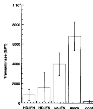
To extend these results, the effects of HD-IFN injection were examined in an additional acute hepatitis model. Among the more commonly used models, injection of ConA appears to be the most specific in inducing hepatic injury by T-cell acti-vation which is associated with the production of different inflammatory cytokines, including TNF-␣, interleukin-2, and IFN-␥ (23). IFN-␣/ was shown to protect mice from liver damage induced by ConA challenge, as indicated by a reduced level of serum transaminases (3).
C57/B6 mice were injected with HD-IFN (3⫻108PFU-E and 6 ⫻ 108 PFU-E) and Ad-IFN (2 ⫻ 109 PFU). Serum mIFN-␣2 was monitored on days 8, 16, and 25, and it was not detected in mice injected with Ad-IFN or with the lower dose
of HD-IFN (data not shown). By contrast, a low level of mIFN-␣2 was detected in the serum of mice injected with 6⫻
108PFU-E of HD-IFN, which ranged between 12.5 and 5 U of mIFN-␣2 per ml. The injected mice were challenged with ConA at day 30 p.i., and GPT levels were measured 24 h after challenge. Mice injected with Ad-IFN at the high dosage (2⫻
109 PFU) were only marginally protected compared with mock-treated mice (Fig. 6). On the contrary, an eightfold re-duction in GPT levels was observed in mice injected with the more effective HD-IFN virus at the higher dose (6 ⫻ 108 PFU-E). Consistently, injection of HD-IFN at the dose of 3⫻
108PFU-E resulted in lower but significant protection. Taken together, these results indicate that HD-IFN exhibits a protec-tive effect against liver injury even at doses that do not yield circulating mIFN-␣2 levels.
DISCUSSION
We have described the construction of an HD Ad expressing the mIFN-␣2 gene. Comparison with a first-generation Ad vector containing the same mIFN-␣2 expression cassette indi-cates that these two vectors function quite differently in vivo, with the HD vector performing more efficiently in inducing an mIFN-␣2-mediated biological response and greater therapeu-tic potency.
The better performance of the HD vector over a first-gen-eration Ad vector carrying the same expression cassette was recently observed with the mouse leptin gene (24). Also in those experiments at 1 week p.i., the transgene mRNA was observed only in the HD-injected mice and not in those in-jected with the Ad vector. Several factors have been shown to contribute to the weaker Ad-mediated gene expression, includ-ing “leaky” viral protein expression, which may trigger an im-mune response against transduced cells (37, 41). The host immune response may be even more relevant to IFN gene delivery because mIFN-␣2 immune modulatory activity may contribute to clearance of the transduced cells. Although we have not examined the extent of clearance of transduced liver cells, the different efficiency of gene expression between the Ad and HD vectors supports the notion that the latter viruses, which do not express Ad proteins, are safer vectors that should guarantee efficient and prolonged expression of the transgene of interest. In this regard, we have observed in preliminary experiments that liver expression of mIFN-␣2 mediated by HD-IFN injection is detectable for 3 months (data not shown). Injection of HD-IFN resulted in high levels of liver mIFN-␣2 (Fig. 2). It has been reported previously that after injection of mice with 105U, homologous IFN-␣levels peaked in the liver at 1 h p.i. (104 U/g) and subsequently decreased to baseline levels after 8 h (15). In contrast, injection of HD-IFN (1.8⫻
109 PFU-E) resulted in a fivefold-higher mIFN-␣2 concen-tration in the liver than in the serum at 1 week p.i. Moreover, the biological response, measured as 2⬘5⬘OAS induction, was dose dependent and observed even at the lowest HD-IFN dose, which did not result in circulating mIFN-␣2. The lack of circulating mIFN-␣2 at the lowest HD-IFN dose may have different explanations. One obvious possibility is the low sen-sitivity of the mIFN-␣2 detection assay. An alternative expla-nation may be that mIFN-␣2 expressed at low levels is trapped by its own receptors in the liver tissue and released into the bloodstream only when produced above a certain threshold, as observed with the highest HD-IFN dose.
Injection of Ad-gal, which resulted in a detectable induc-tion of 2⬘5⬘OAS (Fig. 2), was not associated with protection against MHV-3 challenge (Fig. 3 and 5). It is interesting that a different result was observed in HBV transgenic mice and in
FIG. 4. Hepatic protection in acute hepatitis induced by MHV-3 in the ab-sence of circulating mIFN-␣2. C57/B6 mice were mock injected or injected with HD-IFN at the dose of 3⫻108PFU-E as described in the legend to Fig. 3. Samples were collected at 3 days after MHV-3 infection and analyzed. (A) Transaminase (GPT) levels (expressed as units per liter) present in serum were determined at the time of autopsy (3 days after MHV-3 infection). (B) Northern blot analysis was performed with 20g of total RNA from a liver fraction from each mouse. Membrane was hybridized with a radiolabeled 2⬘5⬘OAS probe. (C) Northern blot analysis with a 250-bp probe from the 3⬘end of the MHV-3 genome, obtained from plasmid DE25 (21). (D) MHV-3 particles present in a liver, expressed per milligram of liver weight. Total liver RNA was also analyzed in an RNase protection assay for expression of inflammatory cytokines such as TNF-␣(E) and IFN-␥(F) and for a marker for T-lymphocyte activation, CD3ε (G). (H) The housekeeping gene GAPDH was used for normalization. cont., controls.
on November 9, 2019 by guest
http://jvi.asm.org/
lymphocytic choriomeningitis virus-infected C57/B6 mice; in-jection of a similar dose of the Ad-gal vector led to transient inhibition of viral replication in both systems (5, 14). In our experiments, protection against the more aggressive MHV-3 was observed only in the presence of IFN-␣expression, indi-cating that HD-IFN not only induced a stronger 2⬘5⬘OAS activation, but it likely elicited a broad spectrum of antiviral pathways which may contribute to the antiviral state observed in the protected mice. Hepatic expression of mIFN-␣2 led to liver protection in 80% of the C57/B6 mice infected with MHV-3 (Fig. 3). The reason why not all of the
HD-IFN-injected mice were protected from MHV-3 challenge remains elusive. The simplest explanation is that MHV-3 replication may have occurred in tissues, including the peritoneum, where the virus was injected, which were not protected by mIFN-␣2. This may have led to greater virus loads in the liver, and gradually the mIFN-␣2-mediated block to virus multiplication may have been overcome in an increasing number of cells. A similar mechanism may also explain the minimal increase in survival time observed in the HD-IFN-treated mice, which may have been limited by MHV-3 replication in extrahepatic tissues (11).
FIG. 5. Liver histology. (A and B) Liver samples from two animals preinjected with HD-IFN. The hepatic parenchyma is normal; no morphologic patterns of hepatic injury are detectable. (C and D) Liver samples from two animals preinjected with Ad-gal. (E and F) Liver samples from two animals mock injected. In C, D, E, and F, several areas of zonal necrosis in the hepatic parenchyma are depicted. A main central core of liquefactive necrosis characterizes these areas where nuclei are undetectable, and apoptotic cells with mainly pyknotic nuclei surround them. Hematoxylin and eosin staining. Bars, 35m.
on November 9, 2019 by guest
http://jvi.asm.org/
In the presence of circulating IFN-␣ (Fig. 3), peripheral effects such as peritoneal macrophage activation may contrib-ute to liver protection. However, injection of a low HD-IFN dose which resulted in IFN-␣ being present only in the liver was equally effective, indicating that viral resistance mecha-nisms activated at the local level are sufficient for liver protec-tion (Fig. 4). In addiprotec-tion, mice protected with a lower dose of HD-IFN showed induction of the 2⬘5⬘OAS antiviral gene as-sociated with an absence of MHV-3 transcripts and viral par-ticles, in line with the notion that viral clearance may occur without destruction of potentially infected cells (14). Further-more, no liver histology alterations were noted in mice pre-treated with HD-IFN, indicating that injection of HD-IFN had no negative effects on the liver tissue which could be ascribed to MHV-3 replication in association with the inflammatory response (Fig. 5). It is known that IFN-␣promotes a variety of antiviral effects, which include induction of antiviral genes and immunomodulatory effects. The absence of MHV-3 mRNAs in the infected liver indicates that antiviral activities such as those induced by 2⬘5⬘OAS may be responsible for MHV-3 clearance. However, we cannot exclude the possibility that virus entry and/or uncoating was also affected by IFN-␣ expression, as observed for simian virus 40 and retroviruses (38), or that IFN-␣ may have induced a local immune response that was ultimately responsible for MHV-3 clearance.
HD-IFN also proved efficient in protecting the liver from damage associated with ConA challenge at 30 days p.i., sug-gesting that transgene expression continued during this period of time (Fig. 6). The anti-inflammatory activity of type I IFNs is thought to be mediated, at least in part, by inhibiting pro-duction of inflammatory mediators (16). This protection may have important implications in cirrhosis of the liver, where
parenchymal cells are progressively destroyed by activated T cells (39).
The results obtained in this study indicate that the HD-IFN vector is quite effective in protecting the liver from the damage associated with acute hepatitis. We therefore believe that local delivery of IFN-␣to the liver tissue with HD Ad vectors might be a promising approach for the treatment of chronic hepatitis. In this context, a more in-depth assessment of the minimal therapeutic dose and potential toxic effects needs to be carried out before this vector can be considered a candidate for the treatment of chronic hepatitis. The development of HD vec-tors for IFN-␣gene delivery to the liver will have to include transcriptional regulatory elements in order to guarantee a minimal effective therapeutic level of IFN-␣expression. This additional modification of the HD vector will further increase the safety features of IFN-␣hepatic gene delivery. To this end, regulated liver gene expression has recently been demon-strated with an HD vector carrying an RU486-inducible pro-moter driving transcription of growth hormone (4). Finally, a regulated HD-IFN vector will have to show therapeutic po-tency in a validated animal model of chronic hepatitis, such as woodchuck hepatitis (32). Utilizing a woodchuck IFN-␣ ho-molog to minimize immune responses against IFN-␣ -trans-duced cells could provide information on the efficacy of IFN-␣
gene delivery in a chronically damaged and probably inflamed liver.
An additional feature that renders the delivery of IFN-␣to the liver an attractive therapeutic strategy is the observation that antiviral effects are observed with no detectable release of IFN-␣ into the bloodstream. The restricted expression of IFN-␣is particularly relevant in light of the side effects asso-ciated with the current treatment of HCV patients, who nor-mally receive 3⫻106U of r-hIFN-␣2 three times per week. Patients receiving higher or more frequent doses suffer from even worse side effects, possibly induced by circulating IFN-␣
(2). It is likely that selective expression of IFN-␣in the liver may render it more tolerable. A further advantage is that liver-specific expression of IFN-␣ could have autocrine and paracrine effects on the target organ, perhaps resulting in very significant bystander effects and thereby maximizing the ther-apeutic potency of IFN-␣.
ACKNOWLEDGMENTS
We thank M. M. C. Lai, S. Makino, S. Stohlman, and F. Taguchi for providing MHV-3 samples and advice, S. Germoni and M. Aquilina for animal care, and E. Fattori and P. Monaci for critically reading the manuscript. We also thank J. Clench for editorial assistance and M. Emili for graphics.
REFERENCES
1.Armstrong, J. A. 1981. Cytopathic effect inhibition assay for interferon: microculture plate assay. Methods Enzymol.78:381–387.
2.Ascione, A., M. De Luca, C. Canestrini, G. G. Di Costanzo, G. Raimondo, G. Longo, M. P. Manns, H. L. Tillmann, G. B. Forte, P. Rocco, O. Biceglia, D. Faleo, F. Vinelli, E. M. Cela, L. Amitrano, L. Addario, and T. Gigliotti.1998. Efficacy of high dose of recombinant alpha 2b interferon on long term response in chronic hepatitis C and cirrhosis: prospective randomized mul-ticentre study. Ital. J. Gastroenterol. Hepatol.30:517–523.
3.Bahr, G. M., P. R. Pouillart, and L. A. Chedid.1996. Enhancement in vivo of the antiinflammatory and antitumor activities of type I interferon by association with the synthetic immunomodulator murabutide. J. Interferon Cytokine Res.16:297–306.
4.Burcin, M. M., G. Schiedner, S. Kochanek, S. Y. Tsai, and B. W. O’Malley. 1999. Adenovirus-mediated regulable target gene expression in vivo. Proc. Natl. Acad. Sci. USA96:355–360.
5.Cavanaugh, V. J., L. G. Guidotti, and F. V. Chisari.1998. Inhibition of hepatitis B virus replication during adenovirus and cytomegalovirus infec-tions in transgenic mice. J. Virol.72:2630–2637.
[image:7.612.75.271.71.299.2]6.Chartier, C., E. Degryse, M. Gantzer, A. Dieterle, A. Pavirani, and M. Mehtali.1996. Efficient generation of recombinant adenovirus vectors by FIG. 6. ConA-induced hepatitis. HD-IFN injection protects C57/B6 mice
from ConA challenge. C57/B6 mice were injected i.v. with HD-IFN at different dosages (6⫻108or 3⫻108PFU-E) or with Ad-IFN at 2⫻109PFU and challenged with ConA (0.3 g/mouse) 30 days p.i. At 24 h after ConA challenge, transaminases (GPT) (expressed as units per liter) present in the serum were measured. The basal level of serum GPT was measured in untreated mice which were used as controls (cont.). Data are the mean⫾standard deviation of serum GPT measured in four animals.
on November 9, 2019 by guest
http://jvi.asm.org/
homologous recombination inEscherichia coli. J. Virol.70:4805–4810. 7.Colamonici, O. R., B. Porterfield, P. Domanski, S. Constantinescu, and
L. M. Pfeffer.1994. Complementation of the interferon alpha response in resistant cells by expression of the cloned subunit of the interferon alpha receptor. A central role of this subunit in interferon alpha signaling. J. Biol. Chem.269:9598–9602.
8.David, M.1995. Transcription factors in interferon signaling. Pharmacol. Ther.65:149–161.
9.Davis, G. L., R. Esteban-Mur, V. Rustgi, J. Hoefs, S. C. Gordon, C. Trepo, M. L. Shiffman, S. Zeuzem, A. Craxi, M. H. Ling, and J. Albrecht.1998. Interferon alfa-2b alone or in combination with ribavirin for the treatment of relapse of chronic hepatitis C. International Hepatitis Interventional Ther-apy Group. N. Engl. J. Med.339:1493–1499.
10. Der, S. D., A. Zhou, B. R. Williams, and R. H. Silverman.1998. Identification of genes differentially regulated by interferon alpha, beta, or gamma using oligonucleotide arrays. Proc. Natl. Acad. Sci. USA95:15623–15628. 11. Ding, J. W., Q. Ning, M. F. Liu, A. Lai, J. Leibowitz, K. M. Peltekian, E. H.
Cole, L. S. Fung, C. Holloway, P. A. Marsden, H. Yeger, M. J. Phillips, and G. A. Levy.1997. Fulminant hepatic failure in murine hepatitis virus strain 3 infection: tissue-specific expression of a novel fgl2 prothrombinase. J. Virol. 71:9223–9230. (Erratum,72:3504, 1998.)
12. Domanski, P., and O. R. Colamonici.1996. The type-I interferon receptor. The long and short of it. Cytokine Growth Factor Rev.7:143–151. 13. Eto, T., and H. Takahashi.1999. Enhanced inhibition of hepatitis B virus
production by asialoglycoprotein receptor-directed interferon. Nat. Med. 5:577–581.
14. Guidotti, L. G., R. Rochford, J. Chung, M. Shapiro, R. Purcell, and F. V. Chisari.1999. Viral clearance without destruction of infected cells during acute HBV infection. Science284:825–829.
15. Heremans, H., A. Billiau, and P. De Somer.1980. Interferon in experimental viral infections in mice: tissue interferon levels resulting from the virus infection and from exogenous interferon therapy. Infect. Immun.30:513– 522.
16. Heremans, H., R. Dijkmans, H. Sobis, F. Vandekerckhove, and A. Billiau. 1987. Regulation by interferons of the local inflammatory response to bac-terial lipopolysaccharide. J. Immunol.138:4175–4179.
17. Huard, J., H. Lochmuller, G. Acsadi, A. Jani, B. Massie, and G. Karpati. 1995. The route of administration is a major determinant of the transduction efficiency of rat tissues by adenoviral recombinants. Gene Ther.2:107–115. 18. Isaacs, A., and J. Lindenmann.1957. I. Virus interference. Proc. R. Soc.
Lond.147:258–267.
19. Kato, Y., Y. Noda, M. Unoura, N. Tanaka, K. Kobayashi, N. Hattori, K. Hatano, and S. Kobayashi.1986. Effect of exogenous mouse interferon on murine fulminant hepatitis induced by mouse hepatitis virus type 2. Dig. Dis. Sci.31:177–180.
20. Maione, D., M. Wiznerowicz, P. Delmastro, R. Cortese, G. Ciliberto, N. La Monica, and R. Savino.Prolonged expression and effective readministration of erythropoietin delivered with a fully deleted adenoviral vector. Hum. Gene Ther. in press.
21. Makino, S., C. K. Shieh, L. H. Soe, S. C. Baker, and M. M. Lai.1988. Primary structure and translation of a defective interfering RNA of murine corona-virus. Virology166:550–560.
22. Minagawa, H., A. Takenaka, S. Mohri, and R. Mori.1987. Protective effect of recombinant murine interferon beta against mouse hepatitis virus infec-tion. Antiviral Res.8:85–95.
23. Mizuhara, H., M. Kuno, N. Seki, W. G. Yu, M. Yamaoka, M. Yamashita, T. Ogawa, K. Kaneda, T. Fujii, H. Senoh, and H. Fujiwara.1998. Strain dif-ference in the induction of T-cell activation-associated, interferon gamma-dependent hepatic injury in mice. Hepatology27:513–519.
24. Morsy, M. A., M. Gu, S. Motzel, J. Zhao, J. Lin, Q. Su, H. Allen, L. Franlin, R. J. Parks, F. L. Graham, S. Kochanek, A. J. Bett, and C. T. Caskey.1998. An adenoviral vector deleted for all viral coding sequences results in en-hanced safety and extended expression of a leptin transgene. Proc. Natl. Acad. Sci. USA95:7866–7871.
25. Parks, R. J., L. Chen, M. Anton, U. Sankar, M. A. Rudnicki, and F. L. Graham.1996. A helper-dependent adenovirus vector system: removal of helper virus by Cre-mediated excision of the viral packaging signal. Proc. Natl. Acad. Sci. USA93:13565–13570.
26. Polyak, S. J., D. M. Paschal, S. McArdle, M. J. Gale, Jr., D. Moradpour, and D. R. Gretch. 1999. Characterization of the effects of hepatitis C virus nonstructural 5A protein expression in human cell lines and on interferon-sensitive virus replication. Hepatology29:1262–1271.
27. Qian, X., U. Samadani, A. Porcella, and R. H. Costa.1995. Decreased expression of hepatocyte nuclear factor 3 alpha during the acute-phase response influences transthyretin gene transcription. Mol. Cell. Biol.15: 1364–1376.
28. Qin, X. Q., N. Tao, A. Dergay, P. Moy, S. Fawell, A. Davis, J. M. Wilson, and J. Barsoum.1998. Interferon-beta gene therapy inhibits tumor formation and causes regression of established tumors in immune-deficient mice. Proc. Natl. Acad. Sci. USA95:14411–14416.
29. Recchia, A., R. J. Parks, S. Lamartina, C. Toniatti, L. Pieroni, F. Palombo, G. Ciliberto, F. L. Graham, R. Cortese, N. La Monica, and S. Colloca.1999. Site-specific integration mediated by a hybrid adenovirus/adeno-associated virus vector. Proc. Natl. Acad. Sci. USA96:2615–2620.
30. Reichard, O., G. Norkrans, A. Fryden, J. H. Braconier, A. Sonnerborg, and O. Weiland.1998. Randomised, double-blind, placebo-controlled trial of interferon alpha-2b with and without ribavirin for chronic hepatitis C. The Swedish Study Group. Lancet351:83–87.
31. Rice, A. P., R. Duncan, J. W. Hershey, and I. M. Kerr. 1985. Double-stranded RNA-dependent protein kinase and 2-5A system are both activated in interferon-treated, encephalomyocarditis virus-infected HeLa cells. J. Vi-rol.54:894–898.
32. Roggendorf, M., and T. K. Tolle.1995. The woodchuck: an animal model for hepatitis B virus infection in man. Intervirology38:100–112.
33. Schiedner, G., N. Morral, R. J. Parks, Y. Wu, S. C. Koopmans, C. Langston, F. L. Graham, A. L. Beaudet, and S. Kochanek. 1998. Genomic DNA transfer with a high-capacity adenovirus vector results in improved in vivo gene expression and decreased toxicity. Nat Genet.18:180–183. (Erratum, 18:298.)
34. Stemmer, W. P., A. Crameri, K. D. Ha, T. M. Brennan, and H. L. Heyneker. 1995. Single-step assembly of a gene and entire plasmid from large numbers of oligodeoxyribonucleotides. Gene164:49–53.
35. Tan, S. L., H. Nakao, Y. He, S. Vijaysri, P. Neddermann, B. L. Jacobs, B. J. Mayer, and M. G. Katze.1999. NS5A, a nonstructural protein of hepatitis C virus, binds growth factor receptor-bound protein 2 adaptor protein in a Src homology 3 domain/ligand-dependent manner and perturbs mitogenic sig-naling. Proc. Natl. Acad. Sci. USA96:5533–5538.
36. Taylor, D. R., S. T. Shi, P. R. Romano, G. N. Barber, and M. M. Lai.1999. Inhibition of the interferon-inducible protein kinase PKR by HCV E2 pro-tein. Science285:107–110.
37. Tripathy, S. K., H. B. Black, E. Goldwasser, and J. M. Leiden.1996. Immune responses to transgene-encoded proteins limit the stability of gene expres-sion after injection of replication-defective adenovirus vectors. Nat. Med. 2:545–550.
38. Vilcek, J., and C. S. Ganes.1996. Interferon and other cytokines, p. 375–399. InB. N. Fields, D. M. Knipe, and P. M. Howley (ed.), Fields virology, 3rd ed. Lippincott-Raven, Philadelphia, Pa.
39. Wermers, G. W., H. Band, and E. J. Yunis.1998. Role of the HLA system in antigen recognition and disease. Raven Press, New York, N.Y.
40. Wills, R. J.1990. Clinical pharmacokinetics of interferons. Clin. Pharmaco-kinet.19:390–399.
41. Yang, Y., F. A. Nunes, K. Berencsi, E. E. Furth, E. Gonczol, and J. M. Wilson.1994. Cellular immunity to viral antigens limits E1-deleted adeno-viruses for gene therapy. Proc. Natl. Acad. Sci. USA91:4407–4411. 42. Yeow, W. S., C. M. Lawson, and M. W. Beilharz.1998. Antiviral activities of
individual murine IFN-alpha subtypes in vivo: intramuscular injection of IFN expression constructs reduces cytomegalovirus replication. J. Immunol.160: 2932–2939.