“A STUDY ONTHE ASSOCIATION OF CLINICAL PROFILE WITH
THE OUTCOMES OF LUPUS NEPHRITIS”
Dissertation Submitted to
THE TAMILNADUDR.M.G.R.MEDICALUNIVERSITY, CHENNAI- 600 032.
In partial fulfillment of the regulation for the award of the degree of
DM (RHEUMATOLOGY) BRANCH - IX
MADRAS MEDICAL COLLEGE
RAJIV GANDHI GOVERNMENT GENERAL HOSPITAL CHENNAI – 600 003.
CERTIFICATE
This is to certify that this dissertation entitled “A study on the association of clinical profile with the outcomes of lupus nephritis” presented here is original
work done by Dr.S.VIDYA, DM Post Graduate in the Department of Rheumatology,
Madras Medical College and Rajiv Gandhi Government General Hospital, Chennai-
600003 in partial fulfillment of the university rules and regulation for the award of
D.M.BranchIX- Rheumatology, under my guidance and supervision during the
academic period from 2010-2013.
Dr.V.KANAGASABAI, MD.,
Dean,
MadrasMedicalCollege and RajivGandhiGovt. GeneralHospital,
Chennai – 600 003.
Dr.S.RUKMANGATHARAJAN, MD., DM., FMMC.,
Professor and HOD, Department of Rheumatology,
Madras Medical College and Rajiv Gandhi Govt. General Hospital,
DECLARATION
I, Dr.S.VIDYAhereby solemnly declare that this dissertation entitled “A study on the association of clinical profile with the outcomes of lupus nephritis”
was done by me in the Department of Rheumatology, Madras Medical College &
Rajiv Gandhi Govt. General Hospital, Chennai-3 during February 2011 to February
2013 under the guidance and supervision of Prof.Dr.S.Rukmangatharajan, MD., DM.,
FMMC., This dissertation is submitted to the Tamil Nadu Dr.M.G.R.Medical
University towards the partial fulfillment of requirement for the award of D.M.,
Degree in Rheumatology.
Signature of the Candidate
ACKNOWLEDGEMENT
I express my heartful gratitude to the Dean, Dr.V.KANAGASABAI,
MD.,MadrasMedicalCollege&RajivGandhi Govt. General Hospital, Chennai -3 for permitting me to do this study.
I gratefully acknowledge and sincerely
thankProf.Dr.S.RUKMANGATHARAJAN, MD., DM.,FMMC.,Professor and Head,
Department of Rheumatology, for his valuable suggestions, guidance, constant
supervision and moral support without which this study would not have been
possible.
I am thankful to Dr.K.MUTHULAKSHMI, MD.,Additional Professor for her
valuable guidance in doing the Biochemical and Immunological workup of patients.
I express my gratitude toDr.S.BALAMEENA, MD., DCH., DM, Asst.
Professor, Department of Rheumatology for the valuable guidance, advice and
suggestions during the study.
I am extremely thankful to Assistant Professors Dr.R.RAVICHANDRAN,
MD., DCH., DM.,Dr.T.N.TAMILSELVAM, MD., DM,andDr.D.THERASAMARY, MD., (Micro) and my FELLOW POSTGRADUATES for their constant support and advice during my study.
I am extremely thankful to the Laboratory Personnel for their invaluable help in carrying out the immunological investigations without which, this work
I thank thePhysiotherapist, theStaff Nurses and all the Paramedical staff members in the Department of Rheumatology, Madras Medical College, Rajiv Gandhi Government General Hospital, Chennai for their full co-operation in
conducting the study.
I thank my parents, my husband and my daughter for their understanding and
co-operation in completion of this work.
I also thank Dr.Pradeepmenon and Mr.Srinivasan from ICMR /NIRT Chetpet
Chennai for lending their helping hands for statistics.
Last but not the least, I owe my sincere gratitude to the patients and their
relatives who co-operated for this study, without whom the study would not have
INDEX
S.NO CONTENTS PAGE NO.
1. INTRODUCTION 1
2. AIMS AND OBJECTIVES 3
3. REVIEW OF LITERATURE 4
4. MATERIALS AND METHODS 33
5. RESULTS OF THE DATA 39
6. DISCUSSION 64
7. CONCLUSION 71
8. BIBLIOGRAPHY
9. ANNEXURE
A) PROFORMA
B) MASTER CHART
C) PATIENT CONSENT FORM
D) PATIENT INFORMATION SHEET
E) ETHICAL COMMITTEE APPROVAL ORDER
ABBREVIATIONS
LN - Lupus Nephritis
SLE - Systemic Lupus Erythematosus
HLA - Human Leucocyte Antigen
EULAR - European League Against Rheumatism
ERA/EDTA - European Renal Association / European Dialysis and
Transplant Association
ISN/RPS - International Society of Nephrology/ Renal Pathology
Society
CR - Complete Response
PR - Partial Response
IMP - Improved
REF - Refractory
LTF - Lost follow up
NIH - National Institute of Health
CYC - Cyclophosphamide
AZA - Azathioprine
MMF - MycophenolateMofetil.
ANA - Anti Nuclear Antibody
ACL - Anti Cardio Lipin
LAC - LupusAnti Coagulant
1 INTRODUCTION
Sytemic lupus erythematosus is a paradigmatic autoimmune
disorder, the manifestations of which are protean sparing few organ systems if
any.1 Such diversity is attributed to its etiopathogenesis wherein antibodies to the
components of cell nucleus have been implicated. One major cause of morbidity
and utilization of health resources is renal involvement. More than half of the
mortality in SLE is due to renal involvement.2-5
As with SLE, heterogeneity, both clinical as well as histological is
the hall mark of lupus nephritis. The disease usually is asymptomatic in its earlier
course thus vigilant screening of SLE patients for renal involvement remains the
important step in reducing the mortality and morbidity.
Even though recent treatments are effective and have reduced the
adverse outcomes the therapeutic options are limited and induce toxicities in the
long term. 6,7 Hence there is a need to identify patients who may have a worse
prognosis so that aggressive treatment can be instituted early.
Prognosis and therefore treatment decisions vary greatly according
to the clinical and pathological forms of lupus nephritis. 8-10 Each individual has a
2 the greater the number of factors with worse prognosis, the less the patient is likely
to respond to therapy and hence needs aggressive therapy.
Considering the above mentioned prognostic factors may help in
improving clinical decision making regarding the type and intensity of immune
suppressive treatment for patients with lupus nephritis.
Studies of long term prognosis in lupus nephritis have focused on
risk factors which are present either at the onset or those that develop during the
course of the illness. These predict the mortality over subsequent 10 years. The
results of these studies may vary but they are useful as they inform us about how
specific manifestations influence the outcome. But these are less helpful in making
treatment decisions than studies of short term prognosis as short term outcome
studies are more likely influenced by timely intervention.
In spite of many years of intense investigations controversies
surrounding the importance of clinical, demographic, laboratory and histologic
features in predicting renal outcomes continue to evolve as current and recent
treatments have altered the prognostic significance of these factors that were
previously considered significant.
Here in this study we have tried to assess the outcomes of lupus
nephritis in fifty patients and the association of clinical and immunologic profile
3
AIMS AND OBJECTIVES
1. To study the outcomes of lupus nephritis in 50 patients during the study period.
2. To study the association of demographic, clinical, laboratory, histopathologic
and treatment profile of these patients with the outcome.
3. To compare the results with the standard data available.
4 REVIEW OF LITERATURE
EPIDEMIOLOGY:
Lupus nephritis affects 40-70% of patients with SLE.11The frequency
of lupus nephritis peaks during the first two years since the onset of SLE and its
incidence follows a decrescendo pattern reaching a trough after five years of SLE.
Asymptomatic urine abnormalities like proteinuria or hematuria is seen in half the
cases. In about 30% nephrotic or nephritic syndrome occurs. Chronic renal
insufficiency or rapidly progressive glomerulonephritis occur in less than 5% of
the individuals.
PATHOGENESIS:
Only a few diseases like lupus nephritis are characterized by immune
complexes detected in all four renal components namely glomeruli, tubules,
interstitium and blood vessels.12 Although IgG is the dominant immunoglobulin
(98%) co-deposits of IgM and IgA are also common. The term ‗full house‘ staining
is applied when all the three immunoglobulin classes are present.
Intraglomerular inflammation and recruitment of leukocytes are the
earliest events in kidney that follow immune complex formation and their
deposition. Activation and proliferation of resident renal cells soon follow the
5 necrosis or apoptosis. In the event of lesser injury there is proliferation of
endocapillary cells and production of extracellular matrix. Rupture of capillary
wall and even the capsule itself occurs in case of severe injury resulting in
accumulation of fibrin over basement membrane along with collagen, mononuclear
cells and epithelial cells in the urinary space resulting in crescentic
glomerulonephritis pattern. Atrophy and scarring is the end result of protracted
inflammation.
The histopathology and the intensity of the inflammatory response are
closely linked to the location of immune complex deposition and formation.
Mesangial lupus nephritis occurs when immune complexes are deposited in
mesangium. Focal or diffuse proliferative lupus nephritis with profuse glomerular
hypercellularity occurs when immune complexes are deposited in the
subendothelial region. Proliferation of endothelial and mesangial cells hand in
hand with leukocytic infiltrates is the cause for this hypercellularity the result of
which is compromised capillary flow and renal function. Membranous
nephropathy occurs when there are epimembranous (subepithelial) deposits along
diffusely thickened peripheral glomerular capillary loops and lack of inflammatory
6 The differences in the composition and properties of the immune
complexes such as size, specificity, charge and immunoglobulin isotype probably
explain the diverse morphological expressions of lupus nephritis. An intermediate
sized, high avidity small immune complex favours a mesangial pattern while large
sized loads can spill into the subendothelial region. Low avidity, smaller, cationic
complexes that dissociate and reform in situ favors sub epithelial deposits.
Traditional thinking is that lupus nephritis is a quintessential type III
hypersensitivity reaction with deposition of immune complexes in the glomeruli
and subsequent complement activation.13 Recently emphasis is also given to the
significance of local formation of immune deposits.14 Positively charged
nucleosomes are attracted towards the negatively charged sites in the glomerular
wall. After getting implanted in the glomerular filter these auto antigens form
immune complexes after reacting with the circulating auto antibodies.
Auto antibodies to normal glomerular constituents like laminin,
heparan sulfate, type IV collagen are also implicated in another theory of in situ
immune complex formation. The role of antigen presentation by T cells, activated
macrophages and Fcγ receptor (FcγR) bearing monocytes are important in
glomerulonephritis.15-17 Mice deficient in FcγR are immune to development of
7
GENETICS:
This can be divided in to HLA and non HLA genes. HLA genes
implicated in lupus nephritis are HLADRB1*1501/DQB1*0602, DQB1* 0201,
DQB1*0301, DR2/DR3. Homozygous deficiencies of early complement
components have also been implicated.19
COMPLEMENT SYSTEM :
The role of complement system in the pathogenesis of lupus nephritis
cannot be under estimated. Presence of complement activation factors in tubules,
glomerulus, interstitium and urine provides support for this. Though classical
component pathway deficiency is viewed as a predisposing factor for lupus
nephritis, great majority of patients have intact alternative especially lectin
pathway. Evidences favouring the role of complement pathway include findings
that an inhibitory anti-C5 mAb hinders the onset of glomerulonephritis in the
(NZB X NZW) F1 model of SLE. The fact that fB-/- and fD-/-MRL/lpr mice are
immune from lupus nephritis also underscores the importance of alternate pathway.
Though role of complement activation is traditionally restricted to
glomerular disease, additional roles in tubulointerstitial inflammation and
proteinuric states have also been suggested. When complement activation
8 capable of being activated since the tubular epithelium lacks complement
regulatory proteins.
ROLE OF ANTI-ds DNA:
Anti-ds-DNA antibodies are present in about 60% of patients with
SLE. Specifically they are associated with nephritis which has the strongest
correlation with it.20 Lot of evidence suggests the role of DNA-anti DNA
complexes in the pathogenesis of LN.21 This includes detection of anti-DNA
antibodies in the kidneys, free DNA in the plasma, alterations in the serum
concentrations of anti DNA antibodies and complement. The theory of molecular
mimicry has also been implicated wherein anti ds DNA may react with glomerular
and mesangial target antigens such as alpha actinin, a matrix protein. Not all
patients with high anti ds DNA develop nephritis. Anti-DNA measurements may
help in monitoring disease activity. Sometimes anti-DNA and complement (C3,
C4) levels vary reciprocally over time.22,23 ELISA and Crithidia luciliae
kinetoplast staining assay detect low affinity interactions whereas Farr assay,
which detects high-affinity antibodies, may predict disease activity more
9
ROLE OF APS NEPHROPATHY:
Anti-phospholipid antibodies are implicated in the pathogenesis of a
unique type of vascular nephropathy (APSN).Features of APSN are present in
20% to 30% of patients with SLE.24 They are fibrous occlusions of
arteries/arterioles, organizing thrombi with recanalisation, thrombotic
microangiopathy, focal cortical atrophy and chronic lesions such as fibrous
intimal hyperplasia.25, 26
CLINICAL FEATURES:
Lupus nephritis is usually asymptomatic unless it is advanced
nephrotic syndrome or renal failure. It is usually discovered during a routine
evaluation. Proteinuria, presence of urinary casts, hematuria, pyuria, increased
serum creatinine and hypertension are the features most commonly seen .
Specifically the revised criteria for classification for SLE includes a) Persistent
proteinuria > 0.5 g per day or > than 3+ if quantitation not performed, OR
b) Cellular casts: red cell, hemoglobin, granular, tubular, or mixed as evidence of
renal disease.Hematuria (>5 RBCs/HPF),pyuria (>5WBCs/HPF) in the absence of
infection and raised serum creatinine concentration have also been recognized as
the renal manifestations. Hence urine analysis must be performed regularly in
10
RENAL BIOPSY
Renal biopsy is necessary to identify the type of kidney involvement as
patient management is determined based on it.27,28 Most patients with lupus
nephritis have abnormal renal biopsy either on light microscopy or special
techniques like immunofluorescence or electron microscopy.
Renal involvement is not only limited to glomerulonephritis but may
also be due to interstitial nephritis, tubular disease, vasculitis, arteriolosclerosis,
thrombotic microangiopathy, and lupus vasculopathy. There may be changes
secondary to co-morbidities such as human immunodeficiency virus (HIV)
infection. A semi quantitativeanalysis of specific histologic features based on a 0-3
scale) is included into the elements of the activity and chronicity indices.
The International Society of Nephrology/Renal Pathology Society
revised the earlier World Health Organization classification in 2003 for a better
description and standardization of the lesions seen on biopsy. The classification is
based on the changes observed on light microscopy, immunofluorescence staining,
and electron microscopy.29 In a study of 46 japenese patients Yakohama et al found
that ISN /RPS 2003 has better prognostic value.30
11
INDICATIONS FOR RENAL BIOPSY IN LUPUS NEPHRITIS
INITIAL BIOPSY (before treatment)
Nephritic urine sediment (glomerular hematuria and cellular casts).
Glomerular hematuria with proteinuria >0.5 to 1.0 gm/day.
Glomerular hematuria with proteinuria <0.3 to 0.5 gm/day and low C3 and/
or positive anti-ds DNA.
Proteinuria >1.0 to 2.0 gm/day (especially if C3 is low and/or positive
anti-ds DNA).
REPEAT BIOPSY (during or after treatment)
Unexplained worsening of proteinuria (>2 gm/day increase if non nephrotic
at baseline, or >50% increase if nephrotic).
Unexplained worsening of renal function (reproducible ≥30% increase in
serum creatinine).
Persistent glomerular hematuria with proteinuria >2 gm/day or proteinuria
>3 gm/day (especially if C3 is decreased).
Nephritic or nephrotic flare.
According to recent EULAR/ ERA-EDTA guidelines any renal involvement is
12
ACTIVITY AND CHRONICITY INDICES *
Activity index (lesions are scored 0-3 with maximum score 24 points) * Hypercellularity: endocapillary proliferation compromising glomerular capillary loops
* Leukocyte exudation: polymorphonuclear leukocytes in glomeruli * Karyorrhexis/fibrinoid necrosis (weighted ×2): necrotizing changes in glomeruli
* Cellular crescents (weighted ×2): layers of proliferating epithelial cells and monocytes lining Bowman capsule
* Hyaline deposits: eosinophilic and PAS-positive materials lining (wire loops) or filling (hyaline thrombi) capillary loops
* Interstitial inflammation: infiltration of leukocytes (predominantly mononuclear cells) among tubules
Chronicity index (lesions are scored 0-3 with maximum score 12 points) * Glomerular sclerosis: collapse and fibrosis of capillary tufts
* Fibrous crescents: layers of fibrous tissue lining Bowman capsule * Tubular atrophy: thickening of tubular basement membranes, tubular epithelial degeneration, with separation of residual tubules
* Interstitial fibrosis: deposition of collagenous connective tissue among tubules
13
Table 1- ISN/RPS CLASSIFICATION OF LUPUS NEPHRITIS
Class I Minimal mesangial lupus nephritis
Normal glomeruli by light microscopy, but mesangial immune deposits by immunofluorescence
Class II Mesangial proliferative
Purely mesangial hypercellularity of any degree or mesangial matrix expansion by light microscopy, with mesangial immune deposits A few isolated subepithelial or subendothelial deposits may be visible by immunofluorescence or electron microscopy, but not by light microscopy
ClassIII Focal lupus nephritis
Active or inactive focal, segmental, or global endocapillary or extracapillary glomerulonephritis involving <50% of all glomeruli, typically with focal subendothelial immune deposits, with or without mesangial alterations
Class IV
Diffuse lupus nephritis
Active or inactive diffuse, segmental or global endocapillary or extracapillary glomerulonephritis involving ≥50% of all glomeruli, typically with diffuse subendothelial immune deposits, with or without mesangial alterations. This class is divided to diffuse segmental (IV-S) lupus nephritis when ≥50% of the involved
glomeruli have segmental lesions and diffuse global (IV-G) when
≥50% of the involved glomeruli have global lesions. Segmental is defined as a glomerular lesion that involves less than half of the glomerular tuft. This class includes cases with diffuse wire loop deposits but with little or no glomerular proliferation
Class V
Membranous lupus nephritis
Global or segmental subepithelial immune deposits or their morphologic sequelae by light microscopy and by
immunofluorescence or electron microscopy, with or without mesangial alterations
Class V nephritis may occur in combination with class III or class IV, in which case both will be diagnosed
Class V nephritis may show advanced sclerotic lesions
Class VI Advanced sclerotic lupus nephritis
14 Renal biopsy may show hematoxylin bodies, the tissue equivalent of
LE cell phenomenon. Mesangial lupus nephritis characterized by the accumulation
of immune complex deposits within the mesangium (class I) and its futher
progression in to mesangial hypercellularity (class II) are at the milder end of the
spectrum of the renal lesions. Most people with mesangial lupus nephritis are
clinically silent and respond well to renoprotective therapies even without
aggressive immunosuppressive regimens though on occasions they may have
severe renal involvement and validate aggressive therapy. Lupus nephritis is
further classified as focal or diffuse depending on the percentage of glomeruli
involved with subendothelial deposits, i.e. class III (<50% involved) and class IV
( ≥50% glomeruli involved) respectively. They are further described according to
the chronicity of the lesions. Membranous lesions (class V) are characterized
mainly by subepithelial deposits.They may also have mesangial involvement.
Advanced sclerosis (class VI) is the end stage or ―burnt out‖ phase of lupus
nephritis. Lupus vasculopathy is characterized by the presence of hyaline thrombi
within the arteriolar lumen and/or intralobular arteries. Tubulointerstitial
disease involves tubular basement membrane atrophy. Other findings that may also
be seen in lupus are lupoid nephrosis, focal segmental glomerulosclerosis, IgM
nephropathy and amyloidosis. 31
15 EVALUATION
ASSESSMENT OF RENAL INVOLVEMENT:
The basic assessment of renal function requires screening for
proteinuria, hematuria, leukocyturia and nitrates to check for infections using a
urine dipstick. While assessing hematuria other conditions like infections, calculi
and menstrual blood loss must be excluded. Urine casts still remain an important
indicator of active renal disease. Analysis of a 24 hr urine specimen is more useful
than a urine dipstick or serum creatinine. Protein/creatinine ratio or albumin
creatinine ratio and glomerular filtration rate (GFR) are also becoming important
tools especially in clinical trials as these require little patient cooperation and are
more robust measurements. Renal biopsy remains the best way to distinguish renal
activity from damage and to establish the type of renal involvement.
The use of serological tests in assessing lupus activity has long been
debated.32 Anti ds DNA antibodies are present in 60% of lupus patients making its
measurement useful only in those with positive antibodies. Usually the levels rise
during the development of a flare. But it may fall during peak clinical activity
because of tissue deposition. Changing levels of ds DNA antibodies must be
viewed with caution especially before reducing therapy. Decreasing levels of
complements C3 or C4 are usually the forerunner of a renal flare. So are the raising
16 MONITORING DISEASE ACTIVITY
URINALYSIS:
Urinalysis is the most useful method to detect and monitor disease
activity in lupus nephritis. The urine sample must be fresh, early morning, mid
stream, clean catch and non refrigerated. Presence of hematuria (usually
microscopic) indicates inflammatory glomerular or tubulointerstitial involvement.
Granular and fatty casts indicate proteinuric states while cellular casts involving
RBCs, WBCs or mixed pattern are indicators of nephritic states. Severe glomerular
and tubular ongoing disease can cause ‗telescopic urine sediment‘ i.e. containing
full range of cells and casts. Resolution of urine sediments is a feature of renal
remission provided it is sustained. Reappearance of cellular casts when associated
with proteinuria is a very early predictor of renal flare and may precede anti ds
DNA titers or decreased complement levels. Spot urine protein creatinine ratio is
an easy method to estimate the severity of proteinuria. It could be used in between
24 hr collections to roughly estimate the response to therapy.
RENAL FUNCTION :
Serum creatinine is a practical test but as it depends on muscle mass,
17 GFR.Rather than absolute values it is the change that is important especially
significant (20-30% increase).Creatinine clearance is being utilized only rarely
since it overestimates the true GFR.
ASSESEMENT OF PROGNOSIS:
There are many demographic and clinical variables which affect the
outcome. In general black race,anemia,antiphospolipid syndrome, azotemia, failure
to respond to initial therapy and flares with deteriorating renal function are
associated with poor outcome.
FACTORS ASSOCIATED WITH ADVERSE PROGNOSIS
DEMOGRAPHIC: Black race, limited access to health care, male sex, children).
CLINICAL: Hypertension; severe extra renal disease affecting major
organ; failure to achieve or marked delay (>2 years) to renal remission; multiple
flares of lupus nephritis; pregnancy.
LABORATORY: Nephritic urinary sediment, azotemia; anemia;
thrombocytopenia; antiphospholipid antibodies; thrombotic microangiopathy;
hypocomplementemia (especially falling levels); high anti-DNA (especially
rising titers); persistent severe nephrotic syndrome.
18 membranous (V) and proliferative (III-IV) glomerulonephritis; cellular
crescents; fibrinoid necrosis; very high activity index; moderate-to-high
chronicity index; combinations of active (cellular crescents) and chronic
histologic features (interstitial fibrosis); extensive subendothelial deposits.
MORBIDITY AND MORTALITY:
Lupus nephritis poses significant morbidity and mortality. In some
studies it was found that the health cost of patients with lupus nephritis was twice
higher than their fellow lupus patients without it. The major cause of morbidity
other than SLE and nephritis include those due to long term corticosteroid use such
as infections and osteoporosis.
The five year survival of LN has improved from 17% for class IV LN
in 1950s to82% in 1990s. The early mortality was due to sepsis and active SLE
while late mortality was due to cardio vascular events and thrombosis. 33.
19
TREATMENT
Recently European League Against
Rheumatism-(EULAR)/ERA-EDTA has published guidelines for treatment of adult and pediatric lupus
nephritis34 wherein the ultimate aims of treatment are preservation of renal
function, prevention of disease flares, avoidance of drug related complications,
and provision of good quality of life. Treatment must aim for complete renal
response.
INDUCTION:
For patients with classIII or class IV(± class V)-MMF 3g/day or low
dose IV cyclophosphamide together with steroids.
In patients with poor prognostic features monthly cyclophosphamide
can be given (0.75-1 gm/m2).This should be combined with three pulses of IV
methylprednisolone (500-750mg) followed by oral steroids. (0.5mg/kg/day of
prednisolone to be given over four weeks and gradually tapered to less than 10 mg
/day by six months.
In patients with class V LN who have nephrotic range of proteinuria
MMF 3g/day over six months along with steroids is the therapy of choice. As an
alternative therapy azathioprine or cyclosporine can also be given in certain
20
MAINTENANCE :
Maintenance therapy is with MMF 2g/day or azathioprine 2mg/kg/day
along with low dose steroids. In those patients who were initially treated with
MMF it should be continued. If pregnancy is desired azathioprine may be an
alternative. In refractory disease treatment can be switched between the drugs.
ADJUNCTIVE TREATMENT 35
ACE inhibitors in patients with hypertension and proteinuria.
Statins for dylipidemia (target LDL 100 mg%).
Chloroquin for decreasing renal flares and to reduce cardiac and kidney
damage.
Aspirin for patients with APS.
Vitamin D and Calcium supplements.
Treatment of comorbidities.
Anti coagulants for patients with nephrotic syndrome (albumin< 2g%) .
LUPUS NEPHRITIS IN PREGNANCY
Pregnancy may be considered if lupus is inactive and Urinary PCR <50
mg/mmol for 6 months, with a GFR > 50 ml/min. Drugs that can be used include
hydroxychloroquine,low dose methylprednisolone, azathioprine and calcineurin
21 Aspirin should be used to reduce the risk of pre-eclampsia. B.P should be treated
with nifedepine or labetolol. Complement levels should be measured to
differentiate lupus nephritis flare from preeclampsia.36, 37
From the above discussion it is clear that lupus nephritis is
characterized by immune complex formation, varied clinical manifestations and
multiple laboratory abnormalities with frequent exacerbations. Renal involvement
being very common is a major determinant of the disease course. 70-80 % of SLE
patients have one or the other form of renal involvement and majority of them
present with class IV lupus nephritis. Worldwide survival rates in LN have
improved remarkably during successive years due to early diagnosis. Better
awareness of risk factors and better treatment modalities also have contributed to
this improved prognosis.
Studies done in yester years (in 1970s) by Ester and Christian38 showed
an estimated five year survival rates for patients with lupus nephritis of 50% as
against 75% for whole SLE series. The rates for severe kidney disease was even
lower,68% for class III and 28% for class IV and V. But in these series patients
were being treated only with steroids as immunosuppressants were not widely
available at that time. In 1980s and 1990s survival rate became much better around
22 As reported by Korbet et al achievement of remission and the type of
treatment given also has considerable effect on outcome.42 The survival rate for
patients in his series was 95% at 5 yrs. In this series complete remission was
achieved in 43% and time taken to achieve complete remission was 18 months.
Stable renal function, lower chronicity index and white race were positive
predictors of remission.
A study conducted in Europe by Houssiau & colleagues 43 has reported
similar outcomes for patients receiving the conventional NIH Vs ELNT protocol.
According to James Tumlin of Emory university Atlanta 44, 45 poor
prognostic factors include persistent anaemia, severity of disease ,time to treatment
and duration of remission. In another study in patients treated with intravenous
cyclophosphamide an age at diagnosis of < 29 years was associated with a higher
risk of progression to LN in 5 years. 46 Also an advanced chronicity index (> 3) at
biopsy and a delay to treatment of greater than 5 months were linked to worse
outcomes. Patients who did not have a flare-up of their disease had only 25% risk
of doubling their serum creatinine in 5 years compared to 75% risk in patients who
experienced flare-ups in the observation period.
On the contrary Austin and colleagues, in 1994 reported that , female
23 crescents,lower C3 (< 76 mg/dL) following therapy and hematocrit of < 26% were
associated with a worse prognosis.47
Renal biopsy is very important in terms of diagnosis, therapeutics and
prognosis. Presence of cellular crescents, interstitial fibrosis was associated with
increased risk of progression.48
Differences in outcomes of patients with lupus nephritis with and
without renal biopsy was studied in a 5 year comparative study by Jakez Ocampo J
published in 2004.49 This study aimed at comparing the 5 year course of patients
treated without biopsy with another group with histologic evidence of diffuse
proliferative glomerulo nephritis, each group consisting of 30 patients. The no
biopsy group had strong clinical and laboratory suspicion of proliferative
glomerulonephritis. In this group biopsy was not done either because of medical
contraindication or patient‘s refusal. The biopsy group consisted of patients with
histologic diagnosis of diffuse proliferative glomerulonephritis. Patients were
regularly followed up from the onset up to 60 months. Results showed that
although both groups had deterioration of renal function, no significant differences
were found in treatment, outcome, survival, renal function tests or development of
renal failure .This study demonstrates that experience in the management of lupus
24 adequately treat patients with proliferative glomerulonephritis even without renal
biopsy.
Studies conducted across the world assessing the outcomes have given
varying results. In a study conducted by Senija et al 50 assessing the long term
outcome of patients with lupus nephritis, complete remission was achieved in
60.9%, partial remission was accomplished in 29.2% pts during a mean period of
follow up of 10.9 yrs.± 4 yrs. This complete remission was sustained for 30.1±
19.1 months and during follow up 29.3% patients developed at least one nephritic
flare.
The very long term prognosis of Lupus nephritis was also analyzed by
Bono L et al in a study published in Quarterly Journal of Medicine in 1999.51 In
this study 110 patients were analyzed over a median follow up of 15 years. Out of
them 40 were dead and 70 alive. Among those alive 38 % had normal renal
function and urinalysis. 62% had persistent proteinuria and 18% had decreased but
stable renal function. But in this study the predictive power of clinical and
histological parameters was not assessed in detail and simple univariate analysis
revealed that there was no correlation between any parameters at onset including
25 The factors which influence the outcome of ESRD in LN are
multifactorial. According to TaK Mao Chan 52 they can be divided into disease
related, treatment related, patient and community related and others.
Disease related Extra renal disease activity
Severe irreversible organ damage
Anti Phospholipid Syndrome
Repeated major flares.
Treatment related
Efficacy of immunosuppression
Timeliness of treatment.
Acute and chronic adverse events related to treatment.
Patient and community related
Ethnicity –genetic variations in progression to renal failure
Geographic variations in health care system and economics.
Socio economic factors that affect access to health services and
education.
Others: Could be related to disease or to treatment like long term
vascular disease.
26 In a study by Faurschou et al 53 where the authors analyse the
outcomes of 91 patients with biopsy proven LN over a median follow up of 6.1
yrs,the cumulative incidence of ESRD after 1,5, and 10 yrs was 3.5%, 15% and
17% respectively. In this study they identified duration of nephritis symptoms
greater than six months prior to biopsy as the strongest independent risk factor for
ESRD. Others being serum creatinine greater than 140 µmol/L,marked
proteinuria, smoking, male sex, higher activity/chronicity index, hypertension,
age, race, ethnicity, low response to initial treatment, frequency of flares, socio
economic factors, treatment modality, low hemoglobin/hematocrit,
thrombocytopenia and histologic features of diffuse proliferative glomerulo
nephritis. This study emphasizes the fact that the timing of renal biopsy and
treatment are critical factors influencing the prognosis of LN.
Specifically in a recent study published in 2011 in the journal Arthritis
Care and Research by Hseih, tubulointerstitial inflammation and not glomerular
inflammation predicted progression to renal failure.54
Others like Estadile 55also have shown that early biopsy and treatment
is an important prognostic factor. Mortality in LN has reduced compared to earlier
decades. Reasons include introduction of immunosuppression in addition to
27 The impact of relapses on the final outcome was studied by M El
Hachmi Et al. 56 Results published in 2003 concluded that renal relapses are
common in patients with lupus nephritis and have a negative impact on outcome
but cannot be always predicted. Even years after initial episode regular blood and
urine examinations are necessary the importance of which was stressed in this
study.
So tight control, frequent monitoring,early diagnosis and treatment
are potent ways to improve the outcomes of LN.
The relationship between clinical renal disease and histologic class
was analysed in a study by Gladman et al published in Oxford Journal Of medicine
in 1989.57 In this study there was no correlation between clinical disease and renal
histology. On the contrary a landmark study by Austin et al 47 discussed the role of
clinical and histological data in the prediction of renal outcomes in patients with
severe lupus nephritis. The study was conducted in Kidney Disease section,
National Institute of Health, Maryland, USA. In this study 65 patients with severe
lupus nephritis treated with intravenous cyclophosphamide or methyl prednisolone.
Five clinical features were associated with increased risk of doubling of serum
creatinine – age more than 30 yrs, black race, hematocrit < 26 %, s.creatine more
than 2.4 mgm%, low c3 < 76mg%.After statistical analysis hematocrit, s.creatinine
28 Other demographic and clinical features like age and C3 levels did not correlate
significantly to outcome prediction when compared to the above mentioned
variables. Renal biopsy offered additional information in that patients with severe
active and chronic changes on biopsy were at increased risk of developing renal
failure. They concluded that outcome prediction based on clinical factors was
significantly enhanced by adding pathology data.
The importance of early diagnosis and treatment was also stressed by
Fiehn et al in 2003.58 In this study which compared outcomes during successive
decades earlier diagnosis and treatment led to better outcomes.
The correlation between clinical and pathological findings was also
studied by Neshad ST and Sepaskhah R at Shiraj medical school, Iran over a
period of five years.59 The study was published in 2008.In this retrospective study
144 patients were analyzed for their clinical features, biopsy class and lab
parameters. Edema, hypertension, low serum albumin, increased proteinuria and
poor renal function were associated with a worse histologic class .It was concluded
that there is a correlation between histologic classification and some of the lab and
clinical findings.
Another study from Iran by Ataei N et al60 in 2008 dealt with
outcomes of LN in the Iranian children and the prognostic significance of certain
29 outcomes of children with lupus nephritis. In this retrospective study 58 children
with biopsy proven LN were followed up between 1989&2005. 58.6% patients had
class IV lupus nephritis. The five year survival rate was 82.5% and specifically
75% in class IV lupus nephritis group. The investigators could not detect any
independent predictor of poor outcome including renal histology by multivariate
analysis.
Reviewing the Indian literature, the long term outcome of lupus
nephritis in Indians has been studied by Dhir V et al 61 which analyses the long
term outcome of patients studied retrospectively over a period of 20 years at a
single center. Here the primary outcome measure was chronic renal failure or death
and secondary outcome was end stage renal disease or death. In this study of 188
patients with lupus nephritis, no difference in survival was observed based on
histologic class. Risk factors for poor outcome were hematuria, hypertension,
creatinine level, low complement, major infection. There was a high rate of
infections. It was concluded that with standard immunosuppression the outcome of
lupus nephritis in Indians is reasonable.
In a very recent study of eastern Indian patients, short term outcome
of 86 cases of proliferative LN was studied .62 64% had CR or PR at one year and
30 of class IV achieved CR in a time of 15 months. 63 In another study of south Indian
patients 69% achieved CR or PR at 15.8 months while 31%were refractory. 64
Among the pediatric patients in India Hari et al 65 from Department of
Pediatrics AIIMS, has analyzed the outcome of lupus nephritis in Indian children
in a study published in Lupus 2009. This study analysed the clinicopathologic
features, treatment and outcome of 54 Indian children. Of the 39 patients who were
followed-up 84.6% achieved complete or partial remission, whereas six (15.4%)
were refractory to therapy. Three year survival rate was 88% .There was no
relation of gender, age of onset, presence of hypertension, haematuria and
proteinuria, glomerular filtration rate, renal biopsy and response to therapy to the
final outcome of death or ESRD. Patient survival rate was lower compared with
the developed countries but similar to developing countries. Serious infections
were an important cause of mortality.
In another study published in 2008 from CMC Vellore by Indira
Aggarwal et al66 where 70 children were analyzed for clinical profile, treatment
and outcome of SLE, 77.1% had renal involvement and were followed up.The
outcomes were defined as i.Remission (normal urinalysis, B.P, s.creatinine, no
extra renal symptoms), ii. Active disease (proteinuria >0.5g/day, hypertension,
extra renal features, microscopic hematuria >5 RBC‘s /HPF) , iii) Death and iv)
31 active disease, 7.5% died and 15% lost follow up. There was no correlation
between gender, age below 10 years, presence of hypertension, impaired renal
function or anemia with renal histopathology. Gross hematuria was significantly
associated with more severe renal histopathology. Nephrotic syndrome at
presentation had no association with adverse outcomes.
A study analyzing the sex differences in Indian patients with lupus
nephritis was published in 2008 by Soni SS et al. 67 This study of 238 patients
compared clinical features, lab investigations and histology in males suffering from
lupus nephritis with females. The study concluded that renal dysfunction and
activity indices were higher in males than females and the difference was
statistically significant.
In another study published by Murali et al 68 from CMC vellore,
prognosis, survival and life expectancy was analyzed in 98 patients with SLE. Here
renal involvement was a poor prognostic factor with proteinuria (>0.5g/day)
carrying a 50% reduction in life expectancy. But there was no correlation between
disease activity at onset and outcome.
The importance of race as a factor influencing the short term outcome
was analyzed in a cohort of 44 patients consisting of African American ethnicity
32 poor prognostic factor in adult patients with severe lupus nephritis. In this study,
consisting predominantly of children of African American ethnicity, 23% achieved
complete remission and 48% had partial remission. It was concluded that the
clinical presentation and short term outcomes did not differ from the studies with
predominantly Caucasians.
From the foregoing discussion it is apparent that studies conducted
across the world regarding the association of clinical profile with outcomes in
lupus nephritis have given conflicting results. Hence this study was undertaken to
33
MATERIALS AND METHODS
SETTING Rheumatic Care Center
Rajiv Gandhi Government General Hospital
Madras Medical College
Chennai
STUDY DESIGN :Prospective analytical study
PERIOD OF STUDY Two years from ethical committee
approval
ETHICAL COMMITTEE
APPROVAL :Attached
CONSENT :Informed written consent was obtained fro
every patient after explaining about the
details of the study in their native language.
34
SELECTION OF SUBJECTS :50 adult cases of new onset lupus nephritis
satisfying the inclusion and exclusion
criteria
INCLUSION CRITERIA :Adult SLE patients who satisfy the 1997
. revised American College of Rheumatology
classification criteria 70 with new onset
lupus nephritis.
EXCLUSION CRITERIA :1.Childhood lupus nephritis
2.End stage renal disease
3.Relapsed lupus nephritis
4.Other causes of chronic kidney disease
LIMITATIONS OF THE STUDY
1. Due to technical and financial constraints anti dsDNA antibody,
ACL antibody and C3, C4 levels could be measured only in few patients.
2. LAC assay was not done in any patient due to laboratory
35 3. Not all patients could be followed up and in one patient outcome
could not be assessed.
4. Biopsy was not done in three patients.
5. The study was only a short term outcome study.
6. Activity /chronicity index was available only for a few patients.
Hence no attempt at correlating these indices with final outcome was made.
METHODS
Selected demographic, clinical, laboratory and histopathologic data
were obtained from the patients and recorded in a proforma (enclosed in annexure).
I. SOCIO DEMOGRAPHIC DATA -Age, sex
II.CLINICAL DATA -Weight, duration , presenting symptoms,
extra renal system involved, SLEDAI, BP, co morbidities, time taken to achieve
complete remission, duration of follow up.
III. LABORATORY DATA -Hemogram, urinalysis, blood sugar, RFT,
LFT,lipid profile, ANA (indirect immunofluorescence method),anti ds DNA
(ELISA) , ACL (ELISA), C3 and C4 (radial immunodiffusion).
36
V.HISTOPATHOLOGIC DATA -Renal biopsy was done by percutaneous
needle biopsy technique at Department of Nephrology and sent to the pathologist
for reporting of classification and analysis by light, electron and
immunofluorescent microscopy.
They were treated with one of the following drugs for induction –
cyclophosphamide (ELNT or NIH protocol), MMF or azathioprine. They were
periodically followed at 3, 6 and 12 months intervals. Outcomes were analyzed at
the end of study period.
STATISTICAL METHODS
Statistical analysis was made using SPSS 20 software. Statistical
methods used include independent samples test, multi step logistic regression
analysis, chi squared test and correlation coefficients. A p value of < 0.05 was
considered statistically significant.
DEFINITIONS USED IN THIS STUDY
COMPLETE RESPONSE: Urinary protein/creatinine ratio < 0.5g/24 hrs and
normal or near normal GFR (within 10% of normal GFR).
PARTIAL RESPONSE: More than 50% reduction in proteinuria with normal or
37
IMPROVED: Any reduction in proteinuria with normalization of GFR.
REFRACTORY: Complete response not achieved by 2 years or partial response
not achieved by 6-12 months or improvement not achieved within 3 to 4 months.
FLARE
NEPHRITIC FLARE: Increase in s.creatinine by >30 % or decrease in GFR by
>10% and active urinary sediments (RBCs >10 /HPF).
NEPHROTIC FLARE: Doubling of UPCR to more than 1g/day after CR or
UPCR to more than 2g/day after PR.
END STAGE RENAL DISEASE: GFR <15 mL/min/1.73 m2, requiring
permanent renal replacement therapy (RRT).
GLOMERULAR FILTRATION RATE: As calculated by Cockroft-Gault
formula.
{140-age X body weight in kg / 72 X serum creatinine } X 0.85 (if female)
HYPERTENSION: According to the Joint National Committee on prevention,
38
SLEDAI: Systemic Lupus Erythematosus Disease Activity Index (SLEDAI)72 An
objective validated global scale to assess the overall disease activity. It contains 24
items and a total score of 105.
NIH PROTOCOL : Monthly induction pulses of cyclophosphamide at a dose of
0.75- 1gm /m2 for seven months followed by quarterly pulses for two years beyond
remission.
ELNT PROTOCOL: Fixed fortnightly pulses of 500 mg cyclophosphamide (6
pulses ) followed by azathioprine or MMF.
39 PATIENT PROFILE
A total of 50 patients were enrolled as part of the study and followed up for a
period of 2 years (Mean : 17.3 months ;range :1week-24months). Among these
forty four were females and six were males. The mean age of the patients was
25.44 years±7.21.The range of age was 16- 47 years.
Table 2
Figure 1
44 6
Sex Distribution
FEMALES
MALES
1 NUMBER OF CASES 50
2 FEMALES 44
3. MALES 6
4. MEAN AGE 25.44 YRS
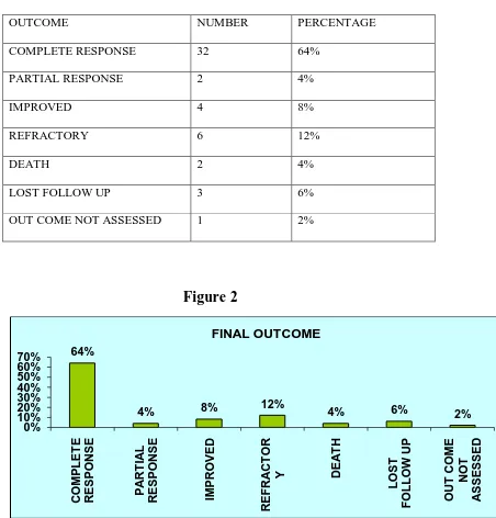
[image:47.612.98.512.301.440.2] [image:47.612.79.542.503.701.2]40 FINAL OUTCOME
The final outcome of these fifty patients were as follows.
Table 3
OUTCOME NUMBER PERCENTAGE
COMPLETE RESPONSE 32 64%
PARTIAL RESPONSE 2 4%
IMPROVED 4 8%
REFRACTORY 6 12%
DEATH 2 4%
LOST FOLLOW UP 3 6%
OUT COME NOT ASSESSED 1 2%
Figure 2
Three patients lost follow up and in one patient the outcome could not be assessed
as the patient was not willing for biopsy and was irregular in follow up.
64%
4% 8% 12% 4% 6% 2%
0% 10% 20% 30% 40% 50% 60% 70% C OM PL ET E R ESPON SE PA R T IA L R ESPON SE IM PR OVED R EF R A C T OR
Y DEA
[image:48.612.64.516.153.625.2]41 AGE DIFFERENCES IN OUTCOME
The mean age of patients who achieved complete response was 25.34 years
(16-47). The mean age of patients who were refractory or dead was 28.75 (range
20-41). On an average, patients who achieved complete response were 3 years
younger than those who remained refractory or died. On multistep logistic
regression analysis age emerged as an important risk factor influencing the final
outcome with p value of 0.047.
Figure 3
25.34
28.75
23 24 25 26 27 28 29 30
CR REF/ Dead
42 GENDER DIFFERENCE IN OUTCOME
Total number of females was 44 and males 6.The mean age of females was 25.27
and 26.67for males ( p value for the two samples 0.2529) .
Table 4
There is no statistically significant difference between the two groups hence
both sexes were matched equally for age.
36 ( 85%) of the females whose outcome was known achieved complete
or partial response or improvement. Only (50%) of males achieved CR. By Chi
squared test sex was an important factor in determining out come with p value
0.047.
FEMALES MALES
NO 44 6
MEAN
25.27 26.67
S.D
43 The outcomes according to gender is tabulated below
Table 5
(Chi square p 0.047 df=1)
Figure 4
0 10 20 30 40
CR/PR/IMP REF/DEATH 36
6
2 2
GENDER DIFFERENCE IN OUTCOME
FEMALES MALES
CR/PR/IMP REF/DEATH
FEMALE 36 6
44 DISEASE DURATION
The table below shows the distribution of duration of SLE at the
onset of lupus nephritis. The mean duration of illness was 23.3 months. Range
1moths to 84 months. 68% of patients presented within two years.
Table 6
Mean disease duration in patients who have achieved complete
response was 22.81 months while that of the refractory/death group was 33
months[ Range 1-84 months in both groups]. Patients who achieved complete
response had lesser disease duration of SLE (10.19 months) before the onset of
lupus nephritis than the other group. But this difference did not have any effect on
the outcome.
MONTHS FEMALE MALE TOTAL
<12 18 3 21
12—23 5 1 6
24—35 7 7
36—47 5 1 6
48—59 4 4
60—71 1 1 2
72—84 4 4
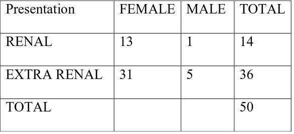
[image:52.612.127.487.258.528.2]45 PRESENTING SYMPTOMS
The following was the distribution of presenting symptoms at the onset of
lupus nephritis. Only 14 patients (28%) presented with renal symptoms. In others
lupus nephritis was asymptomatic and presented with extra renal symptoms.
Table 7
Figure 5
0 5 10 15 20 25 30 35
FEMALE MALE
13
1 31
5 PRESENTING SYMPTOMS
RENAL
Presentation FEMALE MALE TOTAL
RENAL 13 1 14
EXTRA RENAL 31 5 36
[image:53.612.161.454.263.397.2]46 Among those who achieved some response (CR/PR/IMP) 12
presented with renal symptoms and 26 with extra renal symptoms. By Chi square
test the presenting symptom did not have any effect on the outcome.
Table 8
(Chi squared test p value 0.71 df =1)
Figure 6
0 5 10 15 20 25 30
CR/PR/IMP DEATH/REF
12
2 26
6
RENAL NONRENAL
CR/PR/IMP DEATH/REF
RENAL 12 2
[image:54.612.73.546.358.614.2]47 EXTRA RENAL LUPUS
The most common extra renal disease activity affecting the study group was
neurologic involvement which was more in patients who were refractory or dead
than those who achieved CR/PR though the difference was not statistically
significant. Chi squared test p value- 0.27.
Table 9
Figure 7
13 5
25 3
0 5 10 15 20 25 30
CR/PR DEATH/R
EF
NEURO INVOLVEMENT
ABSENT PRESENT
NEURO
INVOLVEMENT CR/PR DEATH/REF
PRESENT 13 5
48 HYPERTENSION
Number of patients with hypertension was 16 ( 13 females, 3 males). The mean
systolic & diastolic BP for CR group was 125.6 & 84 mmHg respectively while
that of the refractory/death group was 137.25 & 92.5.Though by Chi squared test
there was no significant difference between the group (p Value 0.25 ).
Table 10
Figure 8
11
4 27
4
0 5 10 15 20 25 30
CR/PR/IMP REF/DEATH
HYPERTENSION
HT NON HT
CR/PR/IMP REF/DEATH
HT 11 4
[image:56.612.71.546.299.703.2] [image:56.612.141.406.306.408.2]49 COMORBIDITIES
The following were the comorbidities seen.
Table 11
S.No COMORBIDITY NUMBERS
1 HYPERTENSION 16
2 HYPOTHYROIDISM 8
3 INFERTILITY 5
4 DIABETES 3
5 PULM.TB 3
6 EXTRA PULM.TB 1
7 OVARIAN TUMOR 2
8 BENIGN ICT 1
9 PREGNANCY 1
10 POST PARTUM 1
11 NEPHROLITHIASIS 1
12 CATARACT 1
13 INFECTIONS 3
(osteomyl-1,
skin-1,
50 The independent samples test comparing the complete response group with the
refractory /death group with respect to various clinical parameters is given below.
Multistep logistic regression of the variables was also done.
Table 12
INDEPENDENT SAMPLE S TEST
t df Sig.
(2-tailed)
AGE Assumed -1.019 38 .314
not assumed -1.090 11.778 .297
DURATION Assumed -1.054 38 .298
not assumed -.842 8.731 .422
SLEDAI Assumed -3.132 38 .003
not assumed -2.304 8.270 .049
Hb Assumed -.380 38 .706
not assumed -.318 9.055 .758
PLATLET Assumed .222 38 .825
not assumed .232 11.416 .820
S.ALBUMIN Assumed .801 38 .428
not assumed .802 10.808 .440
S.CREAT Assumed -2.727 38 .010
not assumed -1.891 8.000 .095
GFR Assumed 2.870 38 .007
not assumed 3.433 14.124 .004
ESR Assumed .110 38 .913
not assumed .120 12.042 .907
URINE
PROTEIN Assumed -.599 38 .553
not assumed -.512 9.198 .621
TIME TO
CR Assumed -3.449 38 .001
51 Table 13
MULTI STEP LOGISTIC REGRESSION
B S.E. Wald df Sig. Exp(B)
Step 1a
AGE .276 .184 2.259 1 .133 1.318
DURATIO .042 .040 1.087 1 .297 1.043
SLEDAI .254 .115 4.876 1 .027 1.289
Hb .437 .468 .870 1 .351 1.547
PLATLET -2.280 2.043 1.246 1 .264 .102
SALB 1.432 1.462 .959 1 .327 4.185
CREATI -1.799 1.704 1.115 1 .291 .165
GFRI -.158 .111 2.013 1 .156 .854
ESR -.027 .040 .472 1 .492 .973
ur_INITIAL .714 .730 .957 1 .328 2.043
Constant -7.275 9.531 .583 1 .445 .001
Step 9a
AGE .158 .082 3.686 1 .047 1.171
GFRI -.064 .025 6.607 1 .010 .938
Constant -1.902 1.765 1.162 1 .281 .149
[image:59.612.71.483.131.595.2]52 SLEDAI
The mean SLEDAI was 19.78 (range 4-45). Mean SLEDAI for females was
18.95 and for males 25.33.
In patients who achieved CR, mean SLEDAI was 17.94 (range 4-30).
In refractory/dead group the mean SLEDAI was 26.88 (16-45).
Using Independent samples test SLEDAI was an important factor defining
the outcome [ p value 0.003].
Figure 9
17.4
26.88
0 5 10 15 20 25 30
SLEDAI
53
HEMOGLOBIN, PLATELET COUNT & SERUM ALBUMIN
The mean hemoglobin of the study group was 8.49 gm% (range 5-13.4).
The mean platelet count of the study group was 1.5lakhs/mm3. No. of
patients with platelet count less than 1 lakh was 9 out of 50.
The mean serum albumin value of the study group was 2.83g/dl (range
1.6-3.9).
By independent samples test there was no statistically significant
difference between the two groups with respect to hemoglobin, platelet count or
serum albumin. Hence these factors did not influence the outcomes in this study.
INITIAL PROTEINURIA:
The mean initial proteinuria for the CR group was 2.028 (0.3-5.7) and that
of the refractory,dead group was 2.383(0.2-5.2). There was no statistically
significant difference between the two groups.
54
SERUM CREATININE
The mean initial and final serum creatinine of the study group were
1.35 mg% and 0.95 mg% respectively.
The mean initial s.creatinine for the CR group was 1.216 mg%
(0.7-3.5) and that of the refractory,dead group was 2.275(range1.0-5). There was a
statistically significant difference between the two groups. By independent samples
test initial s.creatinine was found to be an important factor influencing the outcome
( p value- 0.010).
Figure 10
0 0.5 1 1.5 2 2.5
CR REF/ Dead
1.216
55 GLOMERULAR FILTRATION RATE
The mean initial and final GFR of the study group were 68.53 ml/min
and89.09 ml/min respectively.
The mean initial GFR for the CR group was 75ml/min (16.8-135.8) and
that of the refractory /dead group was 43.9 ml/min (14.7-74.4). There was a
statistically significant difference between the two groups. By independent samples
test initial GFR was found to be an important factor influencing the outcome ( p
value- 0.007). GFR was also an important factor in determining the outcome in
multistep logistic regression (p = 0.01).
Figure 11 75 43.9 0 10 20 30 40 50 60 70 80
CR REF/ Dead
in
ml
/mi
n
[image:63.612.75.544.434.643.2]56
TREATMENT PROTOCOLS
Patients who were treated with cyclophosphamide under NIH or ELNT
protocol, azathioprine and MMF achieved 80%, 75%, 100%, 80% response
respectively.The treatment protocols used for the induction of treatment did not
influence the outcomes.
Table 14
Chi squared test ‗p‘ value 0.37.
Figure 12 0 2 4 6 8 10 12 14 16
CYC NIH CYC ELNT AZA MMF
16 6 10 5 4 2 0 1 TREATMENT PROTOCOLS CR/PR/IMP REF/DEATH
CR/PR/IMP REF/DEATH
CYC NIH 16(80%) 4
CYC ELNT 6 (75%) 2
AZA 10 (100%) 0