A STUDY ON CLINICAL PROFILE OF CHRONIC OBSTRUCTIVE PULMONARY DISEASES AND CORRELATION OF HIGHLY
SENSITIVE C-REACTIVE PROTEIN WITH INCREASING
SEVERITY OF CHRONIC OBSTRUCTIVE PULMONARY DISEASES
DISSERTATION SUBMITTED FOR
DOCTOR OF MEDICINE
BRANCH - I (GENERAL MEDICINE)
APRIL 2013
THE TAMILNADU
DR.M.G.R.MEDICAL UNIVERSITY
CERTIFICATE
This is to certify that this dissertation titled “A STUDY ON
CLINICAL PROFILE OF CHRONIC OBSTRUCTIVE
PULMONARY DISEASES AND CORRELATION OF HIGHLY
SENSITIVE C-REACTIVE PROTEIN WITH INCREASING
SEVERITY OF CHRONIC OBSTRUCTIVE PULMONARY
DISEASES” submitted by Dr.R.VAIRAKKANI to the faculty of
General Medicine, The Tamil Nadu Dr. M.G.R. Medical University,
Chennai in partial fulfillment of the requirement for the award of MD
degree branch I General Medicine, is a bonafide research work
carried out by him under our direct supervision and guidance.
DR. M.NATARAJAN, M.D., DR. MOSES K. DANIEL, M.D.,
Professor of Medicine, Professor and Head
Chief, V Medical Unit, Department of Medicine,
Department of Medicine, Madurai Medical College,
Madurai Medical College, Madurai
DECLARATION
I, Dr.R.VAIRAKKANI, solemnly declare that the dissertation
titled “A STUDY ON CLINICAL PROFILE OF CHRONIC
OBSTRUCTIVE PULMONARY DISEASES AND
CORRELATION OF HIGHLY SENSITIVE C-REACTIVE
PROTEIN WITH INCREASING SEVERITY OF CHRONIC
OBSTRUCTIVE PULMONARY DISEASES” has been prepared
by me. This is submitted to The Tamil Nadu Dr. M.G.R. Medical
University, Chennai, in partial fulfillment of the regulations for the
award of MD degree (branch I) General Medicine.
Place : Madurai
ACKNOWLEDGEMENT
At the outset, I thank our Dean Dr. N.MOHAN,
M.S.,F.I.C.S.,F.A.I.S., for permitting me to use the facilities of
Madurai Medical College and Government Rajaji Hospital to conduct
this study.
I wish to express my respect and sincere gratitude to my
beloved teacher and Head of the Department of Medicine, PROF.DR.
MOSES K. DANIEL, M.D., for his valuable guidance and
encouragement throughout the study and also during my Post
graduate course. I owe my sincere thanks to him.
I also owe my sincere thanks to my unit chief and my guide
PROF.DR. M.NATARAJAN, M.D., for his guidance throughout the
study.
I express my special thanks to the Professor of Thoracic
medicine, PROF.Dr.C.RAMESH, M.D.,DTCD., for his valuable
guidance.
Dr.R.Balajinathan M.D., Dr.G.Bagyalakshmi M.D.,
Dr.J.Sangumani M.D.
I am extremely thankful to my unit Assistant Professors
Dr.G.Selvarani M.D., Dr.K.Muralidharan M.D., Dr.P.K.Ganesh
Babu M.D., Dr.Kasi Pandiyan M.D., Dr.Peer Mohammed M.D.,
Dr.Sakthi Mohan M.D., Dr.V.N.Alagavenkatesan M.D., for their
constant encouragement, timely help and critical suggestions
throughout the study and also for making my stay in the unit both
informative and pleasurable.
I sincerely thank the assistant professors and the staff of the
Thoracic Medicine department for their timely help and guidance.
I profusely thank the Biochemistry department for their
cooperation and support.
I extend my thanks to my family and friends who have stood by me
during my times of need. Their help and support have been invaluable
Finally, I thank all the patients, who form the most integral part of the
work, were always kind and cooperative. I pray for their speedy
CONTENTS
S
NO.
CONTENTS PAGE NO
1. INTRODUCTION 1
2. AIMS AND OBJECTIVES 3
3. REVIEW OF LITERATURE 4
4. MATERIALS AND METHODS 55
5. OBSERVATION AND RESULTS 58
6. DISCUSSION 74
7. LIMITATIONS OF THE STUDY 83
8. CONCLUSION 84
9. ANNEXURES
BIBLIOGRAPHY
PROFORMA
ABBREVIATIONS
MASTER CHART
ETHICAL COMMITTEE APPROVAL LETTER
INTRODUCTION
Chronic obstructive pulmonary disease, a disorder of expiratory
airflow has been projected to be the 3rd leading cause of mortality and 5th cause for disability as measured by DALY in 2020 by the Global burden of Disease study. It is one of the few diseases whose
morbidity and mortality are increasing at an alarming rate day by day
and imposing a heavy economic burden on the countries particularly
the developing countries like others. Reasons for this troubling
situation include the reduction in other cause mortality increasing the
longeivity of the population, increasing tobacco use & increasing
environmental pollution. The situation is so alarming that the disease
has achieved epidemic proportions. Many advances have been made
recently in the diagnosis and management of this disease but the hard
fact is that the disease is one of gradual progression, increasing
morbidity and incurable and the better way to handle this by
prevention by healthy life style, very importantly avoiding tobacco
use.
years have thrown much light on the systemic component of the
disease largely due to oxidant stress and which in addition to the lung
manifestations has been attributed to the extra pulmonary
manifestations such as coronary artery disease which is the leading
cause of mortality in these patients, osteoporosis, development of
diabetes, loss of muscle mass – these impose health burden on these
patients with already deranged lung function. As the pathophysiology
of the varied manifestations of the disease are being unraveled
everyday recent studies are on to find a unified marker which could
reliably predict the entire disease manifestations, disease course,
which could help in making treatment plan and also studies are on to
find a way of halting the unrelenting inflammation and disease
progression. Among the various markers being tested, one such is
highly sensitive C-Reactive Protein.
In the present study the baseline characteristics of our patients
are analysed and to possibly see whether highly sensitive C-Reactive
AIMS AND OBJECTIVES
The present study was done
1. To study the clinical profile of COPD patients with respect to their
baseline characteristics – age, sex, occupation, symptoms, signs,
X-ray, ECG, ECHO, BMI, pack years of smoking
2. To categorize the patients based on spirometry into different stages
and to study the distribution of the baseline characteristics in each
stage
3. To determine the value of hs-CRP in these patients and to correlate it
with the various stages, smoking pack years, FEV1 and other
variables and its significance
4. To study other markers of inflammation such as ESR, serum albumin
REVIEW OF LITERATURE
DEFINITION 4
COPD is defined as “a disease state characterized by airflow
limitation that is not fully reversible” 25,31
It includes
I. EMPHYSEMA4
Emphysema is defined as “destruction and enlargement of the
lung alveoli”
II. CHRONIC BRONCHITIS4
Chronic bronchitis is clinically defined with chronic cough and
phlegm production.
GOLD criteria for COPD severity4 GOLD
stage
Severity Symptoms Spirometry
0 At risk Chronic cough,
sputum production
Normal
I Mild With or without
chronic cough or sputum production
FEV1/FVC < 0.7 and FEV1 ≥ 80% predicted
II Moderate With or without
chronic cough or sputum production
FEV1/FVC < 0 .7 and 50% ≤ FEV1% < 80% predicted
III Severe With or without
chronic cough or sputum production
FEV1/FVC < 0.7 and 30% ≤ FEV1% < 50% predicted
IV Very severe With or without
chronic cough or sputum production
FEV1/FVC < 0.7 and FEV1 < 30% or FEV1% <50% predicted with respiratory failure or signs of right heart failure
EPIDEMIOLOGY 29,30
PREVALENCE IN INDIA
The exact prevalence in our country could not be ascertained
with certainty because of misdiagnosis, under assessment, lack of
extensive studies, poor statistical information. The prevalence rate
have been variably reported from 2-22% in males and 1.2-19% in
females. Recently ICMR has undertook the INSEARCH study in four
cities and reported prevalence of 5% in males and 3.2% in females in
those more than 35 years of age. The total population affected by the
disease has increased to 14.84 million in 2011 from 6.45 million in
1971. In India the sex ratio and the smoker to non smoker ratio are
not as high when compared to western statistics. This disparity is
because of biomass fuel combustion which is an important risk factor
in women more so in the villages. Data on mortality statistics are
limited; 7% mortality have been attributed to chronic respiratory
illness.29,30
RISK FACTORS
SMOKING 4,5,6,11,25,30,31,35
Cigarette smoking by far is the major cause for COPD with
smokers developing the disease. The effect of smoking on declining
lung function have been proved in many studies. But still not all
smokers develop the disease and even among those who smoke there
is variation in response to the duration of smoking which suggests
that other factors particularly genetic and environmental modulate the
effect of smoking in these patients.
AIRWAY RESPONSIVENESS 4,11
Though the concept of airway hyperresponsiveness have been
proved beyond doubt in bronchial asthma, in COPD there are
conflicting reports as to whether it contributes to the development of
airflow obstruction with recent studies supporting the hypothesis.
RESPIRATORY INFECTIONS 4,30 Infections associated with risk
a. Previous H/O tuberculosis even when adequately treated with ATT
b. Childhood respiratory infections 43
c. Inadequately treated bronchial asthma
INDOOR AIR POLLUTION 4,29,30
contribute to 1/3 to ½ of all cases. Among them chronic exposure to
sulfur dioxide, carbon monoxide, nitric oxide, HCHO and others
released from biomass fuel combustion38 is the important risk factor particularly in female patients.
OCCUPATIONAL DUSTS 4,30
Several occupations are met with chronic exposure to
occupational dust/gas but the impact of these agents in the
development of the disease is much less significant than cigarette use.
INCREASING AGE : 30
Physiological decrease in pulmonary function could also lead to
the disease.
LOW SOCIO-ECONOMIC STATUS 29,30
GENETICS 4,5,6,10,11,30
The considerable variation in smokers developing the disease
could possibly due to various genetic derangements. As of now, the
only proven genetic defect causing the disease is α1-antitrypsin
deficiency which is due to alteration in SERPINA1 locus encoding
Allele α 1 AT
M Normal
S Slightly reduced
Z Markedly reduced
Null Absent
PiZ - most common form.
Treatment for this subset of patients is available as α1-AT
augmentation therapy as weekly intravenous administration.
Apart from α1-AT deficiency, other genetic factors have also
been hypothesized to contribute to the risk factor pool and research on
several other genes are underway.4
PATHOPHYSIOLOGY
AIRFLOW LIMITATION 4,11,30
Airflow limitation due to decreased airway caliber and
increased airway resistance, impaired elastic lung recoil during
expiration manifested in spirometry as reduction in the ratio of Forced
bronchodilator FEV1% predicted value is a cardinal feature for the
disease diagnosis and dividing into stages.
HYPER INFLATION 4,11
In these patients there is hyper inflation as shown by increased
RV and increased ratio of RV/TLC and later increase in TLC.
Mechanism 30
The principal mechanism of airflow during expiration from the
small airways and alveoli is driven by the inward elastic recoil
pressure of the lung which also counteracts thoracic wall outward
pressure. At the end of tidal expiration both these balance each other
resulting in particular amount of air remaining in the lungs named as
FRC (functional residual capacity). In these patients due to the
destruction of lung parenchyma, the inward pressures are low and it
comes into equilibrium with the outward pressure at increased
volumes of FRC resulting in hyperinflation. This state of
hyperinflation also causes flattening of diaphragm resulting in
GAS EXCHANGE 4
Hypercapnea and hypoxia results from deranged gas exchange
due to the reduction in surface area caused by destruction of the
respiratory units. The chronic hypercapnic state decreases the
chemoreceptor sensitivity and hence the respiratory stimulus in these
patients is hypoxia stimulating the peripheral chemoreceptors.30
V/Q MISMATCH 4
It is due to the inhomogeneous disease distribution in the lungs
and airways
PATHOGENESIS
ELASTASE-ANTI ELASTASE HYPOTHESIS 4,10,11 α 1 – AT DEFICIENCY:
Neutrophils in the alveoli are stimulated by various triggers
which get activated and recruited and release their granules which
have elastase, proteinases, cathepsins & MMP and also releases ROS
which inhibit α1 anti trypsin. In patients with the enzyme deficiency,
Smoking
In smokers both neutrophils and macrophages are increased in
alveoli. Neutrophils upon activation releases a number of proteases
mentioned above and activated macrophages release a variety of
cytokines, chemokines, ROS and a variety of matrix
metalloproteinases which are not inhibited by α1 AT rather these
enzymatically degrade α1 AT and thus enhancing pulmonary
destruction several fold.10 CD8 lymphocytes enhance apoptosis of alveoli and CD4 cells trigger autoimmunity against native lung
tissue.30
OXIDANT STRESS
The normal lung contains rich anti-oxidants such as glutathione
to handle the oxidant stress. Smoking by way of its contents and also
by activating inflammatory cells which release free radicals tilt the
balance towards oxidant stress which inactivates anti-proteases
causing lung destruction in patients even with normal α1 AT. Oxidant
stress also results in loss of surfactant, ECM apoptosis, reduction in
CLINICAL PRESNTATION
HISTORY
SYMPTOMS 4,5,6,11
i. Chronic cough
ii. Expectoration
iii. Dyspnoea on exertion – insidious in onset and gradually progressive
over time. Sudden worsening of dyspnoea is due to either an
exacerbation or other complications.
iv. Wheeze
v. Loss of weight – in severe disease. (other causes to be ruled out
before attributing it to the disease per se.)
SIGNS : 4,5,6
The physical examination may be completely normal in patients in the
beginning stages till there is significant deterioration in pulmonary
function.
GENERAL EXAMINATION
c. Clubbing is not a sign of the disease per se and if present a prompt
search for other conditions such as lung Ca has to be made.
d. Pedal edema, ascites, elevated JVP – due to pulmonary hypertension
& cor pulmonale.
e. SPO2 – patients may have hypoxemia.
RESPIRATORY SYSTEM EXAMINATION
a. Patients with severe disease have accessory muscles of respiration
acting with the patient assuming “tripod” position to enhance the
synergistic action of these muscles 4
b. Hoover’s sign 4
c. Signs of hyperinflation – Barrel shaped chest, hyper resonance on
percussion.4
d. On auscultation patient may have prolonged expiratory phase,
diminished intensity of breath sounds, expiratory wheeze.4,6
e. Signs of pulmonary hypertension and cor pulmonale – Loud P2 may
not be present due to hyperinflation, Tricuspid regurgitation murmur
DIFFERENTIAL DIAGNOSIS 5
a. Bronchial asthma – distinguished by near total post bronchodilator
FEV1 reversibility.
b. Bronchiectasis – differentiated by symptoms like hemoptysis,
presence of clubbing & radiography.
c. Cystic fibrosis – history since childhood, recurrent infections,
presence of other features such as cirrhosis.
d. Broncho pulmonary mycosis.
e. Central airway obstruction such as tumours, stenosis or
developmental anamoly – differentiated by history and PFT using
flow-volume loops.
COMPLICATIONS 5,30
i. Recurrent lung/airway infection
ii. PTE
iii. Cor pulmonale and pulmonary hypertension
iv. Bronchogenic carcinoma
RADIOGRAPHY
X-RAY CHEST 4,5
i. Chronic bronchitis – Increased bronchovascular markings
ii. Emphysema – Hyper inflated lung fields
Diaphragm flattening
Decreased peripheral vascular markings
Bulla
iii. Pulmonary hypertension – enlarged main & right descending
pulmonary artery
iv. To look for co morbidities and complications like malignancy,
pneumothorax, etc.
COMPUTED TOMOGRAPHY 4
a. Not routinely done for diagnostic purposes
LABORATORY TESTS
PFT : 4,5,6,31
a. FEV1/FVC < 0.70
b. TLC, FRC, RV may increase often more than normal with
disease progression.
Apart from diagnostic utility, FEV1 is also used to monitor
treatment response and worsening FEV1 is a poor prognostic factor.
ABG : 4,6
a. Normal in early stages
b. To determine the degree of hypoxemia and the acid base status and to
differentiate between acute exacerbations and chronic respiratory
failure.
c. Yearly assessment is recommended.
TREATMENT
STABLE PATIENTS
Survival improving strategies 4,5,6,11,30,35
3. Lung volume reduction surgery
Other management strategies are to the relief of symptoms,
improving the quality of life and to reduce exacerbations. 4
DRUGS
BRONCHODILATORS 4,5,6,11,30
These drugs are the mainstay of management in that they
provide symptom relief and enhances the well being of patient but do
not prevent the decline in FEV1.
Inhaled β2 agonists :
i) short acting β2 : used in all stages of the disease ‘on demand’ basis
to provide immediate relief from dyspnoea and not to be used on a
routine basis.
Drugs used are salbutamol and levosalbutamol. 30
ii) long acting : used on a scheduled basis as maintenance therapy to
provide sustained relief.
Drugs commonly used are formoterol, salmeterol.
Inhaled Anti-cholinergics :
i) short acting : For symptom relief similar to short acting beta 2
agonists but devoid of their sympathetic side effects and also has
prolonged action compared to them.
Drug used ipratropium
ii) long acting : for maintenance therapy
Side effects : dryness of mouth, Primary angle closure glaucoma is an
absolute contra indication, symptoms of BPH may aggravate.
COMBINATION THERAPY 30
As the disease worsens, combination therapy with Long acting
sympathetic and anticholinergic provides maximum symptom benefit
by synergistic mechanisms rather than single drug alone.
CORTICOSTEROIDS
INHALED :
ICS are not to be used as a single agent in management of these
Indication : 30
i. Stage III disease
ii. ≥2 exacerbations / year
Side effects : 30
1. oral candidiasis – prevented by mouth gargling after each use or by
spacer
2. hoarseness of voice
SYSTEMIC STEROIDS 4,5,6,30
Oral steroids are not used in the regular treatment of stable
patients because the adverse effects outweigh the benefits but they are
an important part of acute exacerbation management.
METHYL XANTHINES 5,6,30
Sustained release theophylline is commonly used in patients
who do not have adequate symptom relief with inhalational therapy
because of its effects on diaphragm function, reducing airway
resistance and decreasing inflammation.
Side effects : 6,30
These drugs have a narrow therapeutic window and patients’
CVS : increase in heart rate, arrhythmia.
CNS : tremor, decreases seizure threshold, insomnia.
GIT : gastritis, nausea.
ANTIBIOTICS - No role in stable patients. 5,30
NON PHARMACOLOGICAL MEASURES
OXYGEN 4,5,6,30
Oxygen therapy by various methods undoubtedly prolongs
survival and also provides symptom relief.
Indications 6,42
Arterial partial pressure of O2 ≤ 55 mm Hg or oxygen
saturation by pulse oximetry ≤ 88%
Arterial partial pressure of O2 < 60 mm Hg and oxygen
saturation < 90% in the presence of pulmonary
hypertension, Hct > 55% or cor pulmonale.
VACCINATIONS 6,30,34
For all stages of the disease yearly influenza vaccine and
PULMONARY REHABILITATION 4,5,6,30
It is a multi system modality including respiratory exercises,
adequate nutrition, psychological support that improves exercise
tolerance, improves dyspnoea and quality of life indicated in patients
failing optimal medical therapy and also for severe disease.
SURGICAL MANAGEMENT
I. LUNG TRANSPLANTATION 4,5,6
a. Done either as one lung or sequential double lung transplant
b. Requirements include severe disease inspite of optimal medical
management, absence of other organ system dysfunction, poor quality
of life.
c. PHT and the region of lung involved are not contra indications.
II. LUNG VOLUME REDUCTION SURGERY 4,5,6
Patients with upper lobe emphysema and severely compromised
exercise tolerance are ideal candidates. Presence of PHT/cor
III. BULLECTOMY 5
EXACERBATIONS OF COPD
PRECIPITATING CAUSES 4,30
I. Infections
i. Viral infections
ii. Bacterial infections
a. Pneumococcus
b. Hemophilus influenza
c. Moraxella catarrhalis
d. Pseudomonas in special risk groups
II. Environmental
i. Pollution
ii. Exposure to toxic gases
iii. Climate changes
SYMPTOMS 4,30
iii. Increased sputum with purulence
iv. Worsening general condition
Complete physical examination is mandatory to assess the
severity of the exacerbation. Investigations done include X ray chest
to detect the presence of pneumothorax, consolidation, ECG to detect
rhythm disturbances- most common being MAT, ABG to detect
respiratory failure. The decision as to whether the patient has to be
treated at home or hospital or ICU is based on the severity, patient’s
general health status, supportive care, presence of other co
morbidities like CAD, DM, neurological status, ABG measurements
and the need for assisted ventilation. 4,6,30
DRUGS
BRONCHODILATORS
Short acting beta agonists are used as the primary treatment for
an exacerbation, alone or in addition with an anti cholinergic
delivered through nebulisation or inhalation if patient can perform.6 If patient is already taking methyl xanthines it should not be stopped as
In theophylline naïve patients it should not be prescribed during an
exacerbation. 5,6
GLUCOCORTICOIDS
Systemic steroids via oral or parenteral if patient cannot take
medication orally for short course is an important cornerstone in
management of exacerbation because they decrease hospital stay,
improves lung function and decreases the probability of subsequent
relapse. 4,6,30
ANTIBIOTICS
They are chosen based on the sensivity pattern, the likely
organism and availability and is particularly useful in patients with
purulent sputum production and in those requiring assisted
ventilation. 4,6
SUPPLEMENTAL OXYGEN
To maintain SaO2 ≥ 90%.6 Oxygen should not be withheld for
fear of respiratory depression due to removal of hypercarbic stimulus
because it is essential to prevent tissue hypoxemia but care should be
ASSISTED VENTILATION
Non invasive ventilation indicated in patients with respiratory
failure has shown to decrease mortality, decrease hospital stay, the
need for invasive ventilation and lesser risk of hospital acquired
infections. Invasive ventilation is indicted when NIV fails or patient
has contra indications to NIV or severe acidosis or hypercapnea or
the presence of co-morbidities.4,5,6,30
PULMONARY HYPERTENSION IN COPD :
Mild PHT to some extent develops in most of the patients with
advanced disease and rarely severe pulmonary HT in few. 7,37
CAUSES 7
1. Pulmonary vasoconstriction caused by alveolar hypoxia,37 acidosis, hypercapnia.
2. Increased lung volume causing compression of Pulmonary vessels
3. In patients with emphysema due to terminal air space destruction
there is a decrease in the small vessels in areas of destroyed lung
Pulmonary artery pressure increases by approximately 20 mm
Hg for every acute exacerbation and this can end up in pulmonary
hypertension on repeated occurrence. 9,30
MECHANISM OF PULMONARY HYPERTENSION BY
HYPOXIA: 7
1. Vascular intima thickening
2. Distal vessels muscularisation
3. Hypertrophy of the vascula media of proximal vessels
4. Alveolar hypoxia in order to maintain V/Q and PaO2 is a potent
vasoconstrictor in pulmonary circulation.
TREATMENT 7,44
A.Pulmonary vasodilators produce worsening of V/Q mismatch because
it is not the level of pulmonary hypertension but the degree of
hypoxia which causes the clinical symptoms and that these drugs
further worsen the situation.
B. Only proven treatment of use is O2 which decreases both morbidity
C. To maintain the Hb in upper limit of normal because low Hb is not
tolerated in these patients due to hypoxemia.
Nocturnal or ambulatory SaO2 can guide in optimal O2 concentration.
COR PULMONALE ( RIGHT HEART FAILURE ) : 44
It can occur either acutely during exacerbations or chronically
due to disease progression and worsening gas exchange resulting in
irreversible vascular remodeling.
DIAGNOSIS
i) Echocardiogram
To detect the presence of right ventricular enlargement but the
procedure is difficult due to hyperinflated lungs and rotation of the
heart.8 It can be supplemented by ABG-showing PaO2 < 50 mm Hg
and PCo2 > 50 mm Hg. 44
ii) Electrocardiogram 44
Routine criteria to detect RVH cannot diagnose RVH in these
patients due to
b. Hyper inflation causing increased distance between the skin
and cardiac surface.
c. It is more of RV dilatation rather than hypertrophy.
iii) Cardiac catheterization – gold standard investigation. 44
MANAGEMENT 44
1) Acute cor pulmonale
i) treatment of the precipitating cause
ii) supplemental O2 to maintain adequate oxygenation.
2) Chronic cor pulmonale
i) cautious use of digoxin and diuretics for the fear of precipitating
life threatening rhythm disturbances in the presence of hypoxia and
acidosis.
ii) Oxygen.
SPIROMETRY IN COPD
The investigation to confirm or refute a diagnosis of the disease
is spirometric evaluation as suggested by GOLD. According to them a
absence of post bronchodilator reversibility has been abandoned.
Spirometry has its own limitations and hence a combination of
clinical symptomatology and spirometry improves the diagnostic
accuracy. 30
USES 30
1. Diagnosis
2. Grading of severity
In these patients the rate of decrease in Forced expiratory
volume1 per year is about 75-100 ml in sharp contrast to normal
persons’ 30 ml/year.
3. Lung “age” assessment
By comparing patient’s FEV1( lung age) to the predicted FEV1
of his age matched control. It could be used to encourage smokers to
quit smoking.
4. Detection of Upper airway obstruction
5. Pre-Operative evaluation
These patients have a higher chance of developing post-op
i) Thoracic surgery
Predicted Post operative FEV1 is calculated by applying “rule
of 5” in which 1/5th
function is contributed by each lobe and the PPO
value is calculated by subtracting the lobe to be removed. A PPO
FEV1 < 40% predicted is a contra indication to surgery.
ii) Non thoracic surgery
Factors predicting post-operative complications are
a. FEV1/FVC <50%
b. Maximum voluntary ventilation < 50%
c. FEV1 or Diffusing capacity < 20% predicted
d. High partial pressure of carbon dioxide
e. Short distance of surgical site from the chest
SYSTEMIC INFLAMMATION 30
It has been recognized from the experiences in past decades that
inflammation is no longer confined to lungs in COPD and that GOLD
definition of the disease at present includes “some significant extra
patients” and hence the disease could be renamed as “chronic
systemic inflammatory syndrome”.
MARKERS OF SYSTEMIC INFLAMMATION IN COPD 30
1. hsCRP
2. Fibrinogen
3. Ferritin
4. White blood cell count
5. ROS
6. Interleukins
7. TGF β1
8. TNF α receptor polymorphism
These markers are present even in mild forms of the disease and
their levels increase with increasing disease severity.
HYPOTHESIS 30
1. Probable inflammation spreading over from lungs to other systems.
2. A proinflammatory phenotype in which systemic inflammation
TRIGGERS 30
1. Smoking an important risk factor for the disease per se by way of
causing oxidative stress & endothelial function derangement
2. Hypoxia of severe disease enhances HIF-1 expression.HIF-1
upregulates genes involved in inflammation, vascular remodeling &
new vessel formation. TNF-α levels have been shown to be in relation
with hypoxemia severity and that the enhanced survival of patients on
LTOT might be due to the fact that oxygen decreases inflammation.
3. Increased leptin levels and its receptors
4. Auto-immune process
5. Oxidative stress of the disease accelerates telomere shortening and
causes “ageing” of lungs and other systems.
CONSEQUENCES 30
Consequences of the systemic inflammatory process are the non
pulmonary morbidities that have been shown to have immediate
cause-effect relationship across studies consistently and also have
1. CARDIO VASCULAR SYSTEM : 30
i) Coronary Artery Disease:
These patients have a 3-fold greater risk of developing CAD
and that the mortality rates in these patients due to CAD are
comparable to those due to worsening of the disease per se. Cardiac
injury biomarkers have been shown to be increased in studies during
acute exacerbations when the level of systemic inflammation is high.
ii) Cardiac failure:
Cardiac failure is precipitated by end diastolic pressure changes
induced by pressure variation caused by hyper inflation which
interferes with ventricular remodeling. Cardiac failure and air flow
limitation is a vicious cycle – one perpetuating the other in that
airflow resistance is worsened by the electrolyte changes, mucosal
congestion & decreased lung compliance in heart failure.
iii) Arrythmias:
The common rhythm disturbances include
a. Mutifocal Atrial Tachycardia
b. Atrial Fibrillation
2.BONE: 30
The prevalence of osteoporosis is high in these patients
independent of glucocorticoid use. Inflammatory mediators such as
TNF-α and IL-1 stimulates mesenchymal cells to release receptor
activator of NFКB ligand which mediates macrophage transformation
to osteoclasts & accelerates osteoporosis. Other factors such as old
age, decreased BMI, smoking and sedentary life style may contribute.
3. DIABETES MELLITUS: 30
Undoubtedly there is an increased prevalence of T2DM in these
patients due to systemic inflammation which is strenghthened by the
finding of elevated levels of inflammatory markers such as tumour
necrosis factor α, interleukin 6 & C-reactive protein which further
compounds the risk of developing CVD.
4. MUSCULOSKELETAL SYSTEM : 27,30
Progressive muscle wasting with preserved fat mass is a feature
of the disease.
Mechanism
Endocrine abnormalities
i. Decreased Insulin like Growth Factor-1 expression and its
binding proteins
ii. Decreased testosterone
iii. GH resistance
Hypoxia
Muscle dysfunction-respiratory and non-respiratory muscles
adversely affect the exercise tolerance & health status of the patient &
the loss of skeletal muscle is an important negative predictive factor
for survival in these patients and also the BODE index – validated
index incorporates BMI as one of its four parameters predicting
prognosis of the disease.
5. DEPRESSION : 30
Increased prevalence due to
i. Systemic inflammation
ii. Dependence due to increasing disability
NEED FOR IDENTIFYING CONSEQUENCES: 30
A knowledge of the systemic consequences enlightens us the
need for investigating these patients for the comorbidities and to
intervene early so as to decrease the morbidity and mortality and
improving the QOL and also to look for the disease in patients
presenting with any of these co-morbidities.
C-REACTIVE PROTEIN
It is an acute phase reactant produced by the hepatocytes and to
some extent by vascular endothelial and smooth muscle cells in
response to inflammatory mediators released by macrophages and
adipocytes. It belongs to the pentraxin family of proteins. The name is
derived from its finding as an agent in patients with inflammation that
reacted with the C-polysaccharide of Streptococcus pneumoniae. The
gene encoding is on chromosome 1q21-q23. 16,41
FUNCTION
BENEFICIAL EFFECTS
It binds to phosphocholine exposed on micro organisms and
NEGATIVE EFFECTS
CRP is a pro-inflammatory agent. It directly activates the
complement cascade by binding to C1q causing tissue destruction. It
also enhances NFКB to induce the synthesis of various cytokines
perpetuating inflammation which when unchecked results in
extensive host tissue damage.16
CLINICAL IMPLICATIONS
CRP is increased in acute and chronic inflammation, auto
immune diseases, malignancy, and conditions associated with tissue
damage.13 It is a more sensitive and more accurate marker of acute inflammation than ESR. The degree of increase correlates with the
severity of inflammation. It returns to normal with subsidence of
inflammation rapidly with a t1/2 of 18 hrs and hence can be used to
monitor response to treatment.
Value of Highly Sensitive C-Reactive protein above 3 mg/L is
considered abnormal. 41
Highly sensitive methods for assaying CRP known as hs-CRP
was developed to enhance the sensitivity of detecting the low grade
assays could not detect. Highly Sensitive C-Reactive protein is a
predictor of CVA, sudden cardiac death, CAD and peripheral vascular
diseases.13 It is a prognostic indicator and mortality predictor in acute coronary syndrome. 41
Highly Sensitive C-Reactive protein in COPD:
MECHANISM OF ELEVATION
Interleukin 6 is the important molecule that upregulates the
hepatic production of C-Reactive Protein. 15,16,17
SOURCE OF INCREASED INTERLEUKIN 6 :
i. Interleukin 6 may be released by the activated inflammatory cells –
macrophages in the pulmonary circulation and alveolar epithelial
cells. 14
ii. Oxidant stress induced derangement in muscle metabolism
homeostasis causing enhanced expression of interleukin 6. 40
The increased plasma levels of IL-6 stimulates the hepatocytes to
CLINICAL IMPLICATIONS
Highly Sensitive C-Reactive protein
i. Is increased even in stable patients 16,17,18
ii. Predictor of mortality 14,18,40
iii. Increased significantly in patients with low body mass index than
others signifying malnutrition
iv. Marker of low grade systemic inflammation in these patients 13,16,18,40
v. Marker of increasing severity of the disease
vi. Predicts exacerbations, hospitalization 14,18
vii. Negative correlation with decline in lung function 12,16,18,21,24,28,40
viii. Elevated levels are associated with poor quality of life & reduced
exercise tolerance. 14,18
SMOKING CESSATION 4,30
Smoking cessation is by far the best effective intervention in
halting the disease process.
Approach
a. Counseling
CONSELLING
Counseling either by the clinician or non clinician enhances
cessation rate several fold than self initiated attempts.
5A intervention approach
I. ASK – information about tobacco use at every visit.
II. ADVICE – should be strong and individualized depending on
patient’s health status and other considerations.
III. ASSESS – patient’s willingness. If patient is not willing he/she
should be urged to quit.
IV. ASSIST – by discussing with the patient a quit plan; setting a date to
stop smoking in next 2 weeks, to reduce alcohol use, educate
households and providing psycho social support.
V. ARRANGE – follow-up.
PHARMACOTHERAPY 4,6,30
First line agents
Nicotine Replacement Therapy
Dose : 2 mg – 1-24 cigarettes/d ; 4 mg - >25 cigarettes/d
Duration : 12 weeks
Adverse effects : dyspepsia
Lozenge
Dose : 2 mg – first smoke > 30 min after waking up from bed; 4 mg –
first smoke < 30 min after waking up from bed
Maximum of 20 mg/d
Duration : 12 weeks
Side effects : nausea, insomnia.
Inhaler
Dose : 6 – 16 cartridges of 4 mg each/d
Duration : 6 months with dose reduction in last 3 months
Side effects : rhinitis, local irritation
Spray
Dose : 0.5 mg in each nostril 1-2 doses/hr
Maximum 5/hr
Side effects : local irritation
Patch
Dose : 16 or 24 hr patch for total duration of 8 weeks with dose
reduction
Side effects : local skin reaction, insomnia
Advantage : good compliance, requires less skill.
Contra indication to NRT :
Coronary artery disease – MI or unstable angina
Cerebrovascular accident
Untreated Acid peptic disease
Bupropion sustained release
Mechanism of action
Nor-epinephrine and dopamine reuptake inhibitor
Dose : 150 mg once daily x 3 days; 150 mg bd x 7- 12 weeks
Duration : 6 -12 months
Particularly preferred in patients with concomitant depression
Varnecline
Mechanism of action
Partial agonist of nicotinic Ach receptor
Dose : 0.5 mg od x 1-3 days; 0.5 mg bd x 4-7 days; 1 mg bd from
then up to 8 weeks.
Side effects : CNS – suicidal intention which has resulted in FDA
warning.
Second line agents
1. Clonidine :
Mechanism of action : post synaptic α2 agonist
Dose : 0.1 – 0.4 mg/d x 2 -6 weeks
2. Anxiolytics
Diazepam, buspirone, beta blockers
3. Sensory replacement
Citric acid inhaler, denicotinised tobacco, black pepper extract
Antagonists
a. Mecamylamine
non competitive CNS and PNS nicotine receptor antagonist
b. Naltrexone.
Aversion causing drugs
Silver acetate – poor compliance
SMOKING PREVENTION 4,30
Smoking behavior in around 90% smokers were initiated during
adolescence. So prevention is more important and effective than
cessation after the behavior has begun.
Measures :
1. Health education – target population : adolescents, young adults
2. Public health programs
3. Smoke free public places.
EMERGING THERAPIES
Newer drugs have been developed with the aim of inhibiting
Oxidative stress – imbalance between pulmonary oxidant load
and anti-oxidants causing inflammation and airway modeling – an
important pathogenetic mechanism – probably the reason for steroid
resistance has been the target for newer therapies. 30
SMOKING CESSATION
An important and effective intervention to arrest the decline of
lung function is smoking cessation.
Recent research has developed anti-free nicotine antibodies
which bind with free nicotine denying its access across the BBB and
hence cannot stimulate nicotine receptors causing smoking an
unpleasant experience. 30
NEWER BRONCHODILATORS
Bronchodilators though they do not arrest the disease process –
provide immediate symptom relief and improves well being of the
patient are an important component of treatment plan.
Newer long & ultra long acting bronchodilators have been
developed to meet the short comings of short acting ones – poor
compliance due to multiple dosing. These include ultra-long acting
ULTRA-LONG ACTING MUSCARINIC ANTAGONISTS
These agents besides providing long acting bronchodilatation
have been shown to decrease the number of exacerbations and so they
have the ability to attenuate the disease process partially and hence
provide mortality benefit. 30
Drugs under research:
i. Aclidinium
ii. Dexpironium
iii. TD-4208
iv. Daratropium bromide
v. GSK-573719
vi. Glycopyronium bromide
ULTRA-LONG ACTING β2 AGONISTS 30
This includes
i. Indacaterol – approved in Europe & USA
ii. Vilanterol – safe & efficacious
iv. Milveterol – proved efficacious in asthma, not yet researched on
COPD
v. GSK-642444
vi. UK-503590
vii. BI-1744-CL
viii. Compound X
NOVEL COMBINATIONS
Rationale of combing long acting drugs
a) Synergism between the two agents
b) Patients based on whether beta or muscarinic receptor predominance
in their airway respond to their respective drugs more than the other
and hence their combination can overcome their problem of receptor
variation.
Several combination of these agents along with steroids to
decrease exacerbations are under development but a potential
limitation of using these combination is delivery of these agents at
different site which decrease their synergism. To overcome these
agents at same site – dual acting muscarinic antagonist beta 2 agonist
have emerged.
Other combinations under development are combining a LABA
with an ICS or combining LABA with inhaled steroid and LAMA. 30
ANTI INFLAMMATORY DRUGS
The search for an effective and safe anti inflammatory drug
with the potential of reducing lung inflammation over the decades
have been in vain. One of the recent break through finding is that
oxidative stress causing decrease in HDAC in these patients has been
the reason for corticosteroid unresponsiveness in them and that this
enzyme at cellular levels can be increased by theophylline. Hence
theophylline has staged a comeback in management of the disease
with the potential of additive anti-inflammatory effect when given
with steroids and studies are underway on this aspect. 30
PDE-4 INHIBITORS 30
Phosphodiesterase-4 an enzyme found specifically in most
inflammatory cells have been targeted to reduce the inflammatory
tiotropium. These drugs could be the best adjuvant therapy with
bronchodilators. An inhalational form of this class is under study. But
the side effects of this drug – gastrointestinal and upper respiratory
tract effects have led to search for development for specific inhibitors.
ANTI PROTEASES 30
An imbalance between proteases (matrix metallo proteinases) –
anti proteases is an important pathogenetic mechanism of
emphysema. Anti proteases – MMP inhibitors have shown promising
results in preventing emphysema. Few drugs of this class are under
phase II and III trials.
1. Marimastat – non selective MMP inhibitor
2. AZ11557272 – dual MMP-9/MMP-12 inhibitor
CYTOKINE INHIBITORS 30
Tumour necrosis factor – α and other interleukins – notably
IL-7, IL-1β, IL-6 are important cytokines implicated in the systemic
inflammatory process in the disease. Drugs targeting these cytokines
CHEMOKINE ANTAGONISTS 30
Oral AD28309 – CXCR1/2 receptor antagonist – the receptor
which mediates the effects of chemokines on inflammatory cells has
shown to reduce inflammation in man. Other receptors targeted are
CXCR 3 and 5 and drugs involved in these have finished phase I
trials.
TGF-β INHIBITORS 30
SD-280 which inhibits the fibrogenic cytokine TGF-β causing
small airway fibrosis resulting in decrease in FEV1 and reduced
exercise capacity has been developed but the long term consequences
of this class are yet unknown.
NFКB INHIBITORS 30
Inhibition of NFКB – a transcription factor involved in
upregulating the production of various chemokines, TNF-α and
MMP-9 seems to be a potential option. Such inhibitors are presently
under research.
P38MAPKinase INHIBITORS : 30
PHOSPHOINOSITOL-3-KINASE INHIBITORS under research. 30
PPAR AGONISTS 30
Immunomodulatory effects of PPAR α and γ (also inhibits
TGF-β ) are being exploited and the drugs currently being studied are
rosiglitazone and SB-219994.
ANTIOXIDANTS 30
Anti oxidant drugs being used are
I. NAC and its derivatives
a. N-Acetyl Cysteine
i. Decreases systemic and pulmonary oxidative stress
ii. Mild bronchodilation
iii. Halts decrease in FEV1
iv. Reduces exacerbation
b. N-acestelyn – well tolerated
c. Erdosteine – added advantage of reducing bacterial adhesion
thereby reducing exacerbations.
d. Carbocysteine
f. N – isobutyryl cysteine – less effective
II. NRF-2 Activators
STATINS 30
The pleomorphic action of statins apart from lipid lowering
such as anti-oxidant, endothelial protection and anti inflammatory
properties in retrospective analysis have shown to decrease mortality,
reduce exacerbations, decreasing need for assisted ventilation,
improving functional capacity and reduce FEV1 decline. But
randomized controlled trials are required to confirm their
effectiveness before being part of management plan of the disease.
REGENERATIVE THERAPIES
Currently research is focusing on a modality of treatment that
could actually bring back the destroyed lung tissue and hence
restoring the normal anatomy which in future if discovered can
revolutionize the COPD management. 30
STEM CELLS
Allogenic mesenchymal stem cells have the prospect of
these cells resulting in death is an undesirable outcome. Trials are
currently under research to overcome this side effect. 30
RETINOIC ACID
ATRA can cause regeneration of distal respiratory unit and
hence reverse emphysema but in clinical trials have not shown such
benefits. 30
ANTI-AGEING THERAPY
Oxidative stress causing accelerated ageing by increasing DNA
damage mediated by inactivation by SIRT1 or SIR2 protein1 being
focused as a target for arresting the disease process. 30
Besides these discoveries, research are also on to find a better
way of enhancing the amount of drugs delivered to the desired
segment of lung by using either new carrier formulation,
nanotechnology, liposomal formulations or agents to enhance
absorption such as surfactant, hyaluronic acid, taurocholate,etc.
Several hundreds or thousands of newer treatment
modalities/drugs may emerge but still the best and most effective
MATERIALS AND METHODS
DESIGN OF STUDY : Cross sectional study
PERIOD OF STUDY : April 2012 – October 2012
SELECTION OF STUDY SUBJECTS : Outpatients attending the
medical and thoracic medicine department
STUDY POPULATION : 70 cases
ETHICAL CLEARANCE : Obtained
CONSENT : Informed consent obtained
CONFLICT OF INTEREST : Nil
FINANCIAL SUPPORT : Nil.
INCLUSION CRITERIA
Patients attending the out patient department with symptoms
suggestive of chronic obstructive pulmonary diseases ( chronic cough
with sputum , shortness of breath, wheeze ) who were clinically stable
were subjected to PFT and whose FEV1/FVC < 0.70 (GOLD
guidelines) were chosen.
EXCLUSION CRITERIA
a. PFT showing features of bronchial asthma ( post bronchodilator
c. Pulmonary tuberculosis
d. Diabetes mellitus
e. Ischaemic heart disease
f. Cerebrovascular accident
g. Any acute infection or inflammation ( rheumatoid arthritis,
autoimmune diseases )
h. Malignancy
i. Hepatic or renal impairment
STUDY
70 cases were selected for the study after applying the inclusion
and exclusion criteria as stated above and subjected to the following
Baseline data and clinical characteristics
The baseline characteristics of the patients – age, sex,
occupation, smoking habit and the clinical characteristics – symptoms
and signs by thorough examination were recorded in the proforma
prepared according to the need of study.
Spirometry
Pulmonary function test was done using the spirometer machine
in the department of thoracic medicine after properly instructing the
nebulisation were recorded and the test was repeated twice and the
best of the two was taken and based on FEV1 % predicted value
patients were classified into stages based on GOLD guidelines.
Other investigations
Height, weight were recorded and BMI calculated.
X-ray chest Postero-anterior view was taken for all patients and the
nature of COPD was ascertained
ECG was taken and analysed for signs of pulmonary hypertension,
cor pulmonale or coronary artery disease.
Echocardiography was done in the department of cardiology to assess
for the presence of pulmonary hypertension, its severity and right
heart enlargement/dysfunction
Blood investigations – basic blood investigations, ESR and serum
OBSERVATION AND RESULTS
A total of 70 cases were studied. Data collected from the
patients were entered in Microsoft excel 2007 spread sheet and
[image:65.595.95.516.304.533.2]analysed with simple statistical analysis.
TABLE – 1
STAGES OF COPD
STAGE NO.OF
CASES
PERCENTAGE MEAN
FEV1% PV
S.D
I 8 11.4 84.00 2.39
II 33 47.1 67.94 6.89
III 24 34.3 42.21 4.19
IV 5 7.2 28.20 1.48
TOTAL 70 100
Of the 70 patients, 8 patients (11.4%) were in stage I,
33(47.1%) were in stage II, 24(34.3%) were in stage III and 5(7.2%)
were in stage IV during the period of study. The mean FEV1% of
TABLE – 2
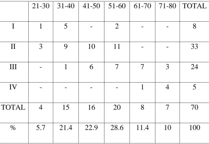
AGE DISTRIBUTION
21-30 31-40 41-50 51-60 61-70 71-80 TOTAL
I 1 5 - 2 - - 8
II 3 9 10 11 - - 33
III - 1 6 7 7 3 24
IV - - - - 1 4 5
TOTAL 4 15 16 20 8 7 70
% 5.7 21.4 22.9 28.6 11.4 10 100
The maximum number of patients belonged to the 51-60 age
group (28.6%) and 21-30 age group (5.7%) constituted the
TABLE – 3
AGE DISTRIBUTION ACCORDING TO STAGE
AGE MEAN S.D
I 39.25 10.58
II 44.91 9.17
III 57.21 10.13
IV 73.20 4.21
p < 0.001 Significant
TABLE – 4
AGE WITH FEV1 % OF PREDICTED VALUE
FEV1 % PV p VALUE
AGE 0.004 Significant
The increase in age with the increasing stage of the disease was
found to be statistically significant with p < 0.001 and the correlation
of age with FEV1% of predicted value was also found to be
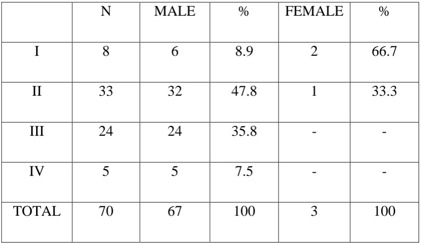
TABLE – 5
SEX
N MALE % FEMALE %
I 8 6 8.9 2 66.7
II 33 32 47.8 1 33.3
III 24 24 35.8 - -
IV 5 5 7.5 - -
TOTAL 70 67 100 3 100
Of the total 70 patients, 67 (95.7%) were males and 3 (4.3%)
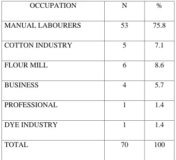
TABLE - 6
OCCUPATION
OCCUPATION N %
MANUAL LABOURERS 53 75.8
COTTON INDUSTRY 5 7.1
FLOUR MILL 6 8.6
BUSINESS 4 5.7
PROFESSIONAL 1 1.4
DYE INDUSTRY 1 1.4
TOTAL 70 100
Of the total 70 patients, 53(75.8%)were manual labourers,
6(8.6%) were working in flour mill, 5(7.1%) in cotton industry,
4(5.7%) were in business,1 (1.4%) in dye industry and 1(1.4%) was a
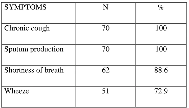
TABLE – 7
SYMPTOMS
SYMPTOMS N %
Chronic cough 70 100
Sputum production 70 100
Shortness of breath 62 88.6
Wheeze 51 72.9
All the patients had chronic cough with sputum production,
88.6% had shortness of breath and 72.9% gave a history of
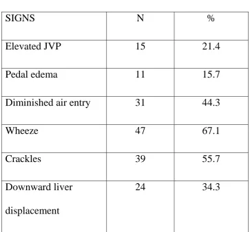
TABLE - 8
SIGNS
SIGNS N %
Elevated JVP 15 21.4
Pedal edema 11 15.7
Diminished air entry 31 44.3
Wheeze 47 67.1
Crackles 39 55.7
Downward liver
displacement
24 34.3
Of the 70 patients, 15(21.4%) had elevated JVP, 11(15.7%) had
pedal edema, 31(44.3%) diminished air entry, 47(67.1%) wheeze,
39(55.7%) crackles on auscultation and 24(34.3%) had downward
TABLE - 9
X-RAY CHEST PA VIEW
Stage N Normal % CB % E % CB + E %
I 8 8 100 - - - -
II 33 5 15.2 10 30.3 10 30.3 8 24.2
III 24 - - 6 25 10 41.7 8 33.3
IV 5 - - 1 20 1 20 3 60
Of the 70 patients, 13 patients had normal X-ray, 17 patients
had features of chronic bronchitis, 21 had findings suggestive of
emphysema and 19 patients showed features of both chronic
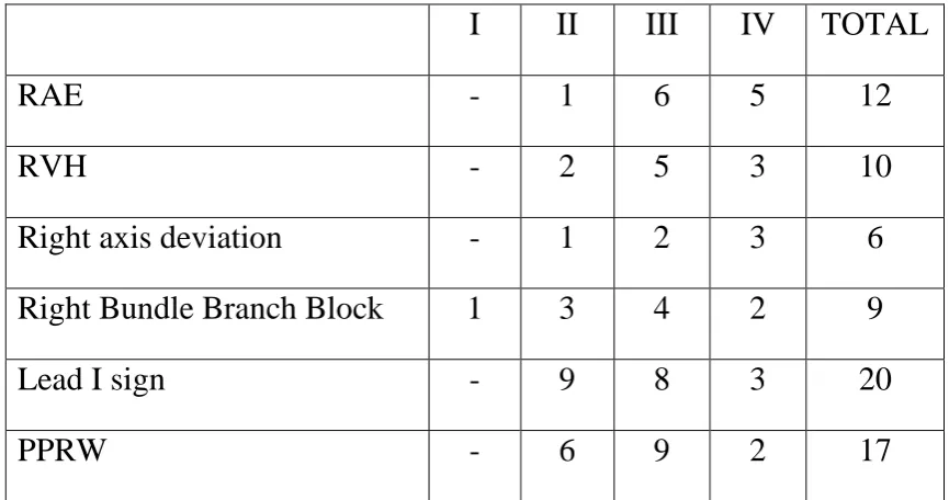
TABLE - 10
ELECTROCARDIOGRAM
I II III IV TOTAL
RAE - 1 6 5 12
RVH - 2 5 3 10
Right axis deviation - 1 2 3 6
Right Bundle Branch Block 1 3 4 2 9
Lead I sign - 9 8 3 20
PPRW - 6 9 2 17
Among patients with stage I disease (8) 1 patient had ECG
criteria for right bundle branch block, stage II disease(33) 1 patient
showed features of right atrial enlargement ( p wave > 2.5mm), 2
patients had right ventricular hypertrophy ( R in V1 > 7mm or R/S in
V1>1), 1 had right axis deviation,3 showed right bundle branch block,
9 had lead I sign ( small equiphasic QRS complex in lead I – due to
hyperinflation of lungs and rotation of heart ) and 6 had poor R wave
progression. Among patients with stage III disease, 6 showed RAE, 5
RVH, 2 right axis deviation, 4 right bundle branch block, 8 lead I sign
and 9 poor progression of R wave. In stage IV disease 5 had right
atrial enlargement,3 right ventricular hypertrophy, 3 had right axis
deviation,2 RBBB,3 lead I sign and 2 had poor R wave
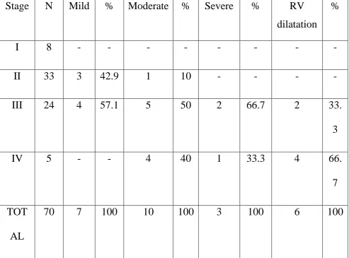
TABLE - 11
ECHOCARDIOGRAPHY
Stage N Mild % Moderate % Severe % RV
dilatation %
I 8 - - - -
II 33 3 42.9 1 10 - - - -
III 24 4 57.1 5 50 2 66.7 2 33.
3
IV 5 - - 4 40 1 33.3 4 66.
7
TOT
AL
70 7 100 10 100 3 100 6 100
Of the total 70 patients, 7 patients – 3 in stage II and 4 in stage
III had mild pulmonary hypertension, 10 patients – 1 in stage II, 5 in
stage III and 4 in stage IV had moderate pulmonary hypertension, 3
patients – 2 in stage III and 1 in stage IV had severe pulmonary
hypertension. Of the 6 patients with RV enlargement 2 were in stage
TABLE –12
BODY MASS INDEX WITH STAGE
BODY MASS INDEX MEAN S.D
I 26.43 4.16
II 23.91 1.84
III 21.50 2.07
IV 18.78 1.98
p < 0.001 Significant
The body mass index showed a decreasing trend with increasing
stage of the disease and the association was found to be statistically
TABLE – 13
PACK YEARS OF SMOKING WITH STAGE
PACK YEARS OF
SMOKING
MEAN S.D
I 13.75 9.91
II 30.61 10.81
III 41.25 6.29
IV 52.00 4.47
[image:87.595.128.493.155.556.2]p < 0.001 Significant
TABLE – 14
FEV1% OF PREDICTED VALUE WITH PACK YEARS
FEV1% OF PV P
PACK YEARS OF SMOKING < 0.001 Significant
The study showed that there is an increase in severity of the
disease as depicted by GOLD stage with the increase in the number of
pack years of smoking and it was statistically significant (p < 0.001).
Also the decline in FEV1 % of predicted value also showed a
significant association with the number of pack years of smoking.(fig
TABLE – 15
ERYTHROCYTE SEDIMENTATION RATE WITH STAGE
ERYTHROCYTE
SEDIMENTATION RATE
Mean S.D
I 22.13 11.13
II 23.18 8.37
III 26.04 6.28
IV 36.00 11.23
p = 0.012 Significant
TABLE – 16
FEV1% OF PREDICTED VALUE WITH ERYTHROCYTE
SEDIMENTATION RATE
FEV1% OF PV P
ERYTHROCYTE
SEDIMENTATION RATE
< 0.001 Significant
The mean erythrocyte sedimentation rate in each stage were
tested for significance and was found to be statistically significant
with p value = 0.012. Similarly the correlation between erythrocyte
sedimentation rate and FEV1% of predicted value was also found to
TABLE – 17
SERUM ALBUMIN WITH STAGE
SR. ALBUMIN MEAN S.D
I 4.43 0.35
II 3.91 0.36
III 3.62 0.35
IV 2.56 0.39
p < 0.001 Significant
TABLE – 18
FEV1% OF PREDICTED VALUE WITH SERUM ALBUMIN
FEV1% OF PV P
SERUM ALBUMIN < 0.001 Significant
Serum albumin concentration was found to decrease among
patients with increasing disease severity and the decrease was found
to be statistically significant (p < 0.001 ). Also with declining FEV1%
of predicted value serum albumin was found to significantly decrease.
[image:91.595.115.492.470.554.2]TABLE – 19
HIGHLY SENSITIVE CRP WITH STAGE
HIGHLY
SENSITIVE CRP
MEAN S.D
I 3.91 1.26
II 5.79 1.23
III 9.01 0.96
IV 11.98 0.73
p < 0.001 Significant
TABLE – 20
HIGHLY SENSITIVE CRP
I II III IV ‘p’ value
≤ 3 mg/L
(3)
2 1 0 0 0.421 Not
Significant
>3 mg/L
(67)
6 32 24 5 < 0.001
[image:93.595.97.513.495.693.2]TABLE – 21
FEV1% OF PREDICTED VALUE WITH HIGHLY
SENSITIVE CRP
FEV1% OF PV P
HIGHLY SENSITIVE CRP < 0.001 Significant
Among the 4 stages, the concentration of highly sensitive
C-Reactive Protein was highest in stage IV disease and the increase in
highly sensitive C-Reactive Protein with disease severity was found
to be statistically significant with p < 0.001.(fig 15). Also among the
total 70 patients, 67 patients had increased highly sensitive
C-Reactive Protein of > 3 mg/L and only 3 patients had normal values.
When statistically tested between the normal vs abnormal groups
among the stages it was found to significant.(fig 16). Similarly, the
correlation between FEV1% of predicted value with this marker of
DISCUSSION
COPD is one of the several diseases whose morbidity and
mortality is increasing day by day at an alarming rate and imposing
economic burden on the nation.34 Despite advances in diagnosis and management the disease still remains untamed. The disease has been
recognized as one with systemic manifestations and several markers
are being tested everyday to possibly reflect the entire disease
process, but the ‘ideal’ marker still remains an enigma. In our study, a
total of 70 patients were studied after applying the inclusion and
exclusion criteria; their clinical status analysed and markers of
inflammation were correlated with severity to determine their
significance.
AGE
In our study, the increase in age with the increasing stage of the
disease was found to be statistically significant with p < 0.001 and the
correlation of age with FEV1% of predicted value was also found to
be statistically significant. The risk of developing the disease has