0022-538X/95/$04.0010
Copyrightq1995, American Society for Microbiology
Genesis of Sindbis Virus by In Vivo Recombination of
Nonreplicative RNA Precursors
RAMASWAMY RAJU,* S. V. SUBRAMANIAM,
ANDMUSTAPHA HAJJOU
Department of Microbiology, School of Medicine, Meharry Medical College,
Nashville, Tennessee 37208
Received 30 June 1995/Accepted 23 August 1995
Genetically engineered RNA transcripts coding for various Sindbis virus (SIN) genes were used to study
structure and sequence requirements of RNA recombination in BHK cells. Three different groups of RNA
transcripts were made: (i) RNAs which retain the ability to replicate and which carry sequences coding for
either viral polymerase or viral structural proteins; (ii) RNAs which lack the complete 3
*
end of the SIN
genome and thus are incapable of replicating; and (iii) RNAs which lack the complete 5
*
end of the SIN genome
and also are incapable of replicating. BHK cells were transfected with specific combinations of these precursor
RNAs, and virus production and RNA synthetic abilities of the released virus were determined. We
demon-strate in vivo generation of infectious SIN by fusion of (i) replicative RNAs to nonreplicative RNAs and (ii) two
nonreplicative RNA precursors. Both homologous and nonhomologous types of recombinations were observed.
In the homologous type of recombination, a 694-nucleotide overlap at the crossover region of the first pair of
precursors resulted in the addition of an A residue converting the UAG stop codon of nonstructural protein P4
to a UAA stop codon. In the nonhomologous type of recombination, the crossover sites contained deletion of
up to 76 nucleotides from one of the precursors and complete preservation of junction sequence from the other
precursor. This is also the first report that a cytoplasmic RNA virus can be generated from biologically
nonreplicative RNA precursors. These results have implications for initiation of viral RNA synthesis and
recombination between RNA viral genomes in general. We favor template switching as a mechanism for the
fusion events described here and suggest inclusion of polymerase scanning of diverse nonreplicative RNAs as
an inherent feature of the copy choice model of RNA recombination. Very importantly, the facile nature of RNA
recombination occurring between nonreplicative RNA precursors should speed up the production and analysis
of targeted mutants of SIN and possibly other RNA viruses.
Sindbis virus (SIN) is one of the best-studied alphaviruses at
the molecular level (54, 59). SIN is transmitted by mosquitoes
to animals and humans and is one of the least pathogenic
member of the Alphavirus genus of the Togaviridae family (59).
The genome of SIN is composed of an 11.7-kb single-stranded
positive-sense RNA (55, 59). The protein-coding potential of
the SIN genome is divided mainly into two large open reading
frames (ORFs). The first ORF, which encompasses a 7.6-kb 5
9
region of the genome, is directly translated into viral
poly-merase proteins (59). The second ORF, which spans the
re-maining 3
9
region of the genome, is actually translated from a
26S subgenomic RNA, giving rise to viral structural (S)
pro-teins (48, 59). Conserved cis-acting sequences located at the 5
9
and 3
9
ends of the viral RNA are known to be necessary and
sufficient to confer replication competency to the viral genome
or its derivatives (26, 38, 39, 59). Genetically engineered cDNA
copies of the SIN genome and its derivatives have been
exten-sively used to study the cis- and trans-acting functions of the
SIN genome (6, 12, 26, 30, 38, 43, 47, 61). Recently, Weiss and
Schlesinger (61) demonstrated the occurrence of RNA
recom-bination between SIN replicons. By making use of
replication-competent engineered SIN RNAs, they showed that
near-wild-type SIN genomes can be produced from two smaller replicons
carrying each of the ORFs (61).
RNA recombination among RNA viruses is a well-studied
biological process (11, 16, 17, 27, 29, 57). Animal (3, 4, 10, 14,
16, 24, 29, 40, 65), plant (2, 7, 8, 9, 19, 35, 36, 37, 60, 62), and
bacterial (5, 40) viruses are known to undergo RNA
recombi-nation. Although RNA recombination is characterized by
pro-duction of new and chimeric RNA viruses by physical exchange
of genetic information, the precise mechanism and forces
gov-erning RNA recombination are not completely understood (9,
20, 27, 36, 37, 49, 61). On the basis of sequence homology at
the crossover sites, both homologous (20, 24, 25, 32, 36, 40)
and nonhomologous (7, 10, 34, 61, 62, 65) types of
recombi-nations were attributed to viral RNA recombirecombi-nations.
Mecha-nistically, RNA recombinations could be due to breakage and
reunion of template RNAs (7, 27) or due to template switching
of polymerase during replicative RNA synthesis (23, 24, 27). In
the absence of any well-characterized enzyme capable of RNA
cleavage, ligation, and editing activities, little attention was
given to the breakage and reunion model of RNA
recombina-tion (7, 27). On the other hand, the copy choice mechanism,
which invokes the template switching of viral polymerase
dur-ing replicative RNA synthesis, is well accepted, since all known
RNA recombinations are known to be associated with
replica-tive RNA synthesis (20, 22, 24, 27, 37, 40, 57, 61).
If the copy choice mechanism is important for RNA
recom-bination, what are the intracellular forces which make the
polymerase jump from one template to another? On the basis
of generation of defective interfering (DI) and rearranged
genomes (41, 53) during high-multiplicity passages of viruses,
and on the basis of the association between active replication
and RNA recombination (24, 27), we hypothesized that
repli-cative polymerase cycling (RPC) and/or high intracellular
con-centrations of cognate template RNAs and polymerase
pro-teins may play a crucial role in facilitating template switching.
* Corresponding author. Mailing address: Department ofMicrobi-ology, Meharry Medical College, School of Medicine, 1005 D.B. Todd Blvd., Nashville, TN 37208. Phone: (615) 327-6687. Fax: (615) 327-6072.
7391
on November 9, 2019 by guest
http://jvi.asm.org/
We define RPC as a process in which large numbers of
poly-merase proteins cycle through template RNAs, generating
abundant levels of nascent RNAs. It is conceivable that RPC is
associated with polymerase crowding on template RNAs,
which might lead to frequent slippage and reinitiation at
alter-nate sites or templates. As indicated by others (9, 27, 37), the
presence of RNA structures on template RNAs might also
influence detachment and reattachment of polymerases.
Events such as mixed hybrid formation between limiting
neg-ative-sense RNAs and excess positive-sense RNAs (or vice
versa) may also result in formation of branched RNAs, leading
to expulsion and rerouting of polymerase travel during RNA
synthesis. In this study, using engineered SIN RNAs as
pre-cursors, we demonstrate that RNA recombination occurs in
the SIN system in the absence of RNA replication and hence
under low concentrations of cognate precursor RNAs. We
postulate multiple modes of template switching by RNA
poly-merases and suggest that template switching may function as
an effective mechanism to generate new replicative RNAs
from nonreplicative RNAs in a protein- and RNA-rich milieu.
MATERIALS AND METHODS
Plasmids.Toto 1002, derivative of Toto 1000 (18, 47), contains an XbaI site at nucleotide (nt) 7613. Toto 1000 is one of the parental SIN plasmids which contain a functional SIN cDNA. An SP6 promoter located at the 59end of the SIN genome drives the synthesis of infectious transcripts. This plasmid contains an SstI site at the 39end of the SIN genomic sequence which is used to linearize the plasmid for in vitro transcription. Cells transfected with RNA derived from this plasmid produce large amounts of infectious virus (47).
TRCAT, a well-characterized SIN vector which lacks all structural proteins (63), contains the complete nonstructural (NS) region, subgenomic promoter, and reporter chloramphenicol acetyltransferase (CAT) gene and all cis-acting sequences essential for replication. TRCAT contains an SstI site for lineariza-tion. Cells transfected with RNA derived from this plasmid express TRCAT genomic RNA, a subgenomic RNA coding for CAT, and high levels of CAT protein. No virus particles are produced from TRCAT RNA-transfected cells.
TRCAT 62 is a replication-competent derivative of TRCAT which lacks all of the 39nontranslated region except for the last 62 nt at the 39end of TRCAT (18). Like TRCAT, TRCAT 62 expresses CAT protein in transfected cells and lacks the ability to produce any virus particles.
GNTR-Lac, a derivative of TRCAT in which the CAT gene is replaced by a
BglII-SacII fragment of the La Crosse virus S genome (44), lacks approximately
90 nt of 39noncoding region of the SIN genome adjacent to and downstream of the S-protein termination codon. In vitro transcripts produced from this plasmid are replication competent, but no virus particles are produced from RNA-transfected cells.
SINrep, one of the latest SIN vectors originally named SINrep 5 (6), lacks all of the S-protein-coding region but contains all cis-acting motifs essential for replication and subgenomic RNA synthesis. An XhoI site located at the 39end of the viral sequence is used to linearize the plasmid. No infectious particles are produced from cells transfected with SINrep RNA.
DI-S is a derivative of DIF20e (43) which contains an SIN DI genome. This plasmid was constructed by cloning the XbaI-SstI fragment of Toto 1002 which contains SIN S region into DIF20e at the same sites. This process resulted in deletion of the CAT gene of DIF20e and introduction of the S region down-stream of the subgenomic promoter located in DIF20e. RNA transcripts pro-duced from this vector are replication competent and express 26S subgenomic RNA when supplemented with NS proteins in trans from another source.
DHBBS (DHBB 59SIN) is a plasmid containing SIN DI sequences, a SIN subgenomic promoter, and SIN S-region-coding sequences (6). The RNA tran-scripts produced from this plasmid are capable of only poor amplification by NS proteins provided in trans as a result of mutational alterations in the 59end of the vector RNA. In spite of its diminished replication ability, this RNA adequately complements SIN structural protein requirements through synthesis of 26S sub-genomic RNA (6).
GNS-2, a plasmid carrying the complete SIN NS region, was made by cloning the NS region from Toto 1002 as an SstI-XbaI fragment into plasmid pGem 3 (Promega) at the same sites. The XbaI site was used to linearize the plasmid for in vitro transcription. The RNA transcripts produced from this plasmid lacks the complete S region, including the 39motifs of the SIN genome. Hence, the GNS-2 transcripts are nonreplicative. However, the GNS-2 transcripts were shown to provide adequate amounts of NS proteins through direct translation in trans-fected cells (this work).
G26S-2 was made by cloning the HpaI-SstI fragment containing all of the SIN S region of Toto 1002 at the HincII-SstI location of pGem 3. Positive-sense runoff transcripts were made by linearizing the plasmid at the SstI site. Negative-sense
transcripts were made by linearizing at the HindIII site and using the T7 pro-moter. The absence of the SIN 59motif makes the RNA nonreplicative, although initiation of negative-strand synthesis is indicated by the present work.
G26S-2N was made by digesting plasmid G26S-2 with NsiI and by end filling with T4 DNA polymerase in the presence of 1 mM deoxynucleoside triphos-phates. The inactivated NsiI site is indicated as a small stalk in Fig. 1. The inactivated Nsi site served as a reporter in the fusion products.
G26S-2SN was made by deleting the NsiI-SstI fragment of G26S-2 which carries the 3P region (SIN 39motif) and religating the plasmid. Runoff tran-scripts were made by linearizing the plasmid at the EcoRI site and using SP6 RNA polymerase. The RNA transcripts produced from this plasmid are non-replicative since they carry neither 59nor 39motifs of the SIN genome, but the RNA transcripts code for functional S proteins of SIN.
G26S-3 was made by cloning the S-region-containing XbaI-SstI fragment of Toto 1002 at the corresponding sites of pGem 3. Positive-sense transcripts were made by linearizing the plasmid at the SstI site and using SP6 RNA polymerase. Negative-sense transcripts were made by linearizing at the XbaI site and using T7 RNA polymerase. Since the RNA transcripts lack the 59region of SIN, they are incapable of replication, although initiation of negative-strand synthesis from the 39end is indicated by the present work.
TT3CAT carries sequences coding for two subgenomic promoters of SIN. The first subgenomic promoter drives the synthesis of the S region, and the second promoter drives the synthesis of the CAT reporter. The plasmid does not carry any SIN 59motifs but does carry functional 39motifs. The plasmid was made by cloning the XbaI-SstI region carrying the two subgenomic promoters, S region, and CAT region of plasmid TT20c (43) into the same sites of plasmid G26S-2. In vitro positive-sense transcripts are made from this plasmid by linearizing the plasmid at the SstI site. Since the transcripts lack 59 motifs of SIN, they are nonreplicative, but initiation and elongation of RNA synthesis from the template RNA were indicated by this work.
As described for TT3CAT, TT19CAT was made by cloning the XbaI-SstI region of TT20c into G26S-3. In vitro transcripts are made by linearizing the FIG. 1. Structures of engineered SIN RNAs. All RNAs are depicted in plus-sense orientation. Common cis-acting sequences are identified uniformly: 5P, 59 promoter or conserved sequence; 3P, 39promoter or conserved sequence; NS, sequences coding for NS proteins which constitute the RNA polymerase com-plex; S, sequences coding for viral S proteins; JP, junction promoter responsible for 26S subgenomic RNA synthesis; 5P1, 59 region of SIN DI DNA which contains a tRNA sequence; 5P2, 59 region of the SIN genome lacking full promoter activity.
on November 9, 2019 by guest
http://jvi.asm.org/
plasmid at the SstI site and using SP6 RNA polymerase. Other properties of TT19CAT transcripts are identical to those of TT3CAT transcripts.
GNS-2BG was constructed by digesting GNS-2 with BglII, end filling the linear DNA with T4 DNA polymerase in the presence of 1 mM deoxynucleoside triphosphate, and recircularizing the plasmid. This procedure resulted in intro-duction of four additional bases in the coding region of the NS polyprotein, leading to translational frameshift and premature termination. RNA transcripts produced from this construct fail to support recombination in vivo.
G3S was constructed by cloning the 2.2-kb region of the SIN S-protein-coding region obtained by StuI digestion of Toto 1002 into the HincII site of pGem 3. Negative-sense RNA transcripts which hybridize to both the SIN genome and SIN 26S subgenomic RNA are produced from T7 promoter-mediated RNA transcription of G3S.
GNS-1EE carries a 1,050-nt-long SIN NS region flanked by SP6 and T7 promoters. Plasmid GNS-1EE was obtained by cloning the 1050-nt EcoRI-HpaI fragment (nt 5870 to 6919 of Toto 1002) in the HincII and EcoRI sites of pGem 3. Linearization of the plasmid with HindIII and transcription with T7 RNA polymerase gives rise to negative sense RNA transcripts which hybridize to positive-sense RNAs carrying the NS region of SIN.
In vitro synthesis of RNA transcripts.Five micrograms of each plasmid was digested with the appropriate restriction enzyme and directly ethanol precipi-tated. One-third of the DNA was used for in vitro transcription by SP6 RNA polymerase in the presence of a fourfold excess of cap structure as described previously (43, 44). Either [3H]UTP or [a-32P]GTP was used as a tracer to quantitate the amount of RNA made. After a 1-h incubation, the template DNA was removed by DNase I digestion, and RNA was purified by phenol-chloroform extraction and ethanol precipitation. The amount of RNA made was quantitated by trichloroacetic acid precipitation. Five percent of the RNA samples was denatured by glyoxal (43) and analyzed on a 1.25% agarose gel.
Cells, viruses, and infection.BHK-21 cells and Vero cells were maintained in minimal essential medium containing 10% fetal bovine serum. Standard (Toto 1101 or Toto 1002) and recombinant SIN stocks were prepared from BHK cells and titered on BHK cells or Vero cells, using a standard plaque assay (61). For virus infections, BHK cells grown in 35-mm-diameter petri plates were infected with 5 to 10 PFU of virus inoculum per cell diluted in serum-free medium and incubated at 378C for the desired times.
Transfection of BHK cells with RNA precursors.Approximately 70 to 90% confluent BHK cells grown in 35-mm-diameter petri plates (Sarstedt) were washed twice with phosphate-buffered saline (PBS) containing no calcium and magnesium and transfected with 200 to 300ml of transfection mixture. The transfection mixture consisted of 20mg of Lipofectin (Gibco-BRL) or Transfec-tace, 50 to 200 ng of each in vitro-transcribed RNA, and PBS (43). The trans-fection mixture was allowed to incubate in ice for 5 to 10 min, layered onto PBS-washed cells, and continually rocked for 20 to 30 min. At the end of the transfection procedure, the transfection mixture was removed from the cells, carefully washed with medium, and layered with 2 ml of medium containing 10% fetal bovine serum. Using TRCAT as a reporter molecule, we routinely observed transfection of up to 0.5 to 2% of cells per ng of RNA. Electroporation of RNA into BHK cells gave comparable efficiencies. The transfected cells were incu-bated for 2 to 3 days at 378C and constantly monitored for development of cytopathic effects. Culture supernatants were recovered from all transfected cells and titered on Vero cells, using a standard plaque assay.
In vivo labeling and analysis of RNA products.Five microliters of the culture supernatant (5 to 15 PFU of virus per cell) derived from transfected cells was diluted to 0.2 ml with serum-free medium and allowed to infect BHK cells grown in 35-mm-diameter petri plates (43). At the end of 1 h, 0.6 ml of medium containing 5mg of dactinomycin per ml was added to the plates. Twenty minutes later, 50mCi of [3H]uridine was added to each plate, and the infection was continued at 378C for 6 h. At the end of infection, cells were harvested and cytoplasmic RNA was isolated (43, 44). Approximately 5mg of RNA was dena-tured with glyoxal, analyzed on a 1.25% agarose gel, and then fluorographed.
Reverse transcription-PCR amplification of cytoplasmic RNA and sequenc-ing.Total cytoplasmic RNA was isolated from infected cells at 6 h postinfection (p.i.) (43), treated with RNase-free DNase (Gibco-BRL), and further purified by proteinase K digestion and phenol extraction. One-fifth of the cytoplasmic RNA was used for reverse transcription and PCR amplification. In brief, the first-strand synthesis involved annealing of 6 pmol of a negative-sense primer (nt 8049 to 8070 of Toto 1000) with 5mg of cytoplasmic RNA in 0.3 M NaCl and subsequent extension using murine leukemia virus reverse transcriptase. In ad-dition to RNA and primer, the reaction mixture consisted of 50 mm Tris-HCl (pH 8.3), 70 mM KCl, 3 mM MgCl2, 10 mM dithiothreitol, 0.35 mM de-oxynucleoside triphosphates, and 400 U of murine leukemia virus in a total volume of 30ml. The reaction mixture was incubated for 1 h at 378C and subsequently for 20 min at 428C. At the end of the reaction, an aliquot of the reaction mixture was diluted to 10-fold and used directly for PCR amplification. The PCR mixture consisted of 2 to 5% of the cDNA products, 20 mM Tris-HCl (pH 8.4), 50 mM KCl, 2 mM MgCl2, 2 mM dithiothreitol, 100mg of gelatin per ml, 5 pmol each of negative-sense primer and positive-sense primer (nt 7248 to 7270), 5 U of Taq polymerase, and 350mM deoxynucleoside triphosphates in a volume of 50ml. After 20 cycles of PCR amplification, the reaction mixture was removed and stored. Ten percent of the PCR products was analyzed on a gel, and the 820-nt band corresponding to the amplified region was isolated from the gel.
The isolated DNA fragment was sequenced by Sanger’s dideoxy method, using an internal primer (nt 7650 to 7666). Identical results were obtained when gel-purified fused RNA was used for reverse transcription-PCR amplification and sequencing.
CAT assay.The CAT assay was performed as described earlier (43, 63). Infected cell monolayers were harvested at indicated times by scraping into 2 ml of PBS. The cells were pelleted at 4,000 rpm in a refrigerated microcentrifuge and suspended in 500ml of lysis buffer (0.25 M Tris-HCl [pH 8]). After three cycles of freeze-thawing, the cell debris was removed by centrifugation at 15,000 rpm in a refrigerated microcentrifuge, and the cytoplasmic supernatant was recovered. The reaction mixture consisted of 20 to 60mg of cytoplasmic proteins, 0.1mCi of [14C]chloramphenicol (50 mCi/mmol), 5ml of 20 mM acetyl coenzyme A, and 90ml of 0.25 M Tris-HCl (pH 8.0). The contents were incubated for 1 h at 378C and extracted with ethyl acetate, and the products were analyzed by silica gel-based thin-layer chromatography. The percent conversion of chlorampheni-col to acetyl chloramphenichlorampheni-col was determined by cutting each of the radioactive spots and determining the radiactivity by liquid scintillation.
Northern (RNA) analysis.Glyoxylated RNA samples were separated on a 1.25% agarose gel for 2 to 3 h. At the end of electrophoretic separation, the gel was soaked in 20 mM NaOH for 10 min and extensively washed with running water. The RNA samples were electrophoretically transferred to a Zetaprobe (Bio-Rad) nylon membrane, using 10 mM Tris–20 mM sodium acetate (pH 7.8) as the transfer buffer for 6 h. Prehybridization and hybridization with RNA probes were done as described earlier (44), using 53SSC (13SSC is 0.15 M NaCl plus 0.015 M sodium citrate), 100mg of RNA per ml, and appropriate RNA probe labeled with [a-32
P]GTP (23106
dpm/10 ml).
RESULTS
Design of substrate RNAs and assay for recombination.
To
study recombination between RNA precursors, three groups
(Fig. 1) of RNAs were made by in vitro transcription of
plas-mid DNAs carrying an SP6 or T7 phage promoter. The group
I RNAs contain both the 5
9
and 3
9
ends of SIN, and hence they
are replication competent. Thus, group I RNAs are amplifiable
in vivo by viral polymerase. The group II RNA is exemplified
by GNS-2. GNS-2 RNA lacks the 39
one-third of the SIN
genome and hence cannot support any RNA synthesis. The
majority of group III RNAs contain an authentic 3
9
end of the
SIN genome, but all of them lack the 5
9
two-thirds of the SIN
genome. None of individual RNAs in groups I, II, and III can
produce live virus when transfected into cells, since they all
lack either NS- or S-protein-coding sequences. Since both 5
9
and 3
9
sequences are essential for replication of SIN-related
RNAs, none of the group II and group III RNAs can replicate
to produce progeny RNAs.
The experimental strategy involved multiple steps: (i)
intro-duction of one or more of the in vitro made RNA precursors
into BHK cells to generate infectious RNA recombinants; (ii)
recovery of culture supernatants containing recombinant
vi-ruses and titration; and (iii) study of gene expression from
recombinant viruses. Although none of the RNAs belonging to
group I, II, and III can produce infectious virus when
trans-fected alone, there is a possibility that live virus can be
pro-duced when two functionally complementing RNAs are
co-transfected. Live virus can be produced either by physical
recombination (24, 61) between RNA precursors or by simple
complementation between virus proteins (12). To demonstrate
recombination between transfected RNA precursors, we
re-sorted to electrophoretic analysis of RNA products (6, 61).
The presence of RNA products of ca. 12 kb or more containing
both NS and S sequences was used as a confirmatory test for
RNA recombination.
Strong SIN replicons expressing NS proteins fuse with weak
replicons expressing S proteins.
Almost all of the RNA
re-combination studies reported in the literature made use of live
virus or RNA substrates which carried both the 59
and 39
ends
of viral sequences. Since the copy choice mechanism is widely
accepted by RNA virologists, and since the copy choice
mech-anism predicts a requirement for replication competency of
substrate RNAs, perhaps it was logical that all of the studies
on November 9, 2019 by guest
http://jvi.asm.org/
made use of replicative viral derivatives. In fact, the first
re-ported study of the recombination between SIN RNAs made
use of highly replicative RNAs (61). Initially, we tested the
ability of strong SIN replicons to recombine with weak
repli-cons. The strong replicons TRCAT, TRCAT 62, and SINrep
code for polymerase proteins. The weak replicon DHBBS
codes for structural proteins (Fig. 1). When DHBBS was used
in conjunction with either one of the strong replicons in
trans-fection experiments, infectious viruses were produced (Table
1, experiments 17 to 19). Cells infected with each of the
re-combinant viruses expressed two genomic and two subgenomic
RNAs (Fig. 2a). The faster-moving genomic RNA and
sub-genomic RNAs are derived from the corresponding strong
replicons (compare Fig. 2a and 3b). The slowly moving
geno-mic RNA which hybridizes to both NS- and S-specific probes of
SIN (data not shown) is the recombinant RNA product. The
slowly moving subgenomic RNA corresponds to 26S
sub-genomic RNA expressed from the recombinant RNA. These
results are reminiscent of the original studies by Weiss and
Schlesinger (61). As demonstrated by Bredenbeek et al. (6),
the parental DHBBS replicon was not packaged from
trans-fected cells. Unlike earlier work reported by Bredenbeek et al.
(6), DHBBS clearly recombined with all of the replicons used
in these studies. These results indicated that recombination
does occur when weakly replicating RNA substrates are used
as substrates. Interestingly, only very low levels of fusion
prod-ucts were demonstrable in cells transfected with precursor
RNAs, by [
3H]uridine labeling of intracellular RNAs, or by
Northern analysis of cytoplasmic RNAs using riboprobes (42).
Strong replicons expressing NS proteins fuse with a
non-replicating RNA.
The weak replicon DHBBS has a defect in
the 5
9
cis-acting sequence (6). In spite of this defect, DHBBS
was shown to serve as a substrate for RNA recombination. To
further test the role of the 5
9
cis-acting sequence in
[image:4.612.61.558.83.435.2]recombi-nation, cells were transfected with one of the strong replicons
such as TRCAT, TRCAT 62, SINrep, or GNTR-Lac and one
of the nonreplicative RNAs (G26S-2 or G26S-3). These
non-replicative RNAs lack the 5
9
end of the SIN genome but carry
genes coding for SIN S proteins (Fig. 1). As shown in Table 1
(experiments 20 to 27), infectious viruses were clearly released
from cells which were transfected with two complementing
RNAs. Infection of cells with these viruses resulted in the
intracellular accumulation of both 49S- and 26S-like RNAs
characteristic of SIN infection (Fig. 2a and b). These results
indicated that 5
9
end of at least one of the substrate RNAs was
dispensable for RNA fusion. G26S-2N RNA, which had an
TABLE 1. Cytopathic effects and release of virus particles from transfected cellsaExpt no. Transfected
RNA-1
Transfected RNA-2
Cytopathic effect Virus titer
(PFU/ml)
Plaque size (%)
24 h 48 h
1 TRCAT None None None 0 NA
2 TRCAT 62 None None None 0 NA
3 GNTR-Lac None None None 0 NA
4 SINrep None None None 0 NA
5 DI-S None None None 0 NA
6 DHBBS None None None 0 NA
7 GNS-2 None None None 0 NA
8 G26S-2 None None None 0 NA
9 G26S-3 None None None 0 NA
10 G26S-2N None None None 0 NA
11 G26S-2SN None None None 0 NA
12 TT3CAT None None None 0 NA
13 TT19CAT None None None 0 NA
14 Toto 1002 None 1111 1111 3.13108 100
15 TRCAT DI-S 1 1111 4.13107 40–80
16 TRCAT 62 DI-S 1 1111 2.23107 20–60
17 TRCAT DHBBS 11 1111 2.13107 40–60
18 TRCAT 62 DHBBS 11 1111 5.63107 20–60
19 SINrep DHBBS 11 1111 6.53106 60–100
20 TRCAT G26S-2 1 1111 8.33106 20–80
21 TRCAT G26S-2 (2b) None None 0 NA
22 TRCAT G26S-2SN None None 0 NA
23 TRCAT 62 G26S-2 11 1111 6.03106 40–60
24 TRCAT 62 G26S-2N 11 1111 2.83106 40–60
25 TRCAT 62 G26S-3 1 111 9.13105 40–60
26 GNTR-Lac G26S-2N 11 1111 8.53106 60–100
27 SINrep G26S-2 11 1111 2.13107 40–60
28 GNS-2 DHBBS 11 111 5.03106 60–100
29 GNS-2 G26S-2 11 1111 9.33106 60–80
30 GNS-2 G26S-3 1 111 5.43105 20–60
31 GNS-2 G26S-2 (2) None None 0 NA
32 GNS-2 G26S-3 (2) None None 0 NA
33 GNS-2 G26S-2SN None None 0 NA
34 GNS-2 TT3CAT 11 1111 7.23106 20–60
35 GNS-2 TT19CAT 1 111 6.03105 20–60
a
Structures of individual RNAs are described in Fig. 1. Cells were periodically monitored to determine necrotic cytopathology. No significant cytopathology was observed in cells which were transfected with any one of the nonreplicative RNAs or RNAs which do not code for structural proteins in comparison with untransfected cells.1, low cytopathology;11, moderate cytopathology;111, severe cytopathology but still adherent;1111, complete cell death and detachment. The plaque size is an approximate estimate with respect to that formed by Toto 1002. NA, not applicable.
b2
, negative strand.
on November 9, 2019 by guest
http://jvi.asm.org/
inactivated Nsi site (nt 11452 of SIN) as a mutational marker in
the 3
9
end, was demonstrable in the fused RNA product (data
not shown). Additionally, G26S-2SN RNA, which lacked both
the 5
9
and 3
9
motifs of SIN, although encoding S-protein
se-quences, failed to support RNA recombination (Table 1,
ex-periment 22; Fig. 2b). Minus-sense RNAs corresponding to
G26S-2 and G26S-3 also failed to serve as templates for RNA
recombination (Fig. 2a). It is interesting that none of the
rep-licative RNAs (TRCAT, TRCAT 62, and SINrep) used in the
original transfection experiment was found to be expressed in
the recombinant virus-infected cells. The original transfected
cells were expected to release particles containing (i)
recom-binant RNA capable of expressing 49S and 26S RNAs and (ii)
the parental replicon (TRCAT, TRCAT 62, or SINrep) which
FIG. 2. (a) Recombination between replicative and nonreplicative RNAs. BHK cells were transfected with template RNAs, and culture supernatant containing released virus was recovered. Fresh BHK cells were infected with 5 to 10 PFU of recovered virus per cell and labeled with uridine, and 5 to 7mg of RNA was separated on a 1.25% gel and fluorographed. The names of RNA substrates used to produce primary virus stocks are indicated above the lanes. Some lanes carry two genomic RNAs and two subgenomic RNAs. In these cases, the slowly moving genomic RNA corresponds to the fusion product. The faster-migrating genomic RNA corresponds to the strong replicon used in transfection. Likewise, the slowly moving subgenomic RNA in each lane corresponds to subgenomic RNA derived from fused genomic RNA. The faster-moving second subgenomic mRNAs are derived from substrate RNAs such as TRCAT, TRCAT 62, and SINrep. (2), negative strand. (b) Recombination between replicative and nonreplicative RNAs. RNA analysis was done as described for panel a. The term genomic RNA corresponds to the fusion product. The subgenomic RNAs are all expressed from fusion products. In addition to 26S subgenomic RNA, two other subgenomic RNA are found. The fastest-moving mRNA labeled CAT mRNA is derived from the second subgenomic promoter found in the TT3CAT and TT19CAT precursors. The slowest-moving subgenomic mRNA is a dicistronic mRNA which contains both the S region of SIN and the CAT region (41), but this dicistronic mRNA is translated to give only S proteins. (c) Expression of CAT protein from recombinant viruses. BHK cells were infected at 20 PFU per cell with recombinant viruses generated from template RNAs indicated above the lanes. After 6 h of infection at 378C, cells were harvested and cytoplasmic extracts were prepared. Ten (lane 2) or 50 (lanes 1, 3, 4, and 5)mg of cytoplasmic proteins was used to assay CAT activity. The percentage conversion of choramphenicol to acetyl chloramphenicol is indicated in parentheses. (d) Intracellular RNA products expressed from plaque-purified viruses. Four plaques each were isolated from duplicate sets of plates p2 and p3 (see below) and were suspended in minimal essential medium containing 2% serum. One-tenth of the diluted plaque virus was used to infect fresh BHK cells, and intracellular viral RNA was labeled with uridine and analyzed as described for panel a. Lanes 1 to 4 correspond to RNA samples corresponding to the four viral plaques from plate p3; lanes 5 to 7 correspond to RNA samples of the four viral plaques from plate p2. (e) Size and morphology of plaques formed by recombinants. Culture supernatants were serially diluted and used to infect Vero cells in culture, overlaid with agarose, and incubated at 378C for 2 to 3 days. Plaques became visible after 36 h. After fixation with 3% paraformaldehyde, the cells were stained with crystal violet and photographed. p1, plaque formation by Toto 1002 (size, 100%); p2, plaque formation by recombinant viruses generated by fusion of GNS-2 and G26S-2 (size, 60 to 80%); p3, plaque formation by recombinant viruses generated by fusion of GNS-2 and G26S-3 (size, 20 to 60%).
on November 9, 2019 by guest
http://jvi.asm.org/
failed to undergo recombination. If these two kinds of
ge-nomes were found in the released particles, then infection of
fresh cells with this population should have led to the
produc-tion of 49S RNA, 26S RNA, the parental replicon, and its
subgenomic RNA. Since only 49S and 26S RNAs are found in
the recombinant virus-infected cells (Fig. 2a and b), it appears
that either the parental replicon is not packaged and released
from the transfected cell or there is a differential gene
expres-sion in recombinant virus-infected cells.
Generation of the SIN genome from a replicative RNA such
as TRCAT and a nonreplicative RNA such as G26S-2
sug-gested that the polymerase proteins produced from TRCAT
initiated RNA synthesis at the 3
9
terminus of G26S-2 and
jumped onto the TRCAT template to make the SIN genome.
This reasoning is based on the copy choice mechanism of RNA
recombination (23, 24). The RNA substrates which function as
templates to initiate RNA synthesis and subsequently donate
the polymerase complex are known as donor templates. Thus,
RNAs such as DHBBS, G26S-2, and G26S-3 can be considered
donor templates (20). The RNA substrates such as TRCAT,
TRCAT 62, and SINrep which accept the jumping polymerase
complex are known as acceptor templates. As depicted in Fig.
4, the donor templates contribute to the 5
9
end of the new
transcript and the acceptor templates contribute to the 3
9
end
of the new transcript.
A nonreplicating RNA expressing NS proteins fuses with a
weak replicon expressing S proteins.
Recombination events
described above made use of amplifiable acceptor templates
with a potential to express high levels of viral polymerase. To
test if low levels of polymerase proteins could still catalyze
RNA recombination, we made use of GNS-2 RNA (Fig. 1).
GNS-2 RNA lacks the complete 4.1-kb 3
9
region of the SIN
genome, and hence it cannot replicate. When cells were
trans-fected with GNS-2 RNA and DHBBS RNA, infectious virus
was produced from transfected cells (Table 1, experiment 28).
Cells infected with the recombinant virus produced both 49S
and 26S RNAs, but as expected, no RNA corresponding to
DHBBS was demonstrable (data not shown). These results
indicated that a low amount of viral polymerase protein
trans-lated from GNS-2 RNA was sufficient to initiate RNA
synthe-sis at the 3
9
end of DHBBS RNA and could effectively switch
to GNS-2 RNA to make recombinant RNAs. Studies carried
out with the brome mosaic virus system indicate that terminal
39
truncations of brome mosaic virus RNA-2 (45) and RNA-3
(36) could be repaired by other cotransfecting wild-type RNAs.
These results confirm the existence of a pathway among plant
and animal alpha-like viruses to repair terminal deletions of
viral genomes.
[image:6.612.89.270.73.354.2]Two nonreplicating RNA precursors recombine.
Results
de-scribed above suggested that a minimally functional donor
template (e.g., G26S-2) must possess at least the 3
9
motif of
SIN, and the minimally functional acceptor template (e.g.,
FIG. 4. Model for nonhomologous recombination of nonreplicative RNA precursors (GNS-2 and G26S-3). In this model, polymerase protein synthesized from one of the templates scans and identifies cognate 39cis-acting sequences to
initiate RNA synthesis, independently of the precise 59and 39sequences of the priming template. The polymerase complex along with its nascent transcript jumps onto a new template some time during transcription. The site of poly-merase jumping may be dictated by base pairing between the donor template and the acceptor template at or near the crossover site. Alternatively, the acceptor template may be selectively recruited from a pool of available substrates by base pairing between 39cis-acting sequences (or an alternate motif) of the donor
template and the 59cis-acting sequences (or an alternate motif) of the receiver
template. Once the acceptor template is recruited to the vicinity of the transcrip-tion complex, the polymerase may switch to the receiver template independently of any local base-pairing requirement, by specific RNA-protein interactions. Alternatively, the polymerase complex may slide through several RNA templates until a cognate RNA motif or template is chosen for reinitiation.
FIG. 3. (a and b) In vivo-made RNAs in transfected BHK cells. BHK cells were transfected with in vitro-transcribed, capped RNAs, and virus-specific RNAs were labeled with uridine in the presence of dactinomycin for 6 to 8 h. Six micrograms of cytoplasmic RNAs was denatured by glyoxal and analyzed on a 1.25% agarose gel. The name of the RNA template used in transfection is indicated above each lane. Note that each template RNA makes a longer genomic RNA and a shorter subgenomic RNA. The gel corresponding to panel a was run for a longer time than the one in panel b. (c) Comparison of in vitro-made RNAs and their in vivo products upon transfection. Lanes corre-sponding to DHBBS and TRCAT represent in vitro-made RNAs loaded on the gel as a marker. Lane TRCAT1DHBBS represents uridine-labeled in vivo RNA products made in cells which were infected with the virus released from cells transfected with TRCAT and DHBBS RNAs. BHK cells were infected with 5 PFU of virus inoculum per cell and labeled for 4 h. Five micrograms of cyto-plasmic RNA was analyzed as in panel a. The gel was run for a shorter duration than in panels a and b. Note the presence of a slowly moving band ahead of TRCAT which is the fusion product between DHBBS and TRCAT and is positive for NS and S regions of SIN. Also note the abundant 26S subgenomic RNA (center band) made from the recombinant product. (d) Some of the in vitro transcripts used as substrates. Five percent of the [a-32P]GTP-labeled transcripts were treated with DNase I to remove the DNA template, purified, denatured with glyoxal, and separated for 1 h on a 1.25% agarose gel. Descriptions of corresponding plasmids are given in Materials and Methods. All of the tran-scripts except G26S-2SN were runoff trantran-scripts at an SstI restriction site of each plasmid. The G26S-2SN plasmid was linearized at the EcoRI site. All plasmids were transcribed with SP6 RNA polymerase.
on November 9, 2019 by guest
http://jvi.asm.org/
[image:6.612.318.555.474.595.2]GNS-2) must possess the 5
9
motif of SIN. The other terminal
motif of SIN may not be needed in the donor and acceptor
templates to support recombination. These predictions were
suggested by results of experiments in which one of the
non-replicative templates was used in conjunction with a
replica-tion-competent template. To test if two nonreplicating
mini-mally functional templates could support recombination, BHK
cells were transfected with G26S-2 and GNS-2 RNAs. As
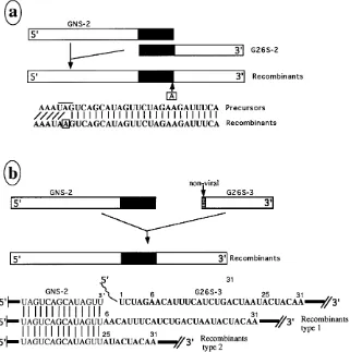
de-picted in fig. 1 and 5a, these two substrate RNAs contain a
694-overlap region at their meeting ends. As shown in Table 1
(experiment 29), infectious virus was released from cells
trans-fected with this pair of nonreplicative RNAs. Both 49S and 26S
RNAs characteristic of SIN were also demonstrable in the
recombinant virus-infected cells (Fig. 2b). Sequence analysis of
four independent recombinants revealed insertion of a single
A residue converting the UAG stop codon of NS protein P4 to
a UAA stop codon. These results indicated that two
nonrep-licative minimally functional RNA substrates could undergo
homologous recombination to evolve into an infectious virus.
Although we were surprised to observe recombination
be-tween two nonreplicative RNA substrates, we thought that the
presence of an overlap region between the two substrate RNAs
[image:7.612.147.468.73.396.2]could have facilitated the recombination. As found for the
poliovirus system (24), the 3
9
growing end of negative-sense
nascent RNA made from the donor template (G26S-2) could
have effectively base paired with the right end of the accetor
template (GNS-2), facilitating homologous recombination. To
determine whether homology at the crossover sites of template
RNAs was required to support recombination, we made use of
GNS-2 and G26S-3 substrate RNAs. GNS-2 and G26S-3
RNAs do not share any homology at their meeting ends (Fig.
5b). In fact, G26S-3 RNA contains a 51-nt nonviral sequence at
its 59
end which is derived from the pGem 3 vector. Except for
this nonviral sequence, the GNS-2 and G26S-3 RNAs
corre-spond to the two fragments of a nicked SIN RNA genome. As
shown in Table 1 (experiment 30), transfection of cells with
these RNAs led to production of infectious virus. Cells
in-fected with the recombinant virus produced both 49S and 26S
RNAs of SIN (Fig. 2b). Both 49S and 26S RNAs continue to
accumulate during the course of infection and, as expected,
contain viral NS and S coding sequences (Fig. 6). These results
clearly demonstrate the apparent ability of two fragments of
nicked SIN genome to recombine in vivo and to produce
in-fectious virus. Sequence analysis of four plaqued recombinants
FIG. 5. Characterization of crossover sites of recombinants. All of the eight cytoplasmic RNA samples used for Fig. 2d were reverse transcribed with murine leukemia virus reverse transcriptase and PCR amplified as described in Materials and Methods. The PCR products were purified with low-melting-temperature agarose and sequenced by using a junction primer. (a) Recombination between GNS-2 and G26S-2 RNAs. The 694-nt overlap region is depicted as solid bars. The open bars represent nonhomologous sequences. The location of the single A insertion in the recombinant is indicated. RNA samples corresponding to all four plaques contained the same insertion. (b) Recombination between GNS-2 and G26S-3. These two template RNAs did not possess any discernible homology at the crossover site. The G26S-3 precursor (donor template) contained 51 nt of nonviral sequences derived from pGem 3 at the 59end, which is denoted as a wavy line. All four recombinant products corresponding to the four plaque-purified viruses contained the precise 39end of GNS-2 used for recombination. None of the four recombinants contained the 51-nt nonviral sequences carried by the G26S-3 precursor. In addition, the type 1 recombinants lost the first 5 nt of the viral noncoding sequence. Thus, type 1 recombinants lack 56 nt of G26S-3 from its 59end. Type 2 recombinants, in addition to lacking 51-nt nonviral sequence, also lack the first 25 nt of the viral noncoding sequence. Thus, type 2 recombinant RNAs lack 76 nt from the 59end of G26S-3 RNAs.
on November 9, 2019 by guest
http://jvi.asm.org/
indicated deletion of all nonviral and some noncoding viral
sequences of the donor template at the crossover site (Fig. 5b).
No nucleotide insertions were detectable within the 92-nt
nu-cleotide vicinity of the crossover site. Interestingly, the precise
3
9
end of the acceptor template, GNS-2, is preserved in all of
the recombinants (Fig. 5b). It is to be noted that these
recom-bination experiments were designed to monitor recomrecom-bination
at noncoding sequences at the intergenic region of the SIN
genome, which is not known to regulate viral replication.
Hence, it is less likely that the virus population released from
RNA-transfected cells represents a skewed population of
re-combinants. This point is also supported by the fact that SIN
vectors (6, 12, 43, 61) carrying many foreign sequences at this
region efficiently undergo replication.
The in vivo fusion of nonreplicative RNA substrates such as
GNS-2 and G26S-3, which carry no known homology,
indi-cated to us that other RNAs which carry SIN 3
9
motifs might
recombine with GNS-2 RNA. To test this proposal, we made
use of RNA substrates which code for the reporter, CAT (43).
As depicted in Fig. 1, TT3CAT and TT19CAT contain
CAT-coding sequences in addition to SIN S-protein-CAT-coding
se-quences and an authentic SIN 3
9
motif. Transfection of cells
with one of these precursor RNAs along with GNS-2 RNA
gave rise to virus particles (Table 1, experiments 34 and 35).
Cells infected with the recombinant virus produced a genomic
RNA which was slightly longer than the 49S RNA of SIN (Fig.
2b). Since the donor template contained two subgenomic RNA
promoters (43), the recombinant genomic RNA expressed two
subgenomic RNAs (Fig. 2b). High levels of CAT activity (Fig.
2c) were also demonstrable in the virus-infected cells,
indicat-ing that the second subgenomic mRNA which codes for the
CAT protein was functional. These results clearly indicate that
the polymerase protein can initiate RNA synthesis on any
RNA template containing a SIN 3
9
motif and then jump onto
a suitable acceptor template and make replicative RNAs.
To authenticate the generation of functional and infectious
SIN from precursor RNAs, GNS-2 and G26S-2 (or G26S-3),
we applied several criteria: (i) production and accumulation of
RNA of ca. 12 kb which contains both NS and S genes of SIN
(Fig. 6); (ii) synthesis and continued accumulation subgenomic
RNA characteristic of SIN during the course of infection (Fig.
6); (iii) protein profile and immunoreactivity pattern of
recom-binant virus-infected cells (42); (iv) characteristic plaque
for-mation by recombinant viruses (Fig. 2e); and (v) sequence
analysis of the putative crossover sites demonstrating
nucle-otide alterations (Fig. 5). We also critically analyzed alternate
possibilities which might explain the generation of new viruses
(Fig. 6). The RNA precursors used in our experiments were all
derived from plasmid DNAs. The plasmid DNAs were all
digested with DNase I, and the RNAs were purified by
phenol-chloroform extraction before transfection. It is possible that
residual plasmid DNAs present in the sample underwent
re-combination in vivo to generate recombinant plasmids which
could have led to the synthesis of full-length SIN RNA. We
ruled out this possibility by two experiments. First,
RNase-treated RNA preparations failed to produce any virus (Fig. 6).
Second, transfection of cells with mock-transcribed plasmids
failed to produce any virus or SIN-specific RNAs. Third, the
production of recombinant virus was insensitive to
dactinomy-cin, which also indicated the absence of any DNA-dependent
RNA synthetic process contributing to the observed
recombi-nation. These studies substantiate the central role of precursor
RNAs in generating recombinant SIN virus. Deletion mutants
in the NS coding region of RNA precursors resulted in loss of
virus production, indicating the importance of exogenously
introduced NS coding sequence in the generation of SIN virus
(Fig. 6) (42).
DISCUSSION
[image:8.612.93.521.70.150.2]Recombination among alphaviruses was originally suggested
by Hahn et al. (14). The first experimental evidence for
recom-bination in alphaviruses was provided by Weiss and
Schle-singer (61). Intrigued by a recombination event between a
severely debilitated SIN DI RNA (DHBBS) and a SIN vector
(TRCAT), we initiated a systematic study of the sequence
requirements of SIN RNAs to support recombination. Our
present results have defined the minimal sequence
require-ments of SIN RNA substrates to participate in RNA
recom-bination. For the first time, we demonstrated that two RNAs
FIG. 6. Controls for RNA-mediated recombination. Ten percent of various control culture supernatants (see below) was used to infect BHK cells, and the intracellular virus-specific RNAs were labeled with uridine as described in Materials and Methods. Cytoplasmic RNAs were isolated from all BHK cells, and triplicate RNA samples (5mg of each) were separated on a 1.25% gel. The first part of the gel was fluorographed (a). The second and third sets were blotted and probed as described below (b and c). The culture supernatants used in this experiment as virus inocula were derived from BHK cells transfected with RNA or DNA. In brief, BHK cells were transfected with 150 ng of appropriate RNA or 2 to 5mg of DNA (see below) and incubated at 378C for 52 h, and the culture supernatants were recovered. Thus, each lane depicts the ability of a previously transfected RNA or DNA to undergo recombination and release of infectious virus. The biological nature and names of the substrates used for the initial transfection experiments are as follows: lanes 1, GNS-2 RNA precursor treated with DNase I and purified by phenol extraction and ethanol precipitation; lanes 2, G26S-3 RNA transcript treated with DNase I and purified by phenol extraction and ethanol precipitation; lanes 3, GNS-2 and G26S-3 RNA transcripts treated with DNase-free RNase and subsequently purified by proteinase K digestion, phenol extraction, and ethanol precipitation; lanes 4, linearized GNS-2 and G26S-3 DNA templates mock transcribed in the presence of only ATP and GTP (omitting UTP and CTP) and purified by phenol extraction and ethanol precipitation (this sample did not receive DNase or RNase treatment, and linearized DNA templates were demonstrable in this preparation); lanes 6, RNA samples derived from GNS-2BG and G26S-3 templates, treated with neither RNase nor DNase, used for transfection; lanes 7 to 9, GNS-2 and G26S-3 RNAs which were treated with RNase-free DNase I and purified and cells treated with 0.5mg of dactinomycin per ml for 20 min prior to transfection and after transfection until virus harvest; lanes 10 to 12, RNA transcripts derived from plasmid Toto 1002, treated with RNase-free DNase I. The cells were labeled from 1 h to 8 h p.i. for samples corresponding to lanes 1 to 6. The labeling times for the remaining lanes are as follows: lanes 7 and 10, 1 to 2 h p.i.; lanes 8 and 11, 1 to 4 h p.i.; lanes 9 and 12, 1 to 8 h p.i. (a) Fluorography of separated RNAs; (b) Northern analysis of samples 1 to 12, using negative-sense RNA probe G3-S RNA, which detects both the SIN genome and 26S RNA; (c) Northern analysis of samples 1 to 12, using negative-sense RNA probe GNS-1EE, which detects the SIN genome and a minor NS-protein-coding RNA which is often found in all SIN-infected cells. The biological significance of the second NS-protein-coding RNA is not known.