0022-538X/96/$04.0010
Copyrightq1996, American Society for Microbiology
Herpes Simplex ICP27 Mutant Viruses Exhibit Reduced
Expression of Specific DNA Replication Genes
SUSAN L. UPRICHARDANDDAVID M. KNIPE*
Committee on Virology and Department of Microbiology and Molecular Genetics, Harvard Medical School, Boston, Massachusetts
Received 5 October 1995/Accepted 1 December 1995
Herpes simplex virus type 1 mutants with certain lesions in the ICP27 gene show a 5- to 10-fold reduction in viral DNA synthesis. To determine how ICP27 promotes amplification of viral DNA, we examined the synthesis, accumulation, and stability of the essential viral replication proteins and steady-state levels of the replication gene transcripts throughout the course of ICP27 mutant virus infections. These studies reveal that in the absence of ICP27, expression of the UL5, UL8, UL52, UL9, UL42, and UL30 genes is significantly
re-duced at the level of mRNA accumulation. In contrast to that of thesebgenes, ICP8 expression is unaltered
in mutant virus-infected cells, indicating that ICP27 selectively stimulates only a subset of herpes simplex virus
bgenes. Analysis of multiple ICP27 mutant viruses indicates a quantitative correlation between the ability of
these mutants to replicate viral DNA and the level of replication proteins produced by each mutant. Therefore, we conclude that the primary defect responsible for restricted viral DNA synthesis in cells infected with ICP27 mutants is insufficient expression of most of the essential replication genes. Of further interest, this analysis also provides new information about the structure of the UL52 gene transcripts.
The human pathogen herpes simplex virus type 1 (HSV-1) can replicate productively in cultured cells, allowing biochem-ical and genetic analysis of complex, biologbiochem-ically significant processes such as DNA replication and gene regulation. HSV provides a particularly valuable model for elucidating para-digms of gene expression because the virus contains over 70 genes that are sequentially activated and repressed in a tightly controlled cascade (36, 37; reviewed in references 66 and 83). These viral genes have been roughly divided into four major kinetic classes based on general prerequisites for expression and maximal times of synthesis.
The temporal program of viral gene expression begins with de novo synthesis of the fivea(or immediate-early) proteins ICP4, ICP0, ICP27, ICP22, and ICP47. In contrast, the activa-tion of viral b (or early) genes, which include those genes involved in viral DNA replication, depends upon the presence of one or more of the a regulatory proteins. As might be expected on the basis of their functions,bgenes attain peak expression levels before or during viral DNA replication; moreover, if viral DNA replication is inhibited,b-gene prod-ucts continue to accumulate to high levels. In addition to
a-gene function(s), the two late gene classes, g-1 and g-2, require viral DNA synthesis for maximal induction. Expression ofg-1 genes begins prior to, but is greatly enhanced by, DNA replication. However, induction ofg-2 genes does not occur to any significant extent in the absence of viral DNA synthesis.
Although HSV-1 uses the host cell RNA polymerase II for transcription of its genes, the virus encodes many of the genes required for DNA amplification. Characterization of DNA synthesis-negative temperature-sensitive (ts) mutant viruses (reviewed in references 41 and 86) and, more recently, tran-sient-expression replication assays (6, 90) have identified a minimal set of sevenb-gene products necessary and sufficient for viral origin-dependent DNA replication. These include a
single-stranded DNA-binding protein (ICP8 or UL29), a viral polymerase (Pol or UL30) and its processivity factor (UL42), an origin-binding protein (UL9), and a helicase-primase com-plex (UL5, UL8, and UL52). Except in the case of the ICP6 gene, which exhibits unique regulation (14, 15, 79, 85, 91), the
aregulatory protein ICP4 appears to be stringently required for transcription of all subsequent viral genes (14, 24, 59, 84). Therefore, during infection ICP4 is most likely necessary for the expression of the seven essential replication genes. ICP4 alone, however, is not sufficient to allow efficient viral DNA synthesis. In addition, the a protein ICP27 is required for significant levels of viral replication to occur during infection. Viruses bearing null mutations in the ICP27 gene replicate between 8 and 23% of the DNA produced by wild-type (WT) virus (49, 62, 65, 68).
Prior to this work, none of the regulatory effects of ICP27 observed during infection could directly account for this di-minished DNA replication. The most pronounced defect of ICP27 mutant viruses is the lack of substantialg-gene expres-sion (68), but genetic analysis has shown that ICP27 stimula-tion ofg-gene activity is distinct from the effects of this protein on DNA replication (34, 62, 63). While protein profiles of some of the ICP27 ts mutant infections suggested a slight reduction in the expression of the essential replication protein, ICP8 (61, 68), this was not seen with ICP27 deletion mutant viruses (49, 62). In fact, between 8 and 15 h postinfection (hpi) several a (ICP4 and ICP27) and b (ICP8 and ICP6) gene products accumulate to higher levels in cells infected with ICP27 deletion mutant viruses, most likely because these gene classes are repressed in WT virus infections following replica-tion of the viral genome (49, 54, 62–64). Indeed, when infec-tions are maintained in the presence of viral DNA synthesis inhibitors, the levels of ICP8 and ICP6 are approximately equal in ICP27 mutant- and WT virus-infected cells (49, 54).
It has become apparent that ICP27 also has detectable ac-tivity early in infection. ICP27 causes a redistribution of host cell splicing factors (57) and inhibits the expression of genes with introns, such as the ICP0 gene (30, 31). In addition, transient-transfection studies (71, 72) and in vitro analysis of * Corresponding author. Mailing address: Dept. of Microbiology
and Molecular Genetics, Harvard Medical School, 200 Longwood Ave., Boston, MA 02115. Phone: (617) 432-1934. Fax: (617) 432-0223. Electronic mail address: [email protected].
1969
on November 9, 2019 by guest
http://jvi.asm.org/
infected-cell extracts (51, 52) suggest that ICP27 may operate in vivo by regulating expression of genes based on 39 untrans-lated region (UTR) or polyadenylation sequences.
In light of the known activities of ICP27 in modulating viral gene regulation, we hypothesized that ICP27 promotes viral DNA replication by augmenting expression of one or more of the essential replication proteins. To test that hypothesis, we have measured the expression of the seven essential replication genes throughout the course of ICP27 mutant virus infections. (This work was presented in part at the International Her-pesvirus Workshop, Vancouver, Canada, July 31 1994.)
MATERIALS AND METHODS
Cells and viruses.All cell lines described below were grown and maintained in Dulbecco’s modified Eagle’s medium (DME) (Irvine Scientific, Santa Ana, Ca-lif.) containing 10% heat-inactivated fetal calf serum (FCS). Experiments were performed with Vero African green monkey kidney cells (American Type Cul-ture Collection, Rockville, Md.) unless otherwise indicated.
The parental HSV-1 WT strains KOS1.1 (received from M. Levine) (38) and KOS (obtained from P. Schaffer) were propagated in Vero cells, using an overlay of medium 199 (GibcoBRL, Gaithersburg, Md.) containing 1% calf serum (199–1% CS). For all experiments, the appropriate WT virus stock was titrated in parallel with each mutant virus stock on the necessary complementing cell line by plaque assay (43). ICP27 mutant viruses derived from KOS1.1, d27 (Fig. 1A),
n59R, n263R, and n504R (62, 65), were grown and titrated on the
ICP27-expressing V27 cell line (62). All the replication gene mutant viruses, except CgalD42, were derived from the KOS strain. Mutant viruses hr114(UL522), hr80(UL82), hr99(UL52), and hr94(UL92), containing lacZ insertions in the
genes indicated, as well as the complementing cell lines 2D6(UL52), SL8(UL8), L5(UL5), and 2B11(UL9) were generously provided by Sandra Weller (5, 25, 46, 94). Don Coen provided the polymerase null mutant, HP66, which contains a 2.3-kbp deletion of HSV sequences and an insertion of the Escherichia coli lacZ into the UL30 open reading frame (ORF), and the complementing cell line DP6 (48). The UL42 null mutant, CgalD42, constructed in the strain 17 syn1
back-ground and the complementing cell line U9 were provided by Paul Johnson (40).
Antibodies.Five of the antisera used for detection of the replication proteins, R252 (anti-UL5), R250 (anti-UL9), R232 (anti-UL42), R213 (anti-Pol), and R219 (anti-ICP8), were kindly provided by Paul Olivo. These are rabbit poly-clonal sera made against peptides or fusion proteins (56). The UL8 antiserum was supplied by Sandra Weller (87). Rabbit 3-83 serum (42) was used for Western blot (immunoblot) detection of ICP8. Western blot analysis of ICP27 and ICP4 was performed with the monoclonal antibodies H1113 (purchased from Goodwin Institute) (1) and 58S (provided by N. DeLuca) (74), respectively.
Metabolic labeling and immunoprecipitation.Vero cells were infected with virus diluted in cold phosphate-buffered saline (PBS) containing 0.1% glucose and 1% heat-inactivated newborn calf serum and absorbed to cells at a multi-plicity of infection (MOI) of 20 PFU per cell. After a 1-h incubation at 378C, the inoculum was removed and cells were overlaid with 199–1% CS medium. When required, sodium phosphonoacetate (PAA) (Sigma Chemicals, St. Louis, Mo.) was included in the overlay media at a concentration of 400mg/ml to inhibit viral DNA replication (8, 44, 47). At the specified times postinfection, the cells were either labeled for 1.5 h with [35
S]methionine (1,186 Ci/mmol) (ICN Biomedicals, Costa Mesa, Calif.) at a concentration of 50mCi/ml in methionine-free minimal essential medium (ICN) containing 10% DME and 1% FCS or pulse-labeled for 12 min with 100mCi of [35
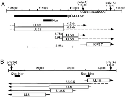
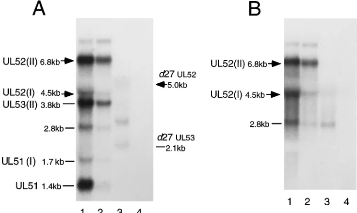
[image:2.612.102.507.73.392.2]S]methionine per ml in methionine-free MEM con-taining 1% FCS, as indicated in the Results. For pulse-chase experiments, the cultures were subsequently chased in fresh 199–1% CS medium containing un-labeled methionine at a concentration of 200 mM. Pulse-labeling was initiated and terminated quickly by submerging flasks in a 378C water bath and a 08C ice bath, respectively, as described previously (43). Cells were harvested in ice-cold FIG. 1. Map of transcripts and probes. Genome location is indicated across the top line of each panel by nucleotide number. Polyadenylation site consensus sequences are marked with downward arrows and the nucleotide position. DNA probes used for Northern analysis are shown as solid black bars. Previously identified ORFs are shown as open rectangles (2, 50). The locations of previously mapped transcripts are shown by solid lines extending from the ORF boxes with arrowheads pointing toward the 39end (2, 88). The approximate locations of transcripts proposed on the basis of these studies are shown by arrows with broken lines (– – – –), or simply by a broken line if a direction for the transcript is not being proposed. Sizes shown for the proposed transcripts were calculated on the basis of a plot derived from mobilities of an RNA size ladder and rRNAs. (A) UL52-UL54 (ICP27) region of the genome; (B) UL8-UL10 region of the genome. The BamHI-StuI fragment deleted from the d27 virus genome is indicated immediately below the genome scale by a striped rectangle.
on November 9, 2019 by guest
http://jvi.asm.org/
PBS containing 100mM phenylmethanesulfonyl fluoride (PMSF), 2mM N-a-p-tosyl-L-lysine chloromethyl ketone (TLCK), and 50 mM N-ethylmaleimide (NEM) (Sigma) and collected by centrifugation. Cell pellets were resuspended in IP buffer (25 mM Tris-HCl, pH 7.7; 150 mM NaCl; 1 mM EDTA; 1% Nonidet P-40; 0.5% sodium deoxycholate; 100mM PMSF; 2mM TLCK; 50 mM NEM) at a concentration of 8.33106cells per ml and sonicated for 1 s 12 times. In some cases, lysates were precleared with preimmune rabbit serum and fixed
Staphylo-coccus aureus cells (CalBiochem, LaJolla, Calif.) before specific
immunoprecipi-tations were performed. Before experiments were performed, antibodies were titrated to ensure that reactions were done under conditions of excess antibody (80). Except for UL8 immunoprecipitations, 5 to 7ml of the appropriate anti-bodies was added to lysate aliquots containing protein from approximately 4.23 105cells. Quantities of antibody and cell lysate were increased threefold for detection of low UL8 protein levels. After a 1-h incubation, immune complexes were collected with fixed S. aureus cells for 40 min on ice. Bound complexes were washed once with 500ml of IP buffer and twice with 500ml of wash buffer (50 mM Tris-HCl, pH 7.6; 150 mM NaCl; 5 mM EDTA; 0.5% Nonidet P-40; 100mM PMSF; 2mM TLCK; 50mM NEM). Denatured immunocomplexes were resolved by sodium dodecyl sulfate-polyacrylamide gel electrophoresis (SDS-PAGE) in a 9.25% polyacrylamide gel. Following electrophoresis, gels were fixed and pro-cessed for fluorography with En3Hance (New England Nuclear [NEN], Boston, Mass.). Dried gels were exposed to Kodak X-ray film for autoradiographic images and to phosphoimager screens for quantitation.
Western blot analysis.Vero and V27 cells were counted and infected at an MOI of 3 PFU per cell. At 1 hpi, the viral inoculum was replaced with 199–1% CS medium supplemented with 400mg of PAA per ml to block DNA replication. At 24 hpi, cells were harvested in SDS-sample buffer (75 mM Tris-HCl, pH 7.6; 2% SDS; 20% glycerol) and boiled. Proteins were separated by SDS-PAGE and transferred by electrophoresis to nitrocellulose (Schleicher & Schuell, Keene, N.H.) overnight in a minigel transfer cell (Bio-Rad, Melville, N.Y.). Membranes were blocked with 4% bovine serum albumin for 45 min at room temperature. Antibodies were applied as previously described (65). Secondary binding of the goat anti-mouse and/or anti-rabbit immunoglobulin G alkaline phosphatase-conjugated antibodies (Promega, Madison, Wis.) allowed detection of immune complexes by enzymatic activity. Color development substrates, Nitro Blue Tet-razolium and 5-bromo-4-chloro-3-indolylphosphate (BCIP), were used according to supplier specifications (Promega).
Northern (RNA) blot analysis.Cytoplasmic RNA for Northern blot analysis was isolated by phenol-chloroform extraction of 0.5% Nonidet P-40 cell lysate supernatants as previously described (62, 70). DNA was removed by digestion with RNase-free DNase I (Boehringer Mannheim, Indianapolis, Ind.). Purified RNA samples (15mg) and an RNA ladder (Bethesda Research Laboratories, Gaithersburg, Md.) for size estimation were resolved by electrophoresis through denaturing formaldehyde-agarose gels and transferred to Hybond-N Nytran membranes (Amersham, Arlington Heights, Ill.).
The plasmids used as probes were SV8.3 (ICP8) (16), pCM-UL42, pCM-pol, pCM-UL5, and pCM-UL52 (32). For increased specificity, the following DNA fragments were used to hybridize to UL52, UL8, and UL9 transcripts: the 540-bp
NcoI-NcoI fragment contained within the UL52 gene (Fig. 1A), the 337-bp XhoI-NarI fragment located in the 39half of the UL8 ORF (Fig. 1B), and the 226-bp SacI-NheI fragment spanning part of the promoter and 59end of the UL9 and UL10 ORFs (Fig. 1B). DNAs were radiolabeled by incorporation of [a-32P] dCTP with a specific activity of 6,000 Ci/mmol (NEN) using a random prime synthesis kit (Boehringer Mannheim). Hybridization of radiolabeled probes was performed by the Church-Gilbert method (10) and quantified by phosphoimage analysis (Molecular Dynamics) of the blots. Before being reprobed for ICP8 gene mRNA, blots were stripped by pouring a boiling solution of 0.1% SDS in 0.013 SSC (13SSC is 0.15 M NaCl plus 0.015 M sodium citrate) over the membranes.
Analysis of viral DNA replication.At 5 hpi, 199–1% CS medium containing [3H]thymidine (87.7 Ci/mmol) (NEN) at a concentration of 25mCi/ml was added to infected cells. After 3 h, the labeling medium was removed and total DNA was isolated (6). Briefly, cells were incubated at 378C overnight in 3 ml of lysis buffer (10 mM Tris-HCl, 10 mM EDTA, 2% SDS, 100mg of proteinase K per ml; pH 8). After addition of 300ml of 3 M Na acetate (pH 5.2), lysates were extracted with phenol-chloroform and DNA was precipitated with ethanol. RNA was removed by digestion with RNase A (25 mg/ml), and 4mg of each purified DNA sample was digested with XbaI and EcoRI. Digested DNA was resolved in a 0.9% agarose gel, and the gel was processed for fluorography with Entensity (NEN). The dried gel was exposed to Kodak (XAR) X-ray film, and bands were quan-titated with a densitometer.
RESULTS
The absence of ICP27 results in decreased accumulation of
viral replication proteins.Because ICP27 null mutant viruses,
[image:3.612.69.553.80.274.2]such as d27 (Fig. 1A), exhibit a defect in viral DNA replication, we hypothesized that ICP27 is necessary for adequate expres-sion of one or more of the essential replication proteins. To test that hypothesis, we compared the levels of UL5, UL9, Pol, UL42, UL8, and ICP8 proteins in cell extracts following infec-tion with WT or ICP27 mutant virus (Fig. 2; Table 1). The notable omission of UL52 in this and subsequent protein stud-ies was due to the lack of a suitable UL52-specific antibody; however, UL52 expression was assayed by other methods which are discussed below. WT virus-, d27 virus-, and mock-infected cells were labeled with [35S]methionine from 4.5 to 6
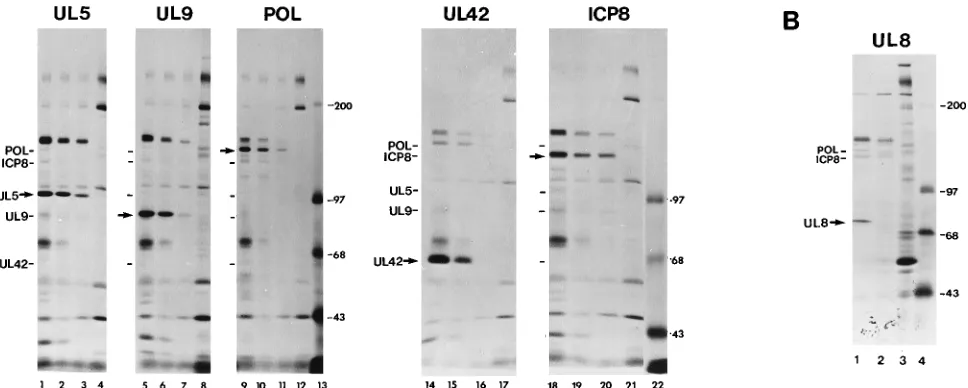
FIG. 2. Replication protein accumulation in d27 virus-infected cells. (A) Vero cells were mock infected or infected at an MOI of 20 with KOS1.1 or d27 in the presence or absence of PAA. Proteins were labeled with [35S]methionine from 4.5 to 6 hpi. Immunoprecipitations for the replication proteins indicated at the top of each gel were performed on equal fractions of lysate, and immune complexes were resolved by SDS-PAGE. Each group of samples was loaded in the order (lanes from left to right) WT, WT plus PAA, d27 plus PAA, and mock plus PAA. Lanes 13 and 22 contain14C-labeled protein molecular size standards. (B) Vero cells were mock infected or infected with KOS1.1 (WT) or d27 virus at an MOI of 20 in the presence of 400mg of PAA per ml. Infected cells were labeled, and UL8 immunopre-cipitations were performed as indicated above. Samples shown are WT plus PAA (lane 1), d27 plus PAA (lane 2), mock plus PAA (lane 3), and14C-labeled protein molecular size standards (lane 4). Positions of HSV-1 proteins are indicated at the left of each gel. Immunoprecipitated protein is designated with an arrow. Molecular sizes of the14C-labeled protein standards are indicated on the right in kilodaltons. (Autoradiograms shown are not of equal exposure times.)
on November 9, 2019 by guest
http://jvi.asm.org/
hpi and harvested for immunoprecipitation. As the status of HSV DNA replication can influence the expression of many viral genes, any potential effects due to DNA replication were ascertained by comparing WT-infected cells in the presence and absence of the viral DNA polymerase inhibitor, PAA. In agreement with results of earlier studies (2, 27, 56), there was a detectable decrease in the expression of several replication proteins when viral DNA synthesis was blocked (Fig. 2A). For this reason, our analysis of ICP27 effects has focused on WT virus- and d27 virus-infected cells under equivalent conditions in which differences in the degree of DNA replication have been eliminated by adding 400 mg of PAA per ml to both infected cultures.
The UL5 antibody precipitated a specific protein with an apparent molecular weight of approximately 102,000 (Fig. 2A, lanes 1 to 3). This corresponded reasonably well with the molecular weight of 99,000 predicted for UL5 and the 95,000 mobility previously reported by Olivo et al. (56). The level of radiolabeled UL5 in d27 virus-infected cells (Fig. 2A, lane 3) was 38% of the WT level (Fig. 2A, lane 2; Table 1). There was also a slight increase in the mobility of the UL5 protein pro-duced in cells infected with the d27 virus (Fig. 2A, lane 3). Hence, in addition to being underexpressed, UL5 may also have been altered posttranslationally in the absence of ICP27. The UL9 antibody bound to a protein with an apparent molecular weight of 89,000 (Fig. 2A, lanes 5 to 7), a size that resembles the molecular weight of 82,000 previously reported by Olivo et al. (49) and the 94,000 predicted molecular weight. Cells infected with the d27 virus (Fig. 2A, lane 7) contained 14% as much labeled UL9 as cells infected with WT virus (Fig. 2A, lane 6; Table 1). Likewise, the amount of UL42 immuno-precipitated from d27-infected cells was extremely small (Fig. 2A, lane 15), measuring approximately 8% of WT levels (Fig. 2A, lane 14; Table 1). Since Pol coprecipitated with UL42 in our assays (Fig. 2A, lanes 14 to 16), we were able to quantify the abundance of UL42-Pol complexes (11, 16, 19, 29, 33, 58, 82). Mutant virus-infected cells (Fig. 2A, lane 16) contained approximately one-fourth the number of UL42-Pol complexes observed following WT virus infection (Fig. 2A, lane 15), even though the amount of UL42 produced by the d27 virus was much smaller than that produced by the WT virus (8%) (Table 1). This difference was in agreement with previous findings that only 5 to 10% of the UL42 present in WT-infected cells is complexed with Pol (29). Consistent with Pol being the limiting factor in the formation of UL42-Pol complexes, the decreased levels of UL42-Pol complexes corresponded more closely with the extent of Pol accumulation in the mutant-infected cells (Fig. 2A, lane 11), which was measured to be 34% of the WT level (Fig. 2A, lane 10; Table 1). The level of UL8 in cells infected with the d27 virus was occasionally reduced below the
limits of detection when assayed by immunoprecipitation of radiolabeled proteins (Fig. 2B, lane 2; Table 1). However, UL8 was always observed to be present at low levels when assayed by Western blot analysis (80).
Because the structure of ICP8 is known to be altered in
d27-infected cells, leading to potential differences in antibody
recognition (13), we measured ICP8 accumulation not only by immunoprecipitation (Fig. 2A, lanes 19 and 20; Table 1) but also directly from infected-cell protein profiles (80). As ex-pected from previous work (49, 62), amounts of ICP8 pro-duced by WT virus (Fig. 2A, lane 19) and d27 virus (Fig. 2A, lane 20) were equivalent at 6 hpi (Table 1). ICP8 levels and examination of infected-cell protein profiles both indicated that all infections were established successfully and that each culture was radiolabeled efficiently. Furthermore, because the absence of ICP27 had no significant effect on ICP8 expression, we conclude that the reduced levels of UL5, UL9, Pol, UL42, and UL8 in d27 virus-infected cells were not due to a general defect inb-gene regulation.
ICP27 provided intransrestores WT expression of the
rep-lication proteins ind27 virus-infected cells.The altered gene
expression observed to occur in d27-infected cells could have been due to the lack of ICP27 or an indirect effect of the deletion or other secondary genomic mutation. To verify that the impaired replication protein expression in cells infected with the d27 virus was due specifically to the absence of ICP27, the effect of ICP27 was tested by two methods.
First, we determined whether providing ICP27 in trans to the
d27 virus from the ICP27-expressing V27 cell line (62) could
restore WT expression of the replication proteins. As expected, ICP27 was detected by Western blot in Vero cells infected with WT virus (Fig. 3A, lane 2) and in V27 cells infected with d27 virus (Fig. 3A, lane 4), but no ICP27 was detected in mock-infected (Fig. 3A, lane 1) or d27 virus-mock-infected (lane 3) Vero cells. Immunoblot analysis confirmed the immunoprecipitation results reported above, showing that accumulation of UL42 was significantly diminished in Vero cells infected with d27 virus (Fig. 3B, lane 3) compared with levels in Vero cells infected with WT virus (Fig. 3B, lane 2). When ICP27 was provided to the d27 virus in trans, however, the expression of UL42 approximated WT levels (Fig. 3B, lane 4). Similar results were obtained for UL5, UL8, UL9, and Pol (80). Importantly, the levels of ICP4 and ICP8 proteins were similar in all three infections, regardless of the presence of ICP27 (Fig. 3A, lanes 2 to 4, and Fig. 3C, respectively).
[image:4.612.57.299.81.174.2]The second way we confirmed that the absence of ICP27 was
[image:4.612.317.555.556.644.2]FIG. 3. ICP27 provided in trans restores WT expression of replication pro-teins in d27 virus-infected cells. Vero cells and the ICP27-expressing V27 cell line were infected at an MOI of 3 in the presence of 400mg of PAA per ml. At 24 hpi cells were harvested in SDS-sample buffer, and equal fractions of each lysate were subjected to SDS-PAGE. Proteins were transferred to nitrocellulose mem-branes and immunoblotted for the HSV-1 protein(s) indicated. Positions of the proteins detected on each blot are indicated on the right. (A and B) Lanes: 1, mock; 2, WT-infected Vero cells; 3, d27-infected Vero cells; 4, d27-infected V27 cells. (C) Lanes: 1, WT-infected Vero cells; 2, infected Vero cells; 3, d27-infected V27 cells.
TABLE 1. Protein levels in d27 mutant virus-infected cellsa
Protein Level relative
to WTb
UL5 ...38
UL8 ... 0c UL9 ...14
UL42 ... 8
Pol ...34
UL42-Pol complex...24
ICP8 ...82
a
Quantitation of experiment shown in Fig. 2.
b
Expressed as a percentage of WT levels in the presence of PAA.
c
Undetectable.
on November 9, 2019 by guest
http://jvi.asm.org/
responsible for the altered replication protein regulation in d27 virus-infected cells was to measure the level of replication proteins produced by other ICP27 mutants. As discussed in detail below, the n59R and n263R viruses, which contain non-sense mutations within the ICP27 gene at codons 59 and 263, respectively (62, 65), have defects in replication gene expres-sion identical to those of the d27 virus. This provided further evidence that the absence of ICP27 was responsible for the decreased replication protein accumulation observed to occur in d27 virus-infected cells.
Null mutation of any one essential replication gene does not
alter expression of the other replication proteins. Because
many of the replication proteins directly interact and function together (3, 4, 9, 12, 28, 33, 53, 55, 67), we wanted to determine if coordinate regulation among the viral replication proteins could explain some of the reduced levels of replication proteins in d27-infected cells. Therefore, we measured the level of rep-lication proteins expressed by six mutant viruses, each bearing a null mutation in one of the replication genes that was un-derexpressed by the d27 virus. Vero cells were infected in the presence of PAA with one of the six mutant viruses or WT
virus and radiolabeled with [35S]methionine from 4.5 to 6 hpi.
Immunoprecipitation of the replication proteins revealed that the absence of any one replication protein did not decrease accumulation of the other replication proteins (Table 2). There was slightly elevated expression of ICP8, Pol, UL42, and UL9 (twofold or less) in cells infected with viruses mutated in the genes encoding Pol (HP66), UL42 (CgalD42), or UL9 (hr94), suggesting possible regulatory effects among these four repli-cation proteins. However, there was no indirepli-cation that the low protein levels observed in d27 virus-infected cells were due to coordinate regulation among any of the essential replication proteins.
Replication protein synthesis, but not stability, is reduced in
cells infected with thed27 virus.As replication structure
for-mation is known to be defective in ICP27 mutant-infected cells (13), we performed pulse-chase labeling experiments to mea-sure the synthesis and stability of the unassembled replication proteins in cells following infection with the d27 virus. WT virus-, d27 virus-, and mock-infected cells were labeled at 5 hpi with [35S]methionine for 12 min. Cells were either harvested
immediately following pulse-labeling or chased in media with unlabeled methionine for 1 h before being harvested. Immu-noprecipitation of the replication proteins revealed greater incorporation of label into UL9 (Fig. 4, cf. lanes 1 and 2), Pol (cf. lanes 7 and 8), UL5 (cf. lanes 13 and 14), UL42 (cf. lanes 20 and 21), and UL8 (data not shown) in WT virus-infected cells than in d27 virus-infected cells. When expressed as a percentage of the WT levels, the protein synthesis levels in
d27-infected cells during the 12-min pulse (Table 3) correlated
[image:5.612.57.298.91.183.2]with the percent protein accumulation levels measured during long labeling periods (Table 1). Furthermore, the ratios of the labeled proteins between d27 virus- and WT virus-infected cells were the same before and after the 1-h chase period, indicating that all these proteins were equally stable in the two infections (Table 3). The apparent increase in labeled protein seen during the 1-h chase was likely due to the completion of
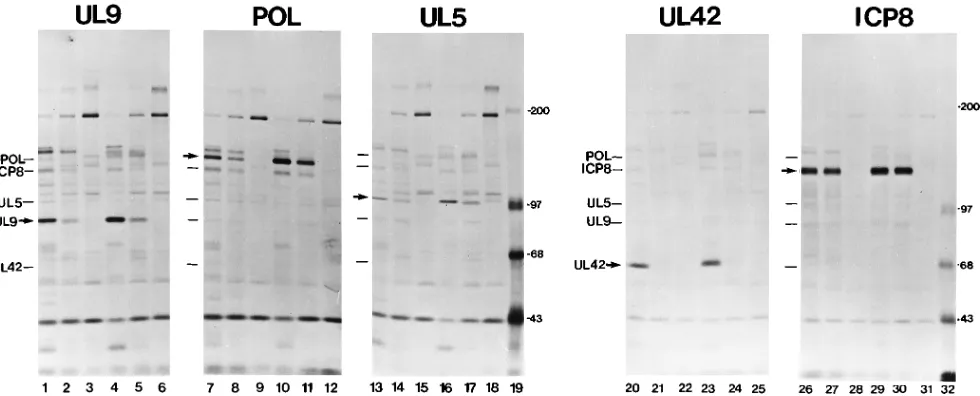
FIG. 4. Pulse-chase analysis of replication proteins in d27 virus-infected cells. Duplicate cultures of Vero cells were mock infected or infected with KOS1.1 (WT) or d27 virus at an MOI of 20 in the presence of 400mg of PAA per ml. Infected cells were pulse-labeled with [35
S]methionine for 12 min at 5 hpi. One set of infected cells was harvested immediately, while cells in parallel infections were incubated with media containing unlabeled methionine for 1 h before being harvested. Immunoprecipitations for the replication protein indicated at the top of each gel were performed on equal fractions of lysate, and immunocomplexes were resolved by SDS-PAGE. The first six lanes of each gel are as follows: WT (pulse), d27 (pulse), mock (pulse), WT (pulse-chase), d27 (pulse-chase), and mock (pulse-chase). Lanes 19 and 32 contain14
C-labeled protein molecular size standards. Positions of HSV-1 proteins are indicated at the left of each gel. The protein being specifically immunoprecipitated is designated with an arrow. Molecular sizes of the14
[image:5.612.64.554.462.660.2]C-labeled protein standards are indicated on the right in kilodaltons. (Autoradiograms shown are not of equal exposure times.)
TABLE 2. Replication protein expression by replication protein mutant virusesa
Mutant virus
Level of proteinb:
UL5 UL9 UL42 Pol ICP8
hr114(UL522) 88 96 88 120 110 hr80(UL82) 73 73 97 110 130 hr99(UL52) 110 150 120 130 hr94(UL92) 120 140 180 200
CgalD42(UL422) 68 180 170 160
HP66(Pol2) 120 150 240 170
a
Quantitation of one representative experiment.
b
Expressed as a percentage of WT levels in the presence of PAA.
on November 9, 2019 by guest
http://jvi.asm.org/
partially translated nascent chains (43). These results indicated that the reduced accumulation of the replication proteins in
d27-infected cells was a consequence of altered protein
syn-thesis.
As expected, the absence of ICP27 had no notable effect on ICP8 synthesis (Fig. 4, cf. lanes 26 and 27) or stability (lanes 26 and 27 versus 29 and 30) as measured by immunoprecipitation (Table 3) and quantitation from infected-cell protein profiles (data not shown). Also in agreement with earlier studies (49, 54, 62), quantitation of ICP6 protein levels from these protein profiles indicated that ICP6 expression was not affected by ICP27, measuring 96 and 108% of WT levels before and after the chase period, respectively.
It should be noted that some of the replication proteins appeared to coimmunoprecipitate in these experiments (Fig. 2 and 4). For example, a protein that comigrated with ICP8 reproducibly bound to polymerase immunocomplexes (Fig. 2A, lanes 9 and 10; Fig. 4, lanes 7 to 11) and two bands migrating at 128 and 135 kDa, believed to be ICP8 and poly-merase, respectively, coprecipitated with UL8 (Fig. 3B, lane 1). Further analysis is being done to verify the identities of the various coprecipitated proteins and to assess the specificity of these interactions.
Steady-state levels of the mRNAs encoding the essential
replication proteins are decreased in the absence of ICP27.To
investigate the basis of the altered replication protein synthesis rates in d27-infected cells, we measured the relative amounts of replication gene transcripts from infected cells in the pres-ence and abspres-ence of ICP27. Vero cells were mock infected or infected with WT or d27 virus, and cytoplasmic RNA was isolated at 5.5 hpi for Northern blot analysis (Fig. 5).
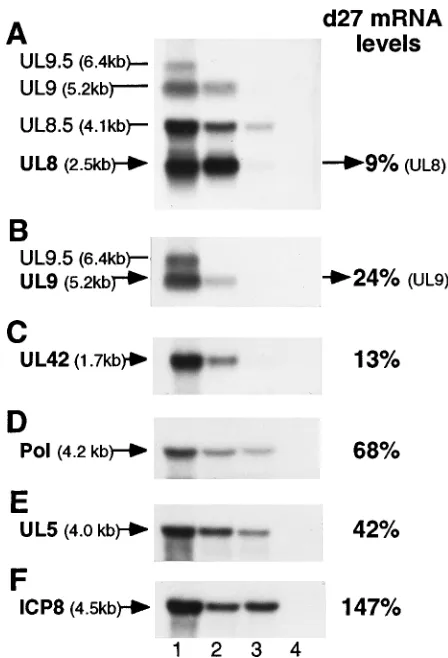
While the UL8 probe (XhoI-NarI; Fig. 1B) revealed a slight decrease in the level of the g-1 UL8.5 transcript when viral DNA synthesis was blocked, the level of the UL8 transcript was unaffected (Fig. 5A, cf. lanes 1 and 2). Under equivalent conditions of blocked viral DNA synthesis, there was a dra-matically lower accumulation of UL8 mRNA in d27-infected cells (Fig. 5A, lane 3) than in WT-infected cells (Fig. 5A, lane 2). Although UL9 mRNA could be detected by the UL8 probe, a second DNA fragment (SacI-NheI; Fig. 1B) was used to hybridize more specifically to the UL9 and UL9.5 mRNAs (Fig. 5B). As expected, the g-2 gene transcripts encoding UL9.5 (Fig. 5B, lane 2) and UL10 (data not shown) were barely detectable in the absence of viral DNA replication. Consistent with a recent report (2), the amount of UL9 mRNA was also decreased in the presence of PAA (Fig. 5B, cf. lanes 1 and 2). The level of UL9 mRNA, however, was further reduced in cells infected with the d27 virus (Fig. 5B, lane 3). Similar to that of UL9, UL42 mRNA accumulation was also diminished by the inhibition of viral DNA synthesis (Fig. 5C, cf. lanes 1 and 2), and the absence of ICP27 resulted in an additional decrease in UL42 mRNA levels (Fig. 5C, lane 3).
Though not as dramatic, the same general expression pattern was observed for Pol mRNA accumulation (Fig. 5D).
Unlike UL8, UL9, UL42, and Pol mRNAs, transcripts from the UL5 region of the genome have not been mapped pre-cisely. The pCM-UL5 plasmid (32) used to detect UL5 mRNA spans the entire UL5 ORF and parts of the adjacent UL4 and UL6 genes. Radiolabeled pCM-UL5 hybridized to several small transcripts in addition to two larger transcripts that had apparent sizes of 2.8 (data not shown) and 4.0 (Fig. 5E) kb. From the ATG translation start site (nucleotide 15133) to the proposed polyadenylation site (nucleotide 11760), the UL5 gene is 3,373 bp (50). Therefore, the only transcript recognized by this probe that is large enough to encode UL5 is the 4.0-kb mRNA. Not only did additional smaller probes verify that this transcript was UL5 (80), but this transcript also appears to correspond with the 4.0-kb b transcript previously identified between map units 0.08 and 0.011 (93). As was observed for the UL5 protein, UL5 mRNA levels in d27-infected cells (Fig.
FIG. 5. Northern blot analysis of replication gene transcripts. Vero cells were mock infected or infected at an MOI of 20 with KOS1.1 (WT) or d27 virus in the presence or absence of PAA. At 5.5 hpi, cytoplasmic RNA was isolated and subjected to Northern blot analysis.32
[image:6.612.321.545.275.604.2]P-labeled DNA probes were used to detect the HSV-1 mRNAs indicated on the left of each blot (panels A to F). Previously reported sizes for the UL8-UL9 transcript family (2), UL42 mRNA (18), Pol mRNA (35), and ICP8 mRNA (60), as well as the calculated size of the UL5 transcript, are shown in parentheses. Sample lanes on each blot are as follows: WT (lane 1); WT plus PAA (lane 2), d27 plus PAA (lane 3), and mock (lane 4). Levels of each mRNA were determined by phosphoimager analysis and d27 mRNA levels are shown as a percentage of the level measured from the WT plus PAA. After phosphoimage quantitation, all blots were stripped and reprobed for ICP8. The ICP8 blot shown is representative of the ICP8 mRNA abundance on all the membranes except for the UL42 blot, which is from a separate experiment in which the d27 ICP8 mRNA level was 85% that of the WT plus PAA.
TABLE 3. Pulse- and pulse-chase labeling of replication proteinsa
Label conditions Level of protein
b
:
UL5 UL8 UL9 UL42 Polc ICP8
Pulse 47 14 30 10 42 86
Pulse-chase 53 11 32 10 64 104
aQuantitation of experiment shown in Fig. 4.
bExpressed as a percentage of WT levels in the presence of PAA. cIn other identical pulse-chase experiments, this change in Pol ratios was not
observed and Pol levels were measured to be approximately 64% before and after the chase period.
on November 9, 2019 by guest
http://jvi.asm.org/
5E, lane 3) were reduced slightly in comparison with WT levels (Fig. 5E, lane 2).
After phosphoimage analysis, all Northern blot membranes were stripped and reprobed for ICP8 mRNA. In this experi-ment, ICP8 mRNA accumulated to slightly higher levels in cells infected with the d27 virus (Fig. 5F, lane 3) than in those infected with WT virus (Fig. 5F, lane 2). These ICP8 mRNA results not only provided an important contrast to the effect of ICP27 on the other replication gene mRNAs but also verified that all membranes contained equivalent amounts of RNA.
Overall, the replication gene mRNA levels measured in d27-infected cells corresponded closely to the protein pulse-label values shown above (Table 3). Hence, the lower rate of repli-cation protein synthesis in cells infected with the d27 mutant virus was likely the result of alterations in mRNA accumula-tion.
To ensure that the differences in cytoplasmic accumulation of UL8, UL9, UL42, Pol, and UL5 mRNAs were not a result of defective transport, Northern blot analysis was performed in parallel on total RNA and cytoplasmic RNA from infected cells. The relative levels of the replication gene transcripts from d27 virus- and WT virus-infected cells were similar in both total and cytoplasmic RNA samples (80). Consequently, the differences in cytoplasmic mRNA levels in d27-infected cells cannot be explained simply by a defect in mRNA trans-port.
Identification and expression of two UL52 transcripts.
Be-ing unable to monitor UL52 protein levels within virus-in-fected cells, we wished to assess the effect of ICP27 on UL52 expression by measuring levels of UL52 mRNA. As mapping of the UL52 transcript has not been reported, it was first necessary to characterize the UL52 gene transcript(s). The radiolabeled pCM-UL52 plasmid initially used to detect UL52 mRNA also spans regions of the UL51 and UL53 genes (Fig. 1A). On the basis of the sequence of the HSV-1 genome, the UL52 and UL53 transcripts should be approximately 4.5 and
1.4 kb, respectively, if the polyadenylation signal at the 39end of the UL53 gene is used. Alternatively, if the UL54 polyade-nylation site is used, the UL52 and UL53 transcripts would each be expected to be 1.9 kb longer (6.4 and 3.3 kb, respec-tively) (Fig. 1A). In addition to the abundant leftward-oriented UL51 transcript (80), four transcripts with sizes of 6.8, 4.5, 3.8, and 1.7 kb were detected from WT-infected cells (Fig. 6A, lane 1). These sizes corresponded reasonably well to the four sizes predicted, suggesting that the polyadenylation signal at the 39 end of the UL53 gene was not used exclusively and that a large percentage of the UL52 and UL53 transcripts terminated at the UL54 (ICP27) polyadenylation site.
Additional evidence that the 6.8- and 4.5-kb RNAs are UL52 transcripts came from Northern blots of RNA from d27 and hr114 mutant virus-infected cells. The deletion in the d27 virus genome removes the polyadenylation site 39of the UL53 gene; hence, all UL52 and UL53 transcripts should terminate at the next available polyadenylation site 39of UL54 (ICP27). The UL52 and UL53 transcripts produced by the d27 virus, however, would be expected to be 1.6 kb smaller than the 6.8-and 3.8-kb transcripts observed from WT-infected cells (5.2 and 2.2 kb, respectively) because of the 1.6-kbp deletion in the viral genome (Fig. 1A). In support of our transcript identifi-cations, shifted transcripts with apparent sizes of approxi-mately 5.0 and 2.1 kb were observed in RNA isolated from
d27-infected cells (Fig. 6A, lane 3). Likewise, the hr114 virus
contains a mutation in this region, specifically, a 4.2-kbp lacZ fragment insertion into the UL52 gene (26). The 6.8- and 4.5-kb RNAs appeared to be shifted accordingly, as two tran-scripts estimated to be in the size range of 11.0 and 8.7 kb were detected from hr114-infected cells with both UL52 probes (80). Thus, the 6.8- and 4.5-kb RNAs behaved as transcripts of the UL52 gene in response to insertions and deletions in this region. As a result, we tentatively designated them UL52(I) and UL52(II) to indicate the UL52 ORF on a transcript ending at the first and second available polyadenylation sites.
Simi-FIG. 6. Analysis of UL52 transcripts and their expression in ICP27 mutant virus-infected cells. For all blots, the name and/or size of transcripts detected from WT-infected cells is shown on the left. (A) Vero cells were infected as described in the legend to Fig. 5, and Northern blot analysis was performed with the pCM-UL52 probe. The UL51 transcript, estimated as approximately 1.4 kb in size, was identified by a strand-specific RNA probe anti-complementary to the UL52 and UL53 transcripts. Transcripts from d27 mutant-infected cells are indicated on the right. Lanes: 1, WT; 2, WT plus PAA; 3, d27 plus PAA; 4, mock. (B) Vero cells were mock infected or infected with WT or n59R mutant virus at an MOI of 20. At 5.5 hpi, cytoplasmic RNA was harvested and subjected to Northern blot analysis with the UL52
Nco fragment probe. Lanes: 1, WT; 2, WT plus PAA; 3, n59R plus PAA; 4, mock.
on November 9, 2019 by guest
http://jvi.asm.org/
[image:7.612.121.489.72.292.2]larly, we designated the 1.7- and 3.8-kb transcripts as UL53(I) and UL53(II), respectively.
To analyze the effects of ICP27 on the accumulation of the UL52(I) and UL52(II) mRNAs, we used a smaller, more spe-cific UL52 probe and the ICP27 point mutant virus, n59R, which exhibits no shifts in UL52 transcript size. Cytoplasmic RNA was harvested from WT-infected (with or without PAA),
n59R-infected (with PAA), and mock-infected cells at 5.5 hpi
and probed by Northern blot analysis with a radiolabeled 540-bp DNA fragment (NcoI-NcoI) contained within the UL52 gene (Fig. 1A). This probe hybridized to three transcripts from cells infected with WT virus, two of which corresponded to the 4.5- and 6.8-kb transcripts (Fig. 6B, lanes 1). The Nco probe also hybridized to a third transcript approximately 2.8 kb in size of unknown origin. Although the amount of the 2.8-kb transcript was increased slightly, the levels of both UL52 mRNAs were reduced in n59R-infected cells (Fig. 6B, lane 3) compared with levels in WT-infected cells (Fig. 6B, lane 2). Interestingly, accumulation of the larger UL52(II) transcript appeared to be more severely decreased than that of UL52(I) in response to the absence of functional ICP27. In contrast, the smaller UL52(I) and UL53(I) transcripts were reduced to a large degree than were the UL52(II) and UL53(II) transcripts when viral DNA synthesis was blocked (Fig. 6A, cf. lanes 1 and 2; Fig. 6B, cf. lanes 1 and 2). Hence, the levels of both UL52 transcripts were decreased in n59R-infected cells, but there were detectable differences in the expression patterns of the two UL52 mRNAs.
Replication gene expression remains low throughout the
course of d27 infection. Because ICP27 mutants have been
reported to exhibit slower infection kinetics, which includes a delay in accumulation of some b-gene products (49, 54), we monitored expression of the replication genes from 4 to 15 hpi in the presence of the viral DNA synthesis inhibitor PAA. Replication gene mRNAs in d27 virus- and WT virus-infected cells were compared at 4, 6, 9, 12, and 15 hpi by Northern blot analysis.
In cells infected with the d27 mutant virus, accumulation of UL8, UL9, UL42, UL5, and UL52 (5.0-kb shifted transcript) mRNAs remained at a low level throughout infection (Fig. 7; UL52 data not shown). Pol mRNA levels, however, increased gradually over the course of d27 infection, in contrast to the shutoff that appeared to occur in WT infection (Fig. 7). This resulted in higher Pol mRNA levels in d27-infected cells by 10 hpi. Relative ICP8 mRNA levels in d27-infected cells fluctu-ated slightly, but as expected they approximfluctu-ated WT infection levels at each time point (Fig. 7). Similar expression patterns were also observed for these genes at the protein level by immunoprecipitation and Western blot analysis (80). Hence, while the ratio of UL9, UL8, UL42, UL5, and UL52 mRNAs between d27 and the WT virus varied at different times during infection, it was clear that the defect in replication gene ex-pression observed with the d27 virus at 6 hpi was not due merely to a delay in synthesis.
Replication protein expression correlates with levels of viral
DNA replication. To determine if there was a relationship
between replication protein production and the ability of var-ious ICP27 mutants to amplify viral DNA, we measured viral DNA synthesis and replication protein expression in cells in-fected with d27, n59R, n263R, or n504R ICP27 mutant viruses (62, 65). Infected-cell proteins were labeled with [35
S]methi-onine for immunoprecipitation analysis, while parallel infected cultures were labeled with [3H]thymidine for measuring DNA
synthesis.
Consistent with previous work, little DNA was replicated by the d27, n59R, and n263R viruses, replication measuring below 10% of WT levels (Fig. 8). At the same time, the n504R virus synthesized considerable amounts of viral DNA, although not equal to WT levels as previously observed (62, 64).
While quantities of UL5 and Pol protein did not closely reflect the viral DNA replication levels in cells infected with the ICP27 mutants, the relative amounts of UL9 and UL8 showed a striking resemblance to the degree of DNA synthesis
[image:8.612.61.298.71.363.2]FIG. 7. Time course of replication gene mRNA accumulation in WT virus-and d27 virus-infected cells. Vero cell cultures were infected at an MOI of 20 with KOS1.1 (WT) or d27 virus. At 4, 6, 9, 12, and 15 hpi, cytoplasmic RNA was isolated and subjected to Northern blot analysis.32P-labeled DNA probes were used to detect the HSV-1 mRNAs indicated at the upper left corner of each graph. Transcripts were quantified by phosphoimage analysis, and relative abun-dance expressed as a fraction of the peak value was plotted for each time point. j, WT;}, d27.
FIG. 8. Protein levels and viral DNA replication in cells infected with ICP27 mutant viruses. Duplicate Vero cell cultures were infected with the HSV-1 strains indicated below. One set of infected cell cultures was labeled with [35
S]methionine for protein analysis, and the other was labeled with [3
H]thymidine to measure DNA replication.■, WT; , d27; , n59R;o, n263R;h, n504R.
on November 9, 2019 by guest
http://jvi.asm.org/
that occurred in each virus infection (Fig. 8). Cells infected with d27, n59R, and n263R contained 14, 6, and 11% of the amount of UL9 in WT-infected cells, respectively. Similarly, the relative levels of UL8 in the d27-, n59R-, and n263R-infected cells were 9, 7, and 8% of WT levels. As would be expected if the availability of these proteins was limiting viral DNA synthesis, cells infected with the n504R replication-com-petent virus contained significantly higher levels of UL9 and UL8, 34 and 57% of WT levels, respectively. Thus, within infected cells the extent of UL9 and UL8 accumulation closely matched the 10% (d27), 6% (n59R), 7% (n263R), and 50% (n504R) levels of DNA replication.
When expressed as a percentage of WT levels, UL42 accu-mulation appeared to be much lower than viral DNA replica-tion levels in all mutant-infected cells; however, as detailed in the Discussion section, only 5 to 10% of UL42 is complexed with Pol (29). Thus, the relative amounts of UL42 present within these mutant-infected cells were not necessarily incon-sistent with the amounts of DNA synthesized. Comparing UL42 levels among the different mutant infections, there was five to nine times more UL42 present in cells following n504R infection (9% of WT levels) than in cells infected with any of the other three ICP27 mutant viruses (1 to 2% of WT levels). Likewise, the n504R mutant replicated five- to eightfold more DNA than did the other mutant viruses. Therefore, when com-parisons among the ICP27 mutant infections are made, relative UL42 levels did correlate with the amount of DNA synthe-sized.
DISCUSSION
It has been known for many years that ICP27 stimulates viral DNA replication (6, 49, 62, 65, 68). These studies were under-taken to determine how this effect is exerted. Because ICP27 has been shown to modulate viral gene regulation, we hypoth-esized that ICP27 may promote viral DNA synthesis by elevat-ing the expression of essential replication genes. To test that hypothesis, we examined the synthesis, accumulation, and sta-bility of the replication proteins and steady-state levels of the replication gene transcripts in cells infected with ICP27 mutant viruses. We have determined that in the absence of ICP27, expression of most of the essential replication genes is signif-icantly reduced within infected cells. The levels of UL5, UL8, UL52, UL9, and UL42 gene products in ICP27 mutant-in-fected cells remain lower than levels in WT-inmutant-in-fected cells throughout the course of infection. In contrast, while Pol ex-pression is decreased in d27-infected cells at 6 hpi, Pol gene products continue to accumulate gradually in the absence of ICP27, ultimately reaching higher levels than in WT-infected cells by 10 hpi. As novel regulation of Pol expression has been reported to occur during HSV infection (89), it is not surpris-ing to find that in the absence of ICP27 additional regulatory factors may be affecting the expression of this gene. The only other evidence that ICP27 might affect b-gene expression, which was published while this manuscript was in preparation, was a report that ICP27 stimulates ICP6 and thymidine kinase (TK) expression in the absence of WT ICP4 (69), a situation clearly different from the conditions studied in this work.
While it has not yet been determined which of the essential proteins is the limiting factor in viral DNA replication, the magnitude of decrease in UL8 and UL9 expression by different ICP27 mutant viruses correlated with the reduced ability of these mutants to replicate DNA. Although these decreases may be sufficient to explain the reduced DNA replication of ICP27 mutants, DNA synthesis levels also corresponded to some extent with UL42 expression. It has been reported that
over 90% of the UL42 produced by WT virus is not required for the maintenance of WT levels of DNA replication (40) and that only 5 to 10% of the UL42 produced in WT-infected cells is complexed with Pol (29). For that reason, availability of UL42 protein may not become limiting to DNA synthesis until levels of the protein become extremely low (approximately 10% of WT levels). Consequently, presenting UL42 accumu-lation in the mutant-infected cells as a percentage of the excess UL42 present in WT-infected cells could obscure a correlation with DNA replication levels. A comparison of UL42 expres-sion among cells infected with the replication-competent and the replication-defective ICP27 mutant viruses revealed a re-lationship between the levels of UL42 present and the amount of viral DNA synthesized. On the basis of these results, we conclude that ICP27 stimulation of several essential bgenes promotes amplification of viral DNA. It should be noted that we believe the lower replication levels measured for the n504R mutant in these experiments (50%) were due to the earlier time at which DNA was labeled. These results and one other published study (63) suggest that the n504R virus might have slightly delayed DNA replication kinetics in comparison with the WT virus.
Diverse regulation among the b genes. As we found no
indication that the observed alterations in replication protein expression could be attributed to coordinated regulation among the various replication proteins, it was curious that the absence of ICP27 did not affect allbgenes to an equal degree. Expres-sion of some genes, such as the UL8 and UL42 genes, was barely detectable without ICP27, while other genes were af-fected moderately (UL5 and Pol genes) or not to any signifi-cant extent (ICP8 and ICP6 genes). This cannot be explained simply by the differences in basal activity of these genes, as the abundantly expressed viral genes encoding UL42 and ICP8 responded very differently to the presence of ICP27. Consis-tent with the work of others (2, 27, 89), we observed that many of the replication genes were sensitive to the status of DNA replication, but again the magnitude of that effect was not the same for all of the b genes. The differing degrees to which these genes were stimulated by ICP27 did not correlate in any consistent way with the observed variations in PAA sensitivity (e.g., UL8). Therefore, ICP27 appears to achieve selective stimulation of a subset ofbgenes independently of these other regulatory effects.
These observations also raise broader questions about whyb genes exhibit such a spectrum of expression patterns. Whileb genes have been grouped together on the basis of general kinetic properties, there is clearly differential regulation occur-ring within this group, as others have previously acknowledged (reviewed in reference 66; 89, 93). Although it is easy to un-derstand why this large and varied assortment of genes might require additional specificity in their expression that cannot be rationalized by their generalbclassification, in the case of the DNA replication proteins which act together, it is less obvious what selective advantage is attained by having separate expres-sion strategies.
One consideration is that the seven replication proteins most likely are needed at different levels for viral replication. That alone, however, would not necessarily result in the evolution of distinct regulatory mechanisms. Alternatively, there have been reports which indicate that excess UL9 may inhibit the viral replication process (46, 75, 77). Furthermore, while UL9 is required early to initiate origin-dependent replication, it is unnecessary for the continuation of rolling-circle replication (75). Proteins such as UL9, which potentially exert negative effects on viral growth, would be expected to be regulated by fail-safe strategies of checks and balances to ensure that their
on November 9, 2019 by guest
http://jvi.asm.org/
expression is properly controlled. This is one possible expla-nation for the low levels of UL8, UL9, and UL52 maintained within virus-infected cells (56). At the other extreme, higher levels of some replication proteins may be required at different times during infection so that they are available for functions other than viral DNA synthesis. One example is ICP8, which has been implicated in regulating the switch from early to late phases of viral infection by down-regulating expression of genes from parental genomes and stimulating late gene tran-scription from progeny templates (8, 21–24). Hence, different programs of regulation may have evolved in response to addi-tional functions or side effects of these proteins.
The effect of ICP27 on replication gene expression is
medi-ated at the mRNA level.Northern blot analysis indicated that
the effects of ICP27 on replication gene expression can be detected at the level of mRNA accumulation. Additional ex-periments will be required to determine why mRNA accumu-lation is decreased and specifically how selectivity is mediated. Other regulatory effects of ICP27 observed during infection have been attributed to alterations in transcription (39, 54, 76) and posttranscriptional processing (30, 76). Several mecha-nisms that could account for these regulatory effects have been proposed.
First, ICP27 may alter transcription rates via its interaction with or effects on ICP4 (17, 73, 78). For instance, ICP4 phos-phorylation is altered when ICP4 is produced in the absence of ICP27 (92). However, Samaniego et al. (69) report that ICP27 exerts an effect on TK and ICP6 mRNA levels during infection, in the absence of WT ICP4 transactivation. Although ICP27 can alter mRNA processing by the redistribution of cellular splicing factors (57) and the inhibition of splicing (30, 31), none of the essential replication genes contain introns, so that this ICP27 activity would also not be expected to directly con-trol the expression of theseb genes. Nevertheless, the possi-bility remains that the effects of ICP27 on spliced transcripts could indirectly alter the expression of the nonspliced replica-tion gene.
A third means of ICP27 regulation, in which ICP27 exerts regulatory effects based on the presence of different 39UTR sequences and/or polyadenylation signals has been proposed (7, 51, 52, 72). Unfortunately, while HSV-1a- andg-gene 39 regions have been assayed for ICP27 responsiveness,b-gene 39 sequences have not been studied. It has been shown, however, that ICP27 increases expression of a chimeric reporter con-struct containing the ICP27 (UL54) polyadenylation site (72). This may be relevant to the ICP27b-gene regulatory effects reported here, as we have found that the UL52 gene, at least in part, appears to use the ICP27 polyadenylation signal. In fact, of the two UL52 transcripts observed, the larger, 6.8-kb UL52(II) transcript that terminates at the ICP27 polyadenyl-ation site appears to be more dependent on ICP27 for expres-sion.
Thus, there are multiple ways in which ICP27 might influ-ence the expression of the replication genes. Because the func-tion of ICP27 may have general applicability in explaining aspects of regulatory selectivity, the study of ICP27 presents an excellent opportunity for extending our understanding of the complexities of gene regulation. As mechanistic explanations for ICP27 effects are sought, it is important to be aware of all the regulatory effects that are exerted by ICP27 during infec-tion. Here we show an essential regulatory effect of ICP27 which is more specific than general b-gene activation. While ICP27 enhancement of replication gene activity may involve coordinated interaction with other a proteins, these results further identify ICP27 as a key factor in HSV gene regulation. The different magnitudes of ICP27 responsiveness measured
among the replication genes not only raise critical questions about the specificity of b-gene regulation but also may ulti-mately provide insights into the nature of the mechanisms at work. The qualitatively distinct regulation observed for ICP8 is a potentially useful comparison which may help identify the properties that render the other genes sensitive to this ICP27 regulation.
Finally, the severe underexpression of the essential replica-tion proteins which we have observed to occur in ICP27 mu-tant-infected cells provides a clear explanation for the DNA replication-deficient phenotype of these mutants. In addition, because UL5, UL8, UL52, and UL9 are required for ICP8 localization (45) and ICP8 conformational changes (81), this regulatory effect also accounts for the altered properties of ICP8 observed in ICP27 mutant infections (13).
ACKNOWLEDGMENTS
We greatly appreciate the generous amounts of replication protein antisera supplied by Paul Olivo, Mark Challberg, and Sandra Weller. In addition, we are grateful to Sandra Weller, Paul Johnson, and Don Coen, who supplied mutant virus strains and the complementing cell lines on which the viruses were propagated. We thank Mei Chen for providing valuable technical advice regarding Northern blot tech-niques. We also thank Don Coen and Cindy Murphy for critical review of the manuscript.
This research was supported by Public Health Service grants AI20530 and CA26345. S.L.U. was supported by Public Health Service training grant AI07245.
REFERENCES
1. Ackerman, M., D. K. Braun, L. Pereira, and B. Roizman. 1984. Character-ization of herpes simplex virus 1aproteins 0, 4, and 27 with monoclonal antibodies. J. Virol. 52:108–118.
2. Baradaran, K., C. E. Dabrowski, and P. A. Schaffer. 1994. Transcriptional analysis of the region of the herpes simplex virus type 1 genome containing the UL8, UL9, and UL10 genes and identification of a novel delayed-early gene product, OBPC. J. Virol. 68:4251–4261.
3. Boehmer, P. E., M. C. Craigie, N. D. Stow, and I. R. Lehman. 1994. Asso-ciation of origin binding protein and single strand DNA-binding protein, ICP8, during herpes simplex virus type 1 DNA replication in vivo. J. Biol. Chem. 269:29329–29334.
4. Boehmer, P. E., and I. R. Lehman. 1993. Physical interaction between the herpes simplex virus 1 origin-binding protein and single-stranded DNA-binding protein ICP8. Proc. Natl. Acad. Sci. USA 90:8444–8448. 5. Carmichael, E. P., and S. K. Weller. 1989. Herpes simplex virus type 1 DNA
synthesis requires the product of the UL8 gene: isolation and characteriza-tion of an ICP6::lacZ insercharacteriza-tion mutacharacteriza-tion. J. Virol. 63:591–599.
6. Challberg, M. D. 1986. A method for identifying the viral genes required for herpesvirus DNA replication. Proc. Natl. Acad. Sci. USA 83:9094–9098. 7. Chapman, C. J., J. D. Harris, M. A. Hardwicke, R. M. Sandri-Goldin, M. K.
Collins, and D. S. Latchman.1992. Promoter-independent activation of heterologous virus gene expression by the herpes simplex virus immediate-early protein ICP27. Virology 186:573–578.
8. Chen, M., and D. M. Knipe. Unpublished data.
9. Chiou, H. C., S. K. Weller, and D. M. Coen. 1985. Mutations in the herpes simplex virus major DNA-binding protein gene leading to altered sensitivity to DNA polymerase inhibitors. Virology 145:213–226.
10. Church, G. M., and W. Gilbert. 1984. Genomic sequencing. Proc. Natl. Acad. Sci. USA 81:1991–1995.
11. Crute, J. J., and I. R. Lehman. 1989. Herpes simplex-1 DNA polymerase. Identification of an intrinsic 59----39exonuclease with ribonuclease H activ-ity. J. Biol. Chem. 264:19266–19270.
12. Crute, J. J., T. Tsurumi, L. A. Zhu, S. K. Weller, P. D. Olivo, M. D.
Challberg, E. S. Mocarski, and I. R. Lehman.1989. Herpes simplex virus 1 helicase-primase: a complex of three herpes-encoded gene products. Proc. Natl. Acad. Sci. USA 86:2186–2189.
13. Curtin, K. D., and D. M. Knipe. 1993. Altered properties of the herpes simplex virus ICP8 DNA-binding protein in cells infected with ICP27 mutant viruses. Virology 196:1–14.
14. DeLuca, N. A., A. M. McCarthy, and P. A. Schaffer. 1985. Isolation and characterization of deletion mutants of herpes simplex virus type 1 in the gene encoding immediate-early regulatory protein ICP4. J. Virol. 56:558– 570.
15. Desai, P., R. Ramakrishnan, Z. W. Lin, B. Osak, J. C. Glorioso, and M.
Levine.1993. The RR1 gene of herpes simplex virus type 1 is uniquely trans