“Efficacy of Simplified Acute Physiological
Scoring II in predicting the Mortality and
Morbidity in Perforation Peritonitis”
Dissertation submitted to
The Tamil Nadu M.G.R Medical University
Chennai- 600032
In partial fulfilment of the
Regulations of the award of degree of
M.S. General Surgery
Department of General Surgery
Coimbatore Medical College Hospital
CERTIFICATE
This is to certify that this dissertation titled “Efficacy of Simplified Acute Physiological Scoring II in predicting the Mortality and Morbidity in Perforation Peritonitis” submitted to the Tamil Nadu Dr. M.G.R. Medical University, Chennai in partial fulfilment of the requirement for the award of
M.S Degree Branch - I (General Surgery) is a bonafide work done by Dr.
Adarsh G., post graduate student in General Surgery under my direct
supervision and guidance during the period of September 2011 to November
2012.
Prof. G. Ravindran, M.S. Associate Professor Dept of General Surgery
Coimbatore Medical College Hospital
Prof. P.V. Vasantha Kumar, M.S. Professor and Head of the Department Dept. of general Surgery
Coimbatore Medical College Hospital
Dr. Vimala, M.D.
Dean,
ACKNOWLEDGEMENT
It gives me immense pleasure to express my deep sense of gratitude to
my Unit Chief Prof. Dr. G. Ravindran, M.S. General Surgery, Department of
General Surgery, Coimbatore Medical College Hospital, for his excellent
guidance and valuable suggestions during the course of study and in preparation
of this dissertation.
I am grateful to Prof. Dr. P.V.Vasantha Kumar, M.S., Professor and Head
of the Department of General Surgery, Coimbatore Medical College Hospital ,
for his guidance throughout this study. I also express my heartfelt thanks for the
associate professors of the college, Dr. Elango, Dr. Swaminathan, Dr.
Ranganathan, Dr. Natarajan, and Dr. Saradha for their suggestions at the apt
time that has helped me in the completion of this work.
I am grateful to my Assistant Professors, Dr. G. Vishwanathan, M.S., Dr.
Muthulaxmi, M.S., for their help and guidance throughout this study.
I express my gratitude to Dr.Vimala., Dean, Coimbatore Medical College
Hospital for permitting me to use the clinical material for the study.
I express my thanks to my friends and all others who have helped me in
the preparation of this dissertation.
Last but not the least; I heartily thank all the patients for their kind
DECLARATION
I hereby declare that the dissertation entitled "Efficacy of Simplified Acute Physiological Scoring II in predicting the Mortality and Morbidity in Perforation Peritonitis" was done by me at Coimbatore Medical College Hospital Coimbatore – 641018 during the period of my post graduate study for
M.S. Degree Branch-1 (General Surgery) from 2010 to 2013.
This dissertation is submitted to the Tamil Nadu Dr. M.G.R. Medical University
in partial fulfilment of the University regulations for award of M.S., Degree in
General Surgery.
Dr. Adarsh G.
Post Graduate Student M.S. General Surgery
CONTENTS
Sl No Topic Page No.
1 Introduction 1
2 Aim of the study 4
3 Review of Literature 5
4 Materials & Methods 46
5 Results 52
6 Discussion 76
7 Conclusion 81
8 Appendix-1 Porforma
9 Appendix-2 Bibliography
Efficacy of Simplified Acute Physiological Scoring II in predicting
mortality and morbidity in Perforation Peritonitis
Abstract:
Introduction: Perforation peritonitis is a very common cause of generalized peritonitis in India. Prognosis of the disease is often difficult and complex. Individual prognosis by treating doctors is subjective and often overstates the chance of survival. We hence intend to find out the performance of simplified acute physiological scoring II (SAPS II) in predicting the mortality and morbidity in patients having perforation peritonitis. SAPSII takes into account 13
physiological variables and presence of chronic illness like AIDS and malignancies in giving the prognosis of the patient in the first 24 hours of admission.
Aims: 1.To evaluate the value of SAPS II scoring in predicting the mortality and morbidity in patients suffering from perforation peritonitis. 2. To provide an objective prognostic system for patients with perforation peritonitis. 3. To provide a risk classification system for patients presenting with perforation peritonitis.
Results: We applied SAPS II score to 100 consecutive patients admitted in our hospital during the study period. 89 were males and 11 were females. We could divide our patients into three groups depending on their SAPS II score as, those having a score less than 20, those having a score of 21-40, and those having a score above 40. The group having a SAPS II score less than 20 had no mortality and lesser hospital stay and were classified as low risk group. Patients with SAPS II score 21-40 had a higher morbidity and mortality and were classified as moderate rick group and patients having SAPS II score more than 40 had maximum
mortality and were classified as high risk group.
Conclusion: SAPS II score is a very good tool in predicting Mortality and morbidity in patients suffering from perforation peritonitis.
1
INTRODUCTION
Over last two centuries the medical field has gone a full circle. For nineteenth
century physicians especially the French, the main goal of medicine was not to
cure but to diagnose the disease and give a satisfactory prognosis of the patients’
chances of survival. Only in the twentieth century did the need to cure the patient
came into the forefront. During this time the prognosis was left to the treating
doctor without any standardization. Towards the end of the twentieth century it
was realized that when a doctor makes a judgment or an estimate on behalf of one
patient it was based on own knowledge, experience or intuition and hence was very
subjective. This led to improper management and poor outcome in some of the
patients. It was thus realized that compared to subjective estimates, objective
estimates based on hard facts and precise measurements would be more accurate,
uniform and reproducible. Patients present with myriad of signs and symptoms,
some complex and some simple. The complex data if it could be presented in a
simple, understandable and in an objective form would help us to teach, evaluate,
review and reproduce it in any situation.
This led to the development of scoring systems which tried to objectively predict
2
taking into account the objective values of various physiological parameters of the
patient and the presence of different chronic diseases in the patient.
The accurate predictive ability would make it possible to measure more precisely,
the quality of intensive care and other new life-saving technologies. Precise
prognosis or risk stratification before treatment would also enable clinical
researchers to use observational studies to contrast the quality of care in various
intensive care units (ICUs) and to identify those components of ICU structure that
are linked to improved patient outcome.
Scoring and grading allows us to understand
1. The pattern of occurrence
2. The complicating or limiting factors, and
3. The various outcomes
Such information could lead to better clinical decision making that would help in
assessing quality of care, identify the deficiencies, enhance patient satisfaction and
guide the rational allocation of health care resources. These risk-adjusted
comparisons can then be made between different surgeons and different hospitals
spanning different geographical areas.
Perforation peritonitis is a common and serious surgical emergency. The famous
3
escape from perforation peritonitis due to which he died following a blunt injury
abdomen.
Perforation of hollow viscus is one of the most important etiological factors in
causing peritonitis in developing countries. Management of perforation continues
to be highly demanding, difficult and complex.
The etiological spectrum of peritonitis in Asia is different from that of western
countries and there is paucity of data from India regarding its prognostic indicators,
mortality and morbidity patterns.
Thus there is a need to properly prognosticate the condition and predict the
mortality and morbidity patterns.
Most of the cases of perforation peritonitis present to peripheral hospitals where
there is a lack of advanced investigative modalities.
There is a need to validate a scoring system which predicts the prognosis of the
patient with minimal investigative modalities.
Simplified acute physiological scoring system II is prognostic system based on
different clinical parameters, physiological parameters and also some basic
investigations recorded within 24 hours of the patients’ admission. It has been used
in different ICU setups in the western countries for patients with different
diagnosis. We intend to apply this scoring system in perforation peritonitis and test
4
OBJECTIVES OF THE STUDY
• To evaluate the value of SAPS II scoring in predicting the mortality and morbidity in patients suffering from perforation peritonitis
• To provide an objective prognostic system for patients with perforation peritonitis
5
REVIEW OF LITERATURE
ANATOMY OF THE PERITONIUM
(1, 2, 3)Peritoneum has 2 layers. Peritoneal cavity is lined by parietal peritoneum and the
intra abdominal organs are covered by visceral peritoneum.
Together its surface area roughly corresponds to the body cutaneous surface area.
Peritoneum consists of a single layer of flattened cells, mesothelium, overlying
areolar tissue which varies in density in different regions. Over expansile parts this
areolar tissue is loose (eg. Transversalis fascia) whereas in nonexpansile regions it
is quite thick (eg. Ileac fascia). Irrespective of the nature these form the layer
between the parietal peritoneum and abdominal wall.
Various folds of peritoneum cover the intra abdominal organs and connect the
viscera to the abdominal wall or one another. These folds form the mesentry of the
bowel as well as various intraperitonial ligaments. The fold of peritoneum between
the stomach and the transverse colon forms the greater omentum which acts as a
policeman of the peritoneal cavity; its functions are described later.
Peritoneum is a semipermeable membrane allowing exchange of fluids between
cavity and the blood.
Usually there is only about 50 ml of peritoneal fluid which is a transudate with
6 • Specific gravity below 0.016;
• protein concentration below 3g/dl;
• white blood cell count below 3000/µL;
• complement mediated antibacterial activity; and
There will be no fibrinogen related clotting inside the peritoneal cavity.
The circulation of peritoneal fluid is directed towards the sub diaphragmatic
lymphatics.
PERITONIAL DEFENCE MECHANISMS
Peritoneal cavity is normally sterile.
Peritonitis ensues if peritoneal defense mechanisms are overwhelmed by massive
or continuous contamination.
Bacterial contamination causes release of many bacterial liposaccharides. These
cause increased expression of tumor necrosis factor (TNF).
Increased TNF causes increased expression of plasminogen activator inhibitor,
thus resulting in decreased plasminogen and persistence of fibrin.
Fibrin clots segregate bacterial deposits, thus reducing the source of endotoxins
that contribute to sepsis, but this may inadvertently shield the bacteria from the
7
Role of omentum in peritonitis is well established.
It helps in
• sealing off a leaking viscus (eg, a perforated ulcer) or an area of infection (eg, appendicitis)
• Carrying collateral blood supply to ischemic viscera.
• It also helps in bacterial scavenging function by absorption of small particles
• Delivery of phagocytes that destroy bacteria.
ACUTE SECONDARY BACTERIAL PERITONITIS
(3, 4, 5)Pathophysiology
Peritonitis is an inflammatory or suppurative response of peritoneal lining due to
direct irritation.
Secondary peritonitis occurs due to bacterial contamination originating from within
the viscera or from external sources (eg, penetrating injuries).
It most often follows disruption of hollow viscus.
The extravasated fluids are often sterile but will provoke a vigorous inflammatory
response once they get infected which is due to bacterial migration.
Gastric juice from a perforated duodenal ulcer remains mostly sterile for several
8
but if left untreated it evolves within 6-12 hours into bacterial peritonitis.
Intraperitonial fluid dilutes opsonic proteins and impairs phagocytosis.
When hemoglobin gets collected in peritoneal cavity, Escherichia.coli growing
within the cavity can elaborate leucotoxins that reduce bactericidal activity.
Continued contamination leads to generalized peritonitis and eventually to
septicemia and multi organ failure. Common causes of peritonitis are illustrated in
the following table.
Causes Mortality rate
Appendicitis <10%
Perforated gastroduodenal ulcers
Acute salpingitis
Diverticulitis (localized perforation) <20%
Non vascular small bowel perforation
Gangrenous cholecystitis
Multiple trauma
Large bowel perforations 20-80%
Ischemic bowel disease
Acute necrotizing pancreatitis
9
Factors influencing the severity of peritonitis include the
• Amount of contamination,
• Duration and nature of injury, and
• Host factors.
Causative organisms
Systemic sepsis in peritonitis depends on the
• virulence of the causative organism
• the bacterial load
• duration of bacterial proliferation
• synergistic interaction between the bacteria Most peritonitis is caused by poly microbial infection.
Cultures usually contain mixture of aerobic and anaerobic organisms.
This usually mimics the microbial contents of the organ involved.
Proximal bowel perforations usually show gram positive organisms.
As it goes to distal bowel there will be more of gram negative and anaerobic
organisms.
Predominant aerobic pathogens include
10 • Streptococci,
• Proteus, and
• The Enterobacter-Klebsiella groups.
The anaerobic group is dominated by
• Bacteroides fragilis,
• Anaerobic cocci, and
• Clostridia.
Any synergism between anaerobic and anaerobic organisms increases the severity
of the peritonitis.
Causative organism in Perforation peritonitis
Upper GI Tract Lower GI Tract
- Streptococci - Bacteroides fragilis
- E. coli - Anaerobic cocci
- Klebsciella - Clostridia
11
Clinical findings
By estimating the severity of the peritonitis from clinical and laboratory findings,
the need for specific treatment and surgery can be determined.
Clinical features reflect the duration and severity of peritonitis.
Age and general health of the patient bear considerably on the outcome of the
disease.
Usual presentation is like an acute abdomen.
Local findings include
1. abdominal pain,
2. tenderness,
3. guarding and rigidity,
4. distension,
5. free air in abdomen,
6. free fluid in abdomen
7. Diminished bowel sounds.
Systemic findings include
1. fever
2. chills or rigors
3. tachycardia
12
5. restlessness
6. dehydration
7. oliguria
8. disorientation
9. refractive shock
Shock is due to combined effect of hypovolemia and septicemia with multi organ
dysfunction.
These signs are difficult to interpret in
• Very young
• Very old, and
• In patients who are chronically debilitated or immunosuppressed.
13
Radiological investigations
These are x-ray, CT scan, and ultrasonogram
The features which may point at peritonitis are
• Free air below diaphragm
• There may be free fluid in pelvic cavity and Morrison’s pouch,
• Dilated bowel loops and absent peristalsis,
• Sometimes it can show the organ involved in the pathology (eg, pancreatitis)
Laboratory investigations
These help to gauge the severity of peritonitis and guide therapy.
Blood studies should include
• Complete blood cell count,
• Arterial blood gas,
• Electrolytes,
• Liver and renal function tests.
Samples for culture for blood, urine, sputum, and peritoneal fluid should be taken
14
Differential Diagnosis
• Specific types of infective peritonitis can be seen (eg, gonococci, Candida).
• In elderly systemic diseases (eg, pneumonia, uremia) can produce intestinal ileus so striking that it may resemble peritonitis or bowel obstruction.
• Familial Mediterranean fever (periodic peritonitis, familial paroxysmal poly serositis)
o rare genetic condition that affects individuals of Mediterranean
genetic background.
o Its cause is unknown.
o Patients have recurrent episodes of abdominal pain with pleuritic and
joint pains.
o Fever and leukocytois are common.
o Colchicines prevent but do not treat the acute attacks.
o Laparoscopy is preferred over laparotomy in suspected individuals.
o Free fluid and inflammation is found but cultures are negative.
15
Treatment of Peritonitis
The mainstay of treatment of peritonitis is
• Fluid and electrolyte replacement,
• operative control of sepsis, and
• systemic antibiotics
Pre Operative Care
Intravenous fluids:
The massive transfer of fluids into the peritoneal cavity should be replaced by an
appropriate amount of intravenous fluid.
If systemic toxicity is evident or if the patient is old or in fragile health, a central
venous line should be started for the purpose of
• Monitoring the central venous pressure as well as
• Infusion of adequate amount of fluids.
A bladder catheter introduced for monitoring the urine output.
Serial body weight measurements are done to monitor fluid requirements.
Ringer lactate or balanced solution is infused rapidly to correct intravascular
hypovolemia and to maintain urine output.
Blood may be required in patients who are anemic or in those who have
16
In advanced septicemia inotropics and mechanical ventilation may be necessary
and should be provided in an intensive care setup.
Antibiotics:
Loading doses of intravenous antibiotics should be given directed against the
anticipated pathogen after the samples for culture and sensitivity are taken.
Initial antibiotics employed are usually
• third generation cephalosporins,
• ampicillin-sulbactam,
• ticarcillin-clavulinic acid,
• aztreonam or imipenem-cilastatin for gram negative coliforms and
• metronidazole or clindamycin for anaerobic organisms.
Inadequate drug dosing in the initial period may contribute for treatment failure.
Aminoglycosides should be used with care because of the fear for renal effects
associated with their use.
Antibiotics should be modified postoperatively according to culture and sensitivity
patterns.
Antibiotics are continued till the patient is afebrile and a differential count of less
17
Operative Management
Control of sepsis:
The surgery should be aimed at
• removing all the infective material,
• correct the underlying cause, and
• prevent late complications.
A midline incision offers the best surgical exposure.
A thorough laparotomy is performed and all the necrotic and infective materials
should be removed.
Special attention should be given to peritoneal recesses where there is a chance of
localized infections.
Adequate samples for cultures are taken and sent for sensitivity tests.
Primary disease is then treated for example closure of a perforation, resection and
anastomosis if the diseased segment is large, appendicectomy in case of ruptured
appendix.
Peritoneal lavage:
A peritoneal wash is usually warranted in diffuse peritonitis.
Copious amounts of warm isotonic crystalloid solutions are used
18 • Fibrin clots and
• Dilute the bacterial load.
Inclusion of antibiotics and antiseptics to the irrigating solutions is generally
useless or even harmful as they may cause adhesions.
Antibiotics given parenterally usually attain bactericidal levels in the peritoneal
fluid thus adding them to lavage fluid will be unnecessary.
After lavage the remaining fluid should be aspirated completely as it may later
dilute the opsonins and hamper the defense mechanisms.
Peritoneal Drainage:
Drainage of free peritoneal cavity is controversial as
• There is a chance of introduction of more infection,
• The drains are sealed off early and
• may predispose to abscess and fistula formation.
When used, closed drains with continuous suction should be used.
Surgical techniques
Gastroduodenal Perforation
Perforations of stomach are most commonly due to, either a gastric ulcer
perforation or a malignant perforation. When the cause of perforation is an
19
perforation usually primarily closed and covered with a live omental patch placed
over it. A larger perforation may need a bypass procedure like a gastrojejunostomy
included in the management.
Management of a malignant perforation depends on the resectability of the tumor
and also on the patient condition. If the tumor is resectable and the patient can
tolerate the surgery then a gastrectomy with either of the Billroth anastomoses can
be used. If the tumor is not resectable and the patient is in debilitated condition
then a closure of perforation and an adjunct bypass procedure of gastrojejunostomy
can be done. For these patients reassessment of the tumor and re-surgery or
chemotherapy should be planned.
Small bowel perforations
Small perforations include the jejunal and ileal perforations. The jejunal
perforations are usually caused due to penetrating injuries to the abdomen. Less
commonly it may be due to inflammatory pathology like diverticular diseases,
chrohn’s disease etc. Ileal perforation may be due to trauma, tuberculosis, typhoid
or other inflammatory disorders. The management of small bowel perforations is
mainly based on the size of perforation or number of perforations or on co existing
strictures. If there is a solitary perforation and it is less than a third of the bowel
20
perforation is larger or there are multiple perforations or they are associated with
strictures then resection and anastomosis of the diseased bowel is indicated.
Large Bowel Perforations
Large bowel perforations are mainly due to trauma, malignancy of inflammatory
bowel disorders. They are usually difficult to treat and have a high morbidity and
mortality. The management is individualized and depends on the presentation.
Traumatic perforations are usually treated with primary closure and a proximal
loop colostomy. When it is due to a tumor which is resectable then resection of the
tumor followed by anastomosis of the ends is attempted. This anastomosis should
be protected by a proximal loop colostomy. Unresectable tumors may be treated
with loop or end colostomy with perforation closure. When the perforation is due
to inflammatory bowel disorders we may have to go with primary closure or the
patient may need extensive resection of the involved large bowel which may
extend to become a total proctocolectomy. But the patient in an emergency set up
may not tolerate extensive procedures and we may have to do a diversion
procedure initially followed by relook surgery once the patients’ general condition
improves.
Post operative care
Intensive care with ventilatory support may be necessary especially for unstable
21
Achieving hemodynamic stability and perfusing major organs is the immediate
objective. Inotropics may be used for this purpose.
Antibiotics are given for 10-14 days depending on the severity of peritonitis.
A favorable response is shown by
• Adequate perfusion
• Good urine output,
• Reduced fever and leukocytosis,
• Resolution of ileus, and
• Returning of sense of wellbeing.
Early removal of non essential catheters is recommended.
Early enteral feeding is advised which has the advantage of improving the sense of
wellbeing as well as restore the gut flora.
Complications
Post operative complications are frequent and can be divided into local and
systemic complications.
Local complications are
• Deep wound infections,
22 • Intraperitonial sepsis,
• Anastomotic breakdown, and
• Fistula formation
These usually manifest by first week.
Persistent fever, hypotension, generalized edema, abdominal distension, prolonged
mental apathy may be the sole indicators of persistent intra abdominal sepsis.
Uncontrolled sepsis leads to multi organ failure and ultimately death of the patient.
Prognosis
Overall mortality of generalized peritonitis is about 40%. Factors contributing to
23
REVIEW OF THE DIFFERENT SCORING SYSTEM
Different scoring systems have been developed over the years to try to accurately
predict mortality and morbidity in patients requiring emergency surgical and
medical care.
THE APACHE SYSTEM:
In 1981, Knaus and others proposed a scoring system to be used for
Classifying patients admitted to intensive care units (6). It consisted of two parts:
1. A physiology score representing the degree of severity of acute illness
( Acute Physiology Score)
2. A preadmission health evaluation indicating a patient’s health status
before the acute illness.
The Acute Physiological Scoring system was developed using a panel of
multidisciplinary physicians who selected laboratory and clinical measurements
important in predicting mortality (7).
The selection of the physiological variables was based on their easy availability at
or shortly after admission to an ICU.
Relative weights of importance were assigned to each variable so that each
24
all the other measurements.
Each researcher in the group was free to suggest additions or deletions of variables
included on an initial list.
Finally , the panel agreed on a list of 34 physiological measurements, and relative
weights of importance were assigned on a scale from 0 to 4.
The weights are neither symmetrical around the normal range nor uniform across
different physiological measures.
In the original APACHE system,
• The greatest degree of abnormality for each variable was recorded in the first 32 hours of admission.
• Score was given to the variables and APACHE score was given to each patient
The original Acute physiological score for a patient was the total points for all 34
variables.
The second part of the original APACHE was the health questionnaire
that assessed health status before admission. On the basis of answers to
questions regarding
1) Number of recent visits to a physician,
2) Work status,
25
4) Presence of carcinoma.
A patient was given a pre- ICU admission classification ranging from `A` for
excellent health and `D` for severe failing health. The end result of APACHE was
a separate APS and chronic disease classification for each patient. (E.g.: 14D, 16C
etc.) (8)
THE APACHE II SYSTEM:
The APACHE II system is a revised version of the original APACHE
and was published in1985.
The number of physiologic measurements was reduced from the original 34 to 12.
Infrequently measured physiologic variables such as serum osmolarity, lactic acid
level, and the skin testing for anergy were deleted, so were potentially redundant
variables.
Each variable was deleted based upon clinical judgement and then evaluated using
a multivariate comparison of the original APACHE system with each
proposed revision, the total R2 and the correct classification rate for hospital
mortality was used standards.
The smallest number of variables that reflected physiologic derangement for all
26
Age and severe chronic health problems reflect diminished physiologic reserve and
hence they have been directly incorporated into APACHE II.
Chronologic age is a well-documented risk factor for death from acute illness that
is independent of the severity of disease.
During the validation, it was found that three of the four chronic health
classifications (B, C, and D) were associated with higher death rates, when age and
acute physiologic derangement were controlled.
However, only the most severe chronic organ system insufficiency or
immunocompromised state (Class D) markedly influenced outcome.
It was also discovered that non-operative and emergency surgery admissions had a
substantially higher risk for death from their prior organ system insufficiency than
elective surgical admissions.
This was probably because patients with the most severe chronic conditions were
not considered to be candidates for elective surgery.
Therefore non- operative or emergency operative admissions with a severe chronic
organ system dysfunction were given an additional five points, while similar
elective surgical admissions were given only two points. The maximum possible
APACHE II score is 719 (9).
The problem with APACHE system is that it uses many investigative modalities
27
Thus there was a need for simplification of the system without altering its
efficiency in predicting the outcome. SAPS is the system which was brought out as
simplified APACHE.
SEPSIS SCORE:
It was developed by Elebute and Stober in 1983.
This system divides the clinical features of the septic state into four classes to
which they ascribed a subjective degree of severity on an analogue scale. The
attributes were
1) Local effects of tissue infection,
2) Degree of temperature elevation,
3) Secondary effects of sepsis and
4) Laboratory data.
The possible range of scores under this system is 0 to at least 45, depending
on how the tables are interpreted. This system has been examined in detail by
Dominioni and associates.
They reported on 135 patients with broad variety of infectious problems, including
peritonitis, pneumonia, wound infection, urinary tract infection, abscess,
septicemia and mediastinitis.
28
In a group of patients with an overall mortality rate of 56%, they observed deaths
of 13 of 64 patients (20%) with scores of 20 or below and 63 of 71(89%) with
scores greater than 20. If a score of 20 is arbitrarily chosen as a point above which
death is predicted, the overall accuracy for this prediction will be 114 of 135
(84%).
THE MANHEIM PERITONITIS INDEX(6, 4, 6):
Wacha and co-workers developed this index which incorporates information
regarding
• age,
• gender,
• organ failure,
• cancer,
• duration of peritonitis,
• involvement of the colon,
• extent of spread within the peritoneum and
• the character of the peritoneal fluid
The possible scores range from 0 to 47, and patients with score above
26 are defined as having peritonitis.
Billing et al evaluated the effectiveness of this system in a multicenter study
29
47. 522 patients had a score of >26 and a mortality rate of 55% which was
significantly greater than the 7% mortality observed in the 1481 patients who had a
score of < 26 (17).
PERITONITIS INDEX ALTONA:
Teichmann and associates, in a report concerning scheduled reoperation for diffuse
peritonitis, referred to this index. In this study, they observed that mean peritonitis
index for patients who died was 1. The index for patients who lived was 0.38 (4).
This index uses
• age,
• extent of infection,
• malignancy,
• cardiovascular risks, and
• leucopenia POSSUM:
Physiological and Operative Severity Score for enUmeration of
Mortality and morbidity (POSSUM) and its Portsmouth modification (P-
POSSUM) were developed to provide risk-adjusted analysis in patients undergoing
surgery.
30
Physiological assessment:
It provides exponential score on 12 variables. The physiological variables are:
• age,
• cardiac signs,
• respiratory signs,
• systolic blood pressure,
• pulse,
• coma score,
• serum urea,
• sodium,
• potassium,
• hemoglobin,
• white cell count, and
• ECG changes Operative severity:
- operative magnitude
- number of operations within 30 days
- blood loss and peritoneal contamination
- presence of malignancy
31
THE SIMPLIFIED ACUTE PHYSIOLOGICAL SCORE (SAPS) (18-24):
This system was developed by Le Gall et al in 1984 as an independent attempt to
simplify APACHE.
It was a European north American study undertaken from September 1991 through
February 1992. The patients were enrolled from September 1991 through
December 1991.
Totally 13152 patients were enrolled from 10 countries from different hospitals.
Patients were followed up for 2 months and any patient remaining in hospital after
February 28 1992 was dropped from the study.
All consecutive admissions, 18 year or older, to adult ICU in the participating
hospitals were eligible for enrollment,
Patients excluded were
- burns,
- coronary care patients, and
- Cardiac surgery patients.
After data collection the validity of data was inspected by random checking, by a
second person.
Data was collected for the first 24 hours of admission.
32
- 65% of patients were selected as developmental data set and
- 35% as validation data set.
For each variable LOWESS smoothening function was used to suggest ranges for
each variable.
For assigning points for each variable, dummy variables were created and multiple
logistic regression analysis was used and resultant coefficients of this analysis
were used to assign the points to the ranges.
The points were multiplied by 10 and rounded off to the nearest integer.
Hosmer-Lemeshow goodness of fit test were performed on both developmental
and validation sets to assess the performance of the system.
Expected outcome within each set of population was compared with actual
outcome to assess the goodness of fit.
Out of 37 initial variables selected using multiple regression technique, 13
variables which individually affected the prognosis of the patient were selected.
33
Type of admission
Emergency surgery 8
Medical 6
Elective surgery 0
Chronic diseases
None 0
Metastatic carcinoma 9
Hematological
malignancy 10
AIDS 17
Glasgow coma scale
<6 26
6-8 13
9-10 7
11-13 5
14-15 0
Age
<40 0
40-59 7
60-69 12
70-74 15
75-80 16
34
Systolic blood pressure
<70 13
70-99 5
100-199 0
>200 2
Heart rate
<40 11
40-69 2
70-119 0
120-159 4
>=160 7
Temperature <39
0
C / <1020 F 0 >390 C/ >1020 F 3
Urine output
<0.5L/24hr 11
0.5L-0.999L/24hr 4
>1L/24hr 0
Serum urea/ BUN
<0.6g/L or <28mg/dl 0 0.6-1.79g/L or
28-83mg/dl 6
35
WBC
<1000/mm3 12
1000-19000/mm3 0
>=20000/mm3 3
Potassium
<3mEq/L 3
3-4.9mEq/L 0
>=5mEq/L 3
Sodium
>=145mEq/L 1
125-144mEq/L 0
<125mEq/L 5
Bicarbonate
<15mEq/L 6
15-19mEq/L 3
>=20mEq/L 0
Bilirubin
<4mg/dl 0
4-5.9mg/dl 4
36
If MV or CPAP PaO2/FIO2(mm Hg)
<100 11
100-199 9
>=200 6
BUN- Blood Urea Nitrogen MV- Mechanical Ventilation
WBC- White Blood Cell count CPAP-Continuous Positive Airway Pressure
From these values the probable hospital mortality was predicted from the
developmental set by deriving a formula.
It was found that distribution of SAPS was highly skewed.
Thus an integration of all the SAPS score was used.
Thus the equation had to accommodate SAPS II and In[ SAPSII +1].
Using these logit was calculated as
Logit = -7.7631+{0.0737x(SAPSII)}+{0.9971xln[(SAPSII)+1]}
this logit was converted to hospital mortality was calculated using following
equation
Predicted Mortality = e(Logit) / (1+e(Logit))
The Reviewer Observation Characteristics (ROC) curve was plotted for SAPS II
was calculated and the area under ROC was found to be 0.88 (95% confidence
37
The only problem with SAPS II scoring is that sedated patients cannot be given a
score, as GCS cannot be calculated. Also neurological considerations are not taken
into account. (17)
Comparisons have been made of the relative predicted accuracy of SAPS versus
APACHE II, both in multidiagnostic data bases and within specific disease
categories.
The results indicate that there is a measurable improvement in predictive accuracy,
defined as percent area under a Receiver-Operator Characteristic curve, for
APACHE II as compared with SAPS when the comparison was performed with
multi diagnostic data.
However, when comparisons were made within a single diagnostic category
virtually equal accuracy was observed. The difference between these results is
explained by the differences in the systems. SAPSII produces probability estimates
without use of specific diagnostic or chronic health variables, therefore
comparisons between it and APACHE II (which does use both these additional
variables) should favour APACHE II. Within a single diagnostic category,
however the two systems perform equally in predictive accuracy. Thus SAPS II is
a very powerful prognostic tool when we are dealing with a single diagnostic
modality. SAPS II is also cheaper and more affordable when compared to the
38
Uses of the prognostic scoring systems
Prognostic scoring systems have proved to be useful in risk stratification of
patients for clinical trials and in the assessment of the quality of care delivered in
ICUs. It is likely that they will assist the decision process regarding ICU
admission. The role they will ultimately have in individual patient care decisions
remains to be determined
1. Clinical studies:
A central problem in conducting a clinical trial with acutely ill patients is the need
to ensure that both the treatment and control groups are at an equivalent baseline
risk of death or another important outcome.
Randomization is used to spread these risks evenly between the patients groups,
but randomization can only ensure that patients are randomly distributed but their
risks are not randomly distributed.
For example, in the evaluation of a new form of therapy for peritonitis, potential
patients could range from a 19 year old with a rupture appendix to a 72 year old
39
Appropriate conclusions regarding the efficacy of a new peritonitis treatment could
not be reached unless the patients and their accompanying risks were evenly
distributed between treatment and control groups.
A prognostic scoring system permits investigators to stratify patients according to
risk before randomization to ensure that risks are evenly distributed (8).
Schein et al in their study on emergency operations for perforated ulcers, divided
their patients based on APACHE II score, into two groups – those with low risk
(score < 10) and those with high risk (score >10). They found that the mortality
rate in the low risk patients was only 8% whereas it was 33.3% in the patients with
a score >1015. Similar stratification of patients was done in numerous other studies
(9, 10, 11, 12)
.
2. Quality of care measurement:
At the costs of medical care, especially hospital care have increased, quality
assessment has become a major priority for
• ICUs,
• Government hospitals, and
40
Adjusting mortality and complication rates for risks before treatment, however, is a
sensitive way to assess a hospital’s or an ICU’s performance.
A suburban shock and trauma unit will have a far different patient population than
an inner city ICU.
A prognostic scoring system that establishes a predicted mortality rate before
treatment for an ICU on the basis of patient-by-patient measurement of risk will
permit the ICUs to compare the predicted outcome to its observed outcome.
The difference between predicted and actual death rates is one direct measure of
quality of care and this technique can also provide unique insights regarding the
usefulness of specific treatments.
Michael Marsh et al in 1990, in a study conducted to assess prediction of mortality
by using the SAPS II scoring system in ICUs, observed that the predicted risk for
hospital death among non-operative patients in Rochester Methodist Hospital was
significantly higher than the risk predicted at Saint Mary’s Hospital (13).
Further evaluation revealed that both the groups of patients had similar mean ages.
When the SAPS II scores were examined, they observed that the mean acute
physiology score of the patients at Rochester Methodist Hospital was significantly
41
Knaus and co-workers in 1982, in a study comparing the outcome of acutely ill
patients treated in French and American ICUs, observed that for patients with
severe gastrointestinal disorders, the French hospital death rate was significantly
higher than the one predicted in American hospitals. Investigations into this
discrepancy led to the conclusion that the disparity may have been due in part to a
more aggressive surgical approach to acute pancreatitis in France (14).
3. Allocation of Resource:
An important issue for every ICU is in deciding which patients to admit. Because
cost containment dominates health care policy, we would like to improve patient
selection to ICU care.
An objective method to identify the relative risk of patients might be useful to
support clinical judgment and to establish priorities for ICU admission during the
periods of limited bed availability (8).
Yet another important issue is to determine which patients have 100% mortality
and further aggressive therapy would be futile.
Borlase et al in their study conducted in 1990, suggested that an SAPS>25, a
Glasgow coma score < 7 and a creatinine > 4.5 mg/dl were good predictors of
42
In considering the daily cost of predicted SICU non-survivors ($ 1500/day), if
treatment had been stopped after10 days of aggressive therapy with no
improvement, the potential savings would have reached almost $ 250,000 or 4% of
the total cost for the 100 patients studied (15).
4. Statistical versus clinical judgment:
One of the interesting aspects of the uses of the scoring systems is a comparison of
the expectations that physicians and patients have regarding their prognosis and
how their clinical and personal assessments compare to probabilities produced by
the application of prognostic scoring systems.
5. Individual Patient care decisions:
For many clinicians, the most important question regarding prognostic scoring
system is how they can help with individual patient care decisions. Prognostic
scoring systems will never be able to predict outcome with 100% specificity, but
accurate risk estimates of death or complications at the 90 to 99% level could be
useful.
Before clinicians actually integrate such risk estimates into their practice, however,
43
The argument frequently used is that group statistics do not apply to single
individuals.
Although individual patients do have unique features they also share many
common features with previous patients and consideration of these common
characteristics permits us to anticipate their response and predict their outcome.
Moreover, if probabilities did not have a role in clinical decision-making, then we
would never be able to use past experience to guide future decisions.
Prognostic scoring systems can assist us in ensuring that clinical predictions
are well calibrated and accurate for a patient. Because they estimate a patient’s
potential to benefit from therapy, they are also estimating, in an unbiased manner,
an individual’s comparative entitlement to medical care (8).
In chronic duodenal ulcer patients, definitive surgery may be performed only if the
SAPS II score was below 20, whereas those with higher scores may be subjected to
simple closure. Likewise in patients with perforated gastric ulcers, closure or
wedge excision of the ulcer may be used, if technically feasible; in the moderate
and high-risk group (SAPS II score > 30). In the low-risk group (score <20),
truncal vagotomy and antrectomy or partial gastrectomy may be performed for
44
LIMITATIONS OF PROGNOSTIC SCORING SYSTEMS
The use of prognostic scoring systems for clinical decision-making raises many
ethical, philosophical and practical issues.
The most important practical requirements are that its predictions must approach
infallibility and it must be reproducible. The original SAPS II score used a single
assessment on first day of ICU admission. While this had been shown to be an
excellent method for stratifying patients into comparable risk groups for audits or
clinical trials, it is inadequate for predicting individual prognosis for several
theoretical and practical reasons.
1. It does not reflect the dynamic pathophysiological changes that occur during the
patient’s stay in the ICU.
2. Although the SAPS II score with the exception of GCS is based on objective
data, derivation of risk of death is based on a subjective choice of a single specific
diagnostic category or major organ system as the primary cause of ICU admission.
The correct choice can sometimes be extremely difficult to make, especially
among patients with multiple organ system failure and high mortality rates,
precisely the group of patients in whom a correct prediction is important. An
incorrect choice can lead to a wrong computation of risk of death and therefore, a
45
3. Therefore, it would be unacceptable to clinicians, patients and relatives to base
major clinical decisions on just one assessment (16).
But in the scoring systems like APACHE II, SAPS II where many clinical criteria
are taken into consideration clinical decisions based on them may prove to be in
sound judgement.
Because of these controversies there is a need for further research and analysis is to
arrive at the ultimate goal of developing ideal and 100% reliable and affordable
46
DESIGN OF
STUDY
:
Prospective observational study
METHODOLOGY
:Study population:
• 100 consecutive patients presenting to surgical ward of Coimbatore medical college Hospital,
• with signs of perforation peritonitis like,
o abdominal tenderness,
o guarding,
o rigidity,
o rebound tenderness,
• x-ray showing air under diaphragm,
• aged 18yrs and above
47
Inclusion criteria:
• Patients with spontaneous perforation peritonitis
• Patients with isolated traumatic perforation peritonitis
• Aged 18years and above
Exclusion criteria:
• Patients who are below 18 years
• Peritonitis of any other cause
• Patients with traumatic perforation peritonitis associated with other organ injury
The SAPS II variables are collected for each patient. The data collection was done
on the Performa attached in the annexure.
The data was then entered in to Microsoft excel sheet.
Data collection was done in all the patient preoperatively within 24hrs of
admission, after informed oral consent.
Points were assigned to each patient based on SAPS II criteria.
48
Logit = -7.7631+{0.0737x(SAPSII)}+{0.9971xln[(SAPSII)+1]}
Predicted Mortality =e(Logit) / (1+e(Logit))
The patients were observed throughout their hospital stay. They were watched for
their recovery as well as development of complications.
The total hospital stay was also calculated in the end which is a good indicator for
the morbidity of the patient.
If the patient expired the cause of the demise was noted and added to the
complications.
Then we compare this mortality score to the actual outcome of the patient
49
Morbidity
definitions
Wound hematoma: It was defined as local hematoma requiring evacuation.
Deep hematoma: Postoperative bleeding occurring in intraperitonial plane
requiring re-exploration.
Respiratory complications: A patient was said to have respiratory complication if
he / she had
- Production of purulent sputum with positive bacteriological cultures,
- Chest radiological changes of consolidation or effusion.
- Unable to maintain saturation even with near normal blood pressure and
hemoglobin.
Wound infection: Patient was said to have wound infection if there were signs of
- Wound cellulitis
- Discharge of purulent exudates from the wound
- Positive wound culture
Urinary tract infection(UTI): The diagnosis of UTI is made when a patient with
50
- symptoms of UTI like burning micturition , hematuria, pain abdomen
radiating to loin
- Positive urine culture that is more than 103 colony forming units/ ml of
urine, of known bacteria which can cause UTI.
Deep infection: This is defined by the presence of an intra-abdominal collection
- pelvic or
- sub diaphragmatic abscesses
confirmed clinically or radiologically.
Septicemia: Positive blood culture of pathological organism.
Wound dehiscence: Defined as either
- Superficial breakdown of wound not exposing bowel
- deep wound breakdown exposing the bowels
Impaired renal function: Arbitrarily defined as an increase in blood urea of more
than 5 mmol / l from preoperative levels.
Hypotension: As indicated by fall in systolic blood pressure below 90 mmHg for
more than 2 Hours as determined by sphygmomanometer measurement or arterial
51
Respiratory failure: Respiratory difficulty requiring emergency ventilation.
Enterocutaneous Fistula: Discharge of bowel content via the drain, wound or
abnormal Orifice.
Relaparotomy: The requirement of a second surgery in the immediate post
operative period due to any reason like intra abdominal abscess or enterocutaneous
fistulas.
The patients were observed in the hospital till their discharge for the above
mentioned complications.
The incidence of these complications, the total hospital stay and the mortality was
recorded and statistics were made using the SAPSII scoring system.
The expected mortality is compared with the actual mortality and the scores are
compared with the actual morbidity of the patients.
We intend to develop a mortality and morbidity classification system based on the
52
Results
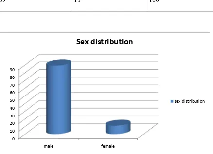
The SAPS II scoring was applied on 100 cases of perforation peritonitis. Of the
100 cases perforation was most common in males accounting for 89 cases while
[image:60.612.74.512.323.639.2]female cases were only 11.
Table 1. Sex distribution
Males Females Total
89 11 100
0 10 20 30 40 50 60 70 80 90
male female
Sex distribution
53
Mortality
Mortality in the study group was found to be 15/100. 13 deaths were from male
population. Females contributed 2 deaths.
Males Females
Death 13 2
Mortality rate was found to be slightly higher in females.
0% 2% 4% 6% 8% 10% 12% 14% 16% 18% 20%
male female
Mortality
54
Anatomical classification
.We could classify the patients according to the site of perforation. The bulk of the
perforations were due to duodenal perforation contributing 57% of cases followed
by Jejunal (19%), Gastric (9%), Ileal (9%), and Colonic perforation (2%). The
Cause was not made out for 4 patients in whom flank drain was put and they did
not require further surgery as they recovered with conservative management.
Gastric Duodenal Jejunal Ileal Colonic Unknown
Number 9 57 19 9 2 4
Death 2 6 4 1 2 0
Hospital
stay
15.42days 8.8 days 18.8 days 13.7days 2 days 15.25days
The patients having duodenal perforations have a lesser hospital stay and early
recovery. Whereas highest hospital stay was found in the patients with jejunal
perforation followed by gastric and patients put on flank drain. Colonic perforation
has a lower hospital stay as the two patients died in immediate post operative
55
Most common etiology was found to be duodenal perforation followed by jejunal
perforation.
Hospital stay was found to be highest in cases of jejunal perforation.
9
57 19
9 2 4
Anatomical classification
gastric duodenal jejunal ileal colonic unknown
0 2 4 6 8 10 12 14 16 18 20
gastric duodenal jejunal ileal colonic unknown
Hospital stay
56
Mortality According to Site of Perforation
The number of death in each type of perforation is shown in table 3 and the
mortality rate in each category is shown in the above chart. The highest mortality
rate was found in the group of colonic perforation followed by jejunal perforation.
Though the duodenal perforations formed the bulk of the diagnosis mortality was
relatively low.
0.00% 10.00% 20.00% 30.00% 40.00% 50.00% 60.00% 70.00% 80.00% 90.00% 100.00%
gastric duodenal jejunal ileal colonic unknown
Mortality
57
Morbidity Analysis
The various complications were recorded. The most common complications were
found to be wound infection and respiratory complications followed by the urinary
tract infections and enterocutaneous fistulas. Intra abdominal abscesses were found
to be lower in number.
WI WD RC UTI IAA ECF
Number 19 4 19 12 2 5
WI- wound infection WD- wound dehiscence
RC- respiratory complications UTI- urinary tract infections
IAA- intra abdominal abscess ECF- enterocutaneous fistula
Males vs. Females
Location of ulcer
Gastric Duodenal Jejunal Ileal Colonic Unknown
Males 7 52 17 9 2 4
58
The chart shows percentage wise distribution of perforations in males and females.
It can be seen that though duodenal perforation is the most common cause in both
males and females, there were higher number of gastric and ileal perforations in
females as compared to males.
Male Female 0%
10% 20% 30% 40% 50% 60%
Perforation distribution according to sex
59
Complications in Males and Females
WI WD RC IAA UTI ECF
Males 18 4 17 4 10 5
Females 0 0 0 0 2 0
WI- wound infection WD- wound dehiscence
RC- respiratory complications UTI- urinary tract infections
IAA- intra abdominal abscess ECF- enterocutaneous fistula
The complications were found to be higher in males. Almost all the complications
were confined to male population while only 2 females suffered from complication
which was urinary tract infection.
SAPS II in study population
The SAPS II scoring was applied to all these cases. The average SAPS II score for
all the cases is 21.56
60
Complications in individual anatomic sites
The complications were found to be highest in the jejunal perforations. Followed
by gastric perforation
Wound infection was found to be highest in jejunal perforations whereas
respiratory complications were highest in the duodenal perforations. 2 cases that
had intra abdominal abscess were both having jejunal perforations.
Enterocutaneous fistula was found to be higher in gastric perforation.
The various complications according to the individual site of perforation are shown
in the following table.
0 5 10 15 20 25 30 35 40 45
average for all patients average for dead
SAPS II Score
61
WI WD RC IAA UTI ECF
Gastric 4 1 2 0 2 2
Duodenal 3 0 8 0 5 0
Jejunal 7 2 2 2 4 2
Ileal 2 1 3 0 1 1
Flank
drain
62
Gastric perforation
Most common complication in gastric perforation was wound infection followed
by UTI and enterocutaneous fistula. 44% of patients with gastric perforation
suffered from wound infection. Respiratory complications, UTI, and
enterocutaneous fistula were found in 22% each. 66% of patients with gastric
perforation suffered from some kind of complication.
4
1 2
2
2
Complications
wound infection wound dehiscence respiratory complications UTI
63
Duodenal perforation
Respiratory complications were the most common complication found in duodenal
perforation there were no cases of wound dehiscence, intra abdominal abscess or
enterocutaneous fistulas. 14% of the patients had respiratory complications and 8%
of them had UTI. Totally only 28% of the patients had any complication at all in
duodenal perforation.
3 0
8 0
5 0
0 2 4 6 8 10
wound infections wound dehiscence respiratory complications intra abdominl abscess UTI enterocutaneous fistula
Complications
64
Jejunal perforation
Wound infection is the most common complication in jejunal perforations. Intra
abdominal abscesses were found only in jejunal perforation. Two patients
developed wound dehiscence and two patients developed enterocutaneous fistula.
Wound infection was found in 32% of the population having jejunal perforation.
Totally 42% of patients having jejunal perforation suffered from some kind of
complication.
complication 0
1 2 3 4 5 6 7
Complications
65
Ileal perforation
Respiratory complications were the most common complication in ileal perforation
followed by wound infection. 33% of patients suffering from ileal perforation had
suffered from respiratory complications, whereas 22% suffered from wound
infection. Totally 44% of patients having ileal perforation suffered from some type
of complication.
2
1
3 0
1
1
Complications
wound infection wound dehiscence respiratory complications intra abdominal abscess UTI
66
Unknown perforation/ Flank Drain
Complications were high in patients who underwent just flank drain. Two patients
that is 50% developed intra abdominal abscess, respiratory complications and
wound infection at the drain site. But the survival in the cases of patients
undergoing flank drain was found to be 100%. This may be attributed to immediate
drainage of the peritoneal sepsis and also these patients may have gained from lack
of stress due to anesthesia and surgery. Further studies may be required to prove
the efficacy of flank drainage without definitive surgery.
0 0.5 1 1.5 2
wound infection wound dehiscence respiratory complication intra abdominal abscess UTI enterocutaneous fistula
Complications
67
Analysis of complications with SAPS II scoring
Wound infection
When we evaluate the incidence of wound infections according to SAPS II score
we find that when SAPS score is less than 20 the wound infection rate is very low
where as with higher SAPS scoring there is an increased wound infection rate. The
wound infection rate in cases with SAPS above 40 is lower than those with 20 to
40 as many cases died in early post operative period before developing wound
infection.
0.00% 10.00% 20.00% 30.00% 40.00% 50.00% 60.00%
0 to 10 11 to 20 21 to 30 31 to 40 >40
Cases having wound infection
68
Respiratory complications
Respiratory complications had a linear relation with SAPS II scoring. Respiratory
complications were actually the cause for most of the deaths.
Thus we see 60 % of patients with SAPS II scoring between 31 and 40 and those
having above 40 developing respiratory complications.
0.00% 10.00% 20.00% 30.00% 40.00% 50.00% 60.00% 70.00%
0 to 10 11 to 20 21 to 30 31 to 40 >40
Respiratory complications
69
Wound dehiscence
This is a cumulative chart which shows increasing number of patients having
wound dehiscence as the SAPS scoring increases. A SAPS score below 20 has no
wound dehiscence whereas when it is 30 to 40, 40% cases have wound dehiscence.
0 0.5 1 1.5 2 2.5 3 3.5 4 4.5
10 20 30 40 50
nu
mb
er
SAPS II Scoring
Wound dehiscence
70
Intra abdominal abscess
Of the 4 cases having intra abdominal abscess all of them came under the group
having SAPS II score as 20 to 40. It was totally absent in cases having a score
below 20.
0 0.5 1 1.5 2 2.5 3 3.5 4
0 to 20
21 to 40
Intra abdominal abscess
71
Urinary tract infections
Urinary Tract Infections were almost uniformly distributed throughout the range
of SAPS scoring. This was probably due to almost universal use of bladder
catheters in patients and indwelling catheter being a single most prominent risk
factor for developing UTI.
0.00% 2.00% 4.00% 6.00% 8.00% 10.00% 12.00% 14.00% 16.00% 0 to 10
11 to 20 21 to 30 31 to 40 >40
UTI
72
Enterocutaneous Fistula
Enterocutaneous fistulas had the similar distribution to the other complications,
being highest in the 30 to 40 range and being absent below a SAPS II score of 20.
Relaparotomy
There were 5 patients who underwent relaparotomy. The cause of re lapar