A STUDY OF PENETRATING
ABDOMINAL INJURIES
Dissertation submitted for
BRANCH I - M.S., (General Surgery)
SEPTEMBER 2006
THE TAMILNADU DR.M.G.R. MEDICAL UNIVERSITY
This is to certify that this dissertation entitled “A STUDY OF
ABDOMINAL PENETRATING INJURIES” submitted by
Dr.G. BALAKRISHNAN to The Tamil Nadu Dr.M.G.R. Medical
University, Chennai is in partial fulfillment of the requirement for the
award of M.S. degree Branch I (General Surgery) and is a bonafide
research work carried out by him under direct supervision and guidance.
Dr. N.Sivaprahasam, M.S., Dr. M.Kalyana Sundaram M.S., FICS,
Additional Professor, Professor and Head,
Department of Surgery, Department of Surgery,
Govt. Rajaji Hospital, Govt. Rajaji Hospital,
Madurai Medical College, Madurai Medical College,
Madurai. Madurai.
I, Dr. G. Balakrishnan declare that I carried out this work on
“A STUDY OF ABDOMINAL PENETRATING INJURIES” at
Department of General Surgery, Government Rajaji Hospital during the
period of January 2005 – January 2006. I also declare this bonafide work or
a part of this work was not submitted by me or any other for any award,
degree, diploma to any university, board either in India or abroad.
This is submitted to the Tamilnadu Dr.M.G.R. Medical University,
Chennai in partial fulfillment of the rules and regulation for the M.S.
Degree examination in General Surgery.
Govt. Rajaji Hospital Dr. G.BALAKRISHNAN
Madurai.
I wish to express my sincere gratitude to my chief
Prof. Dr. N. Sivaprahasam M.S., Additional Professors Department of Surgery
Government Rajaji Hospital and Madurai Medical College for his guidance in
completing this study successfully.
I wish to express my sincere gratitude to my Prof.M.Kalyanasundaram
M.S., FICS, Head of the Department, Department of Surgery Govt. Rajaji
Hospital and Madurai Medical College for his excellent guidance and constant
encouragement in completing this study.
Dr.M.Jebamani M.S., and Dr.R.Prabhakaran M.S., Dr.P.Saravana
Boopathy M.S., Dr.S.Kalirathinam M.S., Assistant Professors of Surgery of
the IV Surgical Unit, have always been helpful to me to complete this study.
I wish to express my thanks to the Dean Government Rajaji Hospital and
Madurai Medical College for using the clinical materials from this hospital.
I also wish to express my thanks to all the patients who have cooperated in
rendering themselves for the study.
PAGE.NO
1. INTRODUCTION 1
2. REVIEW OF ANATOMY 2
3. PATHOPHYSIOLOGY & MANAGEMENT 12
4. AIM OF THE STUDY 37
5. MATERIALS AND METHODS 38
6. OBSERVATIONS 39
7. DISCUSSION 44
8. CONCLUSION 56
9. BIBLIOGRAPHY
10. PROFORMA
11. MASTER CHART
A STUDY OF ABDOMINAL PENETRATING
INJURIES
INTRODUCTION
Trauma is a major worldwide public health problem. It is one of the
leading causes of death and disability in both industrialized and developing
countries. High speed vehicles, decivilization of human race, terrorism and
sports are just few of the predisposing factors of trauma. Evaluation of a
patient with abdominal trauma can be a most challenging task that a surgeon
may be called upon to deal with. Abdominal injuries may be parietal or
visceral injuries. Visceral injuries may be intraperitoneal or retroperitoneal
Liver, spleen, stomach, small bowel, duodenum, Large bowel, pancreas,
Kidney, ureter and retroperitoneal vasculature are the organs included in this
study and pelvic organs are excluded. Multi organ injuries, exsanguinating
hemorrhages delayed presentations and the ominous reputation for high
mortality and morbidity are just few of the many reasons which makes this
REVIEW OF ANATOMY
1,23A revision of the surgical anatomy of the abdominal organs is
necessary at this juncture to appreciate the various aspects of penetrating
abdominal injuries. For evaluation purposes the abdomen is divided into four
areas intrathoracic abdomen, true abdomen, pelvic abdomen, and
retroperitoneal abdomen.
The intrathoracic abdomen is that portion of the upper abdomen that
lies beneath the rib cage. The contents include the diaphragm, liver, spleen
and stomach. Peritoneal lavage becomes useful in evaluating this area.
The pelvic abdomen lies in the hollow of the pelvis. It is surrounded on
all sides by the bony pelvis and its contents include the rectum, bladder,
urethra small bowel and in females the uterus, fallopian tubes and ovaries.
Penetrating injuries of the buttocks may injure the pelvic organs.
The retroperitoneal abdomen contains the kidneys, ureter, pancreas,
second and third portion of the duodenum, the ascending and descending
colon and the great vessels the aorta and vena cava. Evaluation of the
retroperitoneal abdomen requires utilization of radiographic procedures
The true abdomen contains the small and large intestines, the bladder
when distended and uterus when gravid. Injuries to any of these organs are
usually manifested by pain and peritonitis. Peritoneal lavage is useful adjunct
when abdominal film may be helpful when free air is present.
LIVER
The liver is the largest gland in the body weighs about 1500 gms and
receives 1500 ml of blood per minute, developed from ventral mesogastrium
Liver has two surfaces diaphragmatic and visceral surfaces. The
diaphragmatic surface is subdivided into anterior, superior, posterior and right
surfaces. Liver is held in position by attachment of IVC and hepatic veins. The
falciform ligamentum divides the anatomical left and Right lobes. Caudate
Lobe lies back between Inferior venacava and the fissure for the ligamentum
venosum. Quadrate Lobe lies between gall bladder fossa and the fissure for
the ligamentum teres on the basis of blood supply and biliary drainage. Liver
is divided into four anatomical segments in eight sectors. Blood supply is by
hepatic artery and portal vein supplying 25% and 75% of total blood supply
by supplying 50% oxygen each and drained by Right middle and Left hepatic
veins and also about 10 to 15 small veins drain directly into IVC. The
portal vein passes through the free border of lesser omentum. Pringle’s
maneuver is the temporary application of vascular clamp to the free margin of
lesser omentum upto a period of 20mts to 1 hour, indicated in major bleeding
from hepatic or perihepatic injury so that bleeding points can be arrested by
topical cooling.
SPLEEN
Spleen is the largest lymphoid organ in the body developed from dorsal
mesoqastrium. It lies under diaphragm on the left side of the abdomen closely
in contact with 9th, 10th, and 11th ribs. It measures 1x 3x5 inches weighs 7oz.
Spleen is freely mobile organ and held in position by lienorenal ligament and
gastrosplenic ligament, phrenocolic ligament gives additional support. Spleen
is supplied by spleenic artery passes between the layers of lienorenal ligament.
At the hilum it breaks up into four branches which enter hilum separately.
Similar veins leave the hilum and unit to form the splenic vein. The hilum of
spleen is closely related to the tail of pancreas. So concomitant pancreatic and
splenic injuries are common.
Spleen act as an immunological filter. It produces opsonin, tuftsin – a
tetra peptide that coats white cells to promote phagocytosis of particular
matter, bacteria and aged red cells. It is also a source of properdin, a vital
STOMACH
Stomach is the most dilated part of the alimentary tract, interposed
between the esophagus and duodenum in the upper part of abdominal cavity
and Lying mainly in the left hypochondrial, epigastric and umbilical regions
with much of it under cover of the lower ribs. It is loosely suspended in the
abdomen by the gastro hepatic ligament superiorly, the gastrocolic ligament
inferiorly and by its attachment to the spleen laterally. It is relatively fixed at
the gastro esophageal junction and the retroperitoneal duodenum. The gastric
wall consist of an external serosal layer followed by three layers of smooth
muscle outer longitudinal layer, middle circular layer, and inner oblique layer.
A strong sub mucosal layer is followed by a mucosal layer with a rich
capillary network. This network is supplied by arterioles, which originate in
the sub mucosa.
The stomach is supplied by four major nutrient arteries with extensive
collateral circulation between the vascular beds. The left gastric artery most
commonly arises from the celiac axis and usually splits into anterior and
posterior trunks before it reaches the stomach. Branches from the left gastric
artery supply the distal esophagus and cardiac portion of the stomach. The
left gastroepiploic artery is a collateral of the splenic artery and supplies the
greater curvature. It anastamoses with right gastroepiploicartery in about 75%
of cases. The more maximal portion of greater curvature is supplied by the
short gastric vessels which originate from the gastroepiploic artery as well as
from the splenic artery. The right gastroepiploic artery arises from the gastro
duodenal artery and supplies the pyloric area and distal greater curvature.
Venous drainage from the lesser curvature is via the coronary vein to the
portal vein. On the greater curvature drainage is via the short gastric vessels
and right and left gastroepiploic veins to the splenic vein.
SMALL INTESTINE
The small intestine consists of the duodenum, jejunum and Ileum. The
Duodenum extends from the pylorus, lies opposite to the right side of the
spine at the level of the first lumbar vertebra to the duodenojejunal flexure. It
is ‘C’ shaped tube and about 25cm Long. It is divided into four parts and
properly called as superior, descending, horizontal and ascending but simply
called first, second, third and fourth parts of duodenum. The blood supply to
the duodenum is from the celiac and superior mesenteric vessels. The first
portion of the duodenum is intraperitoneal and some what mobile. The
remainder of the duodenum is retroperitoneal owing to the fusion of the
third portion of the duodenum may be easily mobilized through this blood less
fusion plane, a Kocher’s maneuver. An additional point of fixation occurs at
the ligament of Treitz.
Jejunum is wider bored and thicker walled than ileum. Jejunum and
Ileum together lie in the free margin of the mesentery. Total length varies
greatly from about 4 to 6 metres. The jejunum constitutes two fifths and three
fifths for the ileum. Fan shaped mesentery suspends the small bowel and
extends from the left side of the second lumber vertebrae downward to the
right sacroiliac joint, the traversing the transverse colon, duodenum, Aorta,
Inferior venacava, right gonadal vessels and Right writer. The superior
Mesentric Artery supplies the jejunum and Ileum arising from the aorta
approximately 2 cm below the celiac trunk after crossing the uncinate process
of the pancreas, it enters root of the mesentery giving off branches to pancreas
right colic and numerous intestinal vessels before it terminates at the medial
aspect of the caecum.
PANCREAS
The pancreas lies transversely across the upper part of the posterior
head lies with in the concave sweep of the duodenum. The splenic artery runs
along the upper border of the pancreas and the splenic vein runs behind, Just
superior to the lower edge. The superior mesenteric vein and artery lies just
behind the neck of the pancreas and are also enclosed posteriorly by an
extension of the head known as uncinate process. The uncinate process lies
between the Inferior vena cava and portal vein.
The main pancreatic duct of wirsung usually traverses the entire length
of the gland slightly above the line, halfway between the superior and inferior
edges and normally ends by joining the common bileduct. The accessory duct
of santorini branches out from the pancreatic duct in the neck of the pancreas
and empties into the duodenum about 2.5 cm above the duodenal papillae.
Sometimes the anomalous common hepatic artery and right hepatic artery may
pass posterior to the portal vein awareness of this anomaly is useful for
dissection of the portal triad to minimize inadvertent injury.
KIDNEYS
The kidney lies high up on the posterior abdominal wall behind the
peritoneum, largely under cover of the costal margin. Each kidney lies
obliquely, with its long axis parallel with the lateral border of psoas major.
Normal kidney measures about 12x6x3 cms and weighs about 130 gms. The
the transpyloric plane, 5cm from the midline. The relation of kidney
posteriorly mostly the diaphragm and the quadratus lumborum muscles with
overlaps medially on to psoas and laterally or to transversus abdominis. The
right supra renal gland pyramidal inshape surmounts the upper pole of right
kidney whereas the left supra renal is cresentric in shape and is applied to the
medial border above the hilum. The anterior relation of the right kidney are
duodenum, hepatic flexure, coils of jejunum and liver whereas the anterior
relations of the left kidney include the tail of the pancreas, splenic flexure and
stomach. The perinephric fat lies outside the renal capsule. At the hilum vein,
artery and the pelvis lies in the order from anterior to posterior. The constant
anatomy of the origin of the renal arteries on both sides, which is posterior to
the point at which two left renal vein joints the inferiorvenacava. This is very
important in controlling the renal pedicle initially before the Gerota’s fascia is
opened.
URETERS
The ureter is 25cm long. The ureter passes down on the psoas major
under cover of the peritoneum and crosses the genitofemoral nerve being itself
crossed superficially by the gonadal vessels. On the right the upper part is
the left it is lateral to the inferior mesenteric vessels and is crossed by the left
colic vessels and the sigmoid mesocolon. The blood supply is endangered if
the ureter is stripped of the surrounding tissues.
COLON
The right colon is derived from the midgut and is supplied by the
superior mesenteric artery, where as the left colon originates from the hind gut
and is supplied by the Inferior mesenteric vessels. The Right colon has a thin
wall and larger lumen and left colon is thicker and more muscular and has a
smaller lumen. The right colon absorbs and dehydrates the small bowel
contents whereas the left colon functions primarily for storage. Despite the
fact that there are definite anatomical and physiological differences between
the right and left colon both should be treated similarly.
RETROPERITIONEAL SPACE
The Area of the posterior abdominal wall behind the peritoneum that is
not occupied by the major viscera and great vessels. Major structure lie on the
posterior abdominal wall behind the peritoneum includes the aorta and inferior
vene cava with a number of their branches and tributaries, the cysterna chyli,
lymphnodes and nerves including the sympathetic trunk, the kidney’s, ureters,
Having discussed the anatomy of various abdominal organs. It is
important to discuss about the Clinicopathological aspects and management of
penetrating abdominal organs in general, recommended by various
PATHO PHYSIOLOGY AND MANAGEMENT
LIVER INJURIES25,31
Liver is frequently injured in both blunt and penetrating trauma.
Because of its size, injuries sufficient to lacerate liver are associated with
injuries to other organs in about 80% cases. 85% of liver injuries are not
bleeding at the time of laparotomy and patient tolerate these injuries very
well25. Most liver injuries will infact require only documentation and no
drainage. The minority of liver injuries therefore require definitive surgical
care. The history of injury is helpful in that particularly any penetrating injury
to the right rib cage or upper abdomen and a patient, who has a history of
being in shock at eh scene following blunt trauma abdomen should be
suspected of having a major liver injury.
GRADING OF HEPATIC INJURIES31
(Liver injury scale 1994 revision)
Grade* Type of Injury Description of Injury
I Haematoma
Laceration
Sub capsular <10% surface area
capsular tear <1cm parenchymal depth
II Haematoma
Laceration
Sub capsular, 10-50% surface area
intraparenchymal <10 cm in diameter
Capsular, tear, 1-3cm parenchymal
III Haematoma
Laceration
Subcapsular >50% surface area of
ruptured Subcapsular or parenchymal
haematoma, Extraparenchymal
haematoma >10cm or expanding
3cm parenchymal depth
IV Laceration Parenchymal disruption involving
25-75% hepatic lobe or 1-3 couinaud
segments
V Laceration
Vascular
Parenchymal distribution involving
>75% of hepatic lobe or > 3 couinaud
segments with in single lobe
Juxta hepatic venous injuries i.e.
retrohepatic venacava/ central major
hepatic veins
VI Vascular Hepatic avulsion
*advance one grade for multiple injuries upto grade III
After resuscitation the patients plain x-ray abdomen should be taken
and shows altered liver border, hemoperitoneum and associated rib fractures.
Abdominal paracentesis is positive, if large amount of blood presents in the
peritoneal cavity. DPL is diagnostic of minimal hemo peritoneum, but not
specific for liver injury, CT is the investigation of choice in multiple injured
rarely done to document location of biliary fistula after repair of hepatic
injuries.
TREATMENT
311. NON OPERATIVE MANAGEMENT
Indicated in
1. Hemodynamic stability
2. Normal mental status
3. Absence of a clear indication for laparotomy such as peritoneal signs
4. Low grade Liver injuries (grade 1to3)
5. Transfusion requirements of less than 2 units of blood.
These patients – followed by serial hematocrit and vital signs. During
observation if the patient shows signs of bleeding angiography and selective
embolisation can be done.
Indications for laparotomy during observations are
1. Continuing need for blood transfusion, increasing (or) deterioting vital
signs.
2. Peritoneal signs.
3. Progressive expansion of haematoma.
II. OPERATIVE MANAGEMENT25,31
(A) Simple Techniques of repair
1. Drainage of non bleeding injuries rarely performed nowadays
2. Compression: small cracks in the capsules can be treated by
compression for 5 to 10 minutes to stop bleeding.
3. Topical agents: The application of gelfoam, microcrystalline collagen
pad and fibrin glue is used for when avulsion of Glisson’s capsule is
present. After application of topical agent to the raw hepatic surface 5
minutes of compression with pads is applied. After removed
electrocautery can be used for any bleeders.
4. Suture hepatorrhaphy: Horizontal mattress sutures with 1-0 vicryl or
simple continuous suturing with 1/0 vicryl can be done with these
measures most of the bleeding stops.
(B) Advanced Techniques Repair:
1. Extensive hepatorrhaphy:
If simple suturing fails to stop, multiple horizontal but bleeding from
intralobar branches of hepatic artery, portal vein or hepatic vein are not
2. Hepatotomy with selective vascular ligation:
It is indicated when bleeding vessels present deeply. Hepatotomy is
done with finger fracture technique. Bleeding vessels and biliary radicals are
identified and ligated.
3. Omental Pack
In 1975 stone and lamb first described the use of viable pedicle of
omentum placed into deep lobar lacerations to control haemorrhage at the
base. Viable omentum separated from transverse colon can be placed over
liver laceration to control bleeding especially in bare area of liver.
4. Resectional debridement with selective vascular ligation
It is indicated whenever there is friable and partially devascularised
hepatic tissue on the edge of liver or in a hepatic laceration or missile tract. It
is used in preference to anatomic segmentation or lobectomy as these
techniques frequently sacrifice larger amounts of normal hepatic tissue.
5. Resection
It is indicated in the case of total disruption of lobe or segment. In the
6. Selective hepatic artery ligation
When selective vascular ligation fails, ligation of hepatic artery is an
alternative. It may produce dramatic hemostasis without subsequent liver
failure, but this should be done as close to liver as possible and only as a last
resort.
7. Perihepatic packing
This technique involves the insertion of laparotomy pads or rolls of
gauze around the injured liver not into hepatic laceration. Packs can be
removed 12 hrs after packing. Rebleeding and sepsis are common
complications.
8. Drainage
Open penrose drainage has been used after operative treatment but
incidence of intra abdominal sepsis is common.
Complications:
Significant complications following liver injury includes
1. Pulmonary complications
2. Coagulopathy
5. Biliary fistulas
6. Haemobilia
7. Subdiaphragmatic and intraparenchymal abscess formation
Splenic injuries25,31 :
The spleen is the intra abdominal organ most frequently injured in blunt
trauma. In penetrating trauma wound of entry or exit in the left chest, flank or
left upper abdomen should arouse suspicion of splenic injury. The clinical
picture of splenic injury includes left upper quadrant abdominal pain, signs of
blood loss and pain in the left shoulder (Kehr’s sign).
Grading of splenic injury31
Spleen injury scale (1994 revision).
Grade* Type of Injury Description of injury
I Haematoma
Laceration
Subcapsular, <10% surface area
Capsular tear <1cm parenchymal depth
II Haematoma
Laceration
Subcapsular 10-50% surface area
Intra parenchymal <5cm in diameter
Capsular tear, 1 to 3 cm parenchymal depth
that does not involve 9 trabecular vessel.
III Laceration Subcapsular >50% surface area or expanding,
ruptured subcapsular or parenchymal
IV Laceration Laceration involving segmental or hilar vessels
producing major devascularsation (>25% of
spleen)
V Laceration
Vascular
Completely shattered spleen
Hilar vascular injury that devascularizes spleen
* - Advance one grade for multiple injuries upto Grade III
Management
The management of splenic injury has been subject of major
re-examination over the past decade and the recognition of fatal pneumococcal
septicemia in patients undergoing splenectomy has led to an interest in splenic
salvage (D.B.Hyot and A.R. Moossa et al)4.
Plain abdominal films may show
1. Enlargement of splenic shadow
2. Elevation of left hemidiaphragm
3. Medical displacement of splenic shadow or stomach
4. Widening of the space between the splenic flexure and peritoneal pad.
Peritoneal lavage should be performed when there is possibility of
splenic injury, positive indicates laparotomy.
I Non operative management4,31
>70% of all stable splenic injuries are being treated nonoperative
approach.
Criteria for non operative management of splenic injury.
1. Hemodynamic stability.
2. Negative abdominal examination.
3. Absence of contrast extravasation on CT.
4. Absence of other clear indications for explorative laparotomy or
associated injuries requiring a surgical intervention.
5. Absence of associated health conditions that carry on increased risk
of bleeding (coagulopathy, hepatic failure, use of anticoagulants)
and grade 1 to 3 injuries.
The risk of non operative management are missed injury to other
viscera and delayed rupture of subcapsular haematoma. Patient are usually
admitted in ICU and serial abdominal examination and haematocrit are
obtained during the initial 48 to 72 hrs. The patient should be followed
sequentially with CT scan.
II Spleen conserving surgery4,22,31
1. If it is a multiple injury case
2. Patient is in shock (systolic pressure <90mmHg)
3. There are medical contraindications to prolonged surgery (bleeding
disorder, cardiac, pulmonary or hepatic disease)
If the patient condition is favourable the decision to repair is based on
the state of the spleen. Generally, Grade IV and Grade V injuries are not
suitable for repair whereas it can be attempted in Grade I, II and III.
Splenorhaphy4,22,31
The following are the techniques for splenic repair.
1. Local hemostatic agents
Gelatin foam, surgical cellulose, microfibrillar collagens thrombin,
cyanoaerylate, autologous fibrin glue can be used for superficial tears which
are not bleeding actively. But often pressure alone may be sufficient. Non
bleeding tears are best left alone.
2. Suture repair
Deep parenchyma, tears are managed by this technique. After removal
mattress sutures. Vertical or horizontal including the fibrous capsule using
absorble sutures.
3. Partial splenectomy
Polar injury which is grade IV can be managed by segmental
devascularization and debridement by finger fracture technique at the line of
demarcation additional security to the suture line after suture repair or partial
splenectomy can be achieved by omental wrap.
4. Heterotropic auto transplantation of the splenic tissue
If the patients condition permits, the splenic function can be preserved
even after splenectomy by autotransplantation at sites like gastrocolic
omentum, rectus sheath and anterior abdominal wall. To be effective in
preserving adequate splenic function approximately one third of the original
spleen must remain and be nourished by an adequate circulation.
III Splecectomy4,31
Indicated in
1. Shattered or avulsed spleen
2. Severely hypotensive patients
3. Associated with other severe injuries
Complications of splenectomy
1. Early transient thrombocytosis, which resolved spontaneously over 1-3
months.
2. Acute dilatation of stomach
3. Delayed haemorrhage
4. Pancreatitis
5. Subphrenic abscess
6. Left lower lobe atelectasis and pleural effusion
7. Fatal pneumococcal septicemia (Over whelming Post Splectomy
Infection - OPSI)
GASTRIC INJURIES25,31,35
Injuries of stomach are common in penetrating – trauma but very rare
in blunt trauma. The stomach is intrathoracic, partially protected by rib cage
and any penetrating wound in this area should be suspected of causing injury
to stomach. After resuscitation, a nasogastric tube is placed that serves both
diagnostic and therapeutic functions. The return of gross blood on nasogastric
aspirate is suggestive of an upper gastrointestinal injury. Haematemesis or
bright red blood per nasogastric tube was present in 45% of gunshot wounds
parkland memorial hospital25. The nasograstric tube also serves a therapeutic
function by decompressing stomach.
OPERATIVE MANAGEMENT
The intra operative evaluation of stomach injury includes good
visualisation of hiatus, evaluation of anterior portion of the stomach, division
of gastrocolic ligament and complete visualization of the posterior aspect of
the stomach. Penetrating wounds are debrided and primary closure performed
(Moossa A.R. et al)1,31. Injuries with major tissue loss may be best treated by
gastric resection. Post operative complications include bleeding usually from
the submucosal vessels, intrabadominal abscess and more rarely gastric
fistula.
DUODENAL INJURES25,31
The majority of duodenum injuries are caused by penetrating trauma.
The incidence of duodenal injuries varies from 3% to 5%. Most of duodenal
injuries are accompanied by other intraabdominal injuries.
Grade* Type of Injury Description of injury
I Haematoma
Laceration
Involving single portion of duodenum
Partial thickness, no perforation
II Haematoma
Laceration
Involving more than one portion
Disruption <50% of circumference
III Laceration Disruption 50% to 75% of circumference of
D2. disruption of 50% to 100% of
circumference of D1, D3, D4
IV Laceration Disruption >75% of circumference of D2
involving ampulla or distal common bile duct.
V Laceration
Vascular
Massive disruption of duodenopancreatic
complex
Devascularisation of duodenum
* - Advance one grade for multiple injuries upto Grade III.
DIAGNOSIS OF DUODENAL INJURIES
Serum amylase is sensitive but unfortunately not specific for duodenal
injuries. Needle paracentesis or lavage will often be positive for blood, bile or
2. Obliteration of the right psoas shadow
3. Absence of air in the duodenal bulb
4. Air in the retropertitoneum outlining the kidney other
investigation include gastrografin upper gastrointestinal series
and CT of the abdomen.
Treatment31,35
Intra operative evaluation of the duodenum requires an adequate
mobilization of the duodenum by means of a Kocher maneuver. 80 to 85% of
duodenal wounds can be primarily repaired. The remaining 15 to 20% are
severe injuries that require more complex procedures.
Minor injuries (Grade I + II) diagnosed with in 6 hrs of injury, a simple
primary repair is suitable. After 6 hrs, the risk of leak increases and any form
of duodenal decompression like transpyloric nasogastric tube, tube
jejunostomy, tube duodenostomy is advisable.
Grade III injuries are best treated by primary repair, pyloric exclusion
and drainage or alternatively roux-en.Y deudenojejunostomy.
Grade IV injuries are treated by primary repair of the duodenum, repair
limb or choledochoenteric anastomosis can be performed. Grade V injuries are
best treated by pancreatico duodenectomy.
Most common complication after duodenal injury is the development of
duodenal fistula.
PANCREATIC INJURIES31
Pancreatic injuries are rare accounting for 10% to 12% of all abdominal
injuries. The great majority of such injuries are caused by penetrating
mechanism. Major abdominal vascular injuries are present in more than 75%
of cases of penetrating pancreatic trauma. Fistula, pseudocyst, pancreatitis,
anastomotic leak and intraabdominal abscess occur in 1/3rd of the patients and
account for the late mortality. Mortality rates ranges 10 to 25% mostly due to
associated intra abdominal injuries.
GRADING OF PANCREATIC INJURY31
Grade* Type of Injury Description of injury
I Haematoma
Laceration
Minor contusion without duct injury
Superficial laceration without duct injury
II Haematoma
Laceration
Major contusion without duct injury or tissue
loss
Major laceration without duct injury or tissue
III Laceration Distal transaction or parenchymal injury
involving ampulla
IV Laceration Proximal transaction or parenchymal injury
involving ampulla
V Laceration Massive disruption of pancreatic head
* - Advance one grade for multiple injuries upto Grade III
Diagnosis of pancreatic injuries
1. Increased level of serum and urinary amylase after blunt injury are not
diagnostic, but a persistent elevation suggest pancreatic injury.
2. Contrast duodenography may reveal widening of the ‘C” loop.
3. Abdominal CT scan are currently reported as having a sensitivity and
specificity in excess of 80% (Jefery KB et al, 1983)19.
4. ERCP is the most useful in pre operative delineation of ductal anatomy
in patients with delayed presentation or missed injuries (Whitwell et al,
1989).
Treatment
Pancreatic injuries are divided into proximal distal according to the
1. Penetrating wounds to the right of the superior mesenteric vein should
be treated with debridement and direct suture ligation of areas of
bleeding.
2. Extensive injuries to the pancreatic head or to the right of superior
mesenteric vessels best treated by external drainage.
3. Severe trauma to the duodenum and head of pancrease may be treated
with debridement of the pancreas, closure of the duodenal wound and
pyloric exclusion with external drainage.
4. Most distal pancreatic injuries with suspected ductal injuries were
treated by distal resection with or without splenectomy.
The most common complication after pancreatic trauma are pancreatic
fistula and peripancreatic abscess.
SMALL BOWEL INJURIES
Injuries of small bowel are present in approximately 25 to 30% of the
patients who require laparotomy after penetrating trauma (Moossa et al)1. Stab
injuries are usually less severe than gunshot or blunt mechanism of injury. In
most patients who sustain stab wounds the small bowel is spared because the
mobility of small bowel afforded by the redundant mesentery, allows the
EVALUATION AND DIAGNOSIS31,35
Although history and physical examination are valuable in the
diagnosis of small bowel injury following penetrating trauma.
1. Plain films of the abdomen may reveal free air
2. Any patients who has peritoneal signs or hemodynamically unstable
proceeds promptly to exploratory laparotomy. In equivocal cases
peritoneal lavage is employed and look for indicators of hollow visceral
injury like presence of bacteria, food fibres or bile and other criteria
like amylase greater than 200, WBC >500/mm3 and
RBC>100,000/mm3. Gunshot wound present a much greater risk for
significant intra abdominal injury.
Treatment
At laparotomy a careful examination of the entire small bowel should
be performed. Bleeding should be initially controlled and clamps or sutures
should be applied to prevent further leakage of intestinal contents into the
peritoneal cavity.
Penetrating injuries caused by firearms should be debrided and usually
small tears closed primarily. Extensive lacerations, devascularised segments or
multiple lacerations in a short. Segment of bowel are better treated by
Complications
Intra abdominal abscess, anastomatic leakage, enterocutaneous fistula
and intestinal obstruction.
COLONIC INJURIES25,31,35
Colon injuries are usually the result of penetrating trauma. The colon is
the second most frequently injured organ after gunshot wounds and the third
after stab wounds to the abdomen. Morbidity rates after colonic injuries vary
from 20% to 35% and mortality rates from 3% to 15%.
Treatment
General criteria for primary repair include
1. Early diagnosis (within 4 to 6 hrs)
2. Absence of prolonged shock or hypotension
3. Absence of gross contamination of the peritoneal cavity
4. Absence of associated colonic vascular injury
5. Less than 6 units of blood transfusion
6. No requirement for the use of mesh to permanently close the abdominal
wall.
High risk colon injuries or those associated with severe injuries will
benefit from resection and colostomy.
Post operative complications
1. Abscess formation
2. Anastomotic leak
3. Peristomal hernia
4. Morbidity and mortality associated with colostomy closure
RENAL INJURIES32
Penetrating trauma accounts for 84% of renal injuries. The great
majority of penetrating renal injuries were gunshot wounds (99%) and the
remainder were due to stabbing. Gunshot injuries produce multiple
intraabdominal and intrathoracic injuries. Stab wounds to the kidney also were
associated with a high incidence of non renal injuries. The overt mechanism of
renal injury from penetrating trauma is the obvious tissue disruption to the
parenchyma, collecting system and vasculature.
Diagnosis
History is very important a high index of suspicion should be there,
flank pain, haematoma warrants evaluation regardless of the apparent location
haematoma or preexisting mass such as hydronephrosis or tumor. Infusion
pyelography identifies approximately 80% renal injuries.
Renal injury is suggested by the following radiological findings31,32
a. Decreased excretion of contrast
b. Obliteration of psoas shadow or renal outline
c. Scoliosis away from the injury
d. Extravasation of the contrast.
A normal IVU with haematuria in a trauma patient suggest minor renal
contusion and rules out major renal injury. Incomplete or poor visualisation of
a portion of kidney suggest major renal trauma, include deep laceration
avulsion or vascular occlusion. Non visualisation of a kidney on pyelography
require immediate arteriography whenever possible. CT is very useful
investigation helpful in the non operative management.
Treatment32
Surgical exploration of all penetrating renal injuries is recommended
because of the high incidence of associated intra abdominal injuries.
Penetrating wounds causing small parenchymal injuries are generally treated
• Injuries involving the hilum are seldom repaired primarily and in most
circumstances a total nephrectomy is necessary.
• Renal vein laceration may be repaired by venorraphy. Renal arterial
trauma may require a variety of repairs. Lateral arteriorrphy, arterial
resection and repair by primary reanastomosis or autogenous repair.
URETERIC INJURIES31,32
The following classification of ureteral injury helps in organising the
management of ureteral injury
1. Site; upper, middle and lower third
2. Time of recognition: immediate or delayed
3. Nature of injury: blunt trauma with laceration or avulsion, penetrating
trauma
4. Presence of concomitant injuries
Injury to the ureter is an common and occurs mostly after penetrating
trauma. The presence of haematuria in ureteral injury is the exception rather
than the rule.
Diagnosis of ureteric injury
In the majority of cases, intra venous pyelography will confirm the
be required to confirm the diagnosis. In hemodynamically unstable patients
the diagnosis of ureteral injury may be made at the time of laparotomy by
intravenously injecting 5ml of methylene blue. CT scan is more accurate in
detecting the extravasation preoperatively.
Management31,32
The principles of ureteral repair are adequate debridement, tension free
repair, spatulated anastomosis, watertight closure, ureteral stenting and
drainage. Ureteropelvic junction disruption and major urethral injuries (greater
than 2cm laceration) are best treated by nephrostomy and stent after repair
with fine chromic catgut sutures. Drainage should be provided. Lower urethral
injuries usually require tunneled reimplantation into the bladder, if this is not
possible then a flap should be turned cephaled for reconstruction (Boari and
Ockerland) when major ureteral loss is present or when it is necessary not to
have any ureteral leakage post operatively, one may ligate the ureter and
perform nephrostomy either at the time or percutaneouly within 24 hrs. then
later doing a transuretero – ureterostomy. Alternatively one may choose
substitution of a segment of tapered or nontapered ileum for the ureter.
Nephrectomy may be the procedure of choice when there is simultaneous
RETROPERITONEAL HEMATOMA25,31
The exact incidence of retroperitoneal haematoma in patients who have
suffered penetrating abdominal injuries is usually not recorded as the
hematomas are simply a manifestation of a major vascular injury.
In general trauma surgeons recognize retroperitoneal haematoma in
five locations.
1. Midline suprarenal.
2. Midline infra renal.
3. Lateral peri renal.
4. Lateral pelvic and portal.
Anemia and haematoma are constant findings in patients with
retroperitoneal haematoma from pelvic fractures, while hematuria is much less
common inpatients with haematomas caused by penetrating wounds involving
vascular structures. A plain film in a patient with penetrating wound from
missile will localise the site of the missile and give some indication of major
retroperitoneal vascular structure has been injured.
In contrast to the management of retroperitoneal haematoma with blunt
abdominal trauma, all five retroperitoneal hematomas previously listed are
opened in patients with penetrating wounds. In each instance, proximal
vascular control and if possible distal vascular control should be done before
THE AIM OF THE STUDY
The aim of the study is to evaluate the following aspects of penetrating
abdominal injuries excluding pelvic organs.
- The incidence of penetrating injuries abdomen.
- Mode of injury.
- Clinical presentation.
- Associated organ and system involvement.
- Management.
MATERIALS AND METHODS
This study consists of all penetrating abdominal injuries admitted in the
trauma ward of Government Rajaji Hospital, Madurai from January 2005 to
January 2006. Once the patient is admitted the name, age, sex and mode of
injury are noted. The time interval between injury and admission and time
interval between admission and surgery are recorded. After resuscitating the
patients and if time permits necessary investigation are carried out. In those
who are operated, the operative findings and methods of management are
recorded. Cases are followed up till their discharge from the hospital. If death
occurs the case of death is evaluated in those patients who died before surgery
the postmortem findings are noted. The above facts are recorded in a proforma
OBSERVATION
The total number of patients who had sustained penetrating injuries to
abdominal organs were 70. During this period total number of cases of
abdominal trauma managed were 212 cases. Thus penetrating injuries to
abdominal organs account for 33% of the abdominal trauma cases.
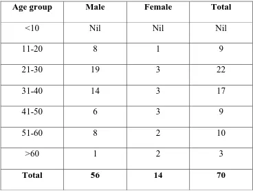
In this study of the 70 patients 56 cases were male and 14 cases were
females. This gives a male to female ratio of 4:1. The high incidence of
trauma in males may probably be due to the relatively high association of
[image:44.612.129.497.425.702.2]males in acts of violence and ventricular accidents.
Table 1: Age and Sex incidence
Age group Male Female Total
<10 Nil Nil Nil
11-20 8 1 9
21-30 19 3 22
31-40 14 3 17
41-50 6 3 9
51-60 8 2 10
Table 1 shows the age and sex incidence in this study. The youngest
patient was a fourteen year old boy who had sustained penetrating. Injuries by
falling on a tree’s branch. More than 50% of the patients belongs to the age
group between 21-40 yrs which is the most productive part of one’s life. The
oldest patient was a 76 yrs old male who had sustained penetrating injuries by
[image:45.612.192.436.339.495.2]bull gore.
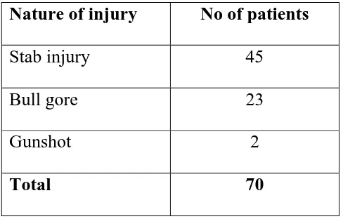
Table 2: penetrating injuries – abdomen
Nature of injury No of patients
Stab injury 45
Bull gore 23
Gunshot 2
Total 70
Penetrating injuries : Abdomen
As given in the Table 2 stab injury is the common penetrating trauma
according for 64%. There were 23 cases of bull gore injury two cases of
gunshot, two cases of RTA and one case of penetrating injury due to falling on
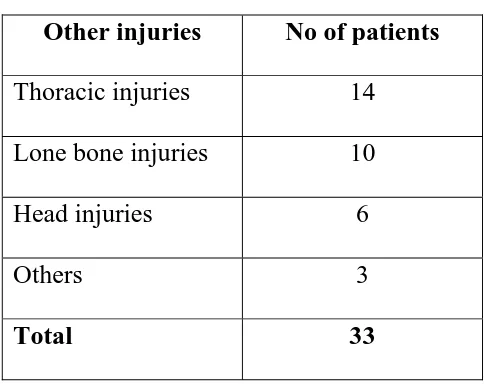
Table 3: Injury of other organs
Other injuries No of patients
Thoracic injuries 14
Lone bone injuries 10
Head injuries 6
Others 3
Total 33
Injury of the other organs
Table 3 shows the associated injuries in penetrating injuries of the
abdomen, fourteen patients sustained associated thoracic injuries. Ten patients
had long bone fractures and six patients suffered from head injury. Two
patients had facial injuries. Totally 33 patients had injuries involving other
organs. This high incidence of polytrauma with penetrating injuries abdomen
Table 4 The analysis of the time interval between injury and admission
and admission and surgery is given in
Time Interval Injury – Admission Admission – surgery
<2 hours 28 20
2-4 hours 18 30
4-6 hours 13 14
6-8 hours 5 2
8-10 hours 3 1
10-12 hours 2 2
>12 hours 1 1
From the Table 4 it can be deduced 59 cases took less than 6 hrs from
the time of injury to admission. The fastest to arrive was with in 30mts from
the injury. The average time duration between admission and surgery was 4
Different structures affected
Structures No of patients
Liver 15
Small bowel 12
Spleen 8
Stomach 10
Duodenum 2
Colon 10
Kidney 6
Ureter 1
Pancreas 2
Retroperitoneal 9
[image:48.612.191.436.112.505.2]Diaphragm 7
Table 5 shows the different organs injured in the study. Liver injury
tops list with 15 cases. This is followed by small bowel, stomach and colon
accounting for 12, 10, 10 cases respectively. These were 8 cases are spleen, 2
cases are duodenal injuries. These were 6 cases are kidney, two cases are
DISCUSSION
LIVER INJURIES
There were totally 15 cases of liver injury. In this 9 cases were due to
stab injury and 6 cases were due to bull gore. The incidence of associated
organ injuries is a significant factor in patients sustaining liver injuries. In this
study only 4 cases were isolated liver injuries and remaining were associated
with other organ injuries. The different ways in which the 15 cases of liver
injuries were managed on follows. Application of gelfoams and suture
hepatorrhaphy was done in 5 cases. In 6 cases there were no active bleeding
hence no repair it was done. In other 2 cases omental pack was kept in deep
lobar laceration to control bleeding. Peritoneal lavage with normal saline was
done in all cases and open drainage was kept in all cases.
In this study 2 out of the 15 cased died, giving a mortality rate of 13%
due to the severe associated injuries and septicemia. Three cases developed
subphrenic abscess. The mortality rate at the ben Taub general hospital in
Houston from 1979 to 1984 was 10 to 15%. The incidence of post operation
perihepatic abscess ranges from 3.5to 22% post operative perihepatic abscess
was diagnosed clinically in the patients who remained continuously febrile
out of open drainage. All the patients were treated conservatively. Two patient
developed pneumonia with hyperpyrexia and it was confirmed by x ray chest
and treated conservatively. In this study no complication of biliary leak.
SPLENIC INJURY
There were totally 8 cases of splenic injury. Of these 8 cases, 5 cases
were due to stab injury and 3 cases were due to bullgore injury. Whereas the
series from Ben Taub General Hospital in Houston has reported an incidence
of gunshot splenic injuries as 7.6% and stab injuries as 7% among penetrating
splenic injuries.
In this study only 2 cases had isolated splenic injuries. 4 cases were
associated with diaphragmatic injuries and one cases associated pancreatic
injury with retroperitoneal haematoma, one cases were associated with
stomach and small bowel injury.
In this study all the injured spleen have undergone splectomy, whereas
the series from Ben Taub Hospital, Houston report 45 to 50% of injured
spleen have undergone repair instead of splenectomy that too splenorrhaphy
was accomplished in 51% of patients with a penetrating mechanism of injury.
impact on treatment. In our study out of 8 patients 6 patients were
hemodynamically unstable and had associated intraabdominal injuries so were
could not perform splenorrhaphy for these patients.
In this study two cases had died in the immediate post operative period
due to hypovolemic shock and multiple organ failure. Four patients had fever
ranging from 99°f to 102°F upto 4th post operative day and one patient had
consolidation of the left lower lobe. All of them were treated conservatively
with antibiotics and antipyretics. Three patients had wound infection.
STOMACH INJURIES
There were totally 10 cases of stomach injuries. In this 6 cases were
due to stab injuries, 3 cases were due to bullgore injuries and one case was due
to accidental gunshot injury whereas the series from Ben Taub General
Hospital report an incidence of gunshot stomach injuries as 17.3% and stab
stomach injuries as 12.6%.
In this study isolated stomach injury was present only 3 cases. All
remaining cases were associated with other organ injuries of which
retroperitoneal hematoma was present 3 cases, liver injury in 2 cases, splenic
injury in 1 case, duodenal injury in one case and diaphragmatic, small bowel
bright red blood through the Rylestube and present of free air on an abdominal
film. In this study in all stomach injuries, the entrance and exit sites of the
penetrating wound was visualised. Then the stomach was closed in two layers
utilizing an inner running row of absorble Vicryl 2/0 placed in full thickness
fashion. This layer is then imbricated with a seromacular of interrupted
lembert sutures using 2/0 silk. One patient after gastrorrhaphy AGJ was done.
That patient died on 4th post operative day due to septicemia. One more patient
died of septicemia due to concomitant colonic injury. One patient developed
consolidation of left lower lobe with left subphrenic abscess and two patients
had wound infection post operatively both of them treated conservatively. In
all cases peritoneal irrigation was done with normal saline and open drainage
was kept in all cases.
DUODENAL INJURIES
These were totally two cases of duodenal injuries of which one case
were due to stab injury, one of bullgore injury. Whereas Ivatuary RR et al,
Levinson MA et al give the following figures gunshot (78%), stab injuries
(16%) and shot gun (6%). According to six recent series the incidence of
gastrojejunostomy with pyloric exclusion. Another patient treated with serosal
jejunal patch. John A Weigelt also states that 80% of patients with duodenal
injuries require simple duodenorrhaphy.
In this study one case died on 4th postoperatively due to septicemia and
concomitant liver and colonic injuries.
But this may not reflect the real problem because the number of cases
studied were toosmall, Levison et al in a recent series reports a mortality of
16.7% for blunt duodenal trauma and a mortality of 7.5% for penetrating
duodenal trauma.
PANCREATIC INJURIES
There were totally 2 cases of pancreatic injury of which one case were
due to stab injuries and one case was due to bull gore injury. Gregory J.
Jurkovich states that penetrating trauma accounts for two third and blunt
trauma accounts for one third of pancreatic injuries.
The patient was treated with debridement, hemostasis and drainage.
Another patient who had multiple stab injury of the bowel with pancreatic
injury, had surgery done outside and was referred to our center with
pancreatic injury was 50%. The combined mortality from several large series
of pancreatic trauma patients range from 10-25% our morbidity rate was 50%.
SMALL BOWEL INJURIES
There were totally 12 cases of small bowel injuries of which 8 cases
were due to stab injury and 3 cases were due to bullgore injury and one case
was due to accidental gun shot injury. Whereas the incidence of small
intestinal injury following penetrating trauma exceeds 80% in gunshot wound
and 30% with stab injuries that penetrate the peritoneum.
In this study only 5 cases had isolated small bowel injury. In the
remaining 3 cases had associated mesenteric tears, 2 cases were associated
with colonic injuries and one case were associated with liver, spleen and
stomach injuries.
After laparotomy thorough search for wounds from the ligament of
Treitz to the Ileocecal valve was done in all small bowel injuries. In this study
8 cases were resection and anastomosis done. Two cases are treated by
primary closure. In all cases thorough peritoneal irrigation with saline and
open drainage was kept. In this study one patient was died due to concomitant
intraabdominal abscess both of them were treated with conservative
management.
COLONIC INJURIES
These were 10 cases of colonic injuries. 8 cases were due to stab
injuries, 2 cases were due to bull gore injury. In this study four patients had
tear in the transverse colon primary repair and defunctioning colostomy done.
Two patients had ascending colon tear and one patient had hepatic fleure tear
why treated by primary closure in two layers. One patient was a diabetic who
had sustained multiple stab injuries in the abdomen. These was injury to
caecum, ascending colon and ileum. The injuries were repaired and tube
cecostomy was done patient died on the second day due to severe sepsis. One
patient had injury to transverse colon duodenum and liver. All injuries were
repaired and defunctioning colostomy was done, patient expired on 4th post
operative day due to septicemia. One patient had injury to sigmoid colon and
was treated by Hartmann’s procedure. Restoration anastomosis done after 6
weeks to this patients. In all cases thorough peritoneal irrigation and open
drainage was kept. In this study 8 patients had wound infection post
RENAL INJURIES
There were totally 6 cases of renal injuries. Three cases were due to
stab injuries and three cases were due to bullgore injuries. Whereas the
commonest cause of penetrating renal trauma in the Parkland Memorial
Hospital study was gunshot wounds accounting for 79% and remaining were
due to stab injury.
The incidence of associated organ injuries is a significant factor in
patients sustaining renal trauma. In this study all penetrating renal trauma had
associated organ injuries. Carlton and associates reported an incidence of non
renal surgeries of 81% in penetrating renal trauma. In recent series from
Parkland Memorial hospital the incidence of non renal injuries. In patients
with blunt renal trauma was 100%. The different ways in which the 6 cases of
renal injuries were managed as follows. Nephrectomy was done in one patient
due to hilar and pedicle injury. Three had class I injuries and the laceration
was sutured and hemostasis was obtained. In two patients, the renal injury was
made out only at autopsy because that patient sustained severe non renal
injury.
URETERIC INJURY
These were one case of ureteric injury was due to stab injury. In this
case the ureteric injury was made out at the initial laparotomy and hence
repaired primarily after keeping a double ‘J’ Stent and drained the site of
anastomosis externally. This type of management has also been recommended
by Hoch et al. in this study there was no mortality in the ureteric injuries.
RETROPERITONEAL HAEMATOMA
These were 9 cases of mild retroperitoneal haematoma with non
expanding which were associated with other injuries. Nothing specific was
done for these hematomas. All patients had uneventful recovery.
DIAPHRAGMATIC INJURIES
These were totally 7 cases of diaphragmatic injuries. Of which 5 cases
were due to stab injury and 2 cases were due to bullgore injury. All cases were
associated with intra abdominal injuries of which 4 cases were splenic
injuries. 2 cases were associated with combined stomach and splenic injuries
and 3 cases were associated with liver injuries.
In this study after laparotomy the rent was closed with horizontal
NEGATIVE LAPAROTOMIES
In this study, these were 14 cases of negative laparotomies. Whereas in
Feliciano et al, Shorr et al series, the negative laparotomy was from 5.8% to
7.4%. In this study after confirmation of peritoneal penetration by wound
exploration, explorative laparotomy was done in all cases. There was no
viscus or vascular injury and there was no missed injury in our study. All were
discharged after an uneventful post operative period.
MORTALITY AND MORBIDITY
These were totally 11 deaths in this study of 70 cases constituting a
mortality rate of 15%. Morbidity in mild to severe forms occurred in all
patients who survived.
The break up of the death cases is as follows:
Duodenal injury : 1
Renal injury : 2
Pancreatic injury : 1
Colonic injury : 2
Retrohepatic venous injury
with severe liver laceration : 2
In the two cases of death due to colonic injury, the cause of death was
mainly due to septicemia. In one case, there was a ascending colon injury in a
diabetic patient and died due to sepsis. In other case of stab injury, there was
transverse colon injury associated with liver. Spleen and duodenal injury and
patient expired on 4th post operative day due to septicemia.
In the two cases of death due to renal injuries, the cause of death was
mainly due to the associated non renal injuries producing hypovolemic shock.
In one case of stab injury it was associated with splenic injury and the patient
was brought to in late. In other case patient expired due to shock and
haemorrhage autopsy revealed injuries to kidney, small bowel, mesentry and
liver.
Among the pancreatic injury deaths in this were operated outside and
referred after complications supervined and patient died due to septicemia.
The two cases of retrohepatic venous injuries with deep liver laceration
had an intraoperative death because of hypovolemic shock secondary to
bleeding.
In the remaining three cases one case of gun shot injury had multiple
small bowel and large bowel perforation and resection anastomosis done and
One case of splenic injury patient died of associated head injury which was
confirmed by autopsy. In other case of small bowel injury patient died of
wound infection and septicemia due to concomitant colonic and stomach
injury.
The severe degree of morbidity occurred in the form of residual
abscess, fistula and post operative lung infections etc. The mild form of
morbidity were due to wound infection.
In our study the mortality rate was 15% and it included only those
patients arriving to the hospital alive. Hence the prehospital mortality having
CONCLUSION
• Penetrating abdominal injuries constitute 33% of the abdominal
injuries.
• In my study stab injury is the common mode of producing penetrating
abdominal injuries.
• In this study more than 50% of the patients belong to the age group
between 26-40 yrs which is the most productive part of one’s life.
• In this study male to female ratio is 4:1 and high incidence of trauma in
male may probably due to relatively high association of males in acts of
violence and vehicular accidents.
• Liver, small bowel, colon and spleen are most frequently injured
organs.
• There was no appreciable delay in the management of majority of the
patients because of the penetrating nature of injuries.
• Pancreatic injuries had delayed presentations in this study.
• Multiple organ injuries were the rule in retroperitoneal trauma.
• The overall mortality of penetrating abdominal injures in this study was
15% and morbidity was 76%.
1. Alfred Cuscherie – The essential surgical practice.
2. Berne CJ, Donovan AJ, White EJ et al: Duodenal “diverticulization” for
duodenal and pancreatic injury. Am.j.surg 127:503-507, 1974.
3. Blaisdell FW, Trunkey DD Trauma Management, Volume 1 Abdominal
trauma. New York, Thieme – Stratton, 1982.
4. Butain WL, Lynn HB; Splenorrhaphy, changing concepts for traumatized
spleen: Surg 86: 148 1979
5. Carlton CE: Jr. Injuries of the kidney and ureter. In Harrison, JH et al
(Eds): Campbell’s Urology. Vol I 5th Edition.
6. Coghill TH, Moore EE, Jurkovich GJ et al, severe hepate injuries
J.Trauma 28 et all, 1988
7. Corton CE, Scot RJ, Guthirie AG. The initial management of Ureteral
injuries.
8. David Richardson, Franklin GA, Lukan JK, et al, Evolution in the
management of hepatic Trauma Ann Surg 232, 2000.
9. Demetriade SD, Murray JA, Chant etd. Penetrating colon injuries
requiring resection. J.Trauma 50, 2001.
10.Flint LM, McCoy M, Richardson JD et al: Duodenal injury: Analysis of
common mis conceptions in diagnosis and treatment Ann Surg 191(6):
11.Feliciano DV: Patterns of injury, Mattox KL, Moore EE, Feliciano DV:
Trauma, I edt. Norwalk, CT Appleton & Lange, 1988.
12.George SM, Fabian TC, Voeller GR et al, Primary repair of colon wounds
Ann Surg 209 1989.
13.Graham JM, Mattox KJ, Jordon GL: Traumatic injuries of the pancreas.
Am J Surg: 136(12): 744-749, 1978.
14.Grieco J Perry J - Retroperitoneal haematoma following trauma. J Trauma
20: 733, 1979.
15.Hol Croft JW, Trunkey DD et al: Renal trauma and retroperitoneal
haematoma indication for exploration J trauma 15:1045 1975.
16.Ivatuary RR, Nallathambi M, Gaudino J et al Penetrating duodenal
injuries: Analysis of 100 consecutive cases. Ann Surg 202(2): 153-158,
1985.
17.Ivatury RR, Cayton CG, Textbook of penetrating trauma 1996.
18.Jones RC: Management of Pancreatic trauma: Ann Surg 187 (5) 555-564,
1978.
19.Jeffrey RE, Federle MD, Craess RA. Computed tomography of Pancreatic
trauma, Radiology 147(5): 491-494, 1983.
20.Jurkovich GJ, Surgical clinics of North America Vol 70; No.3, June 1990.
22.King H, Shumacker HB: Splenic studies, Ann surgery136-239, 1952.
23.Last RJ. Anatomy 9th edition.
24.Levison MA, Peterson SR, Sheldon GF et al: Duodenal trauma experience
of a trauma center. J. Trauma 24(6): 475-480, 1984.
25.Maingot. R. In Maingot’s Abdominal operation 10th edition.
26.Mcaninch JW, Carrol PR: Renal trauma. Kidney preservation through
improved vascular control-A refined approach. J. Trauma. 22:285, 1982.
27.Moore EE, Shacford SR, Pachter HL, et al: Organ injury scaling: spleen,
liver and kidney J. Trauma 29: 1664, 1989.
28.Ochsner JL, Crawford ES, Debakey ME: Injuries of the vena cava caused
by external trauma. Surgery 49:397-405, 1961.
29.Oxford text book of surgery – 2nd Edition
30.Patel J, Williams JS, Shmigel B et al preservation of splenic functions by
auto transplantation of spleen in man. Surg 90:683, 1981.
31.Sabiston Text book of surgery – 17th edition.
32.Sagalowsky I, and Paul C. Peters. Genito urinary trauma. Campbell’s
urology.
33.Snder WH III et al: The Surgical Management of duodenal trauma, Arch
Surg: 115:422-429, 1980.
34.Shannon FL, Moore EE, Primary Colon repair – a sage alternative,
35.Schwartz’s – Principles of surgery – Eighth edition.
36.Steichen FM, Dargan EL et al: The management of retroperitoneal
haematoma secondary to penetrating injuries Surg gynecol obstet 123:581
1966.
37.The new Aird’s companion in surgical studies – 3rd edition.
38.Touloukiann R. Splenic preservation in children World. J Surg
9:214:1985.
39.Vaughu Gg, Frazier OH, Graham D, et al the use of pyloric Exclusion at
the management of severe duodenal injuries AM J.Surgery 134,1977.
40.Weigelt AJ. Duodenal Injuries. SCNA. Vol.70 No.3, 1990.
41.Whitney RF, Peterson NE – Penetrating renal injuries.
PROFORMA
Occupation :
Date and time of Injury :
Date and time of Admission :
Nature of injury : Stab bull gore RTA Others
Clinical parameters on admission :
Consciousness Pulse BP Respiration CVS Urine output
Abdominal Findings :
Associated Injuries :
Head Injury Thoracic Fractures Others
Investigation :
Urine HB Blood Urea Blood Grouping
Blood Sugar Serum Amylase Radiological Findings
No. of Blood transfusions :
Date and time of surgery :
Time interval between injury and surgery:
Operative findings :
Procedure done :
Post operative period :
Complication and its management :
STAB INJURY LEFT ILIAC FOSSA
MULTIPLE TEAR IN ILEUM AND MESENTERY
RESECTION AND TWO LAYER ANASTOMOSIS
STAB INJURY - UMBILICAL REGION
MESENTERIC TEAR
MESENTERIC TEAR