
R E S E A R C H A R T I C L E
Open Access
Optimization of quantitative polymerase chain
reactions for detection and quantification of eight
periodontal bacterial pathogens
Ellen Decat
1,2*, Jan Cosyn
3, Hugo De Bruyn
3, Reza Miremadi
3, Bart Saerens
2, Els Van Mechelen
1, Stefan Vermeulen
1,
Mario Vaneechoutte
2and Pieter Deschaght
2Abstract
Background:The aim of this study was to optimize quantitative (real-time) polymerase chain reaction (qPCR) assays for 8 major periodontal pathogens, i.e.Aggregatibacter actinomycetemcomitans,Fusobacterium nucleatum,
Parvimonas micros,Porphyromonas gingivalis,Prevotella intermedia,Tanerella forsythiaandTreponema denticola, and of the caries pathogenStreptococcus mutans.
Results:Eighteen different primer pairs were analyzedin silicoregarding specificity (using BLAST analysis) and the presence of secondary structures at primer binding sites (using mFOLD). The most specific and efficiently binding primer pairs, according to these analyses, were selected for qPCR-analysis to determine amplification efficiency, limit of quantification and intra-run reproducibility. For the selected primer pairs, one for each species, the specificity was confirmed by assessing amplification of DNA extracts from isolates of closely related species. For these primer pairs, the intercycler portability was evaluated on 3 different thermal cyclers (the Applied Biosystems 7300, the Bio-Rad iQ5 and the Roche Light Cycler 480). For all assays on the different cyclers, a good correlation of the standard series was obtained (i.e. r2≥0.98), but quantification limits varied among cyclers. The overall best quantification limit was obtained by using a 2μl sample in a final volume of 10μl on the Light Cycler 480.
Conclusions:In conclusion, the proposed assays allow to quantify the bacterial loads ofS. mutans, 6 periodontal pathogenic species and the genusFusobacterium.This can be of use in assessing periodontal risk, determination of the optimal periodontal therapy and evaluation of this treatment.
Keywords:QPCR, Periodontal pathogens, Specificity, Quantification limit, Intercycler portability
Background
Periodontitis is a multifactorial infectious disease
whereby an irreversible destruction of periodontal tis-sues occurs. This condition is preceded by a reversible state of inflammation of the periodontal tissues, called gingivitis [1]. From a microbiological point of view, this course is characterized by quantitative and qualitative alterations in the microflora of the subgingival environ-ment [2]. The average surface area of the adult human
oral cavity has been estimated to amount to approxi-mately 215 cm2[3], presenting a vast surface for micro-bial colonization. A total number of around 700 microbial species has been estimated to populate the nu-merous surfaces of the oral cavity [4], and major differ-ences can be observed between subjects and even on a site level within one subject [5]. Although most of these bacteria are commensal microorganisms, numerous bac-terial species, including several that cannot be grown in vitro, have been associated with periodontal health and disease, related to biofilm formation [6-10]. There-fore, assessing the bacterial diversity in the subgingival biofilm may be important for the diagnosis and opti-mized treatment of periodontal diseases. The total num-ber of microbial cells in subgingival plaque from * Correspondence:[email protected]
1Biomedical and Exact Sciences, Faculty of Education, Health & Social Work,
University College Ghent, Keramiekstraat 80, Ghent, Belgium
2Laboratory Bacteriology Research, Department Clinical Chemistry,
Microbiology & Immunology, Faculty of Medicine and Health Sciences, University of Ghent, De Pintelaan 185, Ghent B-9000, Belgium Full list of author information is available at the end of the article
periodontally healthy subjects has been estimated to amount to 3.3 x 10⁹cfu/mg, increased to 1.7 x 10¹⁰cfu/mg for patients with periodontitis, with considerable inter-subject variation [11]. This increase in microbial counts is also accompanied by a certain shift in the microbial species present [12,13]. Basically, the biofilm continues to develop with increasing biodiversity. So-called periodontal patho-gens, mainly including gram negative anaerobic rods and
spirochetes (such as Treponema denticola) benefit from
this phenomenon, especially at the base of the periodontal pocket [13]. Consequently, differences in composition and quantity of the periodontal microflora might be used to explain variations in severity of periodontitis. In spite of the difficulty of cataloguing all the members of the oral microflora and the complexity of their interactions with each other and their human host, certain species have been identified as likely perio-pathogens. For example, there is a strong body of evidence that Aggregatibacter actinomyce-temcomitans, Porphyromonas gingivalis, T. denticola and Tannerella forsythia are periodontal pathogens (Slots et al., [14-19]). WhilstA. actinomycetemcomitanshas been implicated to be responsible for aggressive periodontitis, P. gingivalis, T. forsythia and T. denticola are more asso-ciated with chronic periodontitis [20], although all four species have been implicated in various forms of periodon-titis. In addition to these species, moderately strong evi-dence exists regarding the pathogenicity of certain other bacterial species, such asCampylobacter rectus, Eubacter-ium nodatum, Fusobacterium nucleatum, Parvimonas (Micromonas, Peptostreptococcus) micros, Prevotella inter-media/nigrescens, Streptococcus intermedius and various spirochetes, in some forms of periodontitis [21-29]. Taking these findings into account, the detection and quantifica-tion of a limited number of specific bacterial species in subgingival biofilms might be a helpful tool in periodontal risk assessment, determining the optimal periodontal ther-apy and evaluating the treatment outcome. In this study, we therefore evaluated several qPCR assays for the detec-tion of 8 oral pathogens, i.e. Aggregatibacter actinomyce-temcomitans, Fusobacterium genus, Parvimonas micros, Porphyromonas gingivalis, Prevotella intermedia, Strepto-coccus mutans,Tannerella forsythiaandTreponema denti-cola. S. mutans was also included given its predominant role in the etiology of dental caries [30]. Periodontitis and caries are the most prevalent oral diseases, still resulting in considerable tooth loss [31].
Methods Bacterial strains
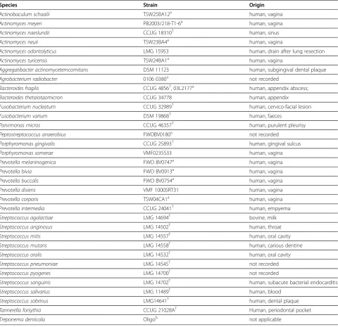
The bacterial strains used in this study for analyzing sen-sitivity and specificity of the primers are listed in Table 1. Clinical isolates, which were not traceable to the patient, and reference isolates were used. The clinical samples used for the study mentioned that was published
elsewhere [32], were covered by the ethical committee approval: B67020097225 (Belgian registration number). These clinical samples were collected from the deepest periodontal pocket per quadrant. A sterile paper point was inserted following supragingival plaque removal and left in situ for about 20 seconds. The paper points were collected in 200μl of a 20 mM Tris–HCl, pH 8 solution
(Merck, Darmstadt, Germany) and stored at−20°C until
DNA extraction.
Extraction of DNA and preparation of standard dilution series
Bacterial genomic DNA used for preparing standard dilution series was extracted with the High Pure PCR Template Preparation Kit (Roche, Basel, Switzerland). Briefly, all strains were grown anaerobically, except for Streptococcus spp., which were grown aerobically, on blood agar. Colonies were scraped from plates and sus-pended in 400μl PBS. To 200μl of bacterial suspension, 2μl mutanolysin (25 U/μl) was added and incubated for 15 min at 37°C. Further DNA extraction was performed according to manufacturers guidelines. The DNA con-centration was quantified by spectrophotometric analysis (Nanodrop, Thermo Scientific, Wilmington, DE) and converted from ng/ml to number of genomes/ml by calculating the molecular weight of the genome (ng/gen-ome) and dividing the concentration (ng/ml) by the molecular weight of the genome in order to assign number of genome values to the standard dilution series. Bacterial DNA used for specificity testing was extracted using alkaline lysis. Briefly, strains were grown on agar plates under appropriate conditions, a single colony was
picked up and dissolved in 20 μl alkaline lysis buffer
(0.25% SDS, 0.05 N NaOH), the mixture was heated for
15 min at 95°C, the tubes were briefly spinned, 180 μl
sterile HPLC water was added to neutralize the pH, and
the tubes were centrifuged during 5 min at 13000g to
spin down the bacterial cell debris. The supernatant was used as DNA extract. Tenfold standard dilution series of reference strains were made from genomic DNA
extracted from A. actinomycetemcomitans DSM 11123,
F. nucleatum CCUG 32989, P. micros CCUG 46357, P. gingivalis CCUG 25893, P. intermediaCCUG 24041, S. mutans LMG 14558T and T. forsythia CCUG
21028AT. Several attempts to grow T. denticola from
different culture collections failed. Therefore, a tenfold standard dilution series was made of a synthetic ds oligonucleotide. We blasted the primers described by Hyvarinen et al. [33] and found that these were located on the coding domain sequence for a glycosyl
transfer-ase, corresponding to region 1470086–147094 of strain
calf thymus DNA (Sigma-Aldrich, St. Louis, MO), according to the MIQE guidelines [34]. Calf thymus DNA was added to decrease adherence of the target DNA to the vials, in order to increase reproducibility, especially of the low concentration standards.
Primers
Primer sequences and amplicons were analysed for spe-cificity using the nucleotide Basic Local Alignment Search Tool and primerBLAST (http://blast.ncbi.nlm. nih.gov/Blast.cgi). The presence of secondary structures
was analyzed using mFOLD (http://mfold.rna.albany. edu/?q=mfold).
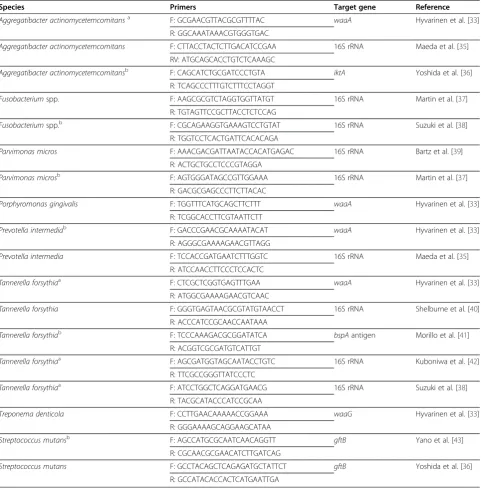
Table 2 lists the primers that were tested.
qPCR
[image:3.595.58.539.111.576.2]Each assay was designed for most efficient amplifica-tion with the same thermocycling program: initial dsDNA denaturation (+ activation of hot start enzyme) for 10 min at 95°C, 40 cycles of 15 s at 95°C and 1 min at 60°C, on an ABI 7300 real time PCR system (Applied Biosystems, Carlsbad, CA). The primer concentrations
Table 1 Bacterial strains and their corresponding collection number used to test sensitivity and specificity of the different primer pairs
Species Strain Origin
Actinobaculum schaalii TSW25BA12a human, vagina
Actinomyces meyeri PB2003/218-T1-6a human, vagina
Actinomyces naeslundii CCUG 18310T human, sinus
Actinomyces neuii TSW23BA4a human, vagina
Actinomyces odontolyticus LMG 15953 human, drain after lung resection
Actinomyces turicensis TSW24BA1a human, vagina
Aggregatibacter actinomycetemcomitans DSM 11123 human, subgingival dental plaque
Agrobacterium radiobacter 0106 0380a not recorded
Bacteroides fragilis CCUG 4856T, 03L2177a human, appendix abscess;
Bacteroides thetaiotaomicron CCUG 34778 human, appendix
Fusobacterium nucleatum CCUG 32989T human, cervico-facial lesion
Fusobacterium varium DSM 19868T human, faeces
Parvimonas micros CCUG 46357T human, purulent pleurisy
Peptostreptococcus anaerobius FWOBV0180a not recorded
Porphyromonas gingivalis CCUG 25893T human, gingival sulcus
Porphyromonas somerae VMF0235S33 human, vagina
Prevotella melaninogenica FWO BV0747a human, vagina
Prevotella bivia FWO BV0913a human, vagina
Prevotella buccalis FWO BV0754a human, vagina
Prevotella disiens VMF 1000SRT31 human, vagina
Prevotella corporis TSW04CA1a human, vagina
Prevotella intermedia CCUG 24041T human, empyema
Streptococcus agalactiae LMG 14694T bovine, milk
Streptococcus anginosus LMG 14502T human, throat
Streptococcus mitis LMG 14557T human, oral cavity
Streptococcus mutans LMG 14558T human, carious dentine
Streptococcus oralis LMG 14532T human, oral cavity
Streptococcus pneumoniae LMG 14545T not recorded
Streptococcus pyogenes LMG 14700T not recorded
Streptococcus sanguinis LMG 14702T human, subacute bacterial endocarditis
Streptococcus salivarius LMG 11489T human, blood
Streptococcus sobrinus LMG14641T human, dental plaque
Tannerella forsythia CCUG 21028AT Human, periodontal pocket
Treponema denticola Oligob not applicable
Legend
a
were the same for all assays, i.e. 300 nM. Assays were performed in a final volume of 25μl with a final MgCl2
concentration of 3 mM and with 2.5 μl DNA extract,
using the SybrGreen qPCR core kit (Eurogentec). Assays carried out on the LightCycler (LC) 480 ther-mal cycling system (Roche) were performed in a final reaction volume of 10μl with 1 or 2 μl of DNA extract (both volumes were tested), using the LightCycler 480
[image:4.595.58.540.111.602.2]SybrGreen I master mix, with the same primer concen-trations and thermocycling program as for the ABI 7300. Assays carried out on the iQ5 thermal cycling system (Bio-Rad Laboratories, Hercules CA) were performed in a final reaction volume of 25μl with 2.5μl DNA extract, using the iQ SYBR Green Supermix, with the same pri-mer concentrations and thermocycling program as for the ABI 7300.
Table 2 Primer sequences evaluated for specificity (BLAST) and primer annealing onto secondary structures (mFOLD) byin silicoanalysis for the eight different species
Species Primers Target gene Reference
Aggregatibacter actinomycetemcomitansa F: GCGAACGTTACGCGTTTTAC waaA Hyvarinen et al. [33]
R: GGCAAATAAACGTGGGTGAC
Aggregatibacter actinomycetemcomitans F: CTTACCTACTCTTGACATCCGAA 16S rRNA Maeda et al. [35] RV: ATGCAGCACCTGTCTCAAAGC
Aggregatibacter actinomycetemcomitansb F: CAGCATCTGCGATCCCTGTA iktA Yoshida et al. [36]
R: TCAGCCCTTTGTCTTTCCTAGGT
Fusobacteriumspp. F: AAGCGCGTCTAGGTGGTTATGT 16S rRNA Martin et al. [37] R: TGTAGTTCCGCTTACCTCTCCAG
Fusobacteriumspp.b F: CGCAGAAGGTGAAAGTCCTGTAT 16S rRNA Suzuki et al. [38] R: TGGTCCTCACTGATTCACACAGA
Parvimonas micros F: AAACGACGATTAATACCACATGAGAC 16S rRNA Bartz et al. [39] R: ACTGCTGCCTCCCGTAGGA
Parvimonas microsb F: AGTGGGATAGCCGTTGGAAA 16S rRNA Martin et al. [37] R: GACGCGAGCCCTTCTTACAC
Porphyromonas gingivalis F: TGGTTTCATGCAGCTTCTTT waaA Hyvarinen et al. [33] R: TCGGCACCTTCGTAATTCTT
Prevotella intermediab F: GACCCGAACGCAAAATACAT waaA Hyvarinen et al. [33] R: AGGGCGAAAAGAACGTTAGG
Prevotella intermedia F: TCCACCGATGAATCTTTGGTC 16S rRNA Maeda et al. [35] R: ATCCAACCTTCCCTCCACTC
Tannerella forsythiaa F: CTCGCTCGGTGAGTTTGAA waaA Hyvarinen et al. [33] R: ATGGCGAAAAGAACGTCAAC
Tannerella forsythia F: GGGTGAGTAACGCGTATGTAACCT 16S rRNA Shelburne et al. [40] R: ACCCATCCGCAACCAATAAA
Tannerella forsythiab F: TCCCAAAGACGCGGATATCA bspAantigen Morillo et al. [41] R: ACGGTCGCGATGTCATTGT
Tannerella forsythiaa F: AGCGATGGTAGCAATACCTGTC 16S rRNA Kuboniwa et al. [42]
R: TTCGCCGGGTTATCCCTC
Tannerella forsythiaa F: ATCCTGGCTCAGGATGAACG 16S rRNA Suzuki et al. [38] R: TACGCATACCCATCCGCAA
Treponema denticola F: CCTTGAACAAAAACCGGAAA waaG Hyvarinen et al. [33] R: GGGAAAAGCAGGAAGCATAA
Streptococcus mutansb F: AGCCATGCGCAATCAACAGGTT gftB Yano et al. [43]
R: CGCAACGCGAACATCTTGATCAG
Streptococcus mutans F: GCCTACAGCTCAGAGATGCTATTCT gftB Yoshida et al. [36] R: GCCATACACCACTCATGAATTGA
Legend
a
: Primer pairs excluded for furtherin vitrotesting on the basis ofin silicoanalysis.
b
Results
The aim of this study was to optimize quantitative PCR assays (qPCR assays) for 8 important oral bacteria, i.e. Aggregatibacter actinomycetemcomitans, Fusobacter-ium nucleatum,Parvimonas micros,Porphyromonas gin-givalis, Prevotella intermedia, Streptococcus mutans, Tanerella forsythia and Treponema denticola. In silico analysis indicated that it was not possible to develop species specific primers forF. nucleatum, based on the 16S rRNA
gene. Therefore, Fusobacterium genus primers were used,
assuming that - when testing oral samples - most signal strength for this qPCR will be caused by the presence of F. nucleatum, because this species is the dominant Fuso-bacteriumspecies in oral microflora [44]. Different primer pairs were tested with regard to amplification efficiency, specificity and intercycler portability (robustness),i.e. port-ability between different thermal cyclers.
Initially, the qPCR formats were developed on an ABI 7300 thermal cycling system (Applied Biosystems), on which we first determined the amplification efficiency of the primers. Thereafter, the primer pairs with the best amplification efficiency were used to test intercycler portability by carrying out the PCRs on a LightCycler 480 thermal cycler (Roche) and on an iQ5 thermal cycler (Bio-Rad), with the same cycling parameters as used on the ABI 7300. The thermal cycler that gave the most reproducible and accurate results, was used to test the specificity of the assays.
Amplification efficiency of different primer pairs
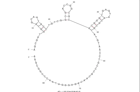
Bioinformatic analysis (PrimerBLAST, mFold) revealed that, at an annealing temperature of 60°C, some of the primers were annealing on secondary structures in the target genes. An example of annealing on secondary structure is shown in Figure 1 for theT. forsythiaforward primer that has been proposed by Kuboniwa et al. [42].
As indicated in Table 2, we omitted these primer pairs from subsequent experiments, because annealing of the primers onto secondary structures has been shown to decrease amplification efficiency [45]. First, the amplifi-cation efficiency and quantifiamplifi-cation limit of the selected primer pairs were tested using a 10-fold standard dilu-tion series. The best performing primer pairs were selected on the basis of amplification efficiency, correl-ation of standard dilution series and quantificcorrel-ation limit, the latter defined as the lowest standard dilution that could be included in the standard series without de-creasing the amplification efficiency below 95% (Table 3). Moreover, intra-run reproducibility was taken into ac-count (data not presented).
Specificity testing
After selection of the primer pairs that enabled amplifi-cation of the target species with the same protocol,
specificity of the different primer sets was tested by in-cluding closely related species (Table 1) in each of the
8 qPCR assays. Assays for A. actinomycetemcomitans,
P. micros, P. gingivalis and P. intermedia detected only the target species for which they were designed. The
assay for the Fusobacterium spp. detected also F.
var-ium, next to F. nucleatum, as expected, since this is a genus specific qPCR. For the assay forT. forsythia,some unspecific amplification was observed during the last cycles (35 < Cq< 40) for strains of the species Fusobac-terium nucleatum, P. bivia,P. intermediaand S. agalac-tiae (Figure 2). This did not affect the specificity of the T. forsythia assay because of the low amplification
effi-ciency. Moreover, the Tm-value of the T. forsythia
amplicon was situated between 81.96 and 82.02°C, whereas Tm-values for all other species were lower. Every strain included in the specificity testing, except
the strains of P. intermedia and A. radiobacter, gave
weak unspecific amplification for theT. denticolaassay. This could possibly be explained by the formation of
pri-mer dipri-mers during the last cycles of the T. denticola
assay, since the NTC had a high Cq value ( > 40). Still, this little affected the specificity of this assay, first be-cause of the low amplification efficiency for these non-target species (i.e., Cq value below the quantification limit of the assay) and second because the melting pro-file of the unspecific PCR products was clearly different from that of the target sequence.”
Intercycler portability (robustness)
After selection of the primer pairs with the highest spe-cificity and amplification efficiency on the ABI 7300 cycler (Table 3), the same assays were carried out on the iQ5 and the LC480 thermal cyclers. In addition, for the LC480, two different DNA extract volumes, i.e. 1 and 2μl were tested. All qPCR’s on the different cyclers gave good correlation of the standard series (i.e. r2 ≥ 0.98), but quantification limits varied between cyclers. The overall best quantification limit was obtained by using a
2 μl sample in a final volume of 10 μl on the LC480
(Table 4).
Discussion
approaches (Socransky et al. [9,46]) offer (semi-)quanti-tative data with an acceptable degree of agreement with quantitative culture for most periodontal pathogens [47]. Although a perfect agreement between microbial enu-meration techniques seems unlikely [48-50], their avail-ability might become relevant for the clinician, especially when conventional therapeutic modalities have failed. Interestingly, microbial data could also become valuable
to predict further periodontal deterioration following ac-tive treatment [51].
[image:6.595.60.539.92.408.2]In order to optimize an assay to detect eight predom-inant oral pathogens, 8 primer pairs were selected that were run on the same thermocycling program with suffi-cient amplification efficiency, specificity and suffisuffi-cient quantification limit. Six of the 8 assays were species spe-cific. For the T. denticola and T. forsythia assays, some
Table 3 Primers used for specificity testing, after selection based on amplification efficiency, quantification limit, and intra-run reproducibility (data not presented)
Species (reference) Correlation
standard curve
Amplification efficiency (%)
Quantification limit (number of bacteria per 25μl reaction)
Aggregatibacter actinomycetemcomitans[35] 0.99 89 25
Fusobacteriumspp. [37] 0.99 94 4
Parvimonas micros[39] 0.99 91 2
Porphyromonas gingivalis[33] 0.99 95 9
Prevotella intermedia[35] 0.99 91 11
Treponema denticola[33] 0.99 95 150
Tannerella forsythia[40] 0.99 93 25
[image:6.595.56.539.596.733.2]Streptococcus mutans[36] 0.98 115 37
unspecific amplification was observed, but only at Cq values of more than 35. This was not an issue, since the last standard included in the standard dilution series,
corresponding to one chromosome/reaction forT.
denti-colaand 2 chromosomes /reactionT. forsythia) had a Cq value below 35, such that all fluorescence signals detected after this Cqvalue are considered as not quanti-fiable. Moreover, melting curve analysis indicated that these unspecific amplification products had melting tem-peratures that were clearly different from that of the tar-get species.
All assays were evaluated for intercycler portability by running the standard dilution series for each species on
three different thermal cyclers, i.e. ABI 7300, Bio-Rad
iQ5 and LightCycler 480. Highly efficient amplification was obtained on all cyclers, but the LightCycler 480 could detect lower bacterial inocula than the other
devices, i.e. on average 3.6 chromosomes /reaction,
compared to 26 chromosomes/reaction for the iQ5 and 33 chromosomes/reaction for the ABI 7300. In addition, the LightCycler 480 has higher throughput (i.e.384 sam-ples) than the ABI 7300 and Bio-Rad iQ5 devices (i.e.96 samples).
[image:7.595.60.540.89.272.2]The optimized assays were implemented to evaluate the microbial effects of an essential oils mouth rinse used by patients in supportive periodontal care [32]. Briefly, during a 3-month double-blind randomized placebo-controlled study, these qPCR assays were used to evaluate the microbial effects of an essential oils mouth rinse used as an adjunct approach to mechanical plaque control by patients in supportive periodontal care. Subgingival plaque samples were collected for the quantification of the 8 bacterial species by means of the qPCR formats described here. No significant differ-ences were observed between treatment and placebo groups. Also, there was no significant change over time
Figure 2Melting curve analysis of unspecific amplification products for theTannerella forsythiaqPCR [40].The melting curves presented were drawn by the software of the LC480 cycler after performing theT. forsythiaqPCR on the species listed in Table 1.
Table 4 Intercycler portability of the different assays on the different thermal cyclers, by comparison of the limits of reliable quantification, i.e. the most diluted standard that could be used to calculate the standard curve, expressed here as number of cells present in the most diluted standard reaction mixture
Assay ABI 7300
(2.5/22.5)a iQ5(2.5/22.5) LC 480(1/9) LC 480(2/8)
Species Reference
Aggregatibacter actinomycetemcomitans Maeda et al. [35] 26 26 10 2
Fusobacteriumspp. Martin et al. [37] 4 4 2 3
Parvimonas micros Bartz et al. [39] 2 1 1 2
Porphyromonas gingivalis Hyvarinen et al. [33] 9 90 36 7
Prevotella intermedia Maeda et al. [35] 11 11 4 9
Streptococcus mutans Yoshida et al. [36] 37 37 15 3
Tannerella forsythia Shelburne et al. [40] 25 25 10 2
Treponema denticola Hyvarinen et al. [33] 150 15 6 1
Legend
a
[image:7.595.55.540.585.718.2]neither in detection frequency nor load for any of the bacterial species.
Conclusion
In summary, we present optimized qPCR assays, with high intercycler portability, for direct quantification of 8 bacterial species that have been associated with peri-odontal disease.
Competing interests
The authors declare that they have no competing interests.
Authors’contributions
Decat E. and Saerens B. carried out the qPCR assays. Decat E. carried out the data analysis and interpretation. Decat E., Vaneechoutte M. and Van Mechelen E wrote the first draft of the manuscript. Decat E., Van Mechelen E. ,Vermeulen S., Cosyn J., Miremadi R., De Bruyn H., Vaneechoutte M. and Deschaght P. conceived the study, participated in its design and coordination and helped to draft the manuscript. All authors read and approved the final manuscript.
Acknowledgements
The authors thank the University College Ghent and the Flemish government for funding this study by means of the PWO project:“Study of Microbiological application for Real Time PCR (SMART)”.
Author details
1Biomedical and Exact Sciences, Faculty of Education, Health & Social Work,
University College Ghent, Keramiekstraat 80, Ghent, Belgium.2Laboratory
Bacteriology Research, Department Clinical Chemistry, Microbiology & Immunology, Faculty of Medicine and Health Sciences, University of Ghent, De Pintelaan 185, Ghent B-9000, Belgium.3Department of Periodontology
and Oral Implantology, Dental School, Faculty of Medicine and Health Sciences, University of Ghent, De Pintelaan 185, Ghent B-9000, Belgium.
Received: 3 July 2012 Accepted: 22 November 2012 Published: 2 December 2012
References
1. Loe H, Anerud A, Boysen H, Morrison E:Natural history of periodontal disease in man. Rapid, moderate and no loss of attachment in Sri Lankan laborers 14 to 46 years of age.J Clin Periodontol1986,13:431–445. 2. Slots J, Rams TE:Microbiology of periodontal disease. In Contemporary oral
microbiology and immunology. St. Louis: Mosby Year Book; 1992:429–431. 3. Collins LM, Dawes C:The surface area of the adult human mouth and
thickness of the salivary film covering the teeth and oral mucosa.J Dent Res1987,66:1300–1302.
4. Paster BJ, Olsen I, Aas JA, Dewhirst FE:The breadth of bacterial diversity in the human periodontal pocket and other oral sites.Periodontol 2000
2006,42:80–87.
5. Hommez GM, Verhelst R, Claeys G, Vaneechoutte M, De Moor RJ:
Investigation of the effect of the coronal restoration quality on the composition of the root canal microflora in teeth with apical periodontitis by means of T-RFLP analysis.Int Endodont J2004,
37:819–827.
6. Kumar PS, Griffen AL, Barton JA, Paster BJ, Moeschberger ML, Leys EJ:New bacterial species associated with chronic periodontitis.J Dent Res2003,
82:338–344.
7. Moore WE, Moore LV:The bacteria of periodontal diseases.Periodontol 20001994,5:66–77.
8. Paster BJ, Boches SK, Galvin JL, Ericson RE, Lau CN, Levanos VA, Sahasrabudhe A, Dewhirst FE:Bacterial diversity in human subgingival plaque.J Bacteriol2001,183:3770–3783.
9. Socransky SS, Haffajee AD:Periodontal microbial ecology.Periodontol 2000
2005,38:135–187.
10. Socransky SS, Haffajee AD, Cugini MA, Smith C, Kent RL:Microbial complexes in subgingival plaque.J Clin Periodontol1998,25:134–144.
11. Sharawy AM, Sabharwal K, Socransky SS, Lobene RR:A quantitative study of plaque and calculus formation in normal and periodontally involved mouths.J Periodontol1966,37:495–501.
12. Gendron R, Grenier D, Maheu-Robert L:The oral cavity as a reservoir of bacterial pathogens for focal infections.Microbes Infect2000,2:897–906. 13. Kolenbrander PE, Palmer RJ Jr, Rickard AH, Jakubovics NS, Chalmers NI, Diaz
PI:Bacterial interactions and successions during plaque development. Periodontol 20002006,42:47–79.
14. Haffajee AD, Socransky SS, Ebersole JL, Smith DJ:Clinical, microbiological and immunological features associated with the treatment of active periodontosis lesions.J Clin Periodontol1984,11:600–618.
15. Haffajee AD, Socransky SS:Microbial etiological agents of destructive periodontal diseases.Periodontol 20001994,5:78–111.
16. Listgarten MA, Lai CH, Evian CI:Comparative antibody titers to
Actinobacillus actinomycetemcomitansin juvenile periodontitis, chronic periodontitis and periodontally healthy subjects.J Clin Periodontol1981,
8:155–164.
17. Slots J:Reynolds HS.Genco RJ: Actinobacillus actinomycetemcomitans in human periodontal disease: a cross-sectional microbiological investigation. Infect Immun1980,29:1013–1020.
18. Tanner A, Maiden MF, Macuch PJ, Murray LL, Kent RL:Microbiota of health, gingivitis, and initial periodontitis.J Clin Periodontol1998,25:85–98. 19. Van Winkelhoff AJ, Loos BG, van der Reijden WA, van der Velden U:
Porphyromonas gingivalis,Bacteroides forsythusand other putative periodontal pathogens in subjects with and without periodontal destruction.J Clin Periodontol2002,29:1023–1028.
20. Socransky SS, Haffajee AD:The bacterial etiology of destructive periodontal disease: current concepts.J Periodontol1992,63:322–331. 21. Booth V, Downes J, Van den Berg J, Wade WG:Gram-positive anaerobic
bacilli in human periodontal disease.J Periodontal Res2004,39:213–220. 22. Choi BK, Park SH, Yoo YJ, Choi SH, Chai JK, Cho KS, Kim CK:Detection of
major putative periodontopathogens in Korean advanced adult periodontitis patients using a nucleic acid-based approach.J Periodontol
2000,71:1387–1394.
23. Colombo AP, Teles RP, Torres MC, Souto R, Rosalem WJ, Mendes MC, Uzeda M:Subgingival microbiota of Brazilian subjects with untreated chronic periodontitis.J Periodontol2002,73:360–369.
24. Herrera D, Roldan S, Gonzalez I, Sanz M:The periodontal abscess (I). Clinical and microbiological findings. J Clin Periodontol2000,27:387–394. 25. Moore WE, Holdeman LV, Cato EP, Smibert RM, Burmeister JA, Palcanis KG,
Ranney RR:Comparative bacteriology of juvenile periodontitis.Infect Immun1985,48:507–519.
26. Papapanou PN, Baelum V, Luan WM, Madianos PN, Chen X, Fejerskov O, Dahlen G:Subgingival microbiota in adult Chinese: prevalence and relation to periodontal disease progression.J Periodontol1997,
68:651–666.
27. Papapanou PN, Neiderud AM, Papadimitriou A, Sandros J, Dahlen G:
“Checkerboard" assessments of periodontal microbiota and serum antibody responses: a case–control study.J Periodontol2000,71:885–897. 28. Rams TE, Feik D, Slots J:Campylobacter rectusin human periodontitis.Oral
Microbiol Immunol1993,8:230–235.
29. Simonson LG, Robinson PJ, Pranger RJ, Cohen ME, Morton HE:Treponema denticolaandPorphyromonas gingivalisas prognostic markers following periodontal treatment.J Periodontol1992,63:270–273.
30. Okada M, Kawamura M, Oda Y, Yasuda R, Kojima T, Kurihara H:Caries prevalence associated withStreptococcus mutansandStreptococcus sobrinusin Japanese schoolchildren.Int J Paediatr Dent2012,22:342–348. Jan 8 [Epub ahead of print].
31. Richards W, Amee J, Coll AM, Higgs G:Reasons for tooth extraction in four general dental practices in South Wales.Br Dent J2005,198:275–278. 32. Cosyn J, Princen K, Miremadi R, Decat E, Vaneechoutte M, De Bruyn H:A
double-blind randomized placebo-controlled study on the clinical and microbial effects of an essential oil mouth rinse used by patients in supportive periodontal care.Int J Dent Hyg2012,10.1111/ idh.12000:10.1111/idh.12000. doi:10.1111/idh.12000.
33. Hyvarinen K, Laitinen S, Paju S, Hakala A, Suominen-Taipale L, Skurnik M, Kononen E, Pussinen PJ:Detection and quantification of five major periodontal pathogens by single copy gene-based real-time PCR.Innate Immun2009,15:195–204.
guidelines: Minimum Information for publication of Quantitative real-time PCR Experiments.Clin Chem2009,55:611–622.
35. Maeda H, Fujimoto C, Haruki Y, Maeda T, Kokeguchi S, Petelin M, Arai H, Tanimoto I, Nishimura F, Takashiba S:Quantitative real-time PCR using TaqMan and SYBR Green forActinobacillus actinomycetemcomitans,
Porphyromonas gingivalis,Prevotella intermedia, tetQ gene and total bacteria.FEMS Immunol Med Microbiol2003,39:81–86.
36. Yoshida A, Suzuki N, Nakano Y, Kawada M, Oho T, Koga T:Development of a 5' nuclease-based real-time PCR assay for quantitative detection of cariogenic dental pathogensStreptococcus mutansandStreptococcus sobrinus.J Clin Microbiol2003,41:4438–4441.
37. Martin FE, Nadkarni MA, Jacques NA, Hunter N:Quantitative
microbiological study of human carious dentine by culture and real-time PCR: Association of anaerobes with histopathological changes in chronic pulpitis.J Clin Microbiol2002,40:1698–1704.
38. Suzuki N, Yoshida A, Nakano Y:Quantitative analysis of multi-species oral biofilms by taqman real-time PCR.Clin Med Res2005,3:176–185. 39. Bartz H, Nonnenmacher C, Bollmann C, Kuhl M, Zimmermann S, Heeg K,
Mutters R:Micromonas(Peptostreptococcus)micros: unusual case of prosthetic joint infection associated with dental procedures.Int J Med Microbiol2005,294:465–470.
40. Shelburne CE, Prabhu A, Gleason RM, Mullally BH, Coulter WA:Quantitation of Bacteroides forsythus in subgingival plaque - Comparison of immunoassay and quantitative polymerase chain reaction.J Microbiol Methods2000,39:97–107.
41. Morillo JM, Lau L, Sanz M, Herrera D, Martin C, Silva A:Quantitative real-time polymerase chain reaction based on single copy gene sequence for detection of periodontal pathogens.J Clin Periodontol2004,31:1054–1060. 42. Kuboniwa M, Amono A, Kimura KR, Sekine S, Kato S, Yamamoto Y, Okahashi N, Lida T, Shizukuishi S:Quantitative detection of periodontal pathogens using real-time polymerase chain reaction with TaqMan probes.Oral Microbiol Immunol2004,19:168–176.
43. Yano A, Kaneko N, Ida H, Yamaguchi T, Hanada N:Real-time PCR for quantification of Streptococcus mutans.FEMS Microbiol Lett2002,
217:23–30.
44. Signat B, Roques C, Poulet P, Duffaut D:Role of Fusobacterium nucleatum in periodontal health and disease.Curr Issues Mol Biol2011,13:25–35. 45. Hoebeeck J, Speleman F, Vandesompele J:Real-time quantitative PCR as an
alternative to Southern blot or fluorescence in situ hybridization for detection of gene copy number changes,Methods in Molecular Biology.Volume 353. New Jersey: Humana Press; 2007:205–226.
46. Persson RG, Hitti J, Verhelst R, Vaneechoutte M, Persson RE, Hirschi R, Weibel M, Rothen M, Temmerman M, Paul K, Eschenbach D:The vaginal microflora in relation to gingivitis.BMC Infect Dis2009,9:6. 47. Haffajee AD, Yaskell T, Torresyap G, Teles R, Socransky SS:Comparison
between polymerase chain reaction-based and checkerboard DNA hybridization techniques for microbial assessment of subgingival plaque samples.J Clin Periodontol2009,36:642–649.
48. Boutaga K, van Winkelhoff AJ, Vandenbroucke-Grauls CM, Savelkoul PH:
Comparison of real-time PCR and culture for detection of
Porphyromonas gingivalisin subgingival plaque samples.J Clin Microbiol
2003,41:4950–4954.
49. Boutaga K, van Winkelhoff AJ, Vandenbroucke-Grauls CM, Savelkoul PH:
Periodontal pathogens: a quantitative comparison of anaerobic culture and real-time PCR.FEMS Immunol Med Microbiol2005,45:191–199. 50. Boutaga K, Savelkoul PH, Winkel EG, Van Winkelhoff AJ:Comparison of
subgingival bacterial sampling with oral lavage for detection and quantification of periodontal pathogens by real-time polymerase chain reaction.J Periodontol2007,78:79–86.
51. Rams TE, Listgarten MA, Slots J:Utility of 5 major putative periodontal pathogens and selected clinical parameters to predict periodontal breakdown in patients on maintenance care.J Clin Periodontol1996,
23:346–354.
doi:10.1186/1756-0500-5-664
Cite this article as:Decatet al.:Optimization of quantitative polymerase chain reactions for detection and quantification of eight periodontal bacterial pathogens.BMC Research Notes20125:664.
Submit your next manuscript to BioMed Central and take full advantage of:
• Convenient online submission
• Thorough peer review
• No space constraints or color figure charges
• Immediate publication on acceptance
• Inclusion in PubMed, CAS, Scopus and Google Scholar
• Research which is freely available for redistribution
![Figure 2 Melting curve analysis of unspecific amplification products for the Tannerella forsythia qPCR [40]](https://thumb-us.123doks.com/thumbv2/123dok_us/8280786.283350/7.595.55.540.585.718/figure-melting-analysis-unspecific-amplification-products-tannerella-forsythia.webp)
