R E S E A R C H A R T I C L E
Open Access
An investigation of horizontal transfer of feed
introduced DNA to the aerobic microbiota of the
gastrointestinal tract of rats
Lise Nordgård
1,2, Lorenzo Brusetti
3, Noura Raddadi
4,6, Terje Traavik
1,2, Beate Averhoff
5and Kaare Magne Nielsen
1,2*Abstract
Background:Horizontal gene transfer through natural transformation of members of the microbiota of the lower gastrointestinal tract (GIT) of mammals has not yet been described. Insufficient DNA sequence similarity for homologous recombination to occur has been identified as the major barrier to interspecies transfer of chromosomal DNA in bacteria. In this study we determined if regions of high DNA similarity between the genomes of the indigenous bacteria in the GIT of rats and feed introduced DNA could lead to homologous recombination and acquisition of antibiotic resistance genes.
Results:Plasmid DNA with two resistance genes (nptI andaadA) and regions of high DNA similarity to 16S rRNA and 23S rRNA genes present in a broad range of bacterial species present in the GIT, were constructed and added to standard rat feed. Six rats, with a normal microbiota, were fed DNA containing pellets daily over four days before sampling of the microbiota from the different GI compartments (stomach, small intestine, cecum and colon). In addition, two rats were included as negative controls. Antibiotic resistant colonies growing on selective media were screened for recombination with feed introduced DNA by PCR targeting unique sites in the putatively recombined regions. No transformants were identified among 441 tested isolates.
Conclusions:The analyses showed that extensive ingestion of DNA (100μg plasmid) per day did not lead to increased proportions of kanamycin resistant bacteria, nor did it produce detectable transformants among the aerobic microbiota examined for 6 rats (detection limit < 1 transformant per 1,1 × 10⁸cultured bacteria). The key methodological challenges to HGT detection in animal feedings trials are identified and discussed. This study is consistent with other studies suggesting natural transformation is not detectable in the GIT of mammals.
Background
Relatively few studies have examined the occurrence of horizontal gene transfer (HGT) by natural transforma-tion in the gastrointestinal tract (GIT) of mammals underin vivoconditions [1-5]. It is unclear if the lack of observable competence among bacteria in the GIT is due to experimental limitations, limited occurrence in the few model bacteria examined, or due to true lack of conditions conductive for the development of compe-tence among members of the GIT microbiota of various mammals.
There are several requirements for natural transforma-tion to take place with non-mobile DNA in the GIT [6,7].
First, bacteria must be able to express a competent stage while in the GIT system. Numerous bacterial species pre-sent in the GIT are known to be able to develop natural competence when grownin vitro[8-10]. For instance, spe-cies within the generaBacillus, Campylobacter, Helicobac-ter, Lactobacillus, Neisseria, Pseudomonas, Streptococcus, and Vibrioare known to express competence under speci-fic laboratory conditions. However, so far only fewin vivo studies have reported on the occurrence of bacterial com-petence in the GIT and those are limited to the upper GIT. The lower part of the GIT has nevertheless been sug-gested to be the most active site for HGT processes due to abundance of nutrients, high bacterial density and slower degradation rates of DNA [11,12].
Second, extracellular DNA of sufficient length and con-centration must be present and accessible to competent * Correspondence: kaare.nielsen@uit.no
1GenØk, Centre for Biosafety, Science Park, 9294 Tromsø, Norway
Full list of author information is available at the end of the article
bacteria. Several experimental studies have investigated the stability of DNA in the GIT of mammals ([2-4,13-20]) suggesting that minor fractions of DNA provided with dif-ferent feed sources may remain in various GIT compart-ments. For a recent review, see Rizziet al.[5]. Moreover, that minor portions of such DNA can remain of sufficient size for the acquisition of functional traits (e.g. new pro-tein coding sequences) by competent bacteria.
Third, chromosomal DNA fragments taken up over the cytoplasmic membrane must be able to recombine with the bacterial host chromosome for stable inheritance and vertical transmission [21-23]. In general, incoming DNA must contain regions of minimum 25-200 bp in length of high similarity to the recipient genome for homologous recombination to occur [24-28]. These prerequisites are met by ribosomal gene sequences, which are sufficient in length and degree of sequence conservation for homolo-gous recombination events [29].
Several studies have investigated conditions for natural transformation in the upper GIT. The oral cavity is the first place feed introduced DNA enters the GIT and therefore also contains the highest amount of DNA ingested. In general, a highly time-limited stability of DNA and ability to transform defined model bacteria introduced to saliva or stomach fluids has been observed in vitroandin vivo[16,30-34]. Only few studies have investigated the occurrence of natural transformation in the compartments of the lower GIT. Wilcks and collea-gues [2,3] reported some persistence of introduced DNA in the GIT of germ-free/gnotobiotic or mono-associated rats, but did not detect uptake of plasmid DNA byE. coli, Bacillus subtilis, orStreptococcus gordonii. In another study, Nordgårdet al., [4] examined the ability of Acine-tobacter baylyicolonized gnotobiotic mice and rats for
potentialin vivotransformation of bacteria with feed
introduced DNA. No transformants were detectedin vivo
orin vitro. In the onlyin vivostudy of natural transfor-mation of bacteria present in the GIT of humans pub-lished so far, only limited persistence of food-derived plant DNA was found [1]. The DNA did not survive pas-sage through the GIT of healthy human subjects as deter-mined by PCR. Three out of these seven human volunteers (ileostomists) showed evidence of
low-frequency HGT of plant DNA (theepspstransgene) to
the microbiota of the small bowel, but this appeared to have occurred before feeding the experimental meal and no single microbial isolate could be obtained for further
analysis. With the exception of the in vivo study in
human described above, most published studies focusing on natural transformation in the GIT have used experi-mental models aimed at detecting HGT events occurring into single bacterial strains introduced into the GIT system.
The aim of this study was to examine if naturally
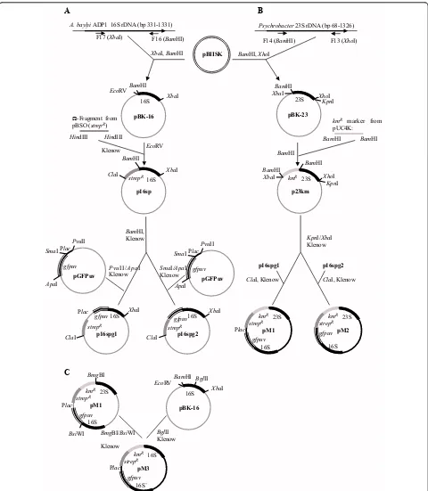
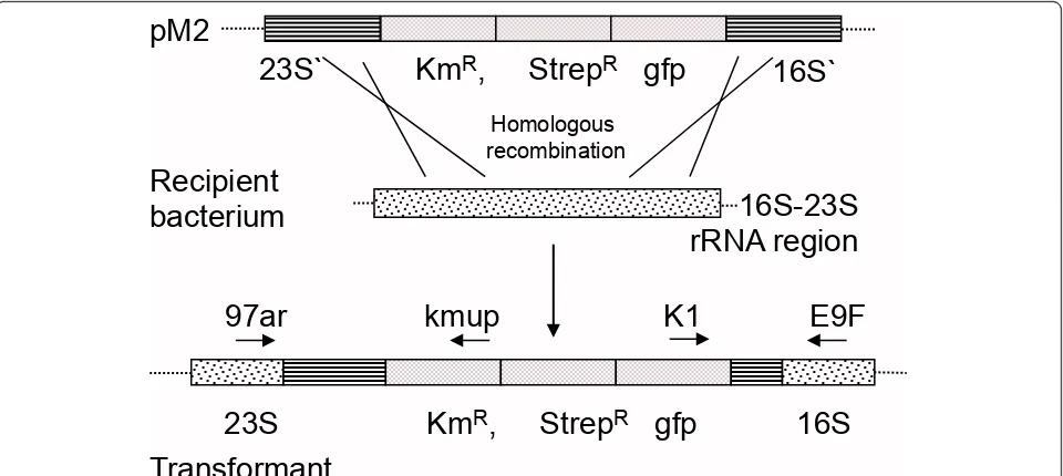
occurring aerobic bacteria in the GIT of rats can undergo natural transformation and recombination with feed introduced DNA. We hypothesized that regions of high DNA similarity between the introduced DNA and the indigenous bacteria in the rat GIT would lead to stable integration of two selectable marker genes (streptomycin or kanamycin resistance) based on homologous recombi-nation. Moreover, that such events occurred at a fre-quency that was detectable by direct selective plating. Plasmid DNA with 16S rRNA and 23S rRNA sequences flanking the two antibiotic resistance marker genes were mixed with standard rat feed pellets (Figure 1). A double-sided homologous recombination event with sequence-similar rRNA genes in competent bacteria in the rat GIT would lead to integration of the antibiotic resistance genes (Figure 2). Recombinant-specific primer annealing sites were chosen to enable unambiguous detection of transformants carrying chromosomal integrations of the resistance genes among the overall population of bacteria recovered from the GIT.
Results
Natural transformation
The plasmids (purified fromE. coli) were highly potent in generating transformants when incubated with naturally
competent A. baylyi cellsin vitro on LB agar plates.
Transformation frequencies of above 1,4 × 10⁻⁵were
obtained per 0,2μg plasmid (data not shown): similar to
previousin vitro studies with the same experimental
setup [35,36].
To address the question whether the plasmids are also sufficient for natural transformation studies of phylogen-etically distant bacteria, natural transformation assays were performed with pM1, pM2 and pM3 using ~30 dif-ferent marine isolates as potential recipients. These studies revealed the ability of different Gammaproteobacteria and Actinobacteria to be naturally transformed with the three DNA constructs, even in the presence of significant differ-ences in 16S rRNA identity (Table 1).
Total CFU and resistant CFU in the gut contents
The total CFU and the number of resistant CFU in sam-ples from the stomach, small intestine, cecum and colon of rats were determined after the 4 days of plasmid expo-sure. The mean numbers of bacteria in the different com-partments are presented in Table 2. The bacterial counts varied between individuals, for instance, in the stomach, the total CFU ranged from < 1 × 10³
A. baylyi ADP1 16S rDNA (bp 331-1331) A
pBIISK
p16sp XbaI
BamHI
pBK-16
p23km pBK-23
XhoI
BamHI
16S 23S
16S 23S
strepR
F16 (BamHI) F17 (XbaI)
Psychrobacter 23S rDNA (bp 68-1326)
F13 (XhoI)
XbaI, BamHI BamHI,XhoI
EcoRV
EcoRV
HindIII HindIII
Klenow
BamHI BamHI
BamHI
BamHI
F14 (BamHI)
BamHI
BamHI
XhoI
XbaI
kmR marker from
pUC4K: Ω-Fragment from
pBSO (strepR)
BamHI, Klenow
SmaI/ApaI, Klenow
XbaI
KpnI/XbaI Klenow
p16spg2
16S
kmR
PvuII/ApaI Klenow
gfpuv SmaI
PvuII Plac
ApaI
pGFPuv
XbaI
p16spg1
16S Plac
strepR
gfpuv SmaI
PvuII Plac
ApaI
pGFPuv ClaI
ClaI ClaI
XbaI
KpnI
XbaI
KpnI
strepR
p16spg2
ClaI, Klenow
p16spg1
ClaI, Klenow
gfpuv gfpuv
pM1
23S
kmR strepR
Plac gfpuv
16S
pM2
23S
kmR strepR gfpuv
16S
pM1
23S
kmR strepR
Plac gfpuv
16S
XbaI
BamHI
pBK-16
16S
EcoRV BglII
BsiWI
BmgBI
BmgBI/BsiWI
Klenow
BglII Klenow
pM3
16S`
kmR strepR
Plac gfpuv
16S` C
[image:3.595.58.538.87.638.2]B
most of the individuals) to up to 4 × 10⁷. Variability in CFU numbers is expected due to different amounts of food material present in the colon of each rat; reflecting differences in activity, eating pattern and digestion.
CFU numbers of resistant colonies determined by plating on selective media containing streptomycin and kanamycin were very low. In most animals we could not detect cultur-able resistant bacteria; with a detection limit of 1 CFU per gram material. However, from some compartments and individuals a few colonies emerged on the selective media (see Table 2). A total of 441 isolates were collected from plates inoculated with GIT material obtained from exposed rats, and 17 isolates from GIT material obtained from non-plasmid exposed rats. All isolates growing on selective
media were picked and frozen in LB media with 15% gly-cerol for further characterization.
PCR analyses of antibiotic resistant colonies
None of the 441 antibiotic resistant bacteria recovered from the GIT content of DNA exposed rats produced a positive PCR signal. The total amount of bacteria plated on selective media from the six exposed individuals exceeded 1 × 10⁸ bacteria. Thus, less than 1 antibiotic resistance gene transfer event was observed per 1,1 × 10⁸culturable bacteria. The transformation frequency of the culturable bacterial fraction with the feed added plasmid was therefore less than 8,8 × 10⁻⁹transformants per culturable bacteria.
pM2
16S-23S
rRNA region
Transformant
Homologous recombination
Km
R, Strep
Rgfp
97ar kmup K1
E9F
16S
23S
23S`
16S`
Km
R, Strep
Rgfp
[image:4.595.59.539.89.304.2]Recipient
bacterium
Figure 2rRNA sequence-based homologous recombination. Schematic presentation of double homologous recombination between the pM2 and the host ribosomal 16S-23S region. The binding sites of the primers 97ar/kmup and K1/E9F used to confirm the chromosomal integration of the constructs are indicated by arrows. KmR; kanamycin-resistance (nptI); strepR: streptomycin resistance; gfp:gfpuv-gene (enhanced
GFP) with native promoter; 16S: 16S rRNA sequence (1 kb) ofAcinetobactersp; 23S: 23S rRNA sequence (1,258 bp) ofPsychrobactersp. Figures are not to scale.
Table 1 Natural transformation of environmental isolates of bacteriaa
Transformable isolateb Phylogenetic position Plasmids tested 16S-rRNA gene identityc
Marinobactersp. Gammaproteobacteria; pM1, pM2, 77
Alteromonadales pM3
Kocuria rosea Actinobacteria; pM2 79
Photobacterium Gammaproteobacteria; pM1, pM2, 86
phosphoreum Vibrionales pM3
Psychrobacter Gammaproteobacteria; pM1, pM2, 96
marincola Pseudomonadales pM3
a
Outcome of a study examining approximately 30 isolates obtained from three sources (Mediterranean, Celtic and north Atlantic seas).
b
Transformants were species identified by 16S sequencing and confirmed by selective plating and PCR. The screen for transformability did not aim to be quantitative, however, the overall CFU numbers indicated a transformation frequency of ~ 10-5to 10-6transformants per total CFU. The utility of the plasmids for transformation studies was analyzed by use of marine isolates since the recombinant plasmids for genomic integration carried rRNA genes from a marine Psychrobacterisolate.
c
[image:4.595.56.538.577.676.2]Discussion
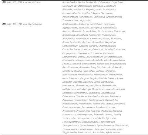
Natural transformation of microbial communities in the GIT requires the presence of bacteria that express compe-tence in gut locations where DNA is present and accessi-ble. Moreover, those competent bacteria must be able to integrate and vertically transfer the DNA over subsequent generations (i.e. through linkage to a replicating unit). Finally, no significant absolute or relative fitness reduc-tions should be experienced by the transformed cells [37]. The model system presented here was optimized such that the requirements for stable inheritance were met through providing the target DNA with flanking regions sharing homology to bacterial rRNA genes harboured by a subset of the bacteria present in the GIT of rats. Table 3 lists the bacterial species with less than 25% DNA sequence diver-gence at the 16S or 23S loci.
The in vivomodel system used also included a
select-able marker system (nptI, aadA) that facilitated
unam-biguous detection of HGT among the diversity of the culturable bacterial populations. Although the model system chosen attempted to circumvent some natural barriers to HGT (introduced DNA sequence similarity) and model based limitations (strong positive selection of transformants, and high levels of DNA exposure to nat-ural bacterial communities) no transformants were
observed among the 6 rats and approx. 108 cultivable,
aerobic bacteria examined. Several factors relevant to these findings may explain the lack of observable uptake of plasmid DNA in this study:
i) Natural transformation does not occur in the GIT of rats due to lack of competence-expressing bacteria or lack of access to extracellular DNA for competent bacteria.
ii) Natural transformation occurs at frequencies below the levels detectable within a limited number of aerobic bacteria per animal GIT and days of DNA exposure. The various parts of the GIT support different inoculum densities [12]. Although total CFU numbers were at rea-sonable levels in the various gut compartments in rats (103-107 CFU per gram GIT content), these population sizes may be too small to enable identification of rare HGT events in individual, single GITs [4,38]. The detec-tion limit per rat and GIT compartment is thus 1
trans-formant per 103 to 107 bacteria, in our study. A
detection limit of 1 transformant per 1 × 10⁸ bacteria is reached when the overall number of CFU tested is sum-marized for the 6 rats. Transformable bacterial species may nevertheless express competence below this fre-quency range and screening of larger bacterial popula-tion sizes should be considered for consistent detecpopula-tion or rare HGT events. The number of species in the GIT
of rats that are expected to be competent based onin
[image:5.595.68.539.111.299.2]vitro data is only a fraction of overall population size and diversity. It can be estimated that it would be neces-sary to sample the combined bacterial populations of the GIT content of 60 to 6 000 rats to identify bacterial transformants produced at lower frequencies of 10-10to 10-13.
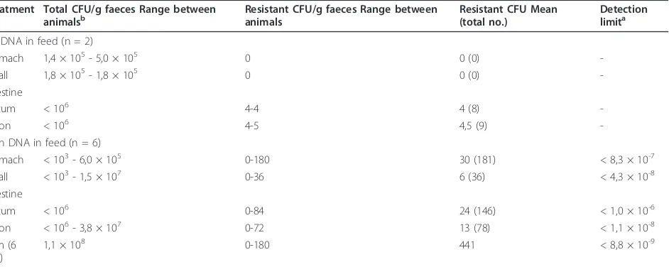
Table 2 CFUs obtained from the gastrointestinal tracts of rats, with a normal microbiota, consuming standard feed with added DNA over 4 days (100μg/day)
Treatment Total CFU/g faeces Range between
animalsb Resistant CFU/g faeces Range betweenanimals Resistant CFU Mean(total no.) Detectionlimita
No DNA in feed (n = 2)
Stomach 1,4 × 10⁵- 5,0 × 10⁵ 0 0 (0)
-Small 1,8 × 10⁵- 1,8 × 10⁵ 0 0 (0)
-intestine
Cecum < 106 4-4 4 (8)
-Colon < 106 4-5 4,5 (9)
-With DNA in feed (n = 6)
Stomach < 103- 6,0 × 10⁵ 0-180 30 (181) < 8,3 × 10⁻⁷
Small < 103- 1,5 × 10⁷ 0-36 6 (36) < 4,3 × 10⁻⁸
intestine
Cecum < 106 0-84 24 (146) < 1,0 × 10⁻⁶
Colon < 106- 3,8 × 10⁷ 0-72 13 (78) < 1,1 × 10⁻⁸
Sum (6 rats)
1,1 × 10⁸ 0-180 441 < 8,8 × 10⁻⁹
a
All the 441 colonies growing on selective-agar plates were tested by PCR for possible uptake of the plasmid DNA and none were found positive. The detection limit is therefore calculated as the reciprocal of the total CFU (summarized for each compartment for all 6 rats); and given as an absolute number. As seen from the range of total CFU obtainable per individual animal, the detection limit can vary 1000-fold. Specifically, the summarized CFU numbers for the compartments of the six DNA feed rats were 1,2 × 10⁶(stomach), 2,3 × 10⁷(small intestine), < 106(cecum) and 9,0 × 10⁷(colon).
b
iii) Natural transformation is limited in frequency due to the low concentrations or lack of accessibility of DNA substrates in the different compartments of the digestive tract. Although a continuous supply of DNA was ensured by daily administration of high amounts (100μg plasmid DNA), purified DNA is known to rapidly fragmentin vivo. However previous studies have shown that minor propor-tions of orally ingested plasmid DNA persist in a biologi-cally active form in the GIT of rats [2,3], and 1-2% of plasmid and bacteriophage DNA can survive passage through the mouse GIT and be detected in the faeces [13-15]. The latter study detected size ranges from a few hundred bp up to about 1700 bp. Similar observations of
short fragment size have also been reported in other ani-mal experiments involving fish, poultry, pig, sheep, cattle and humans [16-19,32,39-41]. See Rizziet al.[5], for a comprehensive review. However, almost all of these stu-dies have biochemically analyzed persistence and degree of fragmentation after recovery of DNA from the gut. So far, little information is available on the extent that DNA frag-ments present in various gut compartfrag-ments are physically accessible to bacteria as templates for natural transforma-tion [4,5]. The study by Nordgård et al. [4] indicates that gut contents may be inhibitory to natural transformation. The study examined the effects of the GIT content of both
[image:6.595.51.539.110.556.2]normal microbiota rats and germfree mice inin vitro
Table 3 Bacterial genera with species that are less than 25% divergent at the 16S or 23S loci compared to the DNA sequences present in plasmids pM2 and pM3
pM2(with 16S rRNA fromAcinetobacter) Aneurinibacillus, Bacillus, Brevibacillus, Caloramator, Caryophanon, Clostridium, Desulfotomaculum, Escherichia, Eubacterium, Filobacillus, Halobacillus, Hespellia, Listeria, Marinibacillus, Oceanobacillus, Paenibacillus, Pelospora, Planococcus, Planomicrobium, Ruminococcus, Salinicoccus, Syntrophomonas, Thermobrachium, Virgibacillus.
pM3(with 23S rRNA fromPsychrobacter) Acidithiobacillus, Acidovorax, Acinetobacter, Aeromonas, Aggregatibacter, Alcanivorax, Alicycliphilus, Alicyclobacillus, Aliivibrio, Alkalilimnicola, Alkaliphilus, Allochromatium, Alteromonas, Anaerococcus, Anaerofustis, Anaerostipes, Anaerotruncus, Anoxybacillus, Aromatoleum, Azotobacter, Bacillus, Baumannia, Blautia, Brevibacillus, Buchnera, Burkholderia, Butyrivibrio, Cardiobacterium, Catonella, Cellvibrio, Chromobacterium, Chromohalobacter, Citrobacter, Clostridium, Colwellia, Comamonas, Congregibacter, Coprococcus, Cronobacter, Cupriavidus,
Dechloromonas, Delftia, Desulfitobacterium, Desulfotomaculum, Dichelobacter, Dickeya, Dorea, Edwardsiella, Eikenella, Enterobacter, Erwinia, Escherichia, Ethanoligenens, Eubacterium, Exiguobacterium, Faecalibacterium, Ferrimonas, Finegoldia, Francisella, Gallionella, Gemella, Geobacillus, Haemophilus, Hahella, Halomonas, Halorhodospira, Halothiobacillus, Heliobacterium, Herbaspirillum, Hydra, Idiomarina, Kangiella, Kingella, Klebsiella, Lachnospiraceae, Laribacter, Legionella, Leptothrix, Listeria, Lysinibacillus,
Macrococcus, Marinobacter, Methylibium, Methylobacillus, Methylococcus, Methylophaga, Methylotenera, Moraxella, Neisseria, Nitrococcus, Nitrosomonas, Nitrosospira, Oceanobacillus, Oribacterium, Oxalobacter, Paenibacillus, Pantoea, Parvimonas, Pasteurella, Pectobacterium, Pelotomaculum, Peptoniphilus, Photobacterium, Photorhabdus, Polaromonas, Proteus, Providencia, Pseudoalteromonas, Pseudomonas, Pseudoxanthomonas, Psychrobacter, Psychromonas, Ralstonia, Rhodoferax, Roseburia, Ruminococcus, Saccharophagus, Salmonella, Serratia, Shigella, Shuttleworthia, Sideroxydans, Simonsiella, Staphylococcus, Stenotrophomonas, Subdoligranulum, Symbiobacterium,
transformation assays of competent cells ofA. baylyi. The study showed that the presence of both types of gut con-tents was inhibitory to transformation [4]. Only purified DNA added to cecum and large intestine content samples from germfree mice was able to transformA. baylyiat low frequenciesin vitro. The sharply reduced DNA uptake fre-quencies also observed in the presence of sterile gut mate-rial from mice indicate that microbially produced DNA nucleases were not responsible for the absence of observa-ble transformation. Bacterial nucleases have also in other studies been found to play a minor role in DNA degrada-tion in the GIT [2,3,42]. Thus, host nucleases or other macromolecules present in the GIT may inhibit natural transformation.
iv) Natural transformation occurs in the GIT but is not observed in our model systems due to other technical limitations. These limitations can include the possibility that bacterial species present in the gut only express competence as a response to certain host physiological conditions or certain (feed) nutrition sources not adopted in our feeding protocol. Moreover, it cannot be excluded that the plasmid DNA used for our transformation study in vivowas not optimal for DNA uptake among all the relevant competent bacterial species in the gut. The DNA similarity, present on the plasmids used, is highest
to theAcinetobacterandPsychobactergenera. However,
thein vitroexperiments with some marine isolates con-firmed the broader applicability of these vectors for nat-ural transformation studies. To our knowledge the results obtained from thein vitrostudy of the marine iso-lates represent the first report of natural transformation
of members of the generaPhotobacterium,Marinobacter
andPsychrobacter(Gammaproteobacteria) and the genus Kocuria(Actinobacteria). These observations suggest that the constructed vectors can be used to detect novel transformable bacteria from various environmental sam-ples. As most bacteria have multiple copies of rRNA genes [43], a lethal effect of an integration of the marker genes into a single rRNA locus is not expected.
Finally, molecular evidence indicates that as little as 10% and up to 40-50% of the GIT population can be identified by differential plating methods [44]. The inabil-ity to isolate the major fraction of the microorganisms in the GIT, and importantly the obligate anaerobic propor-tion, on agar media could also contribute to the failure to detect transformants in our model system. However, it is emphasized that most GIT bacteria that are of high con-cern in the context of resistance development do belong to the culturable fraction (e.g. theEnterobacteriaceae). The lack of provision of an immediate selective advan-tage to the transformants may also have given the net result that rare transformants failed to survive and to expand to numbers that were sufficient to allow their detection [37].
Conclusions
Natural transformation occurring among members of the bacterial community in the lower GIT remains to be demonstrated. Most previous studies have examined nat-ural transformation of single bacterial species introduced into the GIT of various mammalian species. The model system presented here allowed a subset of the aerobic microbial community to be tested for competence devel-opment; however, transformants were not detected among the 6 rat GITs and 108bacteria tested; suggesting a trans-formation frequency below 8.8 × 10⁻⁹(for the combined rat samples). As discussed here, small rodent models may harbour too few culturable bacteria per individual and gut compartment to allow realistic detection limits of natural transformation events in the lower GIT. The design of future studies must consider the opportunities and limita-tions inherent in the population sizes of culturable aerobic and anaerobic bacteria in the model organism used, the transformable fraction and level of competence expressed among these, and the DNA exposure rates expected in the various GIT compartments.
Methods Plasmids
Three chromosome integration vectors, pM1, pM2 and pM3 were derived from theE. colivector pBIISK (oriV of pMB1) (Figure 1). pM1 (9500 bp) and pM2 (9338 bp)
both carry the 16S rRNA gene (bp 331-1331) from
Acine-tobacter baylyiADP1 and the 23S rRNA gene (bp 68-1326) from a marinePsychrobacterisolate. The 16S rRNA gene was amplified with primer pair F16 and F17, and the 23S rRNA gene was amplified with primer pair F13 and F14. The rRNA sequences flank genes encoding the green fluorescent protein (GFP) and streptomycin and kanamy-cin resistance. The two resistance markers can be used for selection of transformants. Thegfpgene enables detection of transformants but was not used throughout this study since transformants were already obtained by selection alone. In pM3 (8281 bp) the resistance genes andgfpgene are flanked by two 16S rRNA gene sequences (bp 362-693 and bp 694-1331) (Figure 1). The plasmids were
main-tained inE. coliTOP10 cells (Invitrogen) grown on LB
agar containing streptomycin (50μg/ml) and kanamycin
(100μg/ml). Plasmid DNA was isolated using the Qiagen Plasmid Maxi Kit (Qiagen, Germany) following the manu-facturer’s protocol and quantified by Nanodrop ND-1000 (Nanodrop Technologies) prior to dilution and mixing in feed. The integrity of the plasmids was also confirmed by agarose gel electrophoresis.
Bioinformatic analyses
The partial nucleotide sequence (331-1331 bp) of the
16S rRNA gene of A. baylyi DSM14961 type strain
(EF611407) and the partial nucleotide sequence (68-1326 bp) of the 23S rRNA gene ofPsychrobacter faecalis strain DSMZ 14664 (HM236417) were used for BLASTN retrieval. The search was limited to the type strain bacteria of the most common genera found in rat and in mouse GIT system [45-49]. For the 16S rRNA fragment, the BLASTN search was to 101 different bac-terial species, while for the 23S rRNA fragment it included 302 different bacterial species. The maximum target sequences parameter was set to 500.
Natural transformation
Cells of the naturally competentA. baylyistrain BD413 were exposed to the plasmids (pM2 and pM3) to confirm that the purified plasmids were able to transform a known naturally competent bacterium at high frequen-cies. For thein vitrofilter transformation assays, 0.2, 2, or
20μg of purified plasmid DNA (Qiagen Miniprep kit,
Germany) was mixed with 100μl of bacterial cells, and incubated on nitrocellulose filters on LB agar plates for 24 h to allow binding and uptake of DNA and replace-ment recombination events of the ribosomal gene sequences (Figure 2.), before serial dilutions and selective plating on LB agar as described by Ray and Nielsen [50]. To confirm the broader functionality of the plasmids for natural transformation, available marine isolates of the Gammaproteobacteria, Actinobacteria and Alphaproteo-bacteria were exposed to pM1, pM2 and pM3. Purified
plasmid DNA (1μg) was linearized withNotI and mixed
with 100μl of overnight culture of a marine isolate and incubated on nitrocellulose filters on marine broth (MB) agar (18.5 g marine broth, 10 g NaCl and 15 g agar per liter, pH 7.6) at room temperature for 24 to 48 h (until growth was visible). After serial dilutions of the cells with saline, selective plating was done on MB agar containing
100 μg/ml streptomycin and 100 μg/ml kanamycin.
Transformation was confirmed by selective plating and PCR using primers E9F and K1 for amplification of the
16S rRNA gene andgfp, and primers kmup and 97ar for
amplification ofnptI and 23S rRNA genes (Table 4 and
Figure 2).
Rat feed preparation
Food pellets were made by mixing 80 ml sterile water with 200 g of standard rat feed powder AIN-93 (Scanbur BK AS, Norway) [55]. After solidification overnight at 4°C, the mass was cut into 1.5 × 1.5 × 1.5 cm feed pellets and left on a tray covered with baking paper to dry overnight in a sterile hood at RT. The pellets were kept at -20°C until
used. A total of 100μg plasmid DNA (pM2 and pM3 at
equal amounts) was pipetted into the dry feed pellet. Con-trol rats received the same pellets, but without DNA.
Previous studies have shown that the feed source is free of DNA and that DNA mixed into the pellet does not degrade or lose its ability to transform bacteria over a 72 h incubation period at RT [56].
Feeding trial
Wistar rats (Mol:WIST Han, M&B Denmark), bred at the rat facility of the Animal Department of the University of Tromsø, Norway were used. The rats (200 ± 20 grams) were referred to two different groups. Group one con-sisted of two rats: one female and one male and group two: three females and three males. Group one was given the pellet meal only (contained no DNA; negative control group), while group two was given the target meal (feed pellets with added plasmid DNA). The total amount of
plasmid DNA ingested per rat in group two was 100μg
per day for four days (50% pM2 and 50% pM3). Plasmid DNA was provided as a purified DNA extract; with no restriction enzyme treatment. The rats were killed in a
CO2 chamber 4 days after starting on the target meal,
and immediately before sampling of contents from the different GI compartments (stomach, small intestine, cecum and colon). The experiments/housing procedures were approved by The National Animal Research Authority, Norway.
Enumeration of bacterial cells
Before starting the feeding trials, the overall background level of antibiotic resistance to streptomycin and kana-mycin were determined for faeces from rats kept at the Animal facility used for the feeding trial. No colonies emerged on the selective plates (data not shown); indicat-ing a low level of background resistance among the aero-bic population and suitability of streptomycin or kanamycin in identifying bacterial transformants.
analysis. The total number of bacterial cells tested for potential uptake of added plasmid DNA was calculated as the sum of the numbers of bacteria (CFUs) obtained under aerobic conditions on non-selective plates for the 6 treated rats combined (1,1 × 10⁸CFUs); as an equal or higher amount of sample was also plated on the selective media for growth of transformed cells. Thus, the detec-tion limit represents the potential for transformadetec-tion occurring in the GIT of a rat population consisting of 6 individuals.
Bacterial DNA isolation and PCR amplification
DNA was isolated from colonies that emerged on the selective agar plates with the GenElute Bacterial Geno-mic Kit (Sigma) following the manufacturer’s protocol. Isolated DNA was quantified with a Nanodrop ND-1000 (Thermo, USA). The DNA from single, re-streaked colonies was subjected to PCR analysis of the bacterial 16S rRNA gene to confirm the general absence of PCR inhibitors. The reactions were
per-formed in a total volume of 25 μl containing the
following: 0,75 μl of each primer at 10 μM, 12,5 μl
HotStarTaqMix (Qiagen, Germany), 10 μl water and 1
μl bacterial DNA template (100 ng/μl). Primers and
cycling conditions used are listed in Tables 4 and 5. The PCR products were run on 1% agarose gels before visualization.
To determine if the emerging antibiotic resistant bac-teria arose from recombination (transformants), primers targeting different regions of the two plasmid constructs were used (Table 4). For each resistant bacterial colony obtained, PCR was done to define if a fragment of the plasmid (pM2 or pM3) was inserted into the genome. The reactions were performed in a total volume of 50μl containing the following: 1μl of each primer at 10 μM,
25μl DYNAzyme™ II PCR Master Mix (Finnzymes Oy,
Finland), 18μl ddH2O and 2μl DNA template (100 ng/
μl). Negative PCR setup controls (no DNA template),
[image:9.595.55.541.100.308.2]negative rat controls (received no DNA in feed) and positive controls (plasmid dilution series) were included in each PCR set-up. Primers and cycling conditions are listed in Tables 4 and 5.
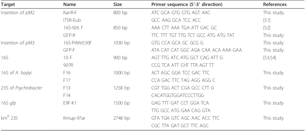
Table 4 PCR primers used in this study
Target Name Size Primer sequence (5’-3’direction) References
Insertion of pM2 KanR-F 800 bp ATC GCA GTG GTG AGT AAC This study,
ITSR-Eub GCC AAG GCA TCC ACC [51]
16S-926 F 850 bp AAA CTT AAA TGA ATT GAC GC [52]
GFP-R TTC TTT TGT TTG TCT GCC ATG ATG TAT This study
Insertion of pM3 16S-PrMir530F 1030 bp GTG CCA GCA GC GCG G This study
GFP-F ATA CAT CAT GGC AGA CAA ACA AAA GAA This study
16S 10 F 900 bp AGT TTG ATC ATG GCT CAG ATT G [53,54]
907R CCG TCA ATT CHT TTR AGT TT
16S ofA. baylyi F16 1000 bp ACT AGC GGA TCC GAC TTC This study
F17 CCA GAC TTC TAG AGG AGG C
23S ofPsychrobacter F13 1258 bp CGT TGG ACT CGA GCC CTT G This study
F14 CACATGGTGGATCCCTTGG
16Sgfp E9F-K1 1500 bp GAG TTT GAT CCT GGA TCA This study
TTG GCC ATG GAA CAG GTA
kmR23S Kmup-97ar 2748 bp GTA TGA GTC AGC AAC ACC TTC This study
CGC TTA GAT GCT TTC AGC
Table 5 PCR cycling conditions
Primers Initial Denaturation Denaturation Annealing Elongation Cycles Terminal Elongation
KanR F and ITSR-Eub R 95°C/5 min 95°C/30 s 54°C/25 s 72°C/70 s 35 72°C/7 min
16S-926 F and GFP R: 95°C/5 min 95°C/30 s 53°C/40 s 72°C/90 s 35 72°C/7 min
16S-Pr.mir-530 F and GFP F 95°C/5 min 95°C/30 s 63°C/25 s 72°C/70 s 35 72°C/7 min
16S 94°C/15 min 94°C/4 min 50°C/45 s 72°C/1 min 5
16S (cont.) 92°C/45 s 55°C/45 s 72°C/1 min 30 72°C/7 min
13 F-14 F 97°C/5 min 97°C/30 s 50°C/45 s 72°C/1.5 min 30 72°C/7 min
16 F-17 F 97°C/5 min 97°C/30 s 50°C/45 s 72°C/1 min 30 72°C/7 min
E9F-K1 97°C/5 min 97°C/30 s 56°C/45 s 72°C/1.5 min 30 72°C/7 min
[image:9.595.57.539.606.731.2]Acknowledgements
We thank the Animal Department, Faculty of Health Sciences, University of Tromsø, for technical assistance and support. We thank Julia Eggert for assistance during the animal experiment and Iris Graf for assistance with the natural transformation experiments of marine isolates. We thank Lise Øverås (University of Bergen, Norway), Ian Joint (Plymouth Marine Laboratory, United Kingdom) and Francisco Rodriguez-Valera (Universidad Miguel Hernandez de Elche, Spain) for providing the marine samples. The work was founded by the Norwegian Research Council, GenØk - Centre for Biosafety and by the European Commission (MIRACLE project).
Author details
1GenØk, Centre for Biosafety, Science Park, 9294 Tromsø, Norway.
2Department of Pharmacy, Faculty of Medicine, University of Tromsø, N-9037
Tromsø, Norway.3Faculty of Science & Technology, Free University of Bozen/
Bolzano, 39100 Bozen/Bolzano, Italy.4Department of Food Science,
Technology and Microbiology, University of Milan, 20133 Milan, Italy.
5Molecular Microbiology & Bioenergetics, Institute of Molecular Biosciences,
Goethe University, 60438 Frankfurt/Main, Germany.6Department of Civil, Environmental and Materials Engineering (DICAM), Faculty of Engineering, University of Bologna, Bologna 40131, Italy.
Authors’contributions
LN participated in the design, coordination, analysis, and drafting of the manuscript. LB and NR participated the molecular and microbiological experiments and analyses, TT participated in the design and drafting of the manuscript. BA designed the plasmids, contributed the data on the marine isolates, and revised the manuscript. KMN contributed to the design, coordination, analysis, and drafting of the manuscript. All authors read and approved the final manuscript.
Competing interests
The authors declare that they have no competing interests.
Received: 24 February 2012 Accepted: 1 April 2012 Published: 1 April 2012
References
1. Netherwood T, Martin-Orue SM, O’Donnell AG, Gockling S, Graham J, Mathers JC, Gilbert HJ:Assessing the survival of transgenic plant DNA in the human gastrointestinal tract.Nat Biotechnol2004,22:204-209. 2. Wilcks A, van Hoek AHAM, Joosten RG, Jacobsen BBL, Aarts HJM:
Persistence of DNA studied in differentex vivoandin vivorat models simulating the human gut situation.Food Chem Toxicol2004,42:493-502. 3. Wilcks A, Jacobsen BB:Lack of detectable DNA uptake by transformation of selected recipients in mono-associated rats.BMC Res Notes2010,3:49. 4. Nordgård L, Nguyen T, Midtvedt T, Benno Y, Traavik T, Nielsen KM:Lack of detectable uptake of DNA by bacterial gut isolates grownin vitroand byAcinetobacter baylyicolonizing rodents in situ.Environ Biosafety Res 2007,6:149-160.
5. Rizzi A, Raddadi N, Sorlini C, Nordgård L, Nielsen KM, Daffonchio D:The stability and degradation of dietary DNA in the gastrointestinal tract of mammals - implications for horizontal gene transfer and the biosafety of GMOs.Crit Rev Food Sci Nutr2012,52:142-161.
6. Nielsen KM, Ray JL, Johnsen PJ:The natural uptake of extracellular DNA in bacteria.InEncyclopedia of Microbiology..3 edition. Edited by: Schaechter M. Oxford: Elsevier; 2009:587-596.
7. Brigulla M, Wackernagel W:Molecular aspects of gene transfer and foreign DNA acquisition in prokaryotes with regard to safety issues.Appl Microbiol Biotechnol2010,86:1027-1041.
8. Lorenz MG, Wackernagel W:Bacterial gene transfer by genetic transformation in the environment.Microbiol Rev1994,58:563-602. 9. De Vries J, Wackernagel W:Microbial horizontal gene transfer and the
DNA release from transgenic crop plants.Plant Soil2004,266:91-104. 10. Van Reenen CA, Dicks LMT:Horizontal gene transfer amongst probiotic
lactic acid bacteria and other intestinal microbiota: what are the possibilities? A review.Arch Microbiol2011,193:157-168.
11. McCracken VJ, Lorenz RG:The gastrointestinal ecosystem: a precarious alliance among epithelium, immunity and microbiota.Cell Microbiol2001, 3:1-11.
12. O’Hara AM, Shanahan F:The gut flora as a forgotten organ.EMBO Rep 2006,7:688-693.
13. Schubbert R, Lettmann C, Doerfler W:Ingested foreign (phage M13) DNA survives transiently in the gastrointestinal tract and enters the bloodstream of mice.Mol Gen Genet1994,242:495-504.
14. Schubbert R, Renz D, Schmitz B, Doerfler W:Foreign (M13) DNA ingested by mice reaches peripheral leukocytes, spleen, and liver via the intestinal wall mucosa and can be covalently linked to mouse DNA.Proc Nat Acad Sci USA1997,94:961-966.
15. Schubbert R, Hohlweg U, Renz D, Doerfler W:On the fate of orally ingested feed-derived DNA in mice: chromosomal association and placental transmission to the fetus.Mol Gen Genet1998,259:569-576. 16. Duggan PS, Chambers PA, Heritage J, Forbes MJ:Fate of genetically
modified maize DNA in the oral cavity and rumen of sheep.Br J Nutr 2003,89:159-166.
17. Reuter T, Aulrich K:Investigations on genetically modified maize (Bt -maize) in pig nutrition: fate of feed-ingested feed-derived DNA in pig bodies.Eur Food Res Technol2003,216:185-192.
18. Einspanier R, Klotz A, Kraft J, Aulrich K, Poser R, Schwägele F, Jahreis G, Flachowsky G:The fate of forage plant DNA in farm animals: a collaborative case-study investigating cattle and chicken fed recombinant plant material.Eur Food Res Technol2001,212:129-132. 19. Einspanier R, Lutz B, Rief S, Berezina O, Zverlov V, Schwarz W, Mayer J:
Tracing residual recombinant feed molecules during digestion and rumen bacterial diversity in cattle fed transgene maize.Eur Food Res Technol2004,3:269-273.
20. Mazza R, Soave M, Morlacchini M, Piva G, Morocco A:Assessing the transfer of genetically modified DNA from feed to animal tissues. Transgenic Res2005,14:775-784.
21. Bensasson D, Boore JL, Nielsen KM:Genes without frontiers.Heredity2004, 92:483-489.
22. Thomas CM, Nielsen KM:Mechanisms of, and barriers to, horizontal gene transfer between bacteria.Nat Rev Microbiol2005,3:711-721.
23. Ray JL, Harms K, Wikmark OG, Starikova I, Johnsen PJ, Nielsen KM:Sexual isolation inAcinetobacter baylyiis locus-specific and varies 10,000-fold over the genome.Genetics2009,182:1165-1181.
24. Shen P, Huang HV:Homologous recombination inEscherichia coli: dependence on substrate length and homology.Genetics1986, 112:441-457.
25. Zawadzki P, Cohan FM:The size and continuity of DNA segments integrated inBacillustransformation.Genetics1995,141:1231-1243. 26. Matic I, Taddei F, Radman M:Genetic barriers among bacteria.Trends
Microbiol1996,4:69-72.
27. Majewski J, Cohan FM:DNA sequence similarity requirements for interspecific recombination inBacillus.Genetics1999,153:1525-1533. 28. Majewski J:Sexual isolation in bacteria.FEMS Microbiol Lett2001,
199:161-169.
29. Strätz M, Mau M, Timmis KN:System to study horizontal gene exchange among microorganisms without cultivation of recipients.Mol Microbiol 1996,22:207-215.
30. Mercer DK, Scott KP, Bruce-Johnson WA, Glover LA, Flint HJ:Fate of Free DNA and transformation of the oral bacteriumStreptococcus gordonii
DL1 by plasmid DNA in human saliva.Appl Environ Microbiol1999, 65:6-10.
31. Mercer DK, Scott KP, Melville CM, Glover AL, Flint HJ:Transformation of an oral bacterium via chromosomal integration of free DNA in the presence of human saliva.FEMS Microbiol Lett2001,200:163-167. 32. Duggan PS, Chambers PA, Heritage J, Forbes MJ:Survival of free DNA
encoding antibiotic resistance from transgenic maize and the transformation activity of DNA in ovine saliva, ovine rumen fluid and silage effluent.FEMS Microbiol Lett2000,191:71-77.
33. Kharazmi M, Bauer T, Hammes WP, Hertel C:Effect of food processing on the fate of DNA with regard to degradation and transformation capability inBacillus subtilis.Syst Appl Microbiol2003,26:495-501. 34. Shedova E, Albrecht C, Zverlov VV, Schwarz WH:Stimulation of bacterial
DNA transformation by cattle saliva: implications for using genetically modified plants in animal feed.World J Microbiol Biotechnol2009, 25:457-463.
bacteriumAcinetobacter calcoaceticusBD413.Theor Appl Genet1997, 95:815-821.
36. Nielsen KM, van Weerelt MD, Berg TN, Bones AM, Hagler AN, van Elsas JD: Natural transformation and availability of transforming DNA to
Acinetobacter calcoaceticusin soil microcosms.Appl Environ Microbiol 1997,63:1945-1952.
37. Pettersen AK, Bøhn T, Primicerio R, Shorten PR, Soboleva TK, Nielsen KM: Modeling suggests frequency estimates are not informative for predicting the long-term effect of horizontal gene transfer in bacteria. Environ Biosafety Res2005,4:223-233.
38. Nielsen KM, Townsend JP:Monitoring and modeling horizontal gene transfer.Nat Biotechnol2004,22:1110-1104.
39. Martin-Orue SM, O’Donnell AG, Arino J, Netherwood T, Gilbert HJ, Mathers JC:Degradation of transgenic DNA from genetically modified soya and maize in human intestinal simulations.Br J Nutr2002, 87:533-542.
40. Sanden M, Bruce IJ, Rahman AM, Hemre GI:The fate of transgenic sequences present in genetically modified plant products in fish feed, investigating the survival of GM soybean DNA fragments during feeding trials in Atlantic salmon,Salmo salarL.Aquaculture2004,237:391-405. 41. Nielsen KM, Berdal KG, Kruse H, Sundsfjord A, Mikalsen A, Yazdankhah S,
Nes I:An assessment of potential long-term health effects caused by antibiotic resistance marker genes in genetically modified organisms based on antibiotic usage and resistance patterns in Norway. VKM-Report, Norwegian Scientific Committee for Food Safety, Oslo, Norway. 42. Maturin L, Curtiss R:Degradation of DNA by nucleases in intestinal tract
of rats.Science1977,196:216-218.
43. Boyer SL, Flechtner VR, Johanson JR:Is the 16S-23S rRNA internal transcribed spacer region a good tool for use in molecular systematics and population genetics? A case study in cyanobacteria.Mol Biol Evol 2001,18:1057-1069.
44. McCartney AL:Application of molecular biological methods for studying probiotics and the gut flora.Br J Nutr2002,88(Suppl 1):S29-S37. 45. Brooks SPJ, McAllister M, Sandoz M, Kalmokoff ML:Culture-independent
phylogenetic analysis of the faecal flora of the rat.Can J Microbiol2003, 49:589-601.
46. Inoue R, Ushida K:Development of the intestinal microbiota in rats and its possible interactions with the evolution of the luminal IgA in the intestine.FEMS Microbiol Ecol2003,45:147-153.
47. Hanske L, Hussong R, Frank N, Gerhäuser C, Blaut M, Braune A: Xanthohumol does not affect the composition of rat intestinal microbiota.Mol Nutr Food Res2005,49:868-873.
48. Dalby AB, Frank DN, St Amand AL, Bendele AM, Pace NR: Culture-independent analysis of indomethacin-induced alterations in the rat gastrointestinal microbiota.Appl Environ Microbiol2006,72:6707-6715. 49. Licht TR, Hansen M, Poulsen M, Dragsted LO:Dietary carbohydrate source
influences molecular fingerprints of the rat faecal microbiota.BMC Microbiol2006,6:98.
50. Ray JL, Nielsen KM:Experimental methods for assaying natural transformation and inferring horizontal gene transfer.InMethods in Enzymology; methods in molecular evolution: producing the biochemical data. Volume 224.Edited by: Zimmer EA, White TJ, Cann RL, Wilson AC. San Diego: Academic; 2005:491-520.
51. Cardinale M, Brusetti L, Quatrini P, Borin S, Puglia AM, Rizzi A, Zanardini E, Sorlini C, Corselli C, Daffonchio D:Comparison of different primer sets for use in automated ribosomal intergenic spacer analysis of complex bacterial communities.Appl Environ Microbiol2004,70:6147-6156. 52. Lane DJ:16S/23S rRNA sequencing. Nucleic acid techniques in bacterial
systematics.InModern microbiological methods.Edited by: Stackebrandt E, Goodfellow M. Chichester: J Wiley 1991:133.
53. Muyzer G:Structure, function and dynamics of microbial communities: the molecular biology approach.InAdvances in molecular ecology.Edited by: Carvalho GR. Amsterdam: Ios Press; 1998:87-117.
54. Munson MA, Baumann P, Clark MA, Baumann L, Moran NA, Voegtlin DJ, Campbell BC:Aphid-eubacterial endosymbiosis: evidence for its establishment in an ancestor of four aphid families.J Bacteriol1991, 173:6321-6324.
55. Reeves PG:Components of the AIN-93 diets as improvements in the AIN-76A diet.J Nutr1997,127:838S-841S.
56. Grønsberg IM, Nordgård L, Fenton K, Hegge B, Nielsen KM, Bardocz S, Pusztai A, Traavik T:The uptake and organ distribution of feed
introduced plasmid DNA in growing or pregnant rats.Food Nutr Sci2011, 2:377-386.
doi:10.1186/1756-0500-5-170
Cite this article as:Nordgårdet al.:An investigation of horizontal transfer of feed introduced DNA to the aerobic microbiota of the gastrointestinal tract of rats.BMC Research Notes20125:170.
Submit your next manuscript to BioMed Central and take full advantage of:
• Convenient online submission
• Thorough peer review
• No space constraints or color figure charges
• Immediate publication on acceptance
• Inclusion in PubMed, CAS, Scopus and Google Scholar
• Research which is freely available for redistribution