T E C H N I C A L N O T E
Open Access
Potential regenerative rehabilitation technology:
implications of mechanical stimuli to tissue health
Colleen L McHenry, Jason Wu and Richard K Shields
*Abstract
Background:Mechanical loads induced through muscle contraction, vibration, or compressive forces are thought to modulate tissue plasticity. With the emergence of regenerative medicine, there is a need to understand the optimal mechanical environment (vibration, load, or muscle force) that promotes cellular health. To our knowledge no mechanical system has been proposed to deliver these isolated mechanical stimuli in human tissue. We present the design, performance, and utilization of a new technology that may be used to study localized mechanical stimuli on human tissues. A servo-controlled vibration and limb loading system were developed and integrated into a single instrument to deliver vibration, compression, or muscle contractile loads to a single limb (tibia) in humans. The accuracy, repeatability, transmissibility, and safety of the mechanical delivery system were evaluated on eight individuals with spinal cord injury (SCI).
Findings:The limb loading system was linear, repeatable, and accurate to less than 5, 1, and 1 percent of full scale, respectively, and transmissibility was excellent. The between session tests on individuals with spinal cord injury (SCI) showed high intra-class correlations (>0.9).
Conclusions:All tests supported that therapeutic loads can be delivered to a lower limb (tibia) in a safe, accurate, and measureable manner. Future collaborations between engineers and cellular physiologists will be important as research programs strive to determine the optimal mechanical environment for developing cells and tissues in humans.
Keywords:Vibration, Mechanical oscillation, Compression, Mechanical load, Spinal cord injury
Background
Vibration and compressive loads are mechanical stimuli that have a powerful influence on biological tissues. Recent studies in animal models demonstrate that certain types of mechanical load regulates bone [1], fat [2,3], skeletal muscle [4,5], and nerve tissues [6]. In addition, it is also well known that“over exposure” to mechanical stimuli is damaging to tissues [7-9]. With the emergence of regenerative medicine in tissue repair, rehabilitation specialists must understand the correct type and dose of mechanical stress that promotes cell survival and cell proliferation in bone, cartilage, ligaments, and muscle. However, to our knowledge, there is no technology that directs specific types of mechanical stimuli to limbs of humans. A method to study mechanical stimuli in humans is necessary to guide future research to determine optimal
rehabilitation prescriptions. The importance is under-scored as multi-potent adult stem cells are harvested and implanted after surgery, injury, disease, and paralysis as regenerative medicine advances. Our long term goal is to establish the extent to which various types of mechan-ical stimuli optimally influence the regenerative capacity of cells in humans. In this technological report, we present an innovative technology that may assist in determining the impact of mechanical stimuli of human tissues and discuss the importance of a partnership between engineers, bioscience researchers, and rehabilitation specialists.
The underlying need to study the value of therapeutic stress in humans is well grounded in the literature. For example, Wolff’s law supports that bone tissue (osteocytes) exposed to high loads triggers osteogenesis [10]. Sub-sequent studies verified that exerting high strain in a dynamic fashion to bone tissue was more effective than delivering a sustained strain [11,12]. For many years, the dynamic delivery of high stress to bone was considered the primary mechanical method to up-regulate osteogenesis * Correspondence:[email protected]
Department of Physical Therapy & Rehabilitation Science, Carver College of Medicine, University of Iowa, 1-252 Medical Education Building, Iowa City, IA 52242-1190, USA
[13-15]. However, more recently, low amplitude vibration stimuli, in the absence of high mechanical loads, were equally effective at up-regulating bone development in mice [5,16-19]. Indeed, regular mechanical stress promotes a healthy environment for bone [1], fat [2,3], skeletal muscle [4,5], nerve tissue [6], and cartilage (articu-lar, menisci) [20,21] in animal or reduced preparations in the laboratory. Translating these findings into human studies has been hampered by the lack of a capacity to dynamically deliver high passive loads and/or low vibra-tion either independently or in various combinavibra-tions with or without muscle activation (electrically or volitionally).
The dose of various mechanical loads has not been carefully examined. For example, most studies evaluat-ing vibration deliver the load to the entire animal [5,16,19,22-24] or human [25-30] which limits the ability to understand adaptive effects of localized vibration dir-ectly on tissues (muscle and bone). This point was empha-sized when whole body vibration of mice had a systemic increase in bone density [5,18,19] and decrease in whole body adipogenesis [3]. The direct effect of the vibration stimuli on bone tissue was confounded by vestibular [31] and/or endocrine system [32] mechanical activation.
The purpose of this technological report is to present a novel method to introduce localized compressive loads and/or vibration into the limbs of humans. The accuracy, repeatability, transmissibility and safety of the instrument will be presented in this report. Future studies are rec-ommended using technology that will assist in better understanding the impact of mechanical stimuli on tissue health. The need for collaborative and inter-disciplinary teams of engineers and cellular physiologists will be emphasized.
Methods
Technology development and testing study subjects Eight individuals with complete paralysis were tested on two occasions to determine the ability to reliably and accurately deliver the mechanical oscillations and loads to the limb of people with spinal cord injury (SCI). A power analysis revealed that 8 participants were required to have power to assess the reproducibility of the system (>80%). Informed written consent was obtained from all subjects prior to participation. All experimental protocols were approved by the University of Iowa Institutional Review Board.
General description of instrumentation
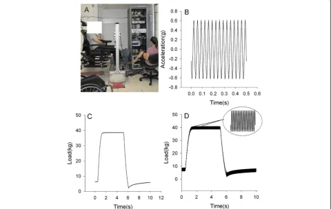
A servo-controlled vibration system (Figure 1) consists of five primary components from the Ling Dynamic Systems (Royston, England): PA1000L power amplifier, FPS10L field power supply, V722 shaker, cooling fan, and Laser USB 6.30 controller. The power amplifier and field power supply are connected in cascade and
generate the required power for the vibration system. A magnetic field within the shaker is generated from the field power supply while the power amplifier drives the shaker and supplies power to the cooling fan. The cooling fan dissipates the heat generated. An acceler-ometer is attached to the shaker and connected to the controller, which is directed to the amplifier creating a feedback loop. The vibration frequency in Hertz (Hz) and acceleration in gravitational force of earth (g = 9.81 m/s2), respectively, are controlled. The software also allows the user to program multiple loops thereby creating a series of on and off cycles of vibration. The controller is also equipped with an abort button designed to stop the vibra-tion quickly. When providing a mechanical intervenvibra-tion to humans it is important to have built in safety mecha-nisms in the event of an emergency.
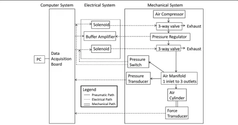
We interfaced a custom designed, pneumatically con-trolled piston that can safely deliver compressive loads to a limb segment either with our without the vibration (Figure 2). The mechanical loading system is driven by a pneumatic compression pump that is controlled by a custom circuit board communicating via the computer interface board. Custom software allows for parameter specification, feedback control, and safety shut down when unwanted loads are inadvertently applied.
is sent to the electrical portion of the system. The desired air pressure continues into the chamber of the air cylinder causing the piston to move downward. A force transducer, 1210ACK-300 lb Load Cell (Interface, Scottsdale, AZ), and pad are attached in series to the piston and allows pressure and force measurements simultaneously.
The limb loading system was designed to introduce a vertical compression load to the tibia via a load applied over the top of the femoral condyles (knee) as a percentage of body weight (%BW). A feedback loop was incorporated
into the software design written in LabVIEW 8.6 (National Instruments, Austin, TX) to continuously monitor the force and pressure through the transducers and adjust accordingly. The user can define the time that the load is on and off in seconds, the number of cycles, and the magnitude of the force. In addition, data is collected with real-time display of force, pressure, electromyography (EMG), vibration, and other mechanical factors.
[image:3.595.58.540.90.234.2]The apparatus that serves to hold the human limb consists of a custom designed frame that was fabricated
[image:3.595.60.537.435.686.2]Figure 2Schematic of the compression system.The mechanical portion consists of a series of hardware components which regulate the amount of air pressure delivered to the air cylinder and subsequently the load applied to the human tibia. The electrical system provides power to many of the mechanical components and links them to the data acquisition (DAQ) board. The personal computer (PC) and the DAQ board control the compression system and allow the user to program the compressive system.
and attached to the shaker so that vibration and load can be delivered concurrently (Figure 3). The novelty of this system is that it enables the load to be applied while the entire limb segment receives vibratory stimuli. Thus, during vibration, a force-time impulse may be delivered to the extremity. The frame is made of an aluminum base plate and foot plate connected with T-slot frames. The uprights and cross bar are also made with aluminum struts. Aluminum housing contains the air cylinder and slides within the frame uprights allowing full adjustability for limb length. In addition, a tilt in space chair was welded to a lift that allows any subject, including indi-viduals with paralysis to be positioned correctly into the device.
Vibration verification and transmissibility testing
We applied an independent external accelerometer, Model 3233A High-Sensitivity triaxial accelerometer (Dytran Instruments, Inc., Chatsworth, CA), to the vibration platform. The Laser vibration software is capable of various vibration parameters. We included settings aligned with those found to be effective in previous studies (0.1 g-10 g
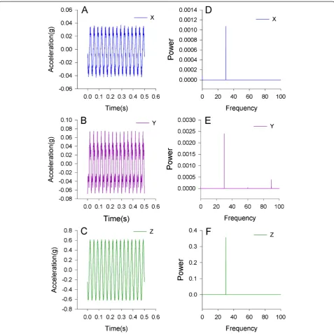
[image:4.595.58.539.379.683.2]at 20–90 Hz) [25-30]. In 2009, Totosy de Zepetnek pre-sented a review of whole body vibration which concluded the optimal vibration parameters for humans have yet to be determined [33]. To test the transmissibility of the vibration signal, the software was programmed for 0.6 g at 30 Hz for 1 minute. The work of Garman and Ozcivici demonstrated the vibration of 0.6 g enhanced the bone of the vibrated limb compared to the contra lateral limb [17,18]. During the vibration, acceleration was collected in all the cardinal directions. The x and y axes were parallel to the platform and the z axis was a perpendicular measure of the acceleration in the vertical direction. A custom MATLAB program (MathWorks, Natick, MA) was written to determine the peak of the acceleration and its frequency content. The peak was defined as the maximum value of the acceleration signal. To determine the frequency of the signal a fast Fourier transform (FFT) was performed. Based on the sampling frequency of 4,000, 32.7680 seconds or 2^17 data points of acceleration data were used for the FFT. This window of data was chosen so the length of data was a power of 2, the recommended length for a FFT.
An accelerometer was attached to the leg, thigh, and head during the vibration protocol of both the vibrated and contra lateral limbs in a single subject. Anatomical locations were defined as tibia tuberosity, distal thigh, and forehead. We defined transmissibility as the ratio of the root mean square (RMS) of acceleration of the anatomical site to the RMS of the acceleration at the mechanical apparatus, consistent with Rubin et al. [34].
Transmissibility¼ RMSaccelerationbody RMSaccelerationplatform
Repeatability, linearity, and accuracy testing of limb load The custom software controls the instrumentation to deliver air pressure to the desired load to the lower leg. The calibration between the air pressure and the deliv-ered force to the limb was determined using five known input pressures (138, 207, 276, 345, 414 kPa). The five input pressures were chosen to calibrate the system. We targeted loads that were able to secure the limb to the device and loads that we previously published to modulate spinal cord activity [35,36]. Ten cycles were collected at each pressure. The accuracy of the limb loading system was measured by determining the linearity, repeatability, and percent error. Linearity was defined as the maximum deviation of the mean difference between the predicted response and the measured load. Repeatability was the maximum difference between measures under the same testing conditions, while percent error was calculated using the following equation, ((measured value-predicted value)/predicted value)*100. Repeatability and percent error were normalized and expressed at percentage of full scale (%FS). Prior to this air pressure validation, the load cell was calibrated. The maximum acceptable error for these three measurement was less than 5%FS.
We delivered limb loads to individuals with complete paralysis to test the reproducibility of the apparatus. Eight individuals underwent two sessions on different days to determine the between day reliability of load delivery to human limbs. Ninety compressive load cycles of 50% of body weight were delivered to one leg of the individuals with paralysis. These five second loading cycles were separated by five second rest periods so ninety cycles took 15 minutes to complete. The peak load was measured after cycles 1, 30, 60, and 90 for each session. The percent difference between days and the intra-class correlation coefficients (ICC) at each time point were calculated (IBM SPSS Statistics Version 19). An ICC > 0.8 indicates that the system has high reproducibility in deliv-ering mechanical load on a day to day basis [37]. Included in this error assessment is the ability to connect the hu-man subject to the mechanical interface system. Any error
less than 10% was considered low for the between day reliability assessment.
Load safety assessment
Since this device is designed to interface with a human tibia, safety is of utmost importance. Although, the vi-bration parameters (0.6 g, 30 Hz) for this intervention are safe for humans, the system is capable of generating much larger vibration signals (66.3 g, 400 Hz) The shaker parameters were altered so that the maximum acceleration is 6 g and the shaker itself has an over travel protection that limits the peak-to-peak excursion to 11 mm. Finally, the vibration controller is equipped with an abort button that will immediately shut down the system.
The compressive system also has several safety fea-tures including an emergency stop switch that removes the load by venting the air to the environment. In addition to a mechanical stop, before starting the com-pression system, the user must input the cycle time, air pressure, and maximum load. The maximum load is the safety parameter which can be set to ensure that an excessive load for human tibia cannot be inadvertently applied.
To assess the safety of the compression an air pressure of 263 kPa or 445 N was programmed into the system while varying the maximum load. Seven maximum load settings, 423 N, 437 N, 441 N, 445 N, 449 N, 454 N, and 467 N were tested. The force was recorded using custom LabVIEW software written to control the compression system. The effectiveness of the maximum load safety setting was determined by examining the force signal and measuring the peak force delivered.
Findings
Transmissibility and quality of vibration signal
Limb load testing results
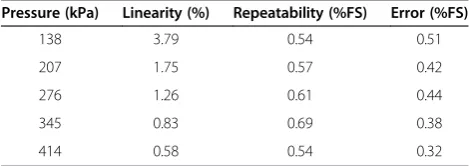
The linearity, repeatability, and error were calculated at each air pressure was 4%, 1%, and 1%, respectively (Table 1). The between session reproducibility assessment using human subjects was excellent with an intra-class correlation of 0.90 (Table 2). The percent change in limb load never exceeded 7% during between day tests. These data support that total error associated with“setting up”a human subject was low.
Vibration and limb load safety results
[image:6.595.59.537.88.565.2]The vibration system consistently shutdown when the acceleration exceeded a 6.1 g, if the platform exceeded 11 mm of displacement, or the user manually pushed the shutdown switch built into the controller. In addition, activating the emergency stop switch consist-ently aborted the limb loading system by exhausting the compressed air into the environment. To formally test the safety mechanisms under software control, we input a
load of 445 N (263 kPa) to the simulated extremity. We then intentionally exceeded the maximum load by pro-gramming in loads in excess of the 445 N threshold. The system consistently exhausted the air by the 3-way valve when the 445 N threshold was exceeded. We next set the threshold at 423 N, 437 N, and 441 N and delivered a load of 445 N. Because of a one second delay in the release of pressure, the limb segment received 435 N, 445 N, and 448 N rather than the 423 N, 437 N, and 441 N that were intended. Thus, the safety shut off was effective to within 3% of the intended load.
We had no subjective complaints from any subjects during this testing. There were no tissue areas of red-dening or indentations that support that mechanical load of vibration and compression can be delivered concurrently to human tissue.
Discussion
Currently, there are no existing devices that can provide isolated mechanical loading to a human limb by delivering controlled vibration and/or compression. There are devices
which can vibrate [18] or compress [38] the limb of a rodent but neither can deliver vibration and compression simultaneously. Many of the existing devices for humans are commercial vibration platforms that are inherently noisy [39] and typically used for whole body vibration and not localized vibration. The vibration system presented in this paper is servo-controlled and therefore provides a constant vibration using the feedback from the accelerom-eter to modulate the vibration. Vibration platforms have been widely used in human research; however, prior to the mechanical system presented in this article, there was not a device capable of delivering limb vibration with or with-out limb compression to an isolated segment.
Bridging the gap: engineering and bioscience
The primary purpose of this technological report was to present the development of an accurate, controlled, repeatable, and safe mechanical system that would be able to induce localized biological stress to tissues within a limb of humans. Based on our presentation of the findings, we are confident that this system can re-liably deliver the stresses within the loads tested based on animal studies and preliminary human reports. Our secondary purpose was to use this report to appeal to the scientific community about the importance of inter-disciplinary teams partnering as cellular therapies are developed in the bioscience laboratories. Our ability to test and learn about the optimal methods to stress tissues is paramount for many new cellular therapies developing today. A brief review of the impact of mech-anical stimuli on various tissues will be presented in the subsequent sections.
Mechanical stimuli and bone tissue adaptation
[image:7.595.56.291.89.247.2]The relationship between mechanical loads and tissue adaptation is long standing. Wolff [10] and Frost [40,41] demonstrated many years ago that bone tissue is highly mutable and adapts to mechanical stress. In recent years it is well documented that the musculoskeletal system deteriorates in people with SCI [42-46], people on bed rest [47], or people exposed to spaceflight [48,49]. In just two years after paralysis, people with spinal cord injury
Figure 5Transmissibility of the vibration.The transmissibility of the vibration signal was calculated as a ratio of the anatomical landmark RMS to the RMS of the platform. An accelerometer was place on the tibia and femur of the vibrated and unvibrated leg as well as the head. A transmissibility of 1.0 indicates that the acceleration of the anatomical site is equal to the vibration platform.
Table 1 The compression system performed with a high level of accuracy which is indicated by the linearity, repeatability, and percent error
Pressure (kPa) Linearity (%) Repeatability (%FS) Error (%FS)
138 3.79 0.54 0.51
207 1.75 0.57 0.42
276 1.26 0.61 0.44
345 0.83 0.69 0.38
414 0.58 0.54 0.32
[image:7.595.306.540.122.191.2]These metrics were calculated using the 10 cycles at each air pressure (%FS = percent full scale).
Table 2 The data of eight spinal cord injury subjects were used to determine the inter-session reliability of the compression system
Cycle Change (%) ± SD ICC
5.07 ± 2.74 0.917
30 3.43 ± 1.43 0.965
60 6.53 ± 3.98 0.899
90 3.06 ± 2.75 0.965
[image:7.595.56.291.633.716.2]have 23%, 25%, and 19% less articular cartilage in the patella, medial tibia, and lateral tibia, respectively [50]. Timely mechanical stress reduced the loss of bone by 32% in people with SCI [40,51], which may ultimately be lifesaving [52]. Even secondary systemic complications like renal failure and metabolic syndrome are linked to deteriorating skeletal muscle and bone tissues [53-60].
Low-magnitude whole body mechanical oscillation (0.2-0.3 g, 30 Hz), which would be well tolerated in people who already have osteoporosis, has been shown to attenuate bone loss in women with low bone mineral density [26,61]. Whole body vibration (0.3 g, 45 Hz), at doses similar to that tested in this study, led to 75% increase in trabeculae of the proximal metaphyses of rats [5,19]. Vibration (0.3 g, 30 Hz) of the sheep hind limb showed 34.2% increase in femur bone density [1]. However, only one animal study delivered direct limb segment vibration in-vivo, but showed the tibia had a 88% higher rate of bone formation using the same 0.6 g force demonstrated in the technology presented in this report [62]. Because the vestibular system was likely ac-tivated during the weight bearing studies, it is possible that reflexes caused muscle activations that contributed to the tissue changes observed. These studies suggest that understanding the effects of mechanical stress on tissue is complicated and the field may benefit from technologies that isolate these mechanical stresses.
Mechanical stimuli and cartilage adaptations
Mechanical loading can alter articular cartilage, inter-vertebral discs, and menisci [63-67]. Knee menisci are particularly susceptible to injury [68] and are often re-sistant to healing [69]. Cyclic loading and intermittent tensile strain up-regulates VEGF (vascular endothelial growth factor), a gene directly involved with blood vessel formation [70]. Importantly, regular mechanical load reduces inflammation initiated by interleukin-1 following menisci injury [21,71]. A torn porcine meniscus exposed to various mechanical compressive loading conditions (1, 10, or 26% strain, and 4 h/day for 14 days) showed a re-duced inflammatory response and repaired mechanical tissue shear strength [21]. The value of cyclic repetitive loads on meniscus health is well documented [72,73]. Importantly, in the absence of natural mechanisms of meniscus tissue repair, regenerative rehabilitation engi-neers have developed a new scaffold consisting of viable undifferentiated cells that require a healthy environment (optimal stress) to proliferate and differentiate cells [74,75]. Injured meniscus cartilage was merely removed from the knee as little as 25 years ago. Today, the emphasis is in preserving and healing the tissue; however, the effect of controlled dynamic loads with vibration has never been explored in humans with menisci injury or repair. Hence there is a need for technologies to better study
the interface between mechanical stimuli and tissue repair in humans.
Mechanical stimuli and muscle/CNS adaptations
Localized limb vibration modulates several central ner-vous system and muscle signaling pathways in people with and without spinal cord injury [35,36,76,77]. During single limb segment vibration, the activity of the soleus muscle was suppressed [76]. Vibration caused an 83% reduction in the Hoffmann reflex (H-reflex), but limb load facilitated segmental excitability (decreased H-reflex post activation depression) [35,77]. Likewise, direct vibration over a muscle tendon increased pre-synaptic inhibition of the H-reflex [78-82] and loading (standing) reduces H-reflex post activation depression [83-85]. Recent re-search has shown that deficiencies in postural control were associated with brain activity during localized vibra-tion of the foot [86].
Vibration platforms for balance control have been reported to cause increased skeletal muscle activity, strength, and power [28,87-89]. These whole body vibra-tion protocols used 2.3 g-30 g at 15–50 Hz, parameters within the range tested in the technology reported in this paper. However, these findings are not supported with dir-ect tendon or muscle vibration; subsequent studies with tendon vibration support a decrease in quadriceps muscle activity and force [90,91]. Some intriguing findings suggest that localized vibration mitigates muscle atrophy during reduced activity [92,93] and regulates certain genes associ-ated with atrophy and synaptic plasticity [4,77]. It may be that the same dose that is optimal for bone is also optimal for cartilage, muscle, and nervous system tissue.
Mechanical stimuli and stem cell stimulation
to establishing the efficacy for preserving health of tissues in the future.
In summary, the instrumentation presented in this technological report is novel, reliable, accurate, and safe for human tissues. To fully translate technology from the laboratory to human studies will require that experts from engineering, rehabilitation, and biosciences, work collaboratively to advance the field of human regenerative rehabilitation.
Conclusions
This report presents a novel example of how to deliver compressive and vibratory loads to the lower limb in humans via a new technology. Mechanical loads such as vibration and direct limb load have not been systematically studied in various combinations in humans. Importantly, the vibration stimuli developed in this report is directed to a single limb, rather than to the whole body, allowing a method to compare the direct effects of load to specific tissue. By delivering isolated therapeutic doses of mechan-ical stress to human tissues, we anticipate that the optimal methods of mechanically and physiologically stressing tissues may be ascertained in future studies.
Availability and requirements
Project name:Human Regenerative Rehabilitation
Project home page:Not applicable
Operating system(s):Platform independent
Programming language:Not applicable
Other requirements:Not applicable
License:Not applicable
Any restrictions to use by non-academics:Not applicable
Availability of supporting data
No supporting data will be submitted with this manuscript.
Abbreviations
SCI:Spinal cord injury; Hz: Hertz; G: Gravitational force of the Earth; kPa: Kilopascal; %BW: Percent body weight; EMG: Electromyography; FFT: Fast Fourier transform; RMS: Root means square; ICC: Intra-class correlation coefficient; VEGF: Vascular endothelial growth factor; H-reflex: Hoffmann reflex; %FS: Percent full scale; DAQ: Data acquisition.
Competing interests
The authors declare that they have no competing interests.
Authors’contributions
CM helped implement the system, collected and analyzed data, and drafted the manuscript. JW provided technical assistance to implement the system and assisted with editing the manuscript. RKS conceived and developed the technology, provided oversight of all testing, procured funding, and edited/ approved the final draft of the manuscript. All authors read and approved the final manuscript.
Acknowledgements
This project was supported by grants from the NIH (R01-HD-062507) and the Neilsen Foundation.
Received: 6 January 2014 Accepted: 21 May 2014 Published: 3 June 2014
References
1. Rubin C, Turner AS, Bain S, Mallinckrodt C, McLeod K:Anabolism. Low mechanical signals strengthen long bones.Nature2001,412:603–604. 2. Maddalozzo GF, Iwaniec UT, Turner RT, Rosen CJ, Widrick JJ:Whole-body
vibration slows the acquisition of fat in mature female rats.Int J Obes (Lond)2008,32:1348–1354.
3. Rubin CT, Capilla E, Luu YK, Busa B, Crawford H, Nolan DJ, Mittal V, Rosen CJ, Pessin JE, Judex S:Adipogenesis is inhibited by brief, daily exposure to high-frequency, extremely low-magnitude mechanical signals.Proc Natl Acad Sci U S A2007,104:17879–17884.
4. Ceccarelli G, Benedetti L, Galli D, Pre D, Silvani G, Crosetto N, Magenes G, Cusella De Angelis MG:Low-amplitude high frequency vibration down-regulates myostatin and atrogin-1 expression, two components of the atrophy pathway in muscle cells.J Tissue Eng Regen Med2012,8:396–406. 5. Xie L, Rubin C, Judex S:Enhancement of the adolescent murine
musculoskeletal system using low-level mechanical vibrations.J Appl Physiol2008,104:1056–1062.
6. Raju SG, Rogness O, Persson M, Bain J, Riley D:Vibration from a riveting hammer causes severe nerve damage in the rat tail model.Muscle Nerve
2011,44:795–804.
7. Heaver C, Goonetilleke KS, Ferguson H, Shiralkar S:Hand-arm vibration syndrome: a common occupational hazard in industrialized countries. J Hand Surg Eur Vol2011,36:354–363.
8. Bovenzi M:Metrics of whole-body vibration and exposure-response relationship for low back pain in professional drivers: a prospective cohort study.Int Arch Occup Environ Health2009,82:893–917. 9. Tiemessen IJ, Hulshof CT, Frings-Dresen MH:Low back pain in drivers
exposed to whole body vibration: analysis of a dose–response pattern. Occup Environ Med2008,65:667–675.
10. Wolff J:The Law of Bone Remodelling.Berlin, Heidelberg, New York: Springer-Verlag; 1986.
11. Akyuz E, Braun JT, Brown NA, Bachus KN:Static versus dynamic loading in the mechanical modulation of vertebral growth.Spine2006,
31(Phila Pa 1976):E952–958.
12. Robling AG, Duijvelaar KM, Geevers JV, Ohashi N, Turner CH:Modulation of appositional and longitudinal bone growth in the rat ulna by applied static and dynamic force.Bone2001,29:105–113.
13. Churches AE, Howlett CR:Functional adaptation of bone in response to sinusoidally varying controlled compressive loading of the ovine metacarpus.Clin Orthop Relat Res1982,168:265–280.
14. Rubin CT, Lanyon LE:Regulation of bone formation by applied dynamic loads.J Bone Joint Surg Am1984,66:397–402.
15. Turner CH, Owan I, Takano Y:Mechanotransduction in bone: role of strain rate.Am J Physiol1995,269:E438–442.
16. Christiansen BA, Silva MJ:The effect of varying magnitudes of whole-body vibration on several skeletal sites in mice.Ann Biomed Eng2006,34:1149–1156. 17. Garman R, Gaudette G, Donahue LR, Rubin C, Judex S:Low-level
accelerations applied in the absence of weight bearing can enhance trabecular bone formation.J Orthop Res2007,25:732–740.
18. Ozcivici E, Garman R, Judex S:High-frequency oscillatory motions enhance the simulated mechanical properties of non-weight bearing trabecular bone.J Biomech2007,40:3404–3411.
19. Xie L, Jacobson JM, Choi ES, Busa B, Donahue LR, Miller LM, Rubin CT, Judex S:Low-level mechanical vibrations can influence bone resorption and bone formation in the growing skeleton.Bone2006,39:1059–1066. 20. Bader DL, Salter DM, Chowdhury TT:Biomechanical influence of cartilage
homeostasis in health and disease.Arthritis2011,2011:979032. 21. McNulty AL, Estes BT, Wilusz RE, Weinberg JB, Guilak F:Dynamic loading
enhances integrative meniscal repair in the presence of interleukin-1. Osteoarthritis Cartilage2010,18:830–838.
22. Flieger J, Karachalios T, Khaldi L, Raptou P, Lyritis G:Mechanical stimulation in the form of vibration prevents postmenopausal bone loss in ovariectomized rats.Calcif Tissue Int1998,63:510–514.
23. Judex S, Lei X, Han D, Rubin C:Low-magnitude mechanical signals that stimulate bone formation in the ovariectomized rat are dependent on the applied frequency but not on the strain magnitude.J Biomech2007,
40:1333–1339.
24. Rubinacci A, Marenzana M, Cavani F, Colasante F, Villa I, Willnecker J, Moro GL, Spreafico LP, Ferretti M, Guidobono F, Marotti G:Ovariectomy sensitizes rat cortical bone to whole-body vibration.Calcif Tissue Int2008,
25. Belavy DL, Beller G, Armbrecht G, Perschel FH, Fitzner R, Bock O, Borst H, Degner C, Gast U, Felsenberg D:Evidence for an additional effect of whole-body vibration above resistive exercise alone in preventing bone loss during prolonged bed rest.Osteoporos Int2011,22:1581–1591. 26. Gilsanz V, Wren TA, Sanchez M, Dorey F, Judex S, Rubin C:Low-level,
high-frequency mechanical signals enhance musculoskeletal development of young women with low BMD.J Bone Miner Res2006,21:1464–1474. 27. Torvinen S, Kannus P, Sievanen H, Jarvinen TA, Pasanen M, Kontulainen S,
Nenonen A, Jarvinen TL, Paakkala T, Jarvinen M, Vuori I:Effect of 8-month vertical whole body vibration on bone, muscle performance, and body balance: a randomized controlled study.J Bone Miner Res2003,
18:876–884.
28. Verschueren SM, Roelants M, Delecluse C, Swinnen S, Vanderschueren D, Boonen S:Effect of 6-month whole body vibration training on hip density, muscle strength, and postural control in postmenopausal women: a randomized controlled pilot study.J Bone Miner Res2004,19:352–359. 29. Ward K, Alsop C, Caulton J, Rubin C, Adams J, Mughal Z:Low magnitude
mechanical loading is osteogenic in children with disabling conditions. J Bone Miner Res2004,19:360–369.
30. Kiel DP, Hannan MT, Barton BA, Bouxsein ML, Lang TF, Brown KM, Shane E, Magaziner J, Zimmerman S, Rubin CT:Insights from the conduct of a device trial in older persons: low magnitude mechanical stimulation for musculoskeletal health.Clin Trials2010,7:354–367.
31. Pollock RD, Woledge RC, Martin FC, Newham DJ:Effects of whole body vibration on motor unit recruitment and threshold.J Appl Physiol2012,
112:388–395.
32. Elmantaser M, McMillan M, Smith K, Khanna S, Chantler D, Panarelli M, Ahmed SF:A comparison of the effect of two types of vibration exercise on the endocrine and musculoskeletal system.J Musculoskelet Neuronal Interact2012,12:144–154.
33. Totosy De Zepetnek JO, Giangregorio LM, Craven BC:Whole-body vibration as potential intervention for people with low bone mineral density and osteoporosis: a review.J Rehabil Res Dev2009,46:529–542. 34. Rubin C, Pope M, Fritton JC, Magnusson M, Hansson T, McLeod K:
Transmissibility of 15-hertz to 35-hertz vibrations to the human hip and lumbar spine: determining the physiologic feasibility of delivering low-level anabolic mechanical stimuli to skeletal regions at greatest risk of fracture because of osteoporosis.Spine2003,28(Phila Pa 1976):2621–2627. 35. Tseng SC, Shields RK:Limb segment load inhibits post activation depression
of soleus H-reflex in humans.Clin Neurophysiol2012,124:982–990. 36. Tseng SC, Shields RK:Limb compressive load does not inhibit post
activation depression of soleus H-reflex in indiviudals with chronic spinal cord injury.Clin Neurophysiol2013,124:982–990.
37. Portney LG, Watkins, Mary P:Foundations of Clinical Research: Applications to Practice.Norwalk: Connecticut: Appleton & Lange; 1993.
38. De Souza RL, Matsuura M, Eckstein F, Rawlinson SC, Lanyon LE, Pitsillides AA:Non-invasive axial loading of mouse tibiae increases cortical bone formation and modifies trabecular organization: a new model to study cortical and cancellous compartments in a single loaded element.Bone
2005,37:810–818.
39. Pel JJ, Bagheri J, Van Dam LM, van den Berg-Emons HJ, Horemans HL, Stam HJ, van der Steen J:Platform accelerations of three different whole-body vibration devices and the transmission of vertical vibrations to the lower limbs.Med Eng Phys2009,31:937–944.
40. Frost HM:Vital biomechanics: proposed general concepts for skeletal adaptations to mechanical usage.Calcif Tissue Int1988,42:145–156. 41. Frost HM:Bone's mechanostat: a 2003 update.Anat Rec A Discov Mol Cell
Evol Biol2003,275:1081–1101.
42. Eser P, Frotzler A, Zehnder Y, Wick L, Knecht H, Denoth J, Schiessl H:
Relationship between the duration of paralysis and bone structure: a pQCT study of spinal cord injured individuals.Bone2004,34:869–880. 43. Wilmet E, Ismail AA, Heilporn A, Welraeds D, Bergmann P:Longitudinal study of the bone mineral content and of soft tissue composition after spinal cord section.Paraplegia1995,33:674–677.
44. Shields RK, Dudley-Javoroski S, Boaldin KM, Corey TA, Fog DB, Ruen JM: Per-ipheral quantitative computed tomography: measurement sensitivity in persons with and without spinal cord injury.Arch Phys Med Rehabil2006,
87:1376–1381.
45. Modlesky CM, Majumdar S, Narasimhan A, Dudley GA:Trabecular bone microarchitecture is deteriorated in men with spinal cord injury.J Bone Miner Res2004,19:48–55.
46. Modlesky CM, Slade JM, Bickel CS, Meyer RA, Dudley GA:Deteriorated geometric structure and strength of the midfemur in men with complete spinal cord injury.Bone2005,36:331–339.
47. Leblanc AD, Schneider VS, Evans HJ, Engelbretson DA, Krebs JM:Bone mineral loss and recovery after 17 weeks of bed rest.J Bone Miner Res
1990,5:843–850.
48. LeBlanc AD, Spector ER, Evans HJ, Sibonga JD:Skeletal responses to space flight and the bed rest analog: a review.J Musculoskelet Neuronal Interact
2007,7:33–47.
49. LeBlanc A, Schneider V, Shackelford L, West S, Oganov V, Bakulin A, Voronin L:Bone mineral and lean tissue loss after long duration space flight. J Musculoskelet Neuronal Interact2000,1:157–160.
50. Vanwanseele B, Eckstein F, Knecht H, Stussi E, Spaepen A:Knee cartilage of spinal cord-injured patients displays progressive thinning in the absence of normal joint loading and movement.Arthritis Rheum2002,46:2073–2078. 51. Dudley-Javoroski S, Saha PK, Liang G, Li C, Gao Z, Shields RK:High dose
compressive loads attenuate bone mineral loss in humans with spinal cord injury.Osteoporos Int2011,23:2335–2346.
52. Dudley-Javoroski S, Shields RK:Muscle and bone plasticity after spinal cord injury: review of adaptations to disuse and to electrical muscle stimulation.J Rehabil Res Dev2008,45:283–296.
53. Bagley WH, Yang H, Shah KH:Rhabdomyolysis.Intern Emerg Med2007,
2:210–218.
54. Welk B, Fuller A, Razvi H, Denstedt J:Renal stone disease in spinal-cord-injured patients.J Endourol2012,26:954–959.
55. Gorgey AS, Shepherd C:Skeletal muscle hypertrophy and decreased intramuscular fat after unilateral resistance training in spinal cord injury: case report.J Spinal Cord Med2010,33:90–95.
56. Vestergaard P, Krogh K, Rejnmark L, Mosekilde L:Fracture rates and risk factors for fractures in patients with spinal cord injury.Spinal Cord1998,
36:790–796.
57. Beck BR:Muscle forces or gravity–what predominates mechanical loading on bone? Introduction.Med Sci Sports Exerc2009,41:2033–2036. 58. Robling AG:Is bone's response to mechanical signals dominated by
muscle forces?Med Sci Sports Exerc2009,41:2044–2049. 59. Kohrt WM, Barry DW, Schwartz RS:Muscle forces or gravity: what
predominates mechanical loading on bone?Med Sci Sports Exerc2009,
41:2050–2055.
60. Judex S, Carlson KJ:Is bone's response to mechanical signals dominated by gravitational loading?Med Sci Sports Exerc2009,41:2037–2043. 61. Rubin C, Recker R, Cullen D, Ryaby J, McCabe J, McLeod K:Prevention of
postmenopausal bone loss by a low-magnitude, high-frequency mechanical stimuli: a clinical trial assessing compliance, efficacy, and safety.J Bone Miner Res2004,19:343–351.
62. Garman R, Rubin C, Judex S:Small oscillatory accelerations, independent of matrix deformations, increase osteoblast activity and enhance bone morphology.PLoS One2007,2:e653.
63. Waldman SD, Spiteri CG, Grynpas MD, Pilliar RM, Kandel RA:Long-term intermittent compressive stimulation improves the composition and mechanical properties of tissue-engineered cartilage.Tissue Eng2004,
10:1323–1331.
64. Fan JC, Waldman SD:The effect of intermittent static biaxial tensile strains on tissue engineered cartilage.Ann Biomed Eng2010,
38:1672–1682.
65. Puetzer JL, Ballyns JJ, Bonassar LJ:The effect of the duration of mechanical stimulation and post-stimulation culture on the structure and properties of dynamically compressed tissue-engineered menisci. Tissue Eng Part A2012,18:1365–1375.
66. Ballyns JJ, Bonassar LJ:Dynamic compressive loading of image-guided tissue engineered meniscal constructs.J Biomech2011,44:509–516. 67. Woiciechowsky C, Abbushi A, Zenclussen ML, Casalis P, Kruger JP, Freymann
U, Endres M, Kaps C:Regeneration of nucleus pulposus tissue in an ovine intervertebral disc degeneration model by cell-free resorbable polymer scaffolds.J Tissue Eng Regen Med2012. [Epub ahead of print].
68. Makris EA, Hadidi P, Athanasiou KA:The knee meniscus: structure-function, pathophysiology, current repair techniques, and prospects for regeneration. Biomaterials2011,32:7411–7431.
69. Arnoczky SP, Warren RF:Microvasculature of the human meniscus.Am J Sports Med1982,10:90–95.
70. Frost HM:Bone "mass" and the "mechanostat": a proposal.Anat Rec1987,
71. Shin SJ, Fermor B, Weinberg JB, Pisetsky DS, Guilak F:Regulation of matrix turnover in meniscal explants: role of mechanical stress, interleukin-1, and nitric oxide.J Appl Physiol2003,95:308–313.
72. Ferretti M, Madhavan S, Deschner J, Rath-Deschner B, Wypasek E, Agarwal S:
Dynamic biophysical strain modulates proinflammatory gene induction in meniscal fibrochondrocytes.Am J Physiol Cell Physiol2006,
290:C1610–1615.
73. Deschner J, Wypasek E, Ferretti M, Rath B, Anghelina M, Agarwal S:
Regulation of RANKL by biomechanical loading in fibrochondrocytes of meniscus.J Biomech2006,39:1796–1803.
74. Rodkey WG, DeHaven KE, Montgomery WH 3rd, Baker CL Jr, Beck CL Jr, Hormel SE, Steadman JR, Cole BJ, Briggs KK:Comparison of the collagen meniscus implant with partial meniscectomy. A prospective randomized trial.J Bone Joint Surg Am2008,90:1413–1426.
75. Monllau JC, Gelber PE, Abat F, Pelfort X, Abad R, Hinarejos P, Tey M:Outcome after partial medial meniscus substitution with the collagen meniscal implant at a minimum of 10 years' follow-up.Arthroscopy2011,27:933–943. 76. Chang SH, Dudley-Javoroski S, Shields RK:Gravitational force modulates
muscle activity during mechanical oscillation of the tibia in humans. J Electromyogr Kinesiol2011,21:847–853.
77. Chang SH, Tseng SC, McHenry CL, Littmann AE, Suneja M, Shields RK:Limb segment vibration modulates spinal reflex excitability and muscle mRNA expression after spinal cord injury.Clin Neurophysiol2012,123:558–568. 78. De Gail P, Lance JW, Neilson PD:Differential effects on tonic and phasic
reflex mechanisms produced by vibration of muscles in man.J Neurol Neurosurg Psychiatry1966,29:1–11.
79. Rymer WZ, Hasan Z:Prolonged time course for vibratory suppression of stretch reflex in the decerebrate cat.Exp Brain Res1981,44:101–112. 80. Hultborn H, Meunier S, Pierrot-Deseilligny E, Shindo M:Changes in
pre-synaptic inhibition of Ia fibres at the onset of voluntary contraction in man.J Physiol1987,389:757–772.
81. Lance JW, De Gail P, Neilson PD:Tonic and phasic spinal cord mechanisms in man.J Neurol Neurosurg Psychiatry1966,29:535–544. 82. Martin BJ, Roll JP, Gauthier GM:Inhibitory effects of combined agonist
and antagonist muscle vibration on H-reflex in man.Aviat Space Environ Med1986,57:681–687.
83. Kawashima N, Sekiguchi H, Miyoshi T, Nakazawa K, Akai M:Inhibition of the human soleus Hoffman reflex during standing without descending commands.Neurosci Lett2003,345:41–44.
84. Nakazawa K, Miyoshi T, Sekiguchi H, Nozaki D, Akai M, Yano H:Effects of loading and unloading of lower limb joints on the soleus H-reflex in standing humans.Clin Neurophysiol2004,115:1296–1304.
85. Hwang S, Jeon HS, Kwon OY, Yi CH:The effects of body weight on the soleus H-reflex modulation during standing.J Electromyogr Kinesiol2011,
21:445–449.
86. Goble DJ, Coxon JP, Van Impe A, Geurts M, Doumas M, Wenderoth N, Swinnen SP:Brain activity during ankle proprioceptive stimulation predicts balance performance in young and older adults.J Neurosci2011,
31:16344–16352.
87. Lamont HS, Cramer JT, Bemben DA, Shehab RL, Anderson MA, Bemben MG:
Effects of a 6-week periodized squat training with or without whole-body vibration upon short-term adaptations in squat strength and whole-body composition.J Strength Cond Res2011,25:1839–1848.
88. Torvinen S, Kannu P, Sievanen H, Jarvinen TA, Pasanen M, Kontulainen S, Jarvinen TL, Jarvinen M, Oja P, Vuori I:Effect of a vibration exposure on muscular performance and body balance. Randomized cross-over study. Clin Physiol Funct Imaging2002,22:145–152.
89. Cardinale M, Bosco C:The use of vibration as an exercise intervention. Exerc Sport Sci Rev2003,31:3–7.
90. Rice DA, McNair PJ, Lewis GN:Mechanisms of quadriceps muscle weakness in knee joint osteoarthritis: the effects of prolonged vibration on torque and muscle activation in osteoarthritic and healthy control subjects.Arthritis Res Ther2011,13:R151.
91. Bongiovanni LG, Hagbarth KE, Stjernberg L:Prolonged muscle vibration reducing motor output in maximal voluntary contractions in man. J Physiol1990,423:15–26.
92. Falempin M, In-Albon SF:Influence of brief daily tendon vibration on rat soleus muscle in non-weight-bearing situation.J Appl Physiol1999,87:3–9. 93. Kyparos A, Feeback DL, Layne CS, Martinez DA, Clarke MS:Mechanical
stimulation of the plantar foot surface attenuates soleus muscle atrophy induced by hindlimb unloading in rats.J Appl Physiol2005,99:739–746.
94. Pre D, Ceccarelli G, Visai L, Benedetti L, Imbriani M, Cusella De Angelis MG, Magenes G:High-Frequency Vibration Treatment of Human Bone Marrow Stromal Cells Increases Differentiation toward Bone Tissue.Bone marrow research2013,2013:803450.
95. Huang CY, Hagar KL, Frost LE, Sun Y, Cheung HS:Effects of cyclic compressive loading on chondrogenesis of rabbit bone-marrow derived mesenchymal stem cells.Stem cells2004,22:313–323.
96. Kaupp JA, Waldman SD:Mechanical vibrations increase the proliferation of articular chondrocytes in high-density culture.Proc Inst Mech Eng H
2008,222:695–703.
doi:10.1186/1756-0500-7-334
Cite this article as:McHenryet al.:Potential regenerative rehabilitation technology: implications of mechanical stimuli to tissue health.BMC Research Notes20147:334.
Submit your next manuscript to BioMed Central and take full advantage of:
• Convenient online submission
• Thorough peer review
• No space constraints or color figure charges
• Immediate publication on acceptance
• Inclusion in PubMed, CAS, Scopus and Google Scholar
• Research which is freely available for redistribution