Open Access
Vol 9 No 3Research article
Correlation of umbilical cord blood hormones and growth factors
with stem cell potential: implications for the prenatal origin of
breast cancer hypothesis
Todd M Savarese
1, William C Strohsnitter
2, Hoi Pang Low
3, Qin Liu
1, Inkyung Baik
1,
William Okulicz
4, David P Chelmow
2, Pagona Lagiou
5, Peter J Quesenberry
6, Kenneth L Noller
2and Chung-Cheng Hsieh
11Department of Cancer Biology, University of Massachusetts Medical School, 364 Plantation Street, Worcester, MA 01605, USA 2Department of Obstetrics and Gynecology, Tufts-New England Medical Center, 750 Washington Street, Boston, MA 02111, USA
3Department of Neurology, University of Massachusetts Medical School, 55 Lake Avenue North, Worcester, MA 01655, USA
4Department of Physiology, ILAT Steroid RIA Laboratory, University of Massachusetts Medical School, 55 Lake Avenue North, Worcester, MA 01655,
USA
5Department of Hygiene and Epidemiology, University of Athens Medical School, 75 M. Asias Street, Goudi, GR-115 27, Athens, Greece
6Division of Hematology/Oncology, Rhode Island Hospital, 593 Eddy Street, Providence, RI 02903, USA
Corresponding author: Chung-Cheng Hsieh, chung.hsieh@umassmed.edu
Received: 26 Feb 2007 Revisions requested: 29 Mar 2007 Revisions received: 26 Apr 2007 Accepted: 14 May 2007 Published: 14 May 2007
Breast Cancer Research 2007, 9:R29 (doi:10.1186/bcr1674)
This article is online at: http://breast-cancer-research.com/content/9/3/R29 © 2007 Savarese et al., licensee BioMed Central Ltd.
This is an open access article distributed under the terms of the Creative Commons Attribution License (http://creativecommons.org/licenses/by/2.0), which permits unrestricted use, distribution, and reproduction in any medium, provided the original work is properly cited.
Abstract
Introduction Prenatal levels of mitogens may influence the lifetime breast cancer risk by driving stem cell proliferation and increasing the number of target cells, and thereby increasing the chance of mutation events that initiate oncogenesis. We examined in umbilical cord blood the correlation of potential breast epithelial mitogens, including hormones and growth factors, with hematopoietic stem cell concentrations serving as surrogates of overall stem cell potential.
Methods We analyzed cord blood samples from 289 deliveries. Levels of hormones and growth factors were correlated with
concentrations of stem cell and progenitor populations (CD34+
cells, CD34+CD38- cells, CD34+c-kit+ cells, and granulocyte–
macrophage colony-forming units). Changes in stem cell concentration associated with each standard deviation change in mitogens and the associated 95% confidence intervals were calculated from multiple regression analysis.
Results Cord blood plasma levels of insulin-like growth factor-1 (IGF-1) were strongly correlated with all the hematopoietic stem and progenitor concentrations examined (one standard-deviation increase in IGF-1 being associated with a 15–19% increase in stem/progenitor concentrations, all P < 0.02). Estriol and insulin-like growth factor binding protein-3 levels were positively and significantly correlated with some of these cell populations. Sex hormone-binding globulin levels were negatively correlated with these stem/progenitor pools. These relationships were stronger in Caucasians and Hispanics and were weaker or not present in Asian-Americans and African-Americans.
Conclusion Our data support the concept that in utero mitogens may drive the expansion of stem cell populations. The correlations with IGF-1 and estrogen are noteworthy, as both are crucial for mammary gland development.
Introduction
It has been hypothesized that the in utero environment and
perinatal factors may influence breast cancer risk of the off-spring later in life [1]. Epidemiological and experimental data have provided support to this hypothesis using surrogate
indi-cators of the in utero and perinatal environment. Birth weight
has thus been positively correlated with lifetime risk of breast cancer [2], as well as other cancers [3-5]; in addition, the off-spring of preeclamptic pregnancies, probably reflecting an
abnormal endocrine environment, have a markedly reduced lifetime risk of breast cancer [6].
A stem cell burden theory has been invoked to explain how in
utero and perinatal factors might impact lifetime breast cancer risk [7-10]. The tenets of this theory include the following. Firstly, the breast cancer risk is related to the pool size of
breast stem cells, which may be determined in utero or early
in life. A second tenet is that individuals with relatively elevated in utero/perinatal levels of growth factors (for example, insulin-like growth factor-1 (IGF-1)) and hormones (for example, estrogens) that act as mammary epithelial cell mitogens will have relatively large and mitotically active pools of breast stem cells. This would increase the probability that oncogenic muta-tions will occur in one of these cells. Thirdly, in the presence of elevated levels of mitogens, such individuals might also have a general increase in various stem cell pools and possibly birth weights. When first proposed, this concept was highly speculative [1,7]. Since that first proposal, however, the exist-ence of stem-like, multipotential breast epithelial cells in both mice [11,12] and humans [13,14] has been established. Addi-tionally, there is evidence for malignant breast 'stem' cells with some properties of normal breast stem/progenitor cells, sug-gesting that the former may be derived from the latter [15].
Preliminary and indirect support for this stem cell-based hypothesis came from a pilot study on 40 umbilical cord blood samples from infants delivered in the Worcester, MA, area [16]. In that study, cord blood plasma levels of several key sex hormones, including estradiol, estriol, testosterone and pro-gesterone, the sex hormone-binding globulin (SHBG) and cer-tain growth factors including prolactin and IGF-1, as well as one of the major IGF-1 binding proteins, insulin-like growth factor binding protein-3 (IGFBP-3), were assayed to deter-mine whether they correlated with the density of cord blood-derived hematopoietic stem cell and progenitor cell popula-tions, serving as surrogates for overall stem cell potential.
Such populations included cord blood CD34+ cells,
repre-senting progenitors of hematopoietic cells, endothelial cells
and possibly other cell types, CD34+CD38- cells,
represent-ing primitive hematopoietic stem/progenitor cells, and granu-locyte–macrophage colony-forming units (GM-CFU), representing a functional measure of multipotential, prolifera-tive hematopoietic precursor cells [17]. There was indication that cord blood plasma levels of IGF-1, and to a lesser extent estriol and testosterone, are positively correlated with the
den-sity of cord blood CD34+cells, CD34+CD38- cells, and
GM-CFU [16].
The current study was carried out to determine whether the correlations between cord blood growth factors and hormone levels and stem cell populations are robust in a larger, more broadly based, population sampling. A total of 289 cord blood samples incorporating 40 samples from the pilot study and
249 samples from the Tufts–New England Medical Center, covering a more racially/ethnically diverse area, were analyzed.
Patients and methods
Study subjects
Study subjects were recruited from one of two sources: par-ticipants in the Worcester, MA-based American Red Cross cord blood program, in which hematopoietic stem cells from umbilical cord blood were collected for transplantation; and pregnant women delivering at the Tufts–New England Medical Center in Boston, MA. In all instances, eligible participants were 18 years of age or older, bearing a single fetus with no anomalies by sonography and with no indication of umbilical cord prolapse or amniotic fluid embolism, and were negative for human immunodeficiency, hepatitis B, and hepatitis C viruses. Each participant signed a consent form approved by the human subjects committee of the institution where deliver-ies occurred.
Umbilical cord blood samples were obtained from full-term
(gestational age ≥37 weeks) infants. Prior to collection, the
cord was cleaned with alcohol and Betadine. Umbilical cord blood was drained from the umbilical vein using a 16-gauge needle and was collected in a collection bag containing 35 ml citrate–phosphate–dextrose anticoagulant (Baxter Health Care, Deerfield, IL, USA). Samples were generally obtained
with the placenta in utero; however, cord blood was collected
after delivery of the placenta in the case of childbirths requiring cesarean section.
Women consenting to the study were asked to provide their age, race, education, number of prior pregnancies and live births, pregnancy complications, and family history. Informa-tion on the birth weight and gestaInforma-tional age of the infant was also recorded. The study protocol was approved by the insti-tutional review boards of the American Red Cross, the Univer-sity of Massachusetts Medical School, the UniverUniver-sity of Massachusetts/Memorial Health Care System, St Vincent's Hospital, and the Tufts–New England Medical Center.
Processing of umbilical cord blood samples
Cord blood samples were kept at room temperature after col-lection, during transportation, and at the initial processing, always within 24 hours of the time of collection. The total vol-ume of the cord blood samples was measured, and a com-plete blood count with automated differential was performed
on an aliquot of the blood using a model ACT-diff Coulter
Counter (Beckman Coulter, Hialeah, FL, USA). Each cord
blood sample was centrifuged for 30 min at 400 × g in a
Canada), then layered onto Ficoll-Paque PLUS® and
centri-fuged for 30 min at 400 × g at room temperature. The
light-density, mononuclear cell (MNC) layer was harvested and washed twice in Iscove's modified Dulbecco's medium + 2% FBS, then washed once in PBS containing 2% FBS. Another complete blood count with automated differential was then performed to determine the MNC count.
Flow cytometric analysis of hematopoietic stem cell populations
We measured the currently recognized, standard populations
of hematopoietic progenitor and stem cells (that is, the CD34+
cells and CD34+CD38- cells) by flow cytometry; in addition,
the CD34+c-kit+ stem/progenitor population was also
quanti-tated by this method. Umbilical cord-derived MNC (see above)
were adjusted to a concentration of 1 × 10⁶ cells in 50 μl PBS
containing 2% FBS and were incubated with the following fluorochrome-conjugated antibodies (all from BD Biosciences Pharmingen, San Diego, CA, USA) for 30 minutes on ice, in the dark: CD34-fluorescein isothiocyanate (FITC),
anti-CD38-phycoerythrin, anti-c-kit-phycoerythrin, or the
combina-tion of anti-CD34-FITC and anti-CD38-phycoerythrin, or the
combination of anti-CD34-FITC and anti-c-kit-phycoerythrin.
Samples treated with mouse IgG-phycoerythrin and IgG-FITC, or no antibody, served as controls.
The treated cells were washed once with PBS, centrifuged,
and then resuspended in 50 μl of 4% paraformaldehyde. After
an overnight incubation at 4°C in the dark, the cell suspension
was brought up to 400 μl in PBS and was analyzed using a
multi-laser flow cytometer (FACSCalibur™; BD Biosciences Immunocytometry Systems, San Jose, CA, USA) at the Univer-sity of Massachusetts Medical School Flow Cytometry Core Facility. Quality control of the flow cytometric analyses was performed on the BD Biosciences FACSCalibur™ on a daily basis. Particles of known fluorescent characteristics (Calbrite particles; BD Biosciences) were utilized to examine instrument alignment and sensitivity. Instrument daily quality control val-ues were maintained and stored by the Flow Cytometry Core
Facility staff. The CD34+, CD34+CD38-, and CD34+c-kit+
hematopoietic progenitor populations were quantitated from the gated MNC population (lymphocytes and monocytes, based on forward versus side light scatter) using the FlowJo™ software program (Tree Star, Inc., Ashland, OR, USA); back-gating demonstrated that all stem/progenitor subpopulations were within the gated MNC population. These progenitor
pop-ulations were then normalized to 103 MNC counts and the
val-ues used in statistical analyses.
These three independently assayed stem/progenitor subpop-ulations were determined to be highly correlated to one
another: Spearman correlation coefficients relating CD34+/
103 MNC levels to CD34+CD38-/103 MNC levels and
CD34+c-kit+/103 MNC levels were 0.86 (P < 0.0001) and
0.93 (P < 0.0001), respectively, and the Spearman correlation
coefficient relating CD34+CD38-/103 MNC levels to CD34+
c-kit+/103 MNC levels was 0.88 (P < 0.0001).
Analysis of hematopoietic stem cell potential by GM-CFU assay
Primitive hematopoietic progenitor cells have the ability to form
colonies in vitro in semisolid medium upon stimulation with
specific growth factors. The growth of colonies derived from GM-CFU in semisolid medium characterizes the proliferative potential of stem cells [17] and is used here for verifying the results from flow cytometry. We have used GM-CFU colony formation in methylcellulose-based semisolid medium as a measurement of stem/progenitor populations within the cord
blood samples. Cord blood MNC (2 × 10⁴ cells) or CD34+
cells (3 × 10² cells, isolated from the MNC population using
the EasySep™ immunomagnetic cell enrichment system; Stem Cell Technologies) were plated in 35-mm tissue culture plates containing 1.1 ml methylcellulose medium (MethoCult™ GF H4534; Stem Cell Technologies). Plates were incubated at
37°C in a 95% air:5% CO2 humidified atmosphere and
colo-nies consisting of 40 cells or greater were quantitated after 14 days using an Olympus model SZ-ILA stereo microscope. Based on a mean of GM-CFU counts from four plates, results
were expressed as the number of GM-CFU/103 MNC.
GM-CFU values were highly correlated to the FACS-based stem/ progenitor measurements, with Spearman correlation
coeffi-cients relating GM-CFU/103 MNC values (n = 175) to
CD34+/103 MNC, CD34+CD38-/103 MNC, and CD34+c-kit+/
103 MNC of 0.88 (P < 0.0001), 0.76 (P < 0.0001), and 0.84
(P < 0.0001), respectively.
Hormone assays
Statistical analysis
Descriptive statistics on the characteristics of study popula-tion and laboratory data were calculated. Spearman rank cor-relation coefficients were estimated for bivariate analyses. Multivariate linear regression was used to examine the associ-ation between hormones (independent variable) and natural log-transformed measures of stem cell potential (dependent variable), adjusting for maternal and neonatal characteristics (mother's age, race of parents, number of live births, gestation duration, baby's gender and birth weight, delivery time, and study site). Maternal age, gestational duration, and birth weight were treated as continuous variables. The fitted coeffi-cients from the regression analyses were exponentiated to obtain the estimated proportional change in outcome associ-ated with each independent variable. Statistical significance was set at 0.05 (two-sided). To conduct statistical analyses, the SAS program (version 9.1; SAS Institute, Cary, NC, USA) was used.
Results
Maternal and newborn characteristics, cord blood hormone
levels, and cord blood cell populations of the total samples (n
= 289) are presented in Table 1. With the exception of ethnic-ity, the maternal and newborn characteristics, including mother's age and education levels, gestation period, time and method of delivery, and infant's birth weight, were similar between the Worcester-derived samples and the Boston-derived samples (data not shown). The Boston cord blood samples had higher GM-CFU concentrations (5.4 ± 3.2
colo-nies/103 MNC) than the previously reported Worcester
sam-ples (0.84 ± 0.47 colonies/103 MNC), but this was due to a
methodological change in which cells were plated in a more efficacious commercial growth medium for obtaining GM-CFUs.
The correlation of the cord blood plasma levels of the eight-factor panel of sex hormones, growth eight-factors, and their asso-ciated binding proteins with measurements of stem cell
poten-tial of the umbilical cord samples (that is, CD34+,
CD34+CD38-, and CD34+c-kit+ pools and GM-CFU values,
all expressed per 103 cord blood MNC) was then assessed by
bivariate analysis. As shown in Table 2, Spearman correlation coefficients between IGF-1 cord blood plasma levels and all four quantitations of stem and progenitor populations were highly significant (coefficients ranging from 0.18 to 0.21). The correlation of IGFBP-3 with these stem/progenitor popula-tions was less strong, with significant correlapopula-tions only
between this protein level and the CD34+ and CD34+c-kit+
subpopulations. Cord blood plasma estriol levels correlated significantly with the concentrations of the more primitive
hematopoietic stem cell pools (that is, the CD34+CD38- and
CD34+c-kit+ populations). Interestingly, the cord plasma levels
of the SHBG, representing a potential negative regulator of sex hormone action, were found to be negatively correlated to
each measurement of stem/progenitor potential (Spearman correlation coefficients of -0.12 to -0.16).
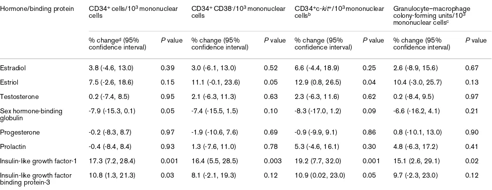
Multiple linear regression analysis was then performed on these data to take into account potential confounding factors involving maternal and neonatal variables including mother's age, race of parents, number of live births, gestation duration, delivery time, birth weight, offspring gender, and study site (Table 3). IGF-1 levels remained strongly associated with all cord blood stem/progenitor concentrations, with each stand-ard-deviation increase in IGF-1 being associated with a 15– 19% increase in stem/progenitor concentrations. IGFBP-3
levels were again positively correlated to CD34+ and CD34+
c-kit+ pools, and estriol levels to CD34+CD38- and CD34+c-kit+
pools, with a one standard-deviation increase in estriol being associated with an 11–12% increase in the concentration of these populations. By this analysis, SHBG remains negatively correlated to these stem/progenitor populations, but only the
association with CD34+ pools was statistically significant.
Different racial and ethnic groups in the United States have dif-fering rates of breast cancer [18]. We explored whether the correlations between cord blood plasma levels of growth fac-tors and hormones and concentration of hematopoietic stem cells would hold across various racial and ethnic groups. We divided the data by racial/ethnic categories, and bivariate anal-ysis was repeated. As shown in Table 4, cord blood plasma levels of IGF-1 are positively and significantly correlated with all four measurements of stem/progenitor cell populations in the Caucasian subgroup, which represent the majority of sam-ples (147 of 252 with known ethnicity). IGF-1 levels are also significantly correlated with some stem/progenitor
measure-ments (CD34+ and CD34+c-kit+ pools) in the
Hispanic-Amer-ican subgroup (n = 15), but not in the African-American (n =
20), Asian-American (n = 39), or mixed (n = 31) subgroups.
Similarly, cord blood plasma estriol levels are positively corre-lated with stem cell pools in the Caucasian subgroup, but not in any of the other subgroups (Table 4). Estradiol levels are positively and significantly correlated only to the CD34+c-kit+
in Caucasians and to the CD34+CD38- in
Hispanic-Ameri-cans, but not in other racial/ethnic groups. Finally, SHBG was negatively but significantly correlated with some of the stem/ progenitor concentrations in the Caucasian samples but, for the most part, not in the other subgroups.
Discussion
Table 1
Summary of the maternal and newborn characteristics, cord blood hormone levels, and cord blood cell populations
Variable n Meana Range
Subject characteristics
Mother's age (years) 289 30.0 ± 5.5 18.0–43.0
Race/ethnicity of mother and biological father
Both Caucasian 147 (58.33%)
Both African-American 20 (7.94%)
Both Asian 39 (15.48%)
Both Hispanic 15 (5.95%)
Mixed 31 (12.30%)
No data 37
Parity
First 118 (43.54%)
Second 77 (28.41%)
Third 46 (16.97%)
Fourth and above 30 (11.07%)
No data 18
Prepregnancy weight (kg) 199 62.7 ± 12.3 38.3–112.5
Gestation duration (weeks) 289 39.6 ± 1.2 36.5–44.0
Gender of the baby
Male 134 (50.57%)
Female 131 (49.43%)
No data 24
Birth weight (g) 288 3,402.4 ± 431.9 2,256–4,660
Cord blood volume (ml) 289 92.5 ± 24.6 44.5–201.5
Cord blood plasma hormone levels
Estradiol (ng/dl) 289 930.9 ± 528.6 40.6–3,305.4
Unconjugated estriol (ng/ml) 289 331.0 ± 141.4 18.1–1,459.5
Testosterone (ng/ml) 289 1.7 ± 0.9 0.3–9.2
Sex hormone-binding globulin (nmol/l) 289 22.8 ± 8.0 6.7–55.0
Progesterone (ng/ml) 289 226.6 ± 134.4 26.5–841.0
Prolactin (ng/ml) 289 254.0 ± 103.6 47.4–686.0
Insulin-like growth factor-1 (ng/ml) 289 81.2 ± 52.5 10.2–306.7
Insulin-like growth factor binding protein-3 (ng/ml) 289 941.1 ± 309.9 300.0–2,534.0
Cord blood cell populations
Initial total nucleated cells × 106/ml 288b 16.1 ± 5.2 6.3–40.6
Initial mononuclear cells × 106/ml 288b 7.6 ± 2.8 2.8–20.8
CD34+ cells/103 mononuclear cells 289 8.9 ± 5.5 0.4–35.5
CD34+CD38- cells/103 mononuclear cells 289 4.1 ± 2.7 0.0–14.4
CD34+c-kit+ cells/103 mononuclear cellsc 249 7.6 ± 4.9 0.2–34.4
Granulocyte–macrophage colony-forming units/103 mononuclear cellsd 175 5.4 ± 3.2 0.5–19.7
and perinatal factors [22] and, speculatively, breast stem cell number and activity [10], is a strong predictor of breast cancer risk [23] and has been associated with both IGF-1 serum lev-els [24] and birth weight [22].
Ethical and practical considerations preclude a direct testing of this hypothesis (that is, establishing whether or not there is an association between prenatal mitogens and breast stem cell quantities) in humans. As an indirect approach, we have used measurements of hematopoietic stem cell and progeni-tor cell concentrations in readily accessible umbilical cord blood samples as surrogates for general stem cell activity or 'stem cell potential'. Although hematopoietic stem cells are capable of giving rise to a wide range of nonhematopoietic cells [9], there has been no report on a direct link with breast stem cells. Levels of hematopoietic stem cells were therefore used in this study as proxy indicators of stem cell burden to correlate with cord blood plasma levels of endocrine factors,
which represent the in utero concentrations of these factors.
A pilot study indicated that cord blood plasma levels of IGF-1, its major binding protein IGFBP-3, and to a lesser degree estriol are positively correlated with hematopoietic stem/pro-genitor concentrations [16]. This finding provided the first evi-dence linking perinatal growth factor and hormone levels to stem cell potential. In the present large study (289 samples), we document more firmly that cord blood plasma levels of IGF-1 are strongly correlated to all cord blood stem and progenitor concentrations examined, and that IGFBP-3 and estriol are
positively correlated to at least some of these stem/progenitor measurements.
The results with respect to IGF-1 are not surprising, as there is considerable evidence that IGF-1 is a positive effector of hematopoiesis. This evidence includes the following: adding
IGF-1 to in vitro cultures of CD34+ or CD34+CD38-
precur-sors augments their proliferation in response to standard col-ony-stimulating factors such as GM-CSF, granulocyte colony-stimulating factor and IL-3 [25], in part by acting as an antiap-optotic factor [26]; in vivo, IGF-1 or the physiologic inducer of IGF-1, growth hormone, can stimulate erythropoieisis, poieisis and granulopoiesis in hypophysectomized or myelo-suppressed rodent model systems [27,28]; and, thirdly, growth hormone administered to patients with growth hormone deficiency coelevates IGF-1 levels and erythroid and myeloid progenitors [29].
There is less evidence linking IGFBP-3 levels with hematopoi-esis. Recently, however, recombinant IGFBP-3 was shown to
directly stimulate the proliferation of human CD34+CD38
-hematopoietic precursors in the absence of IGF-1 [30]. Furthermore, IGFBP-3 is one of the factors elaborated by embryonic stromal feeder-layer cells that can support hemat-opoietic stem cell growth [31]. Why estriol levels correlate with cord blood hematopoietic stem/progenitor concentra-tions is less clear, although estrogens have been used in
con-junction with other growth factors to stimulate the ex vivo
[image:6.612.57.554.129.353.2]proliferation of erythroid precursors from avian bone marrow [32] and human cord blood [33].
Table 2
Spearman correlation coefficients (P values) between cord blood sex hormones, growth factors, and associated binding proteins and cord blood hematopoietic progenitor/stem cell populations
Hormone/binding protein
Total nucleated cells/ml
Mononuclear cells/ml
Measures of stem cell potential, counts/103 mononuclear cells
CD34+ (n = 289) CD34+CD38- (n = 289)
CD34+c-kit+ (n = 249)a
Granulocyte– macrophage colony-forming units (n = 175)b
Estradiol 0.14 (0.02) 0.15 (0.01) 0.05 (0.37) 0.02 (0.79) 0.06 (0.36) 0.03 (0.67)
Estriol 0.20 (0.0006) 0.15 (0.01) 0.09 (0.14) 0.14 (0.02) 0.15 (0.02) 0.11 (0.16)
Testosterone 0.19 (0.001) 0.13 (0.03) -0.03 (0.60) 0.01 (0.80) 0.02 (0.80) 0.06 (0.41)
Sex hormone-binding globulin
0.003 (0.96) 0.05 (0.36) -0.12 (0.04) -0.13 (0.03) -0.13 (0.03) -0.16 (0.04)
Progesterone 0.15 (0.009) 0.15 (0.01) -0.01 (0.84) -0.04 (0.50) -0.008 (0.90) -0.009 (0.91)
Prolactin 0.27 (<0.0001) 0.21 (0.0004) -0.005 (0.93) 0.02 (0.70) 0.09 (0.17) 0.01 (0.90)
Insulin-like growth factor-1
-0.05 (0.41) -0.09 (0.14) 0.18 (0.002) 0.19 (0.001) 0.21 (0.001) 0.18 (0.02)
Insulin-like growth factor binding protein-3
0.10 (0.10) 0.08 (0.20) 0.14 (0.01) 0.11 (0.07) 0.12 (0.05) 0.11 (0.14)
The more relevant question in terms of the stem cell burden theory of breast cancer risk is whether IGF-1 and/or estrogen levels correlate with breast stem cell pool expansion. Although this question cannot be directly answered, it is apparent that both IGF-1 and estrogens are critical to the primitive mammary epithelium during development. In mouse knockout models, IGF-1 null mice have a sharply decreased formation of mam-mary terminal end buds and ductal branching [34], as do
estrogen receptor alpha (ERα) null mice [35]. In sexually
immature female rats that have been hypophysectomized (to eliminate the endogenous growth hormone/IGF-1 axis) and oophorectomized (to remove the major endogenous source of estrogens), treatment with IGF-1 and estradiol were synergis-tic for restoring full mammary gland development [36]; similar results were obtained in IGF-1 null mice [37]. It is reasonable to assume that a factor such as IGF-1 is acting on primitive breast stem and progenitor cells to drive these developmental changes.
Whether or not estrogens act directly on breast stem cells, however, is less clear. At present it is not known whether the
most primitive breast stem cells are ERα- and give rise to more
committed ERα+ cells, or vice versa. Murine mammary stem
cells lack ERα, progesterone receptor, and erbB2 [38]. This
pattern is the same as that found in the so-called 'basal' type of breast cancer, a subclass of breast cancer. Based on micro-array gene expression analysis, this subclass of breast cancer possesses a relatively undifferentiated, stem-like expression profile [39]. In addition, cell culture methods for isolating and expanding breast epithelial precursor cells often do not utilize estrogen. On the other hand, human mammary epithelial cells
with stem-like properties isolated from adult tissues are ERα+
[40], and long-lived, slow cycling, ERα+ cells, have been
described in mouse mammary tissue [41]. Alternatively, a
pop-ulation of ERα+ cells in the developing mammary gland might
serve a paracrine function for adjacent ERα- stem/progenitors.
This would explain the synergy of estrogens and IGF-1 in
mammary development even if breast stem cells lack ERα
[42].
Finally, our data suggest that race/ethnicity may modify the
correlations between in utero/perinatal levels of factors such
as IGF-1 and estrogens and stem cell values. The associations between cord blood plasma levels of IGF-1, IGFBP-3, or estrogens and cord blood hematopoietic stem cell values were stronger in the Caucasian and Hispanic subgroups and were weaker or not present in the Asian-American and African-American subgroups. Comparatively low sample numbers for the latter three subgroups preclude making a definitive conclu-sion on this issue. Caucasians tended to rank high among the groups in terms of hematopoietic stem cell values, whereas Hispanic-Americans ranked low, but again these differences among the subgroups were not significant (data not shown). Other investigators have reported that cord blood from Cau-casians contain relatively high hematopoietic stem cell con-centrations among racial/ethnic groups [43]. Also, since the Asian-American group had the highest levels of estriol (data not shown), it is possible that the nonsignificant relations reflect the lowered study power to document associations within populations at the high or low ends of the hormone lev-els. Factors underlying these racial/ethnic differences in
sign-Table 3
Multiple linear regression analysisa for the association between measurements of hematopoietic stem cell populations and umbilical cord blood plasma levels of hormones and associated binding proteins
Hormone/binding protein CD34+ cells/103 mononuclear
cells
CD34+ CD38-/103 mononuclear
cells
CD34+c-kit+/103 mononuclear
cellsb Granulocyte–macrophage colony-forming units/103
mononuclear cellsc
% changed (95%
confidence interval)
P value % change (95% confidence interval)
P value % change (95% confidence interval)
P value % change (95% confidence interval)
P value
Estradiol 3.8 (-4.6, 13.0) 0.39 3.0 (-6.1, 13.0) 0.52 6.6 (-4.4, 18.9) 0.25 2.6 (-8.9, 15.6) 0.67
Estriol 7.5 (-2.6, 18.6) 0.15 11.1 (-0.1, 23.6) 0.05 12.9 (0.8, 26.5) 0.04 10.4 (-3.0, 25.7) 0.13
Testosterone 0.2 (-7.4, 8.5) 0.95 2.1 (-6.3, 11.3) 0.63 2.3 (-6.3, 11.6) 0.62 0.2 (-8.4, 9.5) 0.97
Sex hormone-binding globulin
-7.9 (-15.3, 0.1) 0.05 -7.4 (-15.5, 1.5) 0.10 -8.3 (-17.0, 1.2) 0.09 -6.6 (-16.2, 4.1) 0.21
Progesterone -0.2 (-8.3, 8.7) 0.97 -1.9 (-10.6, 7.6) 0.69 -0.9 (-9.9, 9.1) 0.86 0.8 (-10.1, 13.0) 0.90
Prolactin -0.4 (-8.4, 8.4) 0.93 1.3 (-7.6, 11.0) 0.78 5.3 (-4.6, 16.1) 0.30 4.8 (-6.3, 17.2) 0.41
Insulin-like growth factor-1 17.3 (7.2, 28.4) 0.001 16.4 (5.5, 28.5) 0.003 19.2 (7.7, 32.0) 0.001 15.1 (2.6, 29.1) 0.02
Insulin-like growth factor
binding protein-3 10.8 (1.3, 21.3) 0.03 8.1 (-2.1, 19.3) 0.12 10.9 (0.02, 23.0) 0.05 9.7 (-2.3, 23.0) 0.12
[image:7.612.60.556.127.319.2]aling molecule/stem cell correlations and breast cancer risk itself will require further study.
Conclusion
Cord blood levels of IGF-1 are strongly positively associated with concentrations of all the cord blood stem and progenitor cells examined. IGFBP-3 and estriol levels are positively asso-ciated with at least some of these cell populations, whereas SHBG tends to show inverse associations with these popula-tions. Our data therefore support the hypothesis that intrauter-ine levels of breast epithelial mitogens modulate stem/ progenitor pools and could, thus, affect long-term risk of breast and perhaps other forms of cancer.
Competing interests
The authors declare that they have no competing interests.
Authors' contributions
[image:8.612.61.555.126.516.2]TMS oversaw the laboratory research protocols and drafted the manuscript. WCS participated in the design of study pro-tocol and collected patient samples and questionnaire infor-mation. HPL participated in the stem cell experiments and conducted analyses on flow cytometry data. QL participated in the design of the study and performed the statistical analysis. IB participated in the design of the questionnaire and coordi-nated the study. WO oversaw the hormonal assays. DPC par-ticipated in the design of clinical protocol and subject enrollment. PL participated in the analyses and interpretation of the results. PQ advised on the stem cell experiments and participated in the interpretation of results. KLN oversaw the clinical research protocols. C-CH conceived the study, partic-ipated in the design, analysis, and result interpretation. All authors read and approved the final manuscript.
Table 4
Spearman correlation coefficients (P values) between cord blood sex hormones, growth factors, and associated binding proteins and hematopoietic stem/progenitor cell populations among different racial/ethnic groups
Stem/progenitor cell
measurement Estradiol Estriol Testosterone Sex hormone-binding globulin Progesterone Prolactin Insulin-like growth factor-1 Insulin-like growth factor binding protein-3
Both parents Caucasian (n = 147)
CD34+/103 MNC 0.12 (0.16) 0.16 (0.06) -0.04 (0.59) -0.18 (0.03) -0.01 (0.87) -0.05 (0.52) 0.24 (0.003) 0.12 (0.15)
CD34+CD38-/103 MNC 0.13 (0.12) 0.25 (0.002) 0.05 (0.53) -0.16 (0.05) -0.02 (0.80) -0.02 (0.80) 0.23 (0.004) 0.05 (0.58)
CD34+c-kit+/103 MNC 0.21 (0.03) 0.30 (0.001) 0.05 (0.61) -0.17 (0.07) -0.001 (0.99) 0.10 (0.29) 0.26 (0.005) 0.09 (0.32) GM-CFU/103 MNC 0.11 (0.32) 0.24 (0.03) 0.13 (0.24) -0.16 (0.16) -0.02 (0.88) 0.13 (0.25) 0.24 (0.03) 0.07 (0.56)
Both parents African-American (n = 20)
CD34+/103 MNC -0.25 (0.28) -0.27 (0.24) -0.22 (0.34) -0.33 (0.16) -0.14 (0.57) -0.23 (0.32) 0.09 (0.71) 0.19 (0.41)
CD34+CD38-/103 MNC -0.25 (0.28) 0.008 (0.97) -0.08 (0.75) -0.32 (0.18) -0.10 (0.67) -0.02 (0.93) -0.003 (0.99) 0.86 (0.72)
CD34+c-kit+/103 MNC -0.40 (0.09) -0.38 (0.11) -0.31 (0.19) -0.42 (0.07) -0.23 (0.34) -0.34 (0.15) 0.12 (0.63) 0.30 (0.20)
GM-CFU/103 MNC -0.39 (0.15) -0.40 (0.14) -0.25 (0.36) -0.52 (0.04) -0.42 (0.12) -0.13 (0.63) -0.02 (0.94) 0.35 (0.20)
Both parents Asian-American (n = 39)
CD34+/103 MNC 0.14 (0.38) 0.09 (0.58) 0.22 (0.19) 0.08 (0.65) 0.15 (0.37) 0.19 (0.25) 0.15 (0.36) 0.06 (0.73)
CD34+CD38-/103 MNC 0.14 (0.93) 0.07 (0.66) 0.17 (0.29) 0.03 (0.86) 0.08 (0.62) 0.14 (0.40) 0.15 (0.35) 0.004 (0.98)
CD34+c-kit+/103 MNC 0.13 (0.42) 0.10 (0.53) 0.19 (0.24) 0.08 (0.64) 0.15 (0.35) 0.19 (0.26) 0.16 (0.35) 0.05 (0.75)
GM-CFU/103 MNC 0.26 (0.25) 0.22 (0.33) 0.24 (0.30) 0.26 (0.26) 0.24 (0.29) 0.09 (0.71) 0.31 (0.17) 0.06 (0.78)
Both parents Hispanic-American (n = 15)
CD34+/103 MNC 0.45 (0.10) -0.16 (0.58) -0.38 (0.17) -0.04 (0.90) -0.06 (0.83) 0.14 (0.63) 0.51 (0.05) 0.01 (0.97)
CD34+CD38-/103 MNC 0.60 (0.02) 0.24 (0.40) 0.01 (0.96) -0.21 (0.45) 0.09 (0.76) 0.14 (0.61) 0.38 (0.16) -0.11 (0.70)
CD34+c-kit+/103 MNC 0.38 (0.19) -0.05 (0.86) -0.35 (0.22) -0.19 (0.51) 0.16 (0.57) 0.23 (0.44) 0.62 (0.02) 0.07 (0.82)
GM-CFU/103 MNC -0.01 (0.98) -0.47 (0.24) -0.49 (0.22) -0.46 (0.26) 0.22 (0.61) -0.25 (0.55) 0.25 (0.55) -0.20 (0.63)
Parents of differing ethnicity/race (mixed) (n = 31)
CD34+/103 MNC -0.06 (0.74) -0.14 (0.46) -0.03 (0.85) -0.07 (0.71) 0.009 (0.96) 0.21 (0.25) 0.03 (0.87) 0.05 (0.80)
CD34+CD38-/103 MNC -0.19 (0.32) -0.18 (0.32) -0.08 (0.68) -0.03 (0.86) -0.05 (0.79) 0.33 (0.07) 0.08 (0.65) 0.25 (0.18)
CD34+c-kit+/103 MNC 0.03 (0.87) 0.005 (0.98) -0.002 (0.99) -0.08 (0.68) 0.004 (0.98) 0.19 (0.31) -0.04 (0.83) -0.02 (0.93)
GM-CFU/103 MNC 0.19 (0.38) 0.23 (0.27) 0.24 (0.27) -0.20 (0.34) 0.20 (0.34) 0.01 (0.95) 0.11 (0.62) 0.08 (0.72)
Acknowledgements
The authors appreciate the effort of the attending physicians and resi-dents of the Department of Obstetrics and Gynecology and Family Prac-tice and the staff on the Labor and Delivery ward at the Tufts–New England Medical Center. This study was supported by a grant (R01CA90902) from the US National Institutes of Health. The authors thank the anonymous reviewers for helpful suggestions.
References
1. Trichopoulos D: Does breast cancer originate in utero? Lancet
1990, 335:939-940.
2. Michels KB, Xue F: Role of birthweight in the etiology of breast cancer. Int J Cancer 2006, 119:2007-2025.
3. Cnattingius S, Zack MM, Ekbom A, Gunnarskog J, Kreuger A, Linet M, Adami HO: Prenatal and neonatal risk factors for childhood lymphatic leukemia. J Natl Cancer Inst 1995, 87:908-914. 4. Ekbom A, Hsieh CC, Lipworth L, Wolk A, Ponten J, Adami HO,
Tri-chopoulos D: Perinatal characteristics in relation to incidence of and mortality from prostate cancer. BMJ 1996, 313:337-341.
5. Andersson SW, Bengtsson C, Hallberg L, Lapidus L, Niklasson A, Wallgren A, Hulthen L: Cancer risk in Swedish women: the rela-tion to size at birth. Br J Cancer 2001, 84:1193-1198. 6. Ekbom A, Hsieh CC, Lipworth LL, Adami HO, Trichopoulos D:
Intrauterine environment and breast cancer risk in women: a population-based study. J Natl Cancer Inst 1997, 89:71-76. 7. Trichopoulos D, Lipworth L: Is cancer causation simpler than we
thought, but more intractable? Epidemiology 1995, 6:347-349. 8. Adami HO, Signorello LB, Trichopoulos D: Towards an under-standing of breast cancer etiology. Semin Cancer Biol 1998, 8:255-262.
9. Baik I, Becker PS, DeVito WJ, Lagiou P, Ballen K, Quesenberry PJ, Hsieh CC: Stem cells and prenatal origin of breast cancer.
Cancer Causes Control 2004, 15:517-530.
10. Savarese TM, Low HP, Baik I, Strohsnitter WC, Hsieh CC: Normal breast stem cells, malignant breast stem cells, and the perina-tal origin of breast cancer. Stem Cell Rev 2006, 2:103-110. 11. Stingl J, Eirew P, Ricketson I, Shackleton M, Vaillant F, Choi D, Li
HI, Eaves CJ: Purification and unique properties of mammary epithelial stem cells. Nature 2006, 439:993-997.
12. Shackleton M, Vaillant F, Simpson KJ, Stingl J, Smyth GK, Asselin-Labat M-L, Wu L, Lindeman GJ, Visvader JE: Generation of a functional mammary gland from a single stem cell. Nature
2006, 439:84-88.
13. Stingl J, Eaves CJ, Zandieh I, Emerman JT: Characterization of bipotent mammary epithelial progenitor cells in normal adult human breast tissue. Breast Cancer Res Treat 2001, 67:93-109.
14. Gudjonsson T, Villadsen R, Nielsen HL, Ronnov-Jessen L, Bissell MJ, Petersen OW: Isolation, immortalization, and characteriza-tion of a human breast epithelial cell line with stem cell properties. Genes Dev 2002, 16:693-706.
15. Ponti D, Costa A, Zaffaroni N, Pratesi G, Petrangolini G, Coradini D, Pilotti S, Pierotti MA, Daidone MG: Isolation and in vitro prop-agation of tumorigenic breast cancer cells with stem/progen-itor cell properties. Cancer Res 2005, 65:5506-5511. 16. Baik I, DeVito WJ, Ballen K, Becker PS, Okulicz W, Liu Q, Delpapa
E, Lagiou P, Sturgeon S, Trichopoulos D, Quesenberry PJ, Hsieh CC: Association of fetal hormone levels with stem cell poten-tial: evidence for early life roots of human cancer. Cancer Res
2005, 65:358-363.
17. Hoffbrand AV, Pettit JE, Moss PAH: Essential Hematology 4th edi-tion. Oxford: Blackwell; 2001:1-11.
18. Smigal C, Jemal A, Ward E, Cokkinides V, Smith R, Howe HL, Thun M: Trends in breast cancer by race and ethnicity: update 2006.
CA Cancer J Clin 2006, 56:168-183.
19. Skalkidou A, Petidou E, Papathoma E, Salvanos H, Tichopoulos D: Growth velocity during the first postnatal week of life is linked to a spurt of IGF-1 effect. Paediatr Perinat Epidemiol 2003, 17:281-286.
20. Hankinson SE, Willett WC, Colditz GA, Hunter DJ, Michaud DS, Deroo B, Rosner B, Speizer FE, Pollak M: Circulating concentra-tions of insulin-like growth factor-I and risk of breast cancer.
Lancet 1998, 351:1393-1396.
21. Li T, Sun L, Miller N, Nicklee T, Woo J, Hulse-Smith L, Tsao MS, Khokha R, Martin L, Boyd N: The association of measured breast tissue characteristics with mammographic density and other risk factors for breast cancer. Cancer Epidemiol Biomar-kers Prev 2005, 14:343-349.
22. Cerhan JR, Sellers TA, Janney CA, Pankratz VS, Brandt KR, Vachon CM: Prenatal and perinatal correlates of adult mam-mographic breast density. Cancer Epidemiol Biomarkers Prev
2005, 14:1502-1508.
23. Byrne C, Schairer C, Wolfe J, Parekh N, Salane M, Brinton LA, Hoover R, Haile R: Mammographic features and breast cancer risk: effects with time, age, and menopause status. J Natl Can-cer Inst 1995, 87:1622-1629.
24. Boyd NF, Stone J, Martin LJ, Jong R, Fishell E, Yaffe M, Hammond G, Minkin S: The association of breast mitogens with mammo-graphic densities. Br J Cancer 2002, 87:876-882.
25. Frostad S, Bjerknes R, Abrahamsen JF, Olweus J, Bruserud O: Insulin-like growth factor-1 (IGF-1) has a costimulatory effect on proliferation of committed progenitors derived from human umbilical cord CD34+ cells. Stem Cells 1998, 16:334-342.
26. Rodriguez-Tarduchy G, Collins MK, Garcia I, Lopez-Rivas A: Insu-lin-like growth factor-I inhibits apoptosis in IL-3-dependent hematopoietic cells. J Immunol 1992, 149:535-540.
27. Kurtz A, Zapf J, Eckardt KU, Clemons G, Froesch ER, Bauer C: Insulin-like growth factor I stimulates erythropoiesis in hypo-physectomized rats. Proc Natl Acad Sci USA 1988, 85:7825-7829.
28. Murphy WJ, Tsarfaty G, Longo DL: Growth hormone exerts hematopoietic growth-promoting effects in vivo and partially counteracts the myelosuppressive effects of azidothymidine.
Blood 1992, 80:1443-1447.
29. Kotzmann H, Riedl M, Clodi M, Barnas U, Kaider A, Hocker P, Luger A: The influence of growth hormone substitution therapy on erythroid and myeloid progenitor cells and on peripheral blood cells in adult patients with growth hormone deficiency.
Eur J Clin Invest 1996, 26:1175-1181.
30. Liu LQ, Sposato M, Liu H-Y, Vaudrain T, Shi MJ, Rider K, Stevens Z, Visser J, Deng HK, Kraus M: Functional cloning of IGFBP-3 from human microvascular endothelial cells reveals its novel role in promoting proliferation of primitive CD34+CD38 -hematopoietic cells in vitro. Oncol Res 2003, 13:359-371. 31. Oostendorp RA, Robin C, Steinhoff C, Marz S, Brauer R, Nuber
UA, Dzierzak EA, Peschel C: Long-term maintenance of hemat-opoietic stem cells does not require contact with embryo-derived stromal cells in cocultures. Stem Cells 2005, 23:842-851.
32. Wessely O, Deiner EM, Beug H, von Lindern M: The glucocorti-coid receptor is a key regulator of the decision between self-renewal and differentiation in erythroid progenitors. EMBO J
1997, 16:267-280.
33. Panzenbock B, Barunek P, Mapara MY, Zenke M: Growth and dif-ferentiation of human stem cell factor/erythropoietin-depend-ent erythroid progenitor cells in vitro. Blood 1998, 92:3658-3668.
34. Ruan W, Kleinberg DL: Insulin-like growth factor I is essential for terminal end bud formation and ductal morphogenesis during mammary development. Endocrinology 1999, 140:5075-5081.
35. Korach K, Couse JF, Curtis SW, Washburn TF, Lindzey J, Kimbro KS, Eddy EM, Migliaccio S, Snedeker SM, Lubahn DB, Schomb-erg DW, Smith EP: Estrogen receptor gene disruption: molec-ular characterization and experimental and clinical phenotypes. Recent Prog Horm Res 1996, 51:159-186. 36. Ruan W, Catanese V, Wieczorek R, Feldman M, Kleinberg DL:
Estradiol enhances the stimulatory effect of insulin-like growth factor-I (IGF-1) on mammary development and growth hormone-induced IGF-1 messenger ribonucleic acid. Endo-crinology 1995, 136:1296-1302.
37. Kleinberg DL, Feldman M, Ruan W: IGF-1: an essential factor in terminal end bud formation and ductal morphogenesis. J Mammary Gland Biol Neoplasia 2000, 5:7-17.
38. Asselin-Labat M-L, Shackleton M, Stingl J, Vaillant F, Forrest NC, Eaves CJ, Visvader JE, Lindeman GJ: Steroid hormone receptor status of mouse mammary stem cells. J Natl Cancer Inst 2006, 98:1011-1014.
Immu-nohistochemical and clinical characterization of the basal-like subtype of invasive breast carcinoma. Clin Cancer Res 2004, 10:5367-5374.
40. Clarke RB, Anderson E, Howell A, Potten CS: Regulation of human breast epithelial stem cells. Cell Prolif 2003, 36:45-58. 41. Zeps N, Dawkins HJ, Papadimitriou JM, Redmond SL, Walters MI: Detection of a population of long-lived cells in the mammary epithelium of the mouse. Cell Tissue Res 1996, 286:525-536. 42. Dontu G, El-Ashry D, Wicha MS: Breast cancer, stem/progeni-tor cells and the estrogen recepstem/progeni-tor. Trends Endocrinol Metab
2004, 15:193-197.