White Rose Research Online URL for this paper:
http://eprints.whiterose.ac.uk/103451/
Version: Accepted Version
Article:
Leong, F, Garbin, N, Di Natali, C et al. (4 more authors) (2016) Magnetic Surgical
Instruments for Robotic Abdominal Surgery. IEEE reviews in biomedical engineering, 9.
pp. 66-78. ISSN 1937-3333
https://doi.org/10.1109/RBME.2016.2521818
(c) 2016 IEEE. This is an author produced version of a paper published in IEEE reviews in
biomedical engineering . Uploaded in accordance with the publisher's self-archiving policy.
Personal use of this material is permitted. Permission from IEEE must be obtained for all
other users, including reprinting/ republishing this material for advertising or promotional
purposes, creating new collective works for resale or redistribution to servers or lists, or
reuse of any copyrighted components of this work in other works.
eprints@whiterose.ac.uk https://eprints.whiterose.ac.uk/
Reuse
Unless indicated otherwise, fulltext items are protected by copyright with all rights reserved. The copyright exception in section 29 of the Copyright, Designs and Patents Act 1988 allows the making of a single copy solely for the purpose of non-commercial research or private study within the limits of fair dealing. The publisher or other rights-holder may allow further reproduction and re-use of this version - refer to the White Rose Research Online record for this item. Where records identify the publisher as the copyright holder, users can verify any specific terms of use on the publisher’s website.
Takedown
If you consider content in White Rose Research Online to be in breach of UK law, please notify us by
For Review Only
" #$ % # & ' & () $ * + ' & * ,- . / 0 + # - &
1 # - 2 / 3 % # 0 - $ + &
, ,
2 - )& / 3 % # 0 - $ + &
, ,
& $$ % - ' 4 / 0 + # - &
& & $- & / 0 + # - " 3 5 $ + " ,
For Review Only
Magnetic Surgical Instruments for Robotic
Abdominal Surgery
Florence Leong
1 ∗, Nicolo Garbin
2, Christian Di Natali
2, Alireza Mohammadi
1, Dhan Thiruchelvam
3, Denny
Oetomo
1, Pietro Valdastri
2Abstract—This paper reviews the implementation of magnetic
based approaches in surgical instruments for abdominal surg-eries. As the abdominal surgical techniques advance towards minimising surgical trauma, surgical instruments are enhanced to support such objective through the exploration of magnetic based systems. With this design approach, the surgical devices are given the capabilities to be fully inserted intra-abdominally to achieve access to all abdominal quadrants, without the conventional rigid link connection with the external unit. The variety of intra-abdominal surgical devices are anchored, guided and actuated by external units, with power and torque transmitted across the abdominal wall through magnetic linkage. This addresses many constraints encountered by conventional laparoscopic tools, such as loss of triangulation, fulcrum effect and the loss / lack of dexterity for surgical tasks. Design requirements in the aspects of clinical considerations are also discussed to aid the successful development of magnetic surgical instruments.
I. INTRODUCTION
Surgical techniques and technologies have progressed sig-nificantly over the past decades with the aim of minimising trauma to the patients. Apart from resulting in less pain and faster recovery, the reduction of trauma also minimises the number and severity of incisions, wound size and blood loss, hence reducing the potential of surgical complications. This has been demonstrated through the trend of the technical and technological development of the surgical procedure: from open surgery to laparoscopic or minimally invasive surgery (MIS) [1], laparoendoscopic single site (LESS) [2], [3] and natural orifice transluminal endoscopic surgery (NOTES) [4]. The concept of decreasing surgical trauma via MIS [5], [6] (as shown in Fig. 1a), gained much popularity throughout the field of abdominal surgery since the 1990s, moving forward from the open surgical procedure. Since the first reported MIS procedure in 1988 [7], the benefits of MIS have been observed across a wide range of surgical disciplines. While MIS does not have the large workspace for surgeons to freely access different quadrants of the abdomen, it allows much smaller incisions, thus reducing the surgical trauma suffered by the patients. Furthermore, using relatively small incisions ranging
1Department of Mechanical Engineering, The University of Melbourne,
Australiafleong@student.unimelb.edu.au, {alirezam, doetomo}@unimelb.edu.au
2Department of Mechanical Engineering, Vanderbilt University, Nashville,
TN, USA {nicolo.garbin.1, christian.di.natali,
pietro.valdastri}@vanderbilt.edu
3St. Vincent’s Department of Surgery, The University of Melbourne,
Mel-bourne, Australiadhant@unimelb.edu.au
from 5mm to 12mm [8], this approach leads to less wound complications, less postoperative pain, shorter recovery period, better cosmetics, and earlier return to employment and daily activities, hence greater patient satisfaction [9]–[11]. More technical advantages of laparoscopy include lower likelihood of infections and blood loss, improving the preservation of normal immune function, thus lower morbidity rate [10], [12]– [14].
The persistent aim in minimising the invasiveness of surgical procedures stimulated further reduction in surgical trauma by introducing the LESS approach, as illustrated in Fig. 1b, with improvements in the aspects of cosmetics and recovery rate [15]–[17]. LESS aims to eliminate the need of multiple surgical incisions, utilising only one incision typically through the umbilicus [18], [19], for the insertion of laparoscopic surgical tools [20], [21]. Currently, LESS devices (e.g. SILSTM port, TriportTM) are widely available, with a plastic disk of 2 to 3 cm connected to a flange for multiple laparoscopic tools to be inserted through the umbilicus [22], [23].
Towards achieving the goal of a completely scarless surgery, NOTES has been proposed [24]–[27]. The NOTES proce-dures (see Fig. 1c) gain access to the abdominal cavity via various transluminal access (e.g. transgastric, transvaginal or transcolonic). Endoscopic-like surgical instruments are used to manoeuvre into the abdomen through an incision made in the stomach wall, uterus or colon to access the abdomen. Due to the cumbersome of procedural setup and manipulation with current available surgical instruments [28], [29] as well as the lack of cases to evaluate the possibility of infection [30], [31], NOTES remains largely experimental.
For Review Only
Fig. 1: Schematic representation of the abdominal surgery techniques. a) Minimally Invasive Surgery (MIS) scheme; each surgical tool requires a single port (trocar). b) Laparoendoscopic Single Site Surgery (LESS) scheme; mirrored surgical tool are used to fit a 2-3 cm multi-port. c) Transcolonic Natural Orifice Transluminal Endoscopic Surgery (NOTES) scheme; endoscopic surgical tool reaches the abdominal cavity though an internal incision.
system for MIS surgery, i.e. the DaVinci Xi [37]. DaVinci Xi has the capability to reach all the abdominal quadrants and 6 or 7 degrees-of-freedom (DOFs) at the end effectors thanks to its cable driven mechanism. Robotic LESS surgery is also explored through a platform that has recently obtained FDA approval, which is the Da Vinci Sp. The Sp platform consists of a single cannula that allows the deployment of three snake-like robotic arms and a stereoscopic vision head into the abdominal cavity. In contrast to the previous version, the DaVinci Single Site, the Da Vinci Sp offers more DOFs per surgical in-strument, improving triangulation, tools congestion avoidance and multi-port dislodgement. Commercial robotic platforms for NOTES are yet to be available for clinical use. The complicated trajectory and the need for highly manoeuvrable and miniaturised robotic manipulators with the capability of endoscopic instruments limit the development of robotic plat-forms. The main technical challenges arise from the connection with long body devices that could deprive the instrument of the rigidity required by surgical tasks. Furthermore, the power available at the end effector is not always sufficient due to the required miniaturisation. A snake-like surgical endoscopic robot, the FlexRRobotic System by Medrobotics [38] that have just recently obtained FDA clearance for transoral surgery could bring future implementation of surgical robotics closer to NOTES as an inner snake-like body greatly caters for the rigidity in supporting the forces at the end effector.
Despite these advances, including those brought about by robotic surgery, challenges arising from having to perform surgery through small access port(s) remain. In all these cases, it has been noted that having to perform task manipulation using instrumentation constrained in its motion to have to pass through the small access ports contributes significantly to the challenges. Investigation into the possibility of removing such rigid body constraints was pioneered by Tillander et al [39], Yodh et al [40] and Montgomery et al [41], where magnets were used to replace rigid mechanical transmission. In these studies, significant advantages in the form of im-proved manipulability and mobility, while maintaining the level of minimally invasive access offered by other approaches, were demonstrated. Magnetic linkages, replacing rigid body
linkages, eliminates the need for the surgical instruments to maintain contact with the access port once inserted into the abdominal cavity. Multiple DOFs can be realised through the use of localised magnetic coupling, thus allowing dexterous manipulation. Magnetic anchoring allows mobility of such platform, providing access to multiple quadrants in the ab-domen. Another potential advantage can be seen in the ability to transmit a large amount of power through the magnetic linkages without compromising the size of the instrument inserted into the body, thus suitable for miniaturisation.
In this paper, the emerging magnetic-based surgical tech-niques and technologies are reviewed, investigating the de-velopment of ideas, the challenges, the current state of the art and the promising potential in future surgical procedures. The reminder of this paper is organised as follows. Section II overviews the application of magnetism in the current abdominal surgery approaches. Section III reviews the imple-mentation of magnetic coupling across the abdominal barrier for anchoring and guidance of surgical tools, both by manual operation and with onboard actuation embedded in the intra-abdominal tool. The advancements into surgical devices with robotic control of magnetic actuation via the means of local magnetic and electromagnetic actuations are discussed in Sec-tion IV, with some insights on the surgical devices employing this strategy. Finally, in the process of developing effective magnetic-based surgical instruments, the technical and clinical considerations are summarised in Section V.
II. MAGNETIC-BASEDAPPROACH INABDOMINAL
SURGERY
For Review Only
adequate visualisation during surgery, which decreases alongwith invasiveness decrease.
A promising approach that can improve both triangulation and dexterity for surgical instruments leverages the magnetic field interactions of two sets of magnets, located on the opposite sides of the abdominal wall. In this paper, sets of magnet located on the outside and the inside of the abdominal wall are referred to as the external unit and the internal unit, respectively. The interaction forces allow the transmission of torques across a physical barrier, and hence the transfer of mechanical power from an external set of driving magnet to the driven magnet embedded into the surgical tool, which is operating inside the human body.
The use of magnets in abdominal surgery was first reported by the successful utilisation of magnetic systems almost 65 years ago in neurosurgery and cardiology in guiding catheter tips through vessels. The very first magnetic guidance of intravascular catheter was experimentally evaluated in 1949 and published in 1951 by Tillander et al [42]. The device consisted of a catheter with an articulated steel tip that was steered by a magnetic field generated by a large electromagnet placed beneath a nonmagnetic surgical table. The advancement in technology and the possibility to regulate magnetic fields led to the translation to clinical use, such as Stereotaxis [43], [44] and Magnetecs [45], with the Stereotaxis obtaining FDA approval for atrial flutter treatment in 2010 [46].
In recent years, more magnetic-based medical devices re-ceived increasing attention, such as the capsule endoscopes (CE) for diagnosis and treatment of the gastro-intestinal (GI) tract [47], [48], with various designs exploiting magnetic-based navigation in colonoscopy [49]–[57], gastroscopy [58]– [61], tissue biopsy [54], controllable drug delivery [62] and tumour detection [63], [64]. Since then, the use of magnetic systems to deploy, place and drive independent surgical units intra-abdominally across the abdominal wall has tremendously intrigued researchers. This created an innovative class of externally actuated surgical instruments which can potentially improve triangulation and tool repositioning and lower surgical invasiveness.
III. MAGNETICANCHORING ANDGUIDANCESYSTEM
(MAGS)
The Magnetic Anchoring and Guidance Systems (MAGS), introduced by Cadeddu and his team in 2007 [65], are surgical tools that benefit from magnetic anchoring between an external permanent magnet (PM) and an internal PM embedded in the internal surgical device. The anchoring serves to guide and reposition the surgical device through manipulation from the outside of the body. The attraction force acting on the internal PM anchors the internal device onto the inside of the abdominal wall. If the external PM is displaced along the surface of the abdominal wall, the magnetic misalignment generates a net force that will act on the internal magnet in the same direction as the displacement resulting in the internal device undergoing the same displacement. By having the magnetic poles of each magnet in parallel to each other, as shown in Fig. 2a, the translation and angular displacement
can be achieved, resulting in 3 DOFs of motion, i.e. translation along x and y and rotation about z (the yaw DOF).
A. MAGS with manual guidance
MAGS was investigated for its ability to perform simple sur-gical tasks [66], to manipulate a sursur-gical camera [65], sursur-gical retractors [65] and surgical cauterizer [67]. Different cameras have been proposed by the same group of authors (Cadedduet al), where the latest version had Ethicon Endo Sugery involved in its development and was tested in clinical trials [65]. It consists of an illumination and vision system embedded in a wired capsule that contains a permanent magnet. The external magnetic handle is hand operated and gives the surgeon the ability to change the field of view of the camera in the internal unit by manually pressing the external PM onto the inflated abdominal cavity. Swain et al[68] studied the possibility to obtain a “stadium view” with a miniaturised (15 mm diameter and 30 mm length) camera and illumination systems attached to the abdominal wall. Trials highlighted the benefits of having a mobile camera hanging from the abdominal wall compared to the use of conventional endoscopes which suffer from tunnel vision, i.e. misperception of the surgical workspace for surgeons.
Surgical retractors in MAGS were designed with manip-ulation capability. Two external PM anchors are arranged such that its relative distance can be varied [66], resulting in the actuation of a device functional unit through simple mechanism (see Fig. 2b). A fan shaped end effector has been successfully used to retract liver portions in a porcine model. Further development on magnetic based retractor was made by Dominquez et al [69] and Cho et al [70], who developed a simpler but functional surgical retractor used in actual abdominal surgeries. Dominguez et al proposed a surgical grasper directly attached to a permanent magnet, while Choet altested a magnet-fixed endoscopic clip. The clip was manipulated by the surgeons using conventional surgical tools. Once attached to the point of interest, the clip was retracted by the magnetic attraction of the external PM.
From the in vivo experiments [67], [71], the potential of MAGS was validated as a technology that emphasises the aspects of self-anchoring capability, instrument guidance within the abdominal cavity and thus decreased invasiveness. However, without the capability to manipulate objects, only relatively simple surgical tasks have been demonstrated with these instruments thus far.
For Review Only
(a)MAGS principle of operation (b)MAGS magnetically actuated by magnetic translation
Fig. 2: Illustration of Magnetic Anchoring and Guidance System (MAGS)
was found to be non-reliable in a surgical scenario. For more reliable and accurate instrument manipulation, the concept of the simple magnetic attraction has to be further capitalised to adapt to more complex surgical tasks and motions.
B. MAGS with onboard conventional actuation
The ideas of incorporating on board actuation units onto MAGS surgical devices grew out of the need to enhance the manipulation capability of the instruments by decoupling the actuation for manipulation from the anchoring tasks. In this case, the MAGS instruments are further enhanced with on board actuators (e.g. miniaturised electrical DC motors) to drive a surgical instrument with higher number of DOFs mounted on the internal unit, for tasks requiring more dexter-ous manipulation within abdominal cavity (see Fig. 3).
[image:6.612.62.287.496.661.2]A magnetically anchored and actuated device was first reported in [66] in the form of a three DOFs robotic cauteriser
Fig. 3: Operation principle of MAGS with on board conventional actuation
(introduced in subsection III-A). The surgical cauteriser, 158 mm long, is pneumatically actuated and embedded with per-manent magnets at the instrument base for its anchoring. The pneumatic actuation did not guarantee a smooth operation of the devicein vivo, but was able to generate sufficient force to elevate tissue and perform incisions on it. The performance of the device in terms of available force at the end effector and attraction forces at the instrument base, was not quantified by the authors.
Vision (camera) systems in surgical instrumentation do not generally require much mechanical power, defined as the product of force and velocity or torque and speed, as no tissue manipulation is required. However, a precise motion in different directions is necessary. In 2009, a camera system with controllable orientation (pan and tilt) and zoom for MIS surgery was proposed [72], incorporating the concept of MAGS enhanced with conventional motors. Two DC elec-trical motors connected with miniaturised gears provided the actuation for the two orientation DOFs while a third motor was used to linearly actuate the camera head thus resulting in zoom feature. The device, 9 mm in diameter, was designed without an anchoring solution andin vivotrials were performed by suturing the device onto the abdominal wall. The study however highlighted the potential use of magnetic links as a way to reposition and dock the device.
For Review Only
approach.A magnetic surgical retractor has to be designed to support the device weight as well as the retracted load / tissue. A mag-netic retraction device for NOTES was proposed by Oleynikov et al. [33] and consists of a MAGS platform that embeds two PMs and an electrical motor that bundle or unbundle a wired surgical clip. No description was given of the performance of the retraction system but the authors reported that limited simple manoeuvres were achieved. Tortoraet al[74] developed a surgical retractor that consist of a retracting arm with two DOFs connected to a magnetic frame for anchoring purposes. The device utilises one DC motor to change the angular position of the retracting arm while the second motor is used to operate the end effector gripper. While sufficient grasping force is achieved at the end effector (5.3 N), the retracting force generated by the motor at the end effector is insufficient for surgical practice (1.53 N). The actuation torque in this case is already leveraged mechanically for the purpose of tissue retrac-tion. The low power of electrical DC micromotors limits their applicability for surgical tasks that demands high power. In [74] the performance of the whole platform is also evaluated on a mock up simulator, consisting of a foldable triangular frame that embeds the PMs for anchoring with different instruments connected to it. The instruments include a four DOF robotic cauteriser, a four DOF robotic manipulators, a two DOF camera and a two DOF surgical retractor. The main benefits of connecting different instruments to the magnetic frame are that (1) the anchoring frame can embed bigger permanent magnets and support larger load and (2) the fixed position of the common base of the instruments improves triangulation, avoids instrument reciprocal interference and provides a known relative location between different instruments. However, the force available at the four DOF robotic tools tip, based on the same modular components, was not sufficient for further investigation in surgical practice (0.65 N).
In parallel, another 3-link miniature surgical robot for NOTES was taken into the in vivo non-survival porcine test beds by Lehman et al [75]. The robot consists of a cen-tral body with stereoscopic vision and PMs for the device anchoring. Attached to it, two arms with three DOFs, each attached and actuated by DC electric motors. The experimental validation demonstrated the possibility of combining magnets with traditional actuators for miniature robotic platform for surgery but the dexterity is found to be lower than using conventional platform (conventional MIS with non-magnetic based approach). To overcome the dexterity limitation en-countered by this study, the same group developed a similar device with four DOFs per arm but without magnetic anchors [76]. The final dimension of the device could not allow trans-gastric deployment of the device; as a 20 cm opening on the abdominal wall was required. This platform is currently being developed for clinical used by Virtual Incision Corp [77].
[image:7.612.333.558.52.203.2]Though the attempts to incorporate actuation capability in MAGS surgical devices using conventional motors for more dexterous manipulation are proven to be feasible for small payloads, the motors with sizes small enough to fit through the incision do not generally provide enough torque and payload capabilities for the surgical instrument manipulation.
Fig. 4: Illustration of MAGS with robotic actuation
Nevertheless, the promising advantages of magnetic coupling were demonstrated in guiding and anchoring of surgical in-struments, with now more complex robotic features, albeit limited in power. Surgical devices can be driven without direct connections between the internal unit and the external actuation. As such, surgical devices with embedded permanent magnets can be manipulated across the physical barrier of the abdominal wall through the appropriate manipulation of the magnetic linkages.
IV. ROBOTIC CONTROL OF MAGNETIC ACTUATION OF SURGICAL DEVICES
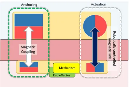
The following type of devices take advantage of the use of magnetic field to develop a generation of surgical devices with improved dexterity, reliable operation and more powerful actu-ation. Mechanical power can be transmitted through a magnetic linkage. The magnetic linkage can in fact be used statically to generate attractive forces for anchoring and guidance or dynamically to produce forces/torque in the internal device, as shown in Fig. 4. The resulting internal device is tether-less, and potentially with no electronics on board.
A. Magnetic actuation solutions
Various magnetic actuation solutions have been investigated for the cases where the absence of rigid link between the surgical tool and the actuation mechanism can improve the outcome for the patients. In the following, different magnetic links for actuation purposes are reviewed.
1) Position control of permanent magnetUsing robotic
For Review Only
since generally a camera system is used as a visualfeedback to the surgeon performing the operation.
2) Permanent magnets with shielding materialBrewer
et al. [82] investigated the possibility of modulating the magnetic field by interposing magnetic shielding material between the external PM and the PM on board the internal device. By controlling the shield position and thus interaction-mitigation of the magnetic field, it is possible to have the force control over the internal device containing an on board PM. Moreover, an interesting result was reported for possible force regulation strategy: the amount of force transmitted to the device becomes linearly dependent on the shielding position.
3) Electromagnetic coils Coil is another magnetic field
generator that has been used by Kummer et al. [83] to actuate and control the position of a miniaturised swimming robot for eye surgery. The magnetic field of a coil can be modulated as a function of the current running through the coil. Moreover, using multiple coils, it is possible to shape the magnetic field in determined areas.
4) Servo control of rotating magnets Diametrically
mag-netised cylindrical magnets can act like mechanical spur gears. The number of the magnetic poles is analogous to the equivalent number of teeth of a gear, while allowing contactless torque and speed transmission. Due to this feature, the magnetic coupling can be referred as mag-netic gear [84]. A motor connected to such magnet can transmit power to another PM across a physical barrier to another magnet. This concept leads to interesting applications in the design of surgical devices reviewed in the following section.
B. Local magnetic and electromagnetic actuation
Local Magnetic Actuation (LMA) is a method for actuating surgical instruments based on the magnetic gear principle. In addition to the magnetic gear principle, LMA surgical instruments also take advantage of magnetic attraction force for anchoring and guidance, as elaborated in MAGS.
In the magnetic gear coupling, the motor is used to drive a cylindrical PM placed external to the body of the pa-tient. Multiple magnetic gears can be used to actuate an equivalent number of DOFs for robotic surgical devices. An LMA magnetic gear unit was controlled with different closed-loop strategies by Di Natali et al. [85]. In this study, it is reported that with the selected PMs, it is possible to deliver an amount of power to the internal PM that is higher than that of a DC micromotor directly embedded in the internal tools. This addresses the main limitation of the traditional actuation techniques based on embedded DC motors on board the internal device.
The efficiency of the transmission however decreases with the increase of the separation distance between the driving (external) PM and driven (internal) one, associated with an increase in the abdominal wall thickness. This is due to the inverse exponential relationship the magnetic field has with the
Abdominal Wall
Permanent Magnet Rotor
Encoder Hall Effect
[image:8.612.324.570.52.160.2]Sensor
Fig. 5: Electromagnet coils replacing permanent magnets in the external actuation unit for higher level control in LEMA [86]
thickness of the abdominal wall separating the driving magnets and the load. It was also observed that pole slipping could exist between the external and internal driving magnets, reducing the amount of transferred torque significantly. Misalignment of the rotational axes of two permanents magnets was also reported as an issue [85]. To account for these limitations, Local Electromagnetic Actuation (LEMA) has been proposed [86], [87] whereby the external permanent magnets in LMA are replaced by electromagnets to generate a rotating magnetic field that produces moments to rotate the internal PM. The use of electromagnetic coils allows the modulation of the intensity of the magnetic field and thus providing a variable parameter which can be controlled to improve the performance of the system. The external electromagnetic coils can be thought of as being analogous to the stator of a DC motor while the driven internal PM as the rotor. In the case of large stator-rotor distance or high load torque, the magnitude of currents in the stators can be increased to compensate for the reduction of torque in permanent magnet inside the abdominal wall. Additionally, the ability to vary the actuation command to the electromagnetic stators would allow the implementation of a controller to compensate for mechanical uncertainties and inaccuracies, such as variable thickness and the misalignment of rotational axes [87]. In the reported design, two sets of electromagnetic windings were used as the stators to generate the magnetic fields to one (internal) rotor. This is required to avoid the ambiguity in the direction of resulting rotor rotation given the generated magnetic field. Additionally, the amount of current to each stator winding can be regulated independently, allowing more DOFs in the control for the resulting magnetic field, which in turn allow the system to compensate for the uncertainties.
For Review Only
as possible kept isolated between the driving external magnetand its intended pair of the driven internal magnet across the abdominal wall, thus allowing multiple magnetic couplings to be utilised to produce independent actuation over multiple DOFs.
C. LMA-based surgical devices
Motivated by the different robotic control strategies of mag-netic field, researchers have already developed some promising device prototypes. Liu and his team [89] studied the optimal design parameters for a surgical camera system based on coil current control. Three electromagnetic coils shape the magnetic field and are used to actuate and anchor a capsule shaped camera with wireless communication for video streaming (Fig. 6). The capsule device embeds multiple magnets with different magnetisation on a hemispherical rotor at the capsule base. The device is yet to be fabricated and tested, but the proposed de-sign can easily be integrated in commercially available vision systems such as Pillcam TMor similar commercial platforms. Moreover, the proposed solution allows the anchoring and actuation functions to fuse into a unique magnetic link.
[image:9.612.333.544.54.205.2]Another camera system based on controlled PM position has been proposed by Simi et al. [90]. With reliable motion and the fine-tuning of one of the DOFs used to change the field of view, the device can be used for LESS scenario. The proposed instrument has one body that contains the anchoring PMs, a flexible joint and a head containing a PM whose angular po-sition is controlled by a DC motor, and a camera-illumination module (Fig. 7). By gradually rotating the PM in the head link, the forces acting on the head changes. The repulsion or attraction force, generated by the magnetic interaction with the corresponding external permanent magnet, is regulated by the rotation generated by the internal motor. The transmitted forces are thus responsible for the deflection of the flexible joint resulting in a change of viewing (angular) position. Bench test demonstrated the reliability of the tilt angle control. A maximum angle of 82 degree is achieved when the magnets are in maximum repulsive state. The dimensions of the device (i.e. 12.7 mm diameter and 120 mm in length) limit itsin vivo
[image:9.612.78.270.508.659.2]Fig. 6: Knoxville camera with external electromagnetic coils for intra-abdominal capsule actuation [89]
Fig. 7: LMA laparoscopic camera with fine tuning capabilities by Simiet al [90]
guidance, but the actuation principle allows a better exploration of the surgical field as well as optimal positioning.
The first LMA-based device is a surgical retractor developed by Garbinet al[91]. The magnetic gear allows the transfer of mechanical power in term of torque and speed to the internal PM from the external magnetic source (Fig. 8). An internal mechanical gearbox attached amplifies the torque available for actuating a retracting lever. Bench test were used to evaluate the device capability in terms of its retracting performance at varying abdominal wall thicknesses. A load of 500 grams was lifted with a 2 cm separation distance between the retractor and the external handle. The anchoring unit was observed to be the limiting component for the device in obese patients (abdominal thickness over 4 cm). When the separation distance is 4 cm, the device was found to only be able to lift 100 grams. The device, tested in porcine model, was able to successfully retract liver portions but demonstrated difficulties in repositioning due to its final dimensions (12.5 mm diameter and 120 mm long).
Driven by the possibility to deliver more power on board an internal surgical device through the LMA approach, Di Natali et al[92] designed a surgical manipulator with four DOFs. The device, combines two modules (as shown in Fig. 9). The left module consists of three magnetic gear coupled to the external powered units to actuate three rotational joints articulating the end effector. The right side consists of the robotic manipulator and its anchoring units, where their relative position is used to actuate the fourth DOF. This prototype is mechanically tested to be functional.
For Review Only
Fig. 8: LMA-based laparoscopic tissue retractor by Garbinet al[91]
V. DESIGN CONSIDERATIONS FOR MAGNETIC SURGICAL INSTRUMENTS
In order to develop effective and efficient surgical instru-ments, it is essential to take into the considerations the surgical environment involved. Furthermore, it is also vital to consider the methods and tools for the design of magnetic surgical instruments given the many technical challenges that magnetic systems are subject to.
A. Technical challenges and considerations
The interaction of magnetic fields are inherently nonlinear and complex, hence rigorous studies are required to ensure the feasible integration of magnetic systems into various applica-tions. Such knowledge is important for the design, analysis and control development stages of the medical devices. Some fundamental models, such as the magnetic-pole [93] and the
Fig. 9: LMA-based surgical manipulator by Di Nataliet al[92]
atomic current [94] modelling approaches have been widely used to design permanent magnets for diverse applications. To further extend the design analysis, the dynamic modelling of magnetic forces and torques is required for a system to function effectively [85], [95]. The results of the modelling and analysis play an important role in the design of the magnetic surgical platform to simulate and foreseen the actual surgical tasks requirements.
Actuator Options and Sizing The physical design of
magnetic surgical instruments depends significantly on the type of magnetic systems required for the surgical applications, i.e. anchoring, guidance, positioning and manipulation. PMs are usually considered for anchoring and guidance due to their simplicity as the magnetic field coupling and interaction of the external and internal magnets are only required for static or quasi-static. For higher attraction force, squared PMs are recommended over the cylindrical PMs [96]. For more complicated tasks that require a varying magnetic actuation, the electromagnetic-based surgical platform combining PM anchoring is considered, with external electromagnetic coils employed to actuate intra-abdominal surgical tools through magnetic coupling with the internal magnets. The design of electromagnetic actuation in the surgical platforms depends on the coil dimension and parameters of windings and magnet core. In order to effectively control the external electromag-netic actuator to produce required force and torque during surgical manipulations, a variation of current flow through the coil wire is desired. The thickness of coil wires becomes a vital parameter to consider. The smaller the wire diameter, the higher the wire resistance, thus generating more heat as current passes through the wire. As a result, a cooling system would need to be incorporated to help dissipate heat from the system. This will result in an undesirable increase of platform dimension. The strength of an electromagnet also depends on the magnetic permeability of the core material. The higher the permeability of the core material, the better the magnetic field generation. Therefore, it is important to find a good match between a core with high permeability and thickness of the coil wires for efficient power transmission.
Design and ModellingChallenges in the design of the
pri-mary coupling (the coupling between an external unit and the intended internal magnet) include the consideration of actuator sizing to take into account various possible uncertainties in the context of surgical task, consideration of sensor feedback, effect of anchoring magnets, design of compact mechanical transmission to the surgical manipulator on board the internal unit. Furthermore, order of the model needs to be taken into consideration. A rigid body such as the internal unit, has a 6 DOFs in 3D space relative to the external unit. A simplified model can consider the only intended motion, such as the rotation of a rotor, as a DOF in its equation of motion and regards the other DOFs as rigid constraints. This approach will ignore the facts that these other DOFs are not perfectly rigid, for example, the distance between the external and internal unit created by the abdominal wall is not rigid, but perhaps better modelled as being viscoelastic.
[image:10.612.54.296.519.666.2]For Review Only
need to be implemented. This creates a challenge in theoptimising of the placement of the magnetic couplings. It is desired that the primary coupling between an external unit and its paired internal unit are as strong as possible. At the same time, it is desired that the effect of secondary coupling, which is the coupling between a magnet on board an internal unit with all other magnetic fields present in its vicinity other than that of the primary coupling is minimised. Shielding can assist in this respect but the placement of the multiple sets need to be well designed.
Task Execution and Control In order to execute a desired
task such as retracting, cutting or suturing, an appropriate control strategy is required for the resulting surgical manip-ulator. The main challenge in the control of these instruments is the high level of nonlinearity in the model of the system, which requires design of controllers that take into account the uncertainties in the model, misalignment problem and variable abdominal wall thickness. Another challenge is that the controllers require feedback measurements such as the position and velocity of the permanent magnet rotor on board the internal unit. However, in many applications, due to limita-tion of the manipulalimita-tion space and sterilisalimita-tion requirements, it may not be practical to place any sensor on board the internal unit making the controller implementation even more complex as robust parameter estimators should be developed and incorporated in control of magnetic instrument.
B. Clinical considerations
For abdominal surgeries, particularly in the MIS and LESS approaches, the main considerations of the design of the surgi-cal instruments are the factors which impose constraints onto the current devices, such as the incision size and the workspace within an insufflated abdomen. Furthermore, the layout of the environment would determine the dexterity required to perform the surgical tasks.
1) Incision size In order to reduce trauma and loss of
insufflated abdominal pressure during surgery, medical practitioners recommended the optimal sizes of abdom-inal incisions in the MIS environment for available surgical port sizes ranging from 5mm to 12mm [8]. In LESS, only a single umbilical insertion point ranging from 2.5 cm to 3 cm [97], [98] is required while incision will only be made on the stomach, colon, rectum and uterus walls for NOTES.
2) Insufflation and workspace Surgical procedures also
recommend an internal abdominal pressure during in-sufflation to be between 12 mmHg to 15 mmHg to avoid disruptions to the blood flow among organs [99], [100], but this is dependent on the size of the patients (i.e. higher pressure might be needed for patients with bigger physique) [101] as well as the surgical position a patient is placed in [102]. Apart from that, the mechanical properties of insufflated abdomen and the volume of workspace within the abdominal cavity have been studied to aid the development of surgical tools. Songet al[103] utilised a motion tracking technique to simulate the motion and position of markers placed on
the abdominal wall during insufflation. From the model analysis performed, it was reported that the abdominal wall experienced a maximum displacement of 40 mm at the highest point (i.e. the centre of the abdomen) among the 18 patients in study. It was also observed that the human abdomen, which is initially cylindrical in shape, becomes a domed shape after intra-abdominal insufflation pressure reaches 12mmHg, with an average abdominal cavity volume to be 1.27×10
−3m3.
3) Abdominal wall thickness Statistical studies showed
that the average abdominal wall thickness among aver-age build patients is approximately 2cm while among obese patients, the average thickness is around 8cm [104]. External PMs of the appropriate sizes are re-quired to cater for different wall thickness. Best et al[105] performed an experiment to evaluate the fur-thest intermagnetic distance the PM magnetic coupling strength can withhold for their current MAGS plat-form. The experimental platform was constructed with a robotic arm attached with various sizes of the external PMs (i.e. Neodymium-iron-boron (NeFeB) magnets) coupled to intra-abdominal surgical devices (i.e. with the heaviest device weighing 39g). Force sensor read-ings were recorded as the robotic arm moves at an incremental distance of 0.5mm over a 10cm range away from the MAGS surgical device using the various sizes of external PMs. The exponential decay of magnetic field coupling force over distance was demonstrated from the force sensor readings. It was reported that the setup (i.e. using a dual stack NeFeB magnets weighing 583g as the external magnet) permitted a maximum intermagnetic distance of 4.78cm with the heaviest load of 39g. For larger intermagnetic distances, bigger and thus heavier external PMs are required. This leads to the considerations on the size constraint during the design of magnetic surgical devices as there could be a limited available space for the external actuation units. Electromagnetic coils have been looked at as the potential solution to providing stronger magnetic fields for a magnetic coupling over larger distances [86].
4) Triangulation and magnetic field interferenceIn the
experimental study performed by Park et al [65], a theoretical distance of 3cm between two sets of external and internal magnets is sufficient to create triangu-lation without significant magnetic field interference. Nevertheless, in the actual laparoscopic procedures, a distance of 5cm was experimentally demonstrated and thus recommended, in order to reduce the possibility of collision and minimise magnetic interference between different magnet pairs.
5) Robotic specification for defined tasks The studies
towards the design of magnetic-based surgical instru-ments have provided some general guidelines and spec-ifications for future improvements. Table I shows some desired tool specifications from the existing develop-ments.
6) Sterilisation processDevices with on board electronics
For Review Only
normally more expensive and time demanding.Mag-nets can be manufactured under Curie temperature, i.e. above which there is loss of magnetic properties, which can withstand autoclave sterilisation (132◦C). The
[image:12.612.50.298.178.213.2]pos-sibility to include only passive parts on the internal unit can potentially allow cheaper and faster sterilisation techniques for magnetic surgical instruments.
TABLE I: Desired DOF, force and speed at the end effector for difference surgical-assist devices
Surgical-Assist Device DOF Force [N] Speed [deg/s] References Tissue Retractor 1 6 N/A [106]–[108] Surgical Camera 2 0.2 18 [109], [110] Surgical Manipulators 6 5 360 [111]–[113]
VI. CONCLUSION
The advances in surgical instruments have played a sig-nificant role in the reduction of surgical trauma on patients. The evolution of surgical applications to the current popular approaches of MIS, LESS and NOTES integrated with the use of magnetic actuation emphasizes the benefits of magnetic systems in the field of abdominal surgery. The transmission of actuation forces and torques across the abdominal wall by means of magnetic coupling between the external mag-netic actuator and the internal surgical device embedded with magnets, enables the surgical devices to be deployed intra-abdominally without a rigid link connection to the outside. This provides the freedom for the placement of the internal device within all quadrants of the abdominal cavity without compromising manipulation dexterity, triangulation and actu-ation forces. The realisactu-ation of such magnetic approaches has been explored through the use of permanent magnets as well as electromagnetic coils for more control variables. The resulting surgical instruments, residing completely within the abdominal cavity during the operation, need to be designed to cater for the requisite of the surgical task and environment. With future advancement in sensing and localisation of multiple DOFs magnetic interaction (e.g. in LMA and LEMA), effective and dexterous magnetic surgical platform will greatly contribute to the abdominal surgical procedures. The concept of magnetic-based techniques in robotic surgery therefore demonstrates great potentials for surgical innovations that could replace conventional abdominal surgery, elevating the surgical robotics field to the next technological level.
ACKNOWLEDGMENT
This work was supported in part by the National Science Foundation under Grants IIS-1453129. Any opinions, findings, and conclusions or recommendations expressed in this material are those of the authors and do not necessarily reflect the views of the National Science Foundation.
REFERENCES
[1] A. G. Harrell and B. T. Heniford, “Minimally invasive abdominal surgery: lux et veritas past, present, and future,”The American Journal of Surgery, vol. 190, no. 2, pp. 239–243, 2005.
[2] M. M. Lirici, “Single site laparoscopic surgery: An intermediate step toward no (visible) scar surgery or the next gold standard in minimally invasive surgery?,”Minimally Invasive Therapy & Allied Technologies, vol. 21, no. 1, pp. 1–7, 2012.
[3] J. R. Romanelli and D. B. Earle, “Single-port laparoscopic surgery: an overview,”Surgical Endoscopy, vol. 23, no. 7, pp. 1419–1427, 2009. [4] A. D. Strickland, M. G. Norwood, F. Behnia-Willison, S. A. Olakkengil, and P. J. Hewett, “Transvaginal natural orifice translu-menal endoscopic surgery (notes): a survey of women’s views on a new technique,”Surgical Endoscopy, vol. 24, no. 10, pp. 2424–2431, 2010.
[5] A. Johnson, “Laparoscopic surgery,”The Lancet, vol. 349, no. 9052, pp. 631–635, 1997.
[6] S. J. Spaner and G. L. Warnock, “A brief history of endoscopy, laparoscopy, and laparoscopic surgery,”Journal of Laparoendoscopic & Advanced Surgical Techniques, vol. 7, no. 6, pp. 369–373, 1997. [7] W. S. Richardson, K. M. Carter, G. M. Fuhrman, J. S. Bolton, and
J. C. Bowen, “Minimally invasive abdominal surgery,”The Ochsner Journal, vol. 2, no. 3, pp. 153–157, 2000.
[8] D. McKay and G. Blake, “Optimum incision length for port insertion in laparoscopic surgery,”Annals of The Royal College of Surgeons of England, vol. 88, no. 1, p. 78, 2006.
[9] A. M. Lacy, J. C. Garc´ıa-Valdecasas, S. Delgado, A. Castells, P. Taur´a, J. M. Piqu´e, and J. Visa, “Laparoscopy-assisted colectomy versus open colectomy for treatment of non-metastatic colon cancer: a randomised trial,”The Lancet, vol. 359, no. 9325, pp. 2224–2229, 2002. [10] K. L. Leung, P. B. Lai, R. L. Ho, W. C. Meng, R. Y. Yiu, J. F. Lee,
and W. Y. Lau, “Systemic cytokine response after laparoscopic-assisted resection of rectosigmoid carcinoma: a prospective randomized trial,” Annals of Surgery, vol. 231, no. 4, p. 506, 2000.
[11] M. Carbajo, J. M. Del Olmo, J. Blanco, C. De la Cuesta, M. Toledano, F. Martin, C. Vaquero, and L. Inglada, “Laparoscopic treatment vs open surgery in the solution of major incisional and abdominal wall hernias with mesh,”Surgical Endoscopy, vol. 13, no. 3, pp. 250–252, 1999.
[12] J. E. Varela, S. E. Wilson, and N. T. Nguyen, “Laparoscopic surgery significantly reduces surgical-site infections compared with open surgery,”Surgical Endoscopy, vol. 24, no. 2, pp. 270–276, 2010. [13] C. Sietses, R. Beelen, S. Meijer, and M. Cuesta, “Immunological
consequences of laparoscopic surgery, speculations on the cause and clinical implications,” Langenbeck’s Archives of Surgery, vol. 384, no. 3, pp. 250–258, 1999.
[14] W. Schwenk, C. Jacobi, U. Mansmann, B. B¨ohm, and J. M¨uller, “Inflammatory response after laparoscopic and conventional colorectal resections–results of a prospective randomized trial,” Langenbeck’s Archives of Surgery, vol. 385, no. 1, pp. 2–9, 2000.
[15] O. Aziz, V. Constantinides, P. P. Tekkis, T. Athanasiou, S. Purkayastha, P. Paraskeva, A. W. Darzi, and A. G. Heriot, “Laparoscopic versus open surgery for rectal cancer: a meta-analysis,”Annals of Surgical Oncology, vol. 13, no. 3, pp. 413–424, 2006.
[16] E. C. Tsimoyiannis, K. E. Tsimogiannis, G. Pappas-Gogos, C. Faran-tos, N. BenetaFaran-tos, P. Mavridou, and A. Manataki, “Different pain scores in single transumbilical incision laparoscopic cholecystectomy versus classic laparoscopic cholecystectomy: a randomized controlled trial,”Surgical Endoscopy, vol. 24, no. 8, pp. 1842–1848, 2010. [17] S. Khandelwal, A. S. Wright, E. Figueredo, C. A. Pellegrini, and B. K.
Oelschlager, “Single-incision laparoscopy: training, techniques, and safe introduction to clinical practice,” Journal of Laparoendoscopic & Advanced Surgical Techniques, vol. 21, no. 8, pp. 687–693, 2011. [18] B. D. Ciprian and R. Daniela, “Appendectomy single incision laparo-scopic surgery (sils) - our early experience,”Colectiv S¸tiin Ific S¸i de Recenzie, p. 25, 2013.
For Review Only
[20] R. Tacchino, F. Greco, and D. Matera, “Single-incision laparoscopic cholecystectomy: surgery without a visible scar,”Surgical Endoscopy, vol. 23, no. 4, pp. 896–899, 2009.
[21] M. A. Cuesta, F. Berends, and A. A. Veenhof, “The “invisible cholecystectomy”: a transumbilical laparoscopic operation without a scar,”Surgical Endoscopy, vol. 22, no. 5, pp. 1211–1213, 2008. [22] J. R. Romanelli, T. B. Roshek III, D. C. Lynn, and D. B. Earle,
“Single-port laparoscopic cholecystectomy: initial experience,” Surgical En-doscopy, vol. 24, no. 6, pp. 1374–1379, 2010.
[23] S. Agrawal, A. Shaw, and Y. Soon, “Single-port laparoscopic totally extraperitoneal inguinal hernia repair with the triport system: initial experience,”Surgical Endoscopy, vol. 24, no. 4, pp. 952–956, 2010. [24] D. Rattner and A. Kalloo, “ASGE/SAGES working group on natural
orifice translumenal endoscopic surgery,” Surgical Endoscopy and Other Interventional Techniques, vol. 20, no. 2, pp. 329–333, 2006. [25] A. Cuschieri, “The spectrum of laparoscopic surgery,”World Journal
of Surgery, vol. 16, no. 6, pp. 1089–1097, 1992.
[26] Y. Song, Y. Kim, C. S. Shin, and D. Hong, “Natural orifice translu-menal endoscopic surgery: Current status and future technical devel-opment,”International Journal of Precision Engineering and Manu-facturing, vol. 14, no. 5, pp. 859–867, 2013.
[27] J. P. Pearl and J. L. Ponsky, “Natural orifice translumenal endoscopic surgery: a critical review,”Journal of Gastrointestinal Surgery, vol. 12, no. 7, pp. 1293–1300, 2008.
[28] A. Rane, J. A. Cadeddu, I. S. Gill, and M. M. Desai, Scar-Less Surgery: NOTES, Transumbilical, and Others. Springer Science & Business Media, 2012.
[29] S. C. Jayasingh, “Comparison of advantages and disadvantages be-tween sils and notes,”World Journal of Laparoscopic Surgery, vol. 4, no. 1, pp. 67–72, 2011.
[30] D. W. Rattner, R. Hawes, S. Schwaitzberg, M. Kochman, and L. Swanstrom, “SAGES/ASGE white paper on natural orifice translu-minal endoscopic surgery: 5 years of progress,”Surgical Endoscopy, vol. 25, no. 8, pp. 2441–2448, 2011.
[31] S. Schwaitzberg, “Identifying and overcoming the potential barriers to the adoption of natural orifice transluminal endoscopic surgery,”Asian Journal of Endoscopic Surgery, vol. 3, no. 2, pp. 53–59, 2010. [32] N. T. Nguyen, M. W. Hinojosa, D. Finley, M. Stevens, and M. Paya,
“Application of robotics in general surgery: initial experience.,” The American Surgeon, vol. 70, no. 10, pp. 914–917, 2004.
[33] D. Oleynikov, “Robotic surgery,”Surgical Clinics of North America, vol. 88, no. 5, pp. 1121–1130, 2008.
[34] P. Dario, E. Guglielmelli, B. Allotta, and M. C. Carrozza, “Robotics for medical applications,” IEEE Robotics & Automation Magazine, vol. 3, no. 3, pp. 44–56, 1996.
[35] E. S. Park, J. W. Shum, T. G. Bui, R. B. Bell, and E. J. Dierks, “Robotic surgery: a new approach to tumors of the tongue base, oropharynx, and hypopharynx,”Oral and Maxillofacial Surgery Clinics of North America, vol. 25, no. 1, pp. 49–59, 2013.
[36] S. Atallah, M. Albert, S. Larach,et al., “Robotic transanal minimally invasive surgery in a cadaveric model,”Techniques in Coloproctology, vol. 15, no. 4, pp. 461–464, 2011.
[37] “Da vinci xi. Intuitive Surgical,” Available: http://www.intuitivesurgical.com/, 2015.
[38] “Magnetecs,”Available: http://medrobotics.com/news/, 2015. [39] H. Tillander, “Magnetic guidance of a catheter with articulated steel
tip,”Acta Radiologica, vol. 35, no. 1, pp. 62–64, 1951.
[40] S. B. Yodh, N. T. Pierce, R. J. Weggel, and D. B. Montgomery, “A new magnet system for ‘intravascular navigation’,”Medical and Biological Engineering, vol. 6, no. 2, pp. 143–147, 1968.
[41] D. Montgomery, R. Weggel, M. Leupold, S. Yodh, and R. Wright, “Su-perconducting magnet system for intravascular navigation,”Journal of Applied Physics, vol. 40, no. 5, pp. 2129–2132, 1969.
[42] H. Tillander, “Selective angiography with a catheter guided by a magnet,”IEEE Transactions on Magnetics, vol. 6, no. 2, pp. 355–358, 1970.
[43] “Stereotaxis,”Available: http://www.stereotaxis.com/, 2015. [44] D. G. Latcu, P. Ricard, N. Zarqane, K. Yaici, J.-P. Rinaldi, A. Maluski,
and N. Saoudi, “Robotic magnetic navigation for ablation of human arrhythmias: initial experience,”Archives of Cardiovascular Diseases, vol. 102, no. 5, pp. 419–425, 2009.
[45] “Magnetecs,”Available: http://www.magnetecs.com/, 2015.
[46] “Magnetecs,” Available: http://www.highbeam.com/doc/1G1-217097319.html, 2015.
[47] M.-H. Meng, T. Mei, J. Pu, C. Hu, X. Wang, and Y. Chan, “Wireless robotic capsule endoscopy: state-of-the-art and challenges,” inWorld Congress on Intelligent Control and Automation, vol. 6, pp. 5561– 5565, 2004.
[48] ASGE Technology Committee and others, “Magnets in the GI tract,” Gastrointestinal Endoscopy, vol. 78, no. 4, pp. 561–567, 2013. [49] M. Sendoh, K. Ishiyama, and K. I. Arai, “Fabrication of magnetic
actuator for use in a capsule endoscope,” IEEE Transactions on Magnetics, vol. 39, no. 5, pp. 3232–3234, 2003.
[50] W. Zhang, Y. Chen, and P. Huang, “Study on the system of a capsule endoscope driven by an outer rotational magnetic field,” inProcs. IEEE International Conference on Mechatronic and Embedded Systems and Applications, pp. 1–5, 2006.
[51] X. Wang and M.-H. Meng, “Guided magnetic actuator for active capsule endoscope,” in Procs. IEEE International Conference on Nano/Micro Engineered and Molecular Systems (NEMS), pp. 1153– 1158, 2007.
[52] F. Carpi, S. Galbiati, and A. Carpi, “Controlled navigation of endo-scopic capsules: concept and preliminary experimental investigations,” IEEE Transactions on Biomedical Engineering, vol. 54, no. 11, pp. 2028–2036, 2007.
[53] A. W. Mahoney and J. J. Abbott, “Generating rotating magnetic fields with a single permanent magnet for propulsion of untethered magnetic devices in a lumen,”IEEE Transactions on Robotics, vol. 30, no. 2, pp. 411–420, 2014.
[54] M. Simi, G. Gerboni, A. Menciassi, and P. Valdastri, “Magnetic torsion spring mechanism for a wireless biopsy capsule,”Journal of Medical Devices, vol. 7, no. 4, p. 041009, 2013.
[55] S. Yim and M. Sitti, “Design and analysis of a magnetically actuated and compliant capsule endoscopic robot,” inProcs. IEEE International Conference on Robotics and Automation (ICRA), pp. 4810–4815, 2011. [56] S. Yim and M. Sitti, “Design and rolling locomotion of a magnetically actuated soft capsule endoscope,” IEEE Transactions on Robotics, vol. 28, no. 1, pp. 183–194, 2012.
[57] M. Gao, C. Hu, Z. Chen, H. Zhang, and S. Liu, “Design and fabrication of a magnetic propulsion system for self-propelled capsule endoscope,”IEEE Transactions on Biomedical Engineering, vol. 57, no. 12, pp. 2891–2902, 2010.
[58] J. Rey, H. Ogata, N. Hosoe, K. Ohtsuka, N. Ogata, K. Ikeda, H. Aihara, I. Pangtay, T. Hibi, S. Kudo,et al., “Feasibility of stomach exploration with a guided capsule endoscope.,”Endoscopy, vol. 42, no. 7, pp. 541– 545, 2010.
[59] J.-F. Rey, H. Ogata, N. Hosoe, K. Ohtsuka, N. Ogata, K. Ikeda, H. Ai-hara, I. Pangtay, T. Hibi, S.-E. Kudo,et al., “Blinded nonrandomized comparative study of gastric examination with a magnetically guided capsule endoscope and standard videoendoscope,” Gastrointestinal Endoscopy, vol. 75, no. 2, pp. 373–381, 2012.
[60] P. Swain, A. Toor, F. Volke, J. Keller, J. Gerber, E. Rabinovitz, and R. I. Rothstein, “Remote magnetic manipulation of a wireless capsule endoscope in the esophagus and stomach of humans (with),” Gastrointestinal Endoscopy, vol. 71, no. 7, pp. 1290–1293, 2010. [61] A. W. Mahoney and J. J. Abbott, “Five-degree-of-freedom
For Review Only
magnet with application in stomach capsule endoscopy,”The Interna-tional Journal of Robotics Research, p. 0278364914558006, 2015. [62] S. Yim and M. Sitti, “Shape-programmable soft capsule robots for
semi-implantable drug delivery,” IEEE Transactions on Robotics, vol. 28, no. 5, pp. 1198–1202, 2012.
[63] M. Beccani, C. Di Natali, M. E. Rentschler, and P. Valdastri, “Wireless tissue palpation: Proof of concept for a single degree of freedom,” in Procs. IEEE International Conference on Robotics and Automation (ICRA), pp. 711–717, 2013.
[64] M. Beccani, C. Di Natali, L. J. Sliker, J. A. Schoen, M. E. Rentschler, and P. Valdastri, “Wireless tissue palpation for intraoperative detec-tion of lumps in the soft tissue,”IEEE Transactions on Biomedical Engineering, vol. 61, no. 2, pp. 353–361, 2014.
[65] S. Park, R. A. Bergs, R. Eberhart, L. Baker, R. Fernandez, and J. A. Cadeddu, “Trocar-less instrumentation for laparoscopy: magnetic po-sitioning of intra-abdominal camera and retractor,”Annals of Surgery, vol. 245, no. 3, p. 379, 2007.
[66] J. Cadeddu, R. Fernandez, M. Desai, R. Bergs, C. Tracy, S.-J. Tang, P. Rao, M. Desai, and D. Scott, “Novel magnetically guided intra-abdominal camera to facilitate laparoendoscopic single-site surgery: initial human experience,” Surgical Endoscopy, vol. 23, no. 8, pp. 1894–1899, 2009.
[67] I. S. Zeltser, R. Bergs, R. Fernandez, L. Baker, R. Eberhart, and J. A. Cadeddu, “Single trocar laparoscopic nephrectomy using magnetic anchoring and guidance system in the porcine model,” The Journal of Urology, vol. 178, no. 1, pp. 288–291, 2007.
[68] P. Swain, R. Austin, K. Bally, and R. Trusty, “Development and testing of a tethered, independent camera for notes and single-site laparo-scopic procedures,”Surgical Endoscopy, vol. 24, no. 8, pp. 2013–2021, 2010.
[69] G. Dominguez, L. Durand, J. De Rosa, E. Danguise, C. Arozamena, and P. A. Ferraina, “Retraction and triangulation with neodymium magnetic forceps for single-port laparoscopic cholecystectomy,” Sur-gical Endoscopy, vol. 23, no. 7, pp. 1660–1666, 2009.
[70] Y. B. Cho, C.-M. Park, H.-K. Chun, L. J. Yi, J. H. Park, S. H. Yun, H. C. Kim, and W. Y. Lee, “Transvaginal endoscopic cholecystectomy using a simple magnetic traction system,”Minimally Invasive Therapy & Allied Technologies, vol. 20, no. 3, pp. 174–178, 2011.
[71] S. L. Best and J. A. Cadeddu, “Development of magnetic anchoring and guidance systems for minimally invasive surgery,”Indian Journal of Urology, vol. 26, no. 3, p. 418, 2010.
[72] T. Hu, P. K. Allen, N. J. Hogle, and D. L. Fowler, “Insertable surgical imaging device with pan, tilt, zoom, and lighting,”The International Journal of Robotics Research, vol. 28, no. 10, pp. 1373–1386, 2009. [73] B. S. Terry, Z. C. Mills, J. A. Schoen, and M. E. Rentschler, “Single-port-access surgery with a novel magnet camera system,”IEEE Transactions on Biomedical Engineering, vol. 59, no. 4, pp. 1187– 1193, 2012.
[74] G. Tortora, M. Salerno, T. Ranzani, S. Tognarelli, P. Dario, and A. Menciassi, “A modular magnetic platform for natural orifice trans-luminal endoscopic surgery,” inProcs. IEEE International Conference of Engineering in Medicine and Biology Society (EMBS), pp. 6265– 6268, 2013.
[75] A. C. Lehman, J. Dumpert, N. A. Wood, L. Redden, A. Q. Visty, S. Farritor, B. Varnell, and D. Oleynikov, “Natural orifice cholecys-tectomy using a miniature robot,”Surgical Endoscopy, vol. 23, no. 2, pp. 260–266, 2009.
[76] A. C. Lehman, N. A. Wood, S. Farritor, M. R. Goede, and D. Oleynikov, “Dexterous miniature robot for advanced minimally invasive surgery,” Surgical Endoscopy, vol. 25, no. 1, pp. 119–123, 2011.
[77] “Virtual incision corp.,”Available: http://virtualincision.com, 2015. [78] A. Arezzo, A. Menciassi, P. Valdastri, G. Ciuti, G. Lucarini,
M. Salerno, C. Di Natali, M. Verra, P. Dario, and M. Morino, “Experimental assessment of a novel robotically-driven endoscopic
capsule compared to traditional colonoscopy,” Digestive and Liver Disease, vol. 45, no. 8, pp. 657–662, 2013.
[79] G. Ciuti, R. Donlin, P. Valdastri, A. Arezzo, A. Menciassi, M. Morino, and P. Dario, “Robotic versus manual control in magnetic steering of an endoscopic capsule.,”Endoscopy, no. 42, pp. 148–52, 2009. [80] G. Ciuti, P. Valdastri, A. Menciassi, and P. Dario, “Robotic magnetic
steering and locomotion of capsule endoscope for diagnostic and surgical endoluminal procedures,”Robotica, vol. 28, no. 02, pp. 199– 207, 2010.
[81] G. Ciuti, R. Donlin, P. Valdastri, A. Arezzo, A. Menciassi, M. Morino, P. Dario,et al., “Robotic versus manual control in magnetic steering of an endoscopic capsule,”Endoscopy, vol. 42, no. 2, p. 148, 2010. [82] R. D. Brewer, K. E. Loewke, E. F. Duval, and J. K. Salisbury, “Force
control of a permanent magnet for minimally-invasive procedures,” in IEEE RAS & EMBS International Conference on Biomedical Robotics and Biomechatronics (BioRob), pp. 580–586, 2008.
[83] M. P. Kummer, J. J. Abbott, B. E. Kratochvil, R. Borer, A. Sengul, and B. J. Nelson, “Octomag: An electromagnetic system for 5-dof wireless micromanipulation,”IEEE Transactions on Robotics, vol. 26, no. 6, pp. 1006–1017, 2010.
[84] K. Ikuta, S. Makita, and S. Arimoto, “Non-contact magnetic gear for micro transmission mechanism,” in Procs. IEEE Micro Electro Mechanical Systems, pp. 125–130, 1991.
[85] C. Di Natali, J. Buzzi, N. Garbin, M. Beccani, and P. Valdastri, “Closed-loop control of local magnetic actuation for robotic surgical instruments,”IEEE Transactions on Robotics, vol. 31, no. 1, pp. 143– 156, 2015.
[86] A. Mohammadi, C. Di Natali, D. Samsonas, P. Valdastri, Y. Tan, and D. Oetomo, “Electromagnetic actuator across abdominal wall for minimally invasive robotic surgery,”Journal of Medical Devices, vol. 9, no. 3, p. 030937, 2015.
[87] A. Mohammadi, D. Samsonas, C. Di Natali, Y. Tan, P. Valdastri, and D. Oetomo, “Speed control of non-collocated stator-rotor synchronous motor with application in robotic surgery,”Asian Control Conference, pp. 1–6, 2015.
[88] P. Vartholomeos, C. Bergeles, L. Qin, and P. E. Dupont, “An mri-powered and controlled actuator technology for tetherless robotic in-terventions,”The International Journal of Robotics Research, vol. 32, no. 13, pp. 1536–1552, 2013.
[89] X. Liu, G. J. Mancini, and J. Tan, “Design of a unified active locomo-tion mechanism for a capsule-shaped laparoscopic camera system,” in IEEE International Conference on Robotics and Automation (ICRA), pp. 2449–2456, 2014.
[90] M. Simi, R. Pickens, A. Menciassi, S. D. Herrell, and P. Valdastri, “Fine tilt tuning of a laparoscopic camera by local magnetic actua-tion two-port nephrectomy experience on human cadavers,” Surgical Innovation, vol. 20, no. 4, pp. 385–394, 2013.
[91] N. Garbin, C. Di Natali, J. Buzzi, E. De Momi, and P. Valdastri, “Laparoscopic tissue retractor based on local magnetic actuation,” Journal of Medical Devices, vol. 9, no. 1, p. 011005, 2015. [92] C. Di Natali, A. Mohammadi, D. Oetomo, and P. Valdastri, “Surgical
robotic manipulator based on local magnetic actuation,” Journal of Medical Devices, vol. 9, no. 3, p. 030936, 2015.
[93] T. L. Chow, Introduction to electromagnetic theory: a modern per-spective. Jones & Bartlett Learning, 2006.
[94] J. D. Jackson and J. D. Jackson,Classical electrodynamics, vol. 3. Wiley New York etc., 1962.
[95] J. J. Abbott, O. Ergeneman, M. P. Kummer, A. M. Hirt, and B. J. Nelson, “Modeling magnetic torque and force for controlled manipula-tion of soft-magnetic bodies,”IEEE Transactions on Robotics, vol. 23, no. 6, pp. 1247–1252, 2007.
For Review Only
[97] A. A. Saber, M. H. Elgamal, E. A. Itawi, and A. J. Rao, “Single incision laparoscopic sleeve gastrectomy (sils): a novel technique,” Obesity Surgery, vol. 18, no. 10, pp. 1338–1342, 2008.
[98] F. Muneharu, K. Hidejiro, W. Kazuhiro, U. Takuro, T. Yoichi, Y. Satoru, K. Susumu, and Y. Katsuhiko, “Single-incision laparoscopic appendectomy with the SILS port,”(Case Report), 2011.
[99] Y. Hashikura, S. Kawasaki, Y. Munakata, S. Hashimoto, K. Hayashi, and M. Makuuchi, “Effects of peritoneal insufflation on hepatic and renal blood flow,” Surgical Endoscopy, vol. 8, no. 7, pp. 759–761, 1994.
[100] N. J. Soper, “Access to abdomen,” inThe SAGES Manual, pp. 16–30, Springer, 2006.
[101] B. Abu-Rafea, G. A. Vilos, A. G. Vilos, J. Hollett-Caines, and M. Al-Omran, “Effect of body habitus and parity on insufflated co 2 volume at various intraabdominal pressures during laparoscopic access in women,”Journal of Minimally Invasive Gynecology, vol. 13, no. 3, pp. 205–210, 2006.
[102] F. Obeid, A. Saba, J. Fath, B. Guslits, R. Chung, V. Sorensen, J. Buck, and H. M. Horst, “Increases in intra-abdominal pressure affect pulmonary compliance,”Archives of Surgery, vol. 130, no. 5, pp. 544– 548, 1995.
[103] C. Song, A. Alijani, T. Frank, G. Hanna, and A. Cuschieri, “Mechan-ical properties of the human abdominal wall measured in vivo during insufflation for laparoscopic surgery,”Surgical Endoscopy and Other Interventional Techniques, vol. 20, no. 6, pp. 987–990, 2006. [104] M. P. Milad and M. F. Terkildsen, “The spinal needle test effectively
measures abdominal wall thickness before cannula placement at la-paroscopy,”The Journal of the American Association of Gynecologic Laparoscopists, vol. 9, no. 4, pp. 514–518, 2002.
[105] S. L. Best, R. Bergs, M. Gedeon, J. Paramo, R. Fernandez, J. A. Cadeddu, and D. J. Scott, “Maximizing coupling strength of magneti-cally anchored surgical instruments: how thick can we go?,”Surgical Endoscopy, vol. 25, no. 1, pp. 153–159, 2011.
[106] M. Cadeddu, M. Boyd, and P. Swain, “Retraction force measure-ment during transgastric and transvaginal notes,” Gastrointestinal Endoscopy, vol. 67, no. 5, p. AB119, 2008.
[107] B. E. Padilla, G. Dominguez, C. Millan, and M. Martinez-Ferro, “The use of magnets with single-site umbilical laparoscopic surgery,” in Seminars in Pediatric Surgery, vol. 20, pp. 224–231, Elsevier, 2011. [108] M. Ryou and C. Thompson, “Magnetic retraction in natural-orifice
transluminal endoscopic surgery (notes): addressing the problem of traction and countertraction.,”Endoscopy, vol. 41, no. 2, pp. 143–148, 2009.
[109] M. Simi, G. Sardi, P. Valdastri, A. Menciassi, and P. Dario, “Magnetic levitation camera robot for endoscopic surgery,” inProcs. International Conference on Robotics and Automation (ICRA), pp. 5279–5284, 2011. [110] M. Simi, N. Tolou, P. Valdastri, J. Herder, A. Menciassi, and P. Dario, “Modeling of a compliant joint in a magnetic levitation system for an endoscopic camera,”Mechanical Sciences, vol. 3, no. 1, 2012. [111] G.-P. Haber, M. A. White, R. Autorino, P. F. Escobar, M. D. Kroh,
S. Chalikonda, R. Khanna, S. Forest, B. Yang, F. Altunrende,et al., “Novel robotic da vinci instruments for laparoendoscopic single-site surgery,”Urology, vol. 76, no. 6, pp. 1279–1282, 2010.
[112] M. Piccigallo, U. Scarfogliero, C. Quaglia, G. Petroni, P. Valdastri, A. Menciassi, and P. Dario, “Design of a novel bimanual robotic system for single-port laparoscopy,” IEEE/ASME Transactions on Mechatronics, vol. 15, no. 6, pp. 871–878, 2010.


![Fig. 5: Electromagnet coils replacing permanent magnets in the externalactuation unit for higher level control in LEMA [86]](https://thumb-us.123doks.com/thumbv2/123dok_us/7859752.179780/8.612.324.570.52.160/electromagnet-coils-replacing-permanent-magnets-externalactuation-higher-control.webp)
![Fig. 7: LMA laparoscopic camera with fine tuning capabilities by SimiFor Review Only et al[90]](https://thumb-us.123doks.com/thumbv2/123dok_us/7859752.179780/9.612.78.270.508.659/fig-laparoscopic-camera-ne-tuning-capabilities-simifor-review.webp)
![Fig. 9: LMA-based surgical manipulator by Di Natali et al [92]](https://thumb-us.123doks.com/thumbv2/123dok_us/7859752.179780/10.612.105.245.52.267/fig-lma-based-surgical-manipulator-by-di-natali.webp)

