Retrospective Theses and Dissertations Iowa State University Capstones, Theses and Dissertations
2006
Studies of hypoxia response and regulation of
hypoxia-inducible factor HIF-1 in Caenorhabditis
elegans
Chuan Shen
Iowa State UniversityFollow this and additional works at:https://lib.dr.iastate.edu/rtd
Part of theGenetics Commons, and theMolecular Biology Commons
This Dissertation is brought to you for free and open access by the Iowa State University Capstones, Theses and Dissertations at Iowa State University Digital Repository. It has been accepted for inclusion in Retrospective Theses and Dissertations by an authorized administrator of Iowa State University Digital Repository. For more information, please contactdigirep@iastate.edu.
Recommended Citation
Shen, Chuan, "Studies of hypoxia response and regulation of hypoxia-inducible factor HIF-1 in Caenorhabditis elegans " (2006). Retrospective Theses and Dissertations. 3022.
in Caenorhabditis elegans
by
Chuan S hen
A dissertation submitted to the graduate faculty
in partial fulfillment of the requirements for the degree of
DOCTOR OF PHILOSOPHY
Major: Toxicology
Program of Study Committee: Jo Anne Powell-Coffman, Major Professor
Marit Nilsen-Hamilton Jeffrey Beetham
Drena Dobbs Clark Coffman
Iowa State University
Ames, Iowa
2006
UMI Number: 3229124
INFORMATION TO USERS
The quality of this reproduction is dependent upon the quality of the copy
submitted. Broken or indistinct print, colored or poor quality illustrations and
photographs, print bleed-through, substandard margins, and improper
alignment can adversely affect reproduction.
In the unlikely event that the author did not send a complete manuscript
and there are missing pages, these will be noted. Also, if unauthorized
copyright material had to be removed, a note will indicate the deletion.
UMI
UMI Microform 3229124
Copyright 2006 by ProQuest Information and Learning Company.
All rights reserved. This microform edition is protected against
unauthorized copying under Title 17, United States Code.
ProQuest Information and Learning Company 300 North Zeeb Road
Graduate College Iowa State University
This is to certify that the doctoral dissertation of
Chuan Shen
has met the dissertation requirements of Iowa State University
Major Professor
'or the Major Prog
Signature was redacted for privacy.
iii
TABLE OF CONTENTS
CHAPTER 1 GENERAL INTRODUCTION 1
Literature Review 2
Dissertation Organization 22
References 24
CHAPTER 2 ROLES OF THE HIF-1 HYPOXIA-INDUCIBLE FACTOR DURING
HYPOXIA RESPONSE IN CAENORHABDITIS ELEGANS 34
Abstract 34
Introduction 35
Experimental Procedures 37
Results 40
Discussion 48
Acknowledgements 53
References 53
CHAPTER 3 THE RHY-1 INTEGRAL MEMBRANE PROTEIN SUPPRESSES HIF-1 HYPOXIA-INDUCIBLE FACTOR ACTIVITY IN CAENORHABDITIS ELEGANS IN A NEGATIVE FEEDBACK LOOP THAT DOES NOT
INCLUDE VHL-1 70
Abstract 70
Introduction 71
Experimental Procedures 74
Results 78
Discussion 85
Acknowledgements 91
References 91
CHAPTER 4 GENERAL CONCLUSIONS 107
Summary 107
References 115
CHAPTER 1 GENERAL INTRODUCTION
All aerobic organisms require molecular oxygen to generate metabolic energy for
normal growth and survival. During evolution, multi-cellular organisms have developed and
refined complex networks for adaptation to hypoxic environments at both systemic and
cellular levels (1,2). Adaptation to hypoxia largely results from changes in the activity and
expression of key proteins. These include proteins involved in increasing oxygen delivery to
hypoxic tissues and proteins that facilitate glycolysis for anaerobic metabolism (2). The
mammalian transcription factor hypoxia-inducible factor (HIF) is a master regulator of
oxygen homeostasis. More than 100 target genes of HIF mediate broad systemic and local
responses to hypoxia, including angiogenesis/vascular remodeling, erythropoiesis, glucose
transport, glycolytic metabolism, and cell proliferation (3). Many human diseases such as
myocardial ischemia, stroke, cancer, and chronic lung disease cause hypoxic stress, and HIF
is a critical mediator for pathophysiological responses to hypoxia (4). Elucidating cellular
and molecular mechanisms underlying regulation of HIF activity may enable novel
therapeutic approaches (5). The C. elegans hif-1 gene is orthologous to mammalian
HIF-alpha gene, and C. elegans has proven to be a powerful system for the study of
hypoxia-inducible factor regulation and function (6). In this dissertation, I studied the role of HIF-1 in
hypoxia response and initiated genetic studies to identify HIF-1 regulators in C. elegans. I
demonstrate that C. elegans hif-1 regulates the majority of early transcriptional responses to
hypoxia. My studies also provide clear evidence for HIF-1-independent pathways for
adaptation to oxygen deprivation (7). Finally, I discovered a novel membrane-bound protein
2
Literature Review
Hypoxia-inducible factors
Discovery and molecular cloning of HIF-la and |3 subunits
HIF-1 was first identified as the transcriptional complex that increased erythropoietin
(EPO) in response to hypoxia. In 1992, Semenza and Wang discovered that a nuclear factor
interacted with the 3' hypoxia response element (HRE) of EPO in an oxygen-dependent
manner (8). They named this DNA-binding complex as "hypoxia-inducible factor 1" or
"HIF-1". Later studies showed that HIF-1 DNA binding activity was presented in various
non-erythropoietin-producing cell lines under hypoxic conditions (9), indicating that HIF-1
might have a general role in activating hypoxia-response gene expression. By using
biochemical purification, Wang and Semenza revealed that HIF-1 was a heterodimeric factor
consisting of a 120KDa a-subunit and a 91-94 KDa 3-subunit (10). Then, they partially
sequenced the purified protein fragments, and they cloned HIF-la cDNA by comparison
with sequence databases (11). Wang and Semenza's studies showed that both HIF-1 subunits
contain basic helix-loop-helix (bHLH) and PER-ARNT-SIM (PAS) domains. While HIF-la
subunit was a novel protein, HIF-1(3 had been identified previously as aryl hydrocarbon
receptor nuclear translocator (ARNT), which dimerized with the aryl hydrocarbon receptor
after its activation by aryl hydrocarbons such as dioxin (12). Later on, HIF-la cDNA was
independently cloned by others during studies of ARNT (13) and the transcriptional
coactivator p300/CBP (14).
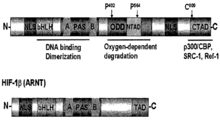
HIF-1 is heterodimeric protein consisting of HIF-la and HIF-ip subunits (10). While
HIF-ip is constitutively expressed as a subunit of several different heterodimers, expression
of HIF-la is tightly regulated by oxygen concentration (15). Both subunits contain a basic
helix-loop-helix (bHLH) motif and two Per-ARNT-Sim (PAS) domains designated PAS-A
and PAS-B (11) (Figure 1). The basic domain is essential for DNA binding, and the
HLH-PAS domain is required for dimerization of HIF-la and HIF-1(3 (16). HIF-1 heterodimer
binds to the consensus DNA sequences 5-RCGTG-3% which are located in the DNA major
groove (9).
HIF-la contains an oxygen-dependent degradation (ODD) domain, which is
responsible for oxygen-mediated regulation of HIF-la protein stability (17) (Figure 1).
When oxygen levels are sufficiently high, the conserved proline residues P402 and P564
within the ODD domain are modified by HIF prolyl hydroxylases (Figure 1), which trigger
VHL tumor suppressor protein-mediated ubiquitination and subsequent proteosomal
degradation (18). The well elucidated HIF/HIF prolyl hydroxylase/pVHL-mediated pathway
will be further described later in this chapter. A recent study showed that the HIF-la ODD
domain can bind to tumor suppressor p53 under physiological conditions, indicating the
existence of new potential regulatory mechanisms (19).
HIF-la also contains two transactivation domains, which are required for the
transcriptional regulation of HIF-1 targets (16). The N-terminal TAD is overlapped with the
ODD domain (Figure 1). Both TADs recruit transcriptional co-activators including
CBP/p300, Ref-1, SRC-1 and TIF-2 (14). It has been found that cysteine 800 in HIF-la is
4
HIF-1a
DNA binding Oxygen-dependent Dimerization degradation
p300/CBP,
SRC-1, Ref-1
HIF-1p{ARNT)
M =™r- --F sro™ =;
N- NL& bHLH A PAS 6 TADI-C
Figure 1. Protein domains of HIF-1 a and HIF-ip.
contains a TAD domain. However, the function of this domain on HIF-l's transcriptional
activity is unclear. Recruitment of p300/CBP to HIF-1 TAD domains is essential for
regulating HIF-1 transcriptional activity. I will discuss these regulatory mechanisms later in
the chapter.
HIF-la contains two nuclear localization signals (NLS). One is located at the
N-terminus within the bHLH domain, and one is located within the C-terminal region of
HIF-la (21) (Figure 1). Studies showed that the C-terminal NLS was functional, and a missense
mutation within this region could prevent the nuclear localization of GFP-HIF-la under
hypoxia (21). Later studies indicated that the functional C-terminal NLS of HIF-la is a
bipartite-type NLS, which is characterized by two sets of adjacent basic residues separated by
a spacer of about 10 amino acids (22). The nuclear translocation of HIF-la is independent of
the presence of HIF-ip (23). However, heterodimerization is required for nuclear
stabilization of both subunits (23). HIF-ip also contains a NLS signal at the N-terminus of
the protein, which causes HEF-ip to be expressed constitutively in the nucleus (Figure 1).
Additional mammalian HIFs
In addition to la, there are two other members in this family in mammals:
2a and 3a. 2a is also known as endothelial PAS domain protein 1 (EPAS1),
HIF-la-like factor (HLF), HIF-related factor (HRF), and member of PAS super-family 2 (MOP2)
(24). HIF-2a has high similarity with HIF-la in the main functional domains. Like HIF-la,
it exhibits oxygen-dependent regulation, dimerization with ARNT, DNA
recognization/binding, and activation of hypoxia-mediated gene expression (25). Little is
6
also regulated by ubiquitination and proteasome-mediated pathway for degradation (26). One
of the splice variants of HIF-3a, named IPAS (inhibitory PAS domain protein), was found to
inhibit the transcriptional activity of HIF-la by competing for binding with HIF-1(3 (27).
While HIF-la is expressed ubiquitously, both HIF-2a and HIF-3a are expressed in
more restricted, but partially overlapping, cell types. Studies of HIF-la and HIF-2a have
shown that the functions of these two proteins are not redundant. HIF-la knockout mouse
embryos died around midegestation with neural tube defects, cardiovascular malformations,
and marked cell death within the cephalic mesenchyme (28). Loss of function of HIF-2a
caused embryonic lethality in mouse with distinct defects in the cardiovascular system, such
as defects in catecholamine homeostasis (29) or aberrant vascular remodeling (30). Recent
studies also have shown that HIF-la and HEF-2a regulate overlapping but non-identical
target genes even in same cell types (31). Raval et al. (2005) reported that in renal carcinoma
cells, the protumorigenie genes encoding cyclin Dl, transforming growth factor alpha, and
vascular endothelial growth factor were specifically regulated by HEF-2a, but not HIF-la.
Also, the proapoptotic gene encoding BNip3 responded positively to HIF-la and negatively
to HIF-2a in renal carcinoma cells. Other studies showed that only overexpression of HIF-2a,
but not HIF-la, promotes growth of renal carcinoma cells (32).
Roles of HIF-1 in human cancers and therapeutic applications for targeting HIF-1
Under hypoxia, HIF-la dimerizes with HIF-1(3 and the heterodimer binds to core
binding site 5'-RCGTG-3' to activate transcription of target genes. The number of target
shown to be putative HIF-1 target genes in mammals. The HIF-1 target genes encode protein
products which are involved in angiogenesis/vascular remodeling, energy metabolism,
erythropoiesis, cell proliferation and viability, which makes HIF-1 a major mediator of
physiological and pathophysiological responses to hypoxia. HIF-1 has been shown to be
involved in many hypoxia characterized human diseases such as ischemic cardiovascular
disorders, pulmonary hypertension, and cancers (Reviewed in (4)). The role of HIF-1 in
tumor progression and therapeutic strategies for targeting HIF-1 will be described here.
Role of HIF-1 in tumor development
In solid tumors, rapid cell proliferation, severe structural abnormalities of tumor
microvessels, and disturbed microcirculation lead to a hypoxic microenvironment (33). Low
oxygen partial pressures in tumor cells correlates with resistance to radiotherapy and
chemotherapy (34). A critical molecular mechanism by which tumor cells adapt to hypoxic
microenviroments is to increase the level of HIF-1. Several lines of evidence reveal the
essential role of HIF-1 in tumor progression. First, HIF-la is overexpressed in a variety of
human cancers, including escophageal, brain, breast, lung, ovarian, cervical, and colon
cancers. In early-stage cervical carcinoma, HIF-la overexpression is correlated with patient
mortality (35). In oropharyngeal squamous cell carcinoma, the degree of HIF-la expression
is correlated with both radiation resistance and patient mortality (36). Second, HIF-1
activates the expression of target genes such as angiogenic signaling genes and glycolytic
enzyme genes which allow the tumor to adapt to hypoxia. Angiogenesis, the process by
which new blood vessels form from the existing vasculature, is critical for the growth of
tumors. New vascularization provides fresh nutrients and oxygen to rapidly proliferating
8
the angiogenic switch and induction of neovascularization at the early stage of tumorigenesis
(39). The ability to induce angiogenesis is associated with the rapid tumor growth and
metastasis (40). Also, tumor cells are generally characterized by low oxygen supply and high
glucose consumption rate, and anaerobic glycolysis becomes the major way for ATP
generation (25). HIF-1 promotes anaerobic glycolysis in tumor cells by activation of
metabolic enzymes such as aldolase A, phosphoglycerate kinase 1, and pyruvate kinase (41).
Third, tumor hypoxia renders a physiological stress that activates cell death pathways (42).
Meanwhile, hypoxia selects death-resistant cells, which in turn contribute to cancer
progression (43). HIF-1 can mediate hypoxic control of cell death in tumor growth.
Embryonic stem (ES) cells with inactive HIF-la genes (HIF-la v") have decreased
hypoxia-induced apoptosis (44). Induction of the cell death factors such as BNIP3 and NIX in human
tumors is HIF-1-dependent (45). IAP-2, an apoptosis inhibiting protein mediated by HIF-1,
has been shown to be strikingly induced by severe hypoxia in various types of cells (46).
Fourth, It has been reported that HIF-la expression and activity were affected by the tumor
suppressor gene p53. The p53 gene was found mutated in over 50 % of human cancers (47),
indicating its critical role in tumor suppression. Several studies have shown direct interaction
between HIF-la and p53 (48). Recent studies also further defined the direct interaction
between HIF-la ODD domain and p53 by using biophysical techniques (19). The interaction
of these two proteins supports the previous studies indicating that p53 can repress HIF-la
stimulated signaling by competing for binding to p300 (49), and can facilitate
Mdm2-dependent degradation of HIF-la (50).
The essential roles of HIF-1 for the adaptation to hypoxic tumor microenvironments
point to a promising therapeutic approach. Currently, a variety of anticancer drugs have been
reported to inhibit HIF-1 (Reviewed in (51)). In 1999, the Giovanni Melillo lab at the
National Cancer Institute began to screen for HIF-1 inhibitors. They successfully found that
the camptothecins, well known topoisomerase I inhibitors, could inhibit HIF-1 by a
topoisomerase I-dependent inhibition of translation (52). Currently, topotecan, an
FDA-approved semisynthetic camptothecin analogue, is in phase I human trials for treatment of
human cancers characterized by high HIF-1 levels such as ovarian cancer and small cell lung
cancer (53). Another promising HIF-1 inhibitor to enter the clinic is PX-478. PX-478
increases ubiquitination and degradation of HIF-la by an undefined mechanism. PX-478
shows potent antitumor effects with decreased expression of HIF-1 target genes such as
VEGF and glucose transporter-1, with tumor regression and long-term growth delay in mice
(54). There are also a number of other potential HIF-1 inhibitors, including Radicicol
(Hsp-90 inhibitor that reduces HIF-la DNA binding activity), FK228 (Histone deacetylase
inhibitor for that decreases HIF-1 transcription), and Rapamycin (PI-3-kinase pathway
inhibitor that reduces HIF-1 translation) (Reviewed in (53)). For the future, it is essential to
elucidate the functions of these small inhibitors on HIF-1 expression or transactivating
activity in tumors in vivo.
Pharmacological manipulation of HIF-la has promising clinical applications in
hypoxia characterized diseases. Fully understanding of regulation of HIF-1 expression and
activity would provide foundational knowledge for its therapeutic targeting.
10
pVHL tumor suppressor protein and ubiqutination of HIF-1
Von Hippel-Lindau (VHL) disease is a hereditary cancer syndrome characterized by
central nervous system and retinal hemangioblastomas, clear cell renal carcinoma, and
pheochromocytomas. It was first described in the medical literature in 1894 by Treacher
Collins. A decade later, Eugene von Hippel recorded observations of blood vessel tumors of
the retina in patients. Later, the Swedish pathologist Arvid Lindau reported that such patients
were at high risk of developing blood vessel tumors of the brain and spinal cord, known as
hemangioblastomas. In memory of these founders, the disease was called "von
Hipple-Lindau disease" in 1936. A variety of other tumors have been associated with VHL disease
including visceral cysts, clear-cell renal carcinomas, pheochromocytomas,
endolymphatic-sac tumors and islet-cell tumors of the pancreas (Reviewed in (55)).
The VHL gene was mapped on chromosome 3 in 1988 by Seizinger et al (56).
Individuals with VHL disease are VHL heterozygotes with one wild-type allele and one
defective allele (56). The VHL gene encodes two proteins (pVHL) due to alternative
translation initiation, and both isoforms retain tumor suppressor activity (57). Biochemical
studies revealed that pVHL was a component of a multiprotein complex including elongin B,
elongin C, Cul2, and Rbxl, which resemble so called SCF ubiquitin ligases (58). SCF
complexes target specific proteins for degradation by a post-translational modification called
polyubiquitylation (59). The potential function of pVHL promoting polyubiquitination was
supported by the fact that purified pVHL contained an ubiquitin ligase activity (60).
The inactivation of pVHL in tumors always causes the overexpression of
hypoxia-responsive genes such as vascular endothelial growth factor (VEGF) (61). In 1999, Maxwell
HIF-la can bind to each other (62). Very soon after Maxwell's observation, pVHL protein was
found to bind directly to the ODD domain of HIF-la via its beta-domain, which results in the
polyubiqutination and subsequently proteasomal degradation of HIF-la in the presence of
oxygen (63) (Figure 2). Thus, these studies proved the mechanistic explanation for
over-expression of hypoxia-response mRNAs in tumors lacking of pVHL.
Prolyl hydroxylation and discovery of EGL-9/PHD enzymes
Studies showed that the HIF-la ODD domain was sufficient to confer instability of
HIF-la in the presence of oxygen (64). In 2001, three groups independently showed that an
oxygen-dependent posttranslational modification was critical for interaction between pVHL
and HIF-la ODD domain, and for the ubiqutination and proteasomal degradation of HIF-la
under normoxic conditions (65-67). Their studies showed that the hydroxylation of
evolutionary conserved proline residues at the motif LXXLAP within the HIF-la ODD
domain resulted in binding of pVHL to HIF-la (Figure 2).
Soon after these great findings, another big breakthrough was made by Ratcliffe's
group at Oxford. They successfully identified HEF prolyl hydroxylases which are responsible
for hydroxylation of the conserved proline residues in HIF-la ODD by studying egl-9 gene
in the nematode C. elegans (68) (Figure 2).
First, Ratcliffe and colleagues demonstrated hydroxylation of the LXXLAP motif in C.
elegans HIF-1 allows direct binding of VHL-1, the ortholog of VHL. Then they isolated
mutant worms carrying a deletion in the vhl-1 gene, and they found that the VHL-1-deficient
worms expressed high level of HIF-1 at both normoxia and hypoxia. Secondly, they
12
Cellular
Oxygen Concentration
HIF-1 a"
HIF-1 a HIF-1 a
Degradation
ÇcôactjT^
RCGlW
Hypoxia adaptation
Î
Protein
mRNA 3'
series of mutant strains. They found that animals carrying loss-of-function mutations in the
egl-9 gene failed to downregulate HIF-1 expression in normoxic conditions. Additional
experiments demonstrated that the EGL-9 protein acted directly on HIF-1 to hydroxylate the
proline in the LXXLAP motif in vitro. C. elegans gene egl-9 was originally identified from
the genetic screen for egg-laying-defective phenotypes (69). Later studies showed that egl-9
encode a protein belonging to the iron and 2-oxoglutarate-dependent dioxygenase
superfamily (70). Finally, through secondary structure prediction algorithms, Ratcliffe and
colleagues identified mammalian homologs of EGL-9 in the same dioxygenase superfamily
(termed as prolyl hydroxylase domain (PHD) 1, PHD2, & PHD3) and demonstrated that the
mammalian PHDs hydroxylate human HIF-la or HCF-2a in vitro. Soon after Ratcliffe and
colleagues' studies, mammalian HIF prolyl hydroxylases were also identified by other groups
using biochemical technologies, which were named as EglN or HPH proteins (71,72).
Regulation of PHD enzymes
Since the discovery that the HIF/PHD/pVHL pathway was the central mechanism for
regulating HIF-la protein degradation, extensive studies have showed that factors
regulations of PHD enzymes have essential roles for hypoxia signaling in organisms
(Reviewed in (73)). Major known regulators of PHD enzymes will be summarized here.
PHD enzymes use oxygen as their co-substrate. Studies using VHL capture by a short
HIF-la polypeptide as a measure of hydroxylation have demonstrated that PHD enzyme
activity was greatly affected by oxygen concentration in vitro (68). Studies also confirmed
the direct incorporation of oxygen into the HIF-la substrate by monitoring the incorporation
of 180 into the sites of prolyl hydroxylation by mass spectrometry (74). In keeping with this,
values for molecular oxygen in the range 230-250 pM (75). Nitric oxide (NO) also has been
shown to cause accumulation of HIF under normoxic conditions (76,77). One explanation is
the ability of NO to inhibit 20G-dependent oxygenases by acting as an analogue of
molecular oxygen (78).
Studies also have shown that multiple factors affect the availability of other
co-substrates such as Fe (II) and 2-oxoglutarate (20G), thereby regulating the activities of PHD
enzymes. Studies showed molecules that reduce or inhibit Fe(II), such as iron chelators and
metals such as Co(II), Ni(II), and Mn(II), can enhance HIF-lot levels (68). It remains to be
elucidated whether changes in cellular iron availability play a physiological role in the
regulation of PHD enzymes. Several studies have demonstrated that cellular level of reactive
oxygen species (ROS) has roles in regulating HIF-1 activity (Reviewed in (79)). A recent
study provided the evidence that junD reduces ROS levels as part of a defense against
oxidative stress (80). PHD2 activity was found to be reduced in junD deficient cells, and the
potential mechanism involved Fenton's reaction: the conversion of Fe(II) to Fe(III) by
elevated ROS levels (80). The citric cycle intermediate 20G is also a co-substrate for PHD
enzymes. Levels of other citric cycle intermediates such as succinate or fumarate have been
shown to affect PHD enzymes activities, as they are competitive inhibitors of 20G (81).
Molecules that inhibit PHDs would be predicted to be tumorigenic. Indeed, certain tumors
have defects in succinate dehydrogenase (subunits B, C or D), such as hereditary paraglioma.
Other tumors have defects in fumarate hydratase, such as leiomyomata of the skin, uterine
fibroids and papillary renal cell carcinoma. In these cancers, levels of succinate and fumarate
are increased, and this appears to inhibit the PHD enzymes activities as competitive
PHD associated proteins could also play roles in the regulation of these enzymes. The
ring-finger E3 ligases Siahla/2 have been found to associate with PHD3 in
immunoprecipitates (82). In Siahla/2 deficient cells PHD3 is highly expressed, and HIF-1 is
downregulated. Also, in Siah2 deficient mice, hypoxia-induced gene expression is defective.
Using the yeast two-hybrid system, the protein OS9 has been found to interact with PHD2
(83). Interaction with OS9 enhanced the activity of PHD2 and downregulated the HIF-la
protein level. In a recent study, the tumor suppressor ING4 was found to interact with PHD2,
and unexpectedly, the interaction did not affect HIF-la stability, but HIF-la transactivation
(84). The underlying mechanism is unclear.
Both PHD2 and PHD3 have been shown to be transcriptionally regulated by HIF,
forming a negative feedback loop (85). The induction of PHD2 and PHD3 in hypoxia
contributes to the enhanced rates of HIF degradation during reoxygenation (86). Studies also
showed that PHD2 is the most abundant PHD enzyme under normoxia, and it plays
important roles for setting normoxic levels of HIF-la (87). PHD3 plays important roles for
setting hypoxic levels of HIF-la (87). These studies further indicate that the oxygen level
determines the function of PHD enzymes.
Other regulatory mechanisms of HIF-1 activity
Organisms also use a number of other hypoxia-dependent and -independent strategies
to regulate HIF-1 activity at multiple levels, including translation, protein stabilization,
posttranslational modifications, nuclear localization, DNA binding capacity, and
transcriptional co-activator recruitment (Reviewed in (25)). Some mechanisms related to
HIF-la protein level
The well-characterized VHL-mediated ubiquitin-proteasome pathway as described
above plays essential roles for HIF-1 degradation. Several post-translational modifications
have been shown to regulate the interaction between pVHL and HIF-la, including
acetylation and phosphorylation. Acetyltransferase ARD1 aceylates lysine residue in the
HIF-la ODD domain, which results in the enhancement of the interaction between HIF-la
and E3-ligase pVHL (88). Researchers have had difficulty repeating this observation, so this
finding remains controversial. In human pancreatic cancer cells, p38 MAPK-mediated
phosphorylation of HIF-la contributed to the inhibition of HIF-la and pVHL interaction
(89).
Other potential VHL-independent degradation pathways maybe involved in HIF-1
protein stability (90). The nuclear expression of forkhead transcription factor F0X04 has
been shown to suppress the hypoxia response by down-regulation of HIF-1 a protein level.
F0X04 inhibition of HIF-1 appears to involve in a proteasome-dependent degradation
pathway which is independent of VHL-mediated function (90). A recent study showed that
hi stone deacetylase inhibitors produced a dose- and time-dependent inhibition of
accumulated la levels (91). Histone deacetylase inhibitors-induced degradation of
HIF-la was reported to be ubiquitination independent and mediated by a proteasome system
which was independent of VHL function. These studies further suggested that histone
deacetylse 6 (HDAC6) mediated hyperacetylation of Hsp90, which affected HIF-la
stabilization. Another recently published study showed the existence of
independent mechanism (92). Demidenko et al. showed that inhibition of transcription using
transcription inhibitors had no effect on HIF-la induction under normoxia, but caused
super-induction of HIF-la under hypoxia, indicating that accumulation of HIF-la under hypoxia
could transcriptionally activates a feedback loop for the degradation of HIF-la.
There is growing evidence that multiple cellular signaling pathways can lead to the
induction of HIF-1 in oxygen-independent manner (Reviewed in (3)). HER2 activation
increases the rate of HIF-la protein synthesis via phosphatidylinositol 3-kinase (PI3K) and
the downstream serine-threonine kinases AKT (protein kinase B) and mTOR (mammalian
target of rapamycin) (93). IGF-1 enhanced the HIF-la protein synthesis, and this was
dependent upon both the PI3K and MAPK pathways (94). Current models suggest that
mTOR phosphorylates and activates the translational regulatory protein p70 S6 kinase, which
phosphorylates the 40S ribosomal protein S6, resulting in the elevated HIF-la protein
translation.
Transcriptional co-activator recruitment
The HIF-1 complex recruits a number of coactivators, including p300/CBP, Ref-1,
Jabl, SCR-1 and TIF2, to transactivate the expression of a multitude of targets genes
(Reviewed in (25)). The regulation of the interaction between p300/CBP and HIF-la has
great impact for HIF-1 transactivation under hypoxia. Oxygen-dependent asparagine
hydroxylation has a critical role in this. Under normoxia, an asparagine residue (Asn-803) in
the CTAD domain of HIF-la is hydroxylated by an asparaginyl hydroxylase enzyme called
FIH (factor-inhibiting HIF). Like the PHDs, FIH belongs to the oxygen and iron dependent
18
CBP/p300, thereby inhibiting transcriptional activity of HIF-1 (95). Under hypoxia, the
enzymatic activity of FIH is inhibited, which enables the recruitment of CBP/p300 to HIF-1.
There are also number of other mechanisms involved in the interaction of p300/CBP and
HIF-la. Hypoxia-induced CITED2/p35srj and CITED4 proteins have shown to bind to
CBP/p300, and they down-regulate HIF-1 transactivation by blocking the interaction
between HIF-la and p300 (96). The phosphorylation of p300 by MAPK increased the
interaction between the HIF-la C-TAD and p300 (97). The p53 tumor suppressor could
repress HIF-la activity by competing co-activator p300 binding (49).
Important unanswered questions about HIF regulation
The HIF hydroxylases appear to function as oxygen sensors, as the PHD and FIH
enzymes require molecular oxygen. Important questions remaining to be answered include: 1)
Are these potential oxygen sensors involved in other non-HIF-mediated hypoxia sensitive
signaling pathways? 2) Recent studies showed the potential roles of PHDs on HIF-la
transactivation, in addition to HIF-la protein stabilization. What are the molecular
mechanisms underlying the VHL-independent functions of PHD enzymes? 3) The relation of
in vitro characteristics of these PHD enzymes with their physiological functions in intact
organisms needs the further elucidation. It's important to answer the question of whether
pharmacological inhibition of the HIF hydroxylases is an effective therapeutic strategy in
ischaemic/hypoxic disease.
In addition to HIF hydroxylases-regulated oxygen sensing machinery, there are many
expression. This suggests the existence of additional pathways that signal changes in the
cellular oxygen concentration. The big questions raised are: 1) Are there other oxygen
sensors regulating HIF-1 activity, and what are these potential oxygen sensors? 2) How are
these multiple regulatory mechanisms integrated to generate the appropriate physiological
response, and how do these pathways impact the development of HIF-targeted therapeutic
strategies in clinic?
VHL-dependent ubiquitination and proteasomal degradation is a central mechanism
for regulation of HIF-la protein stability. However, there are several reports showing the
existence of VHL- independent degradation pathways. The underlying mechanisms are less
understood. The questions raised are: 1) What are the important proteins mediating
degradation of HIF-1 in a VHL-independent manner? 2) Is there condition specificity
(normoxia or hypoxia) and/or tissue specificity for these degradation pathways, and what is
the relationship with the VHL-mediated pathway?
Hypoxia response in C. elegans
The genetic model system C. elegans
The nematode C. elegans is a powerful animal model system for the dissection of
evolutionary conserved signaling pathways. C. elegans are small (~1 mm in length as
adults), and they can be cultured on agar plates with a bacterial food source. There are two
sexes, self-fertilizing hermaphrodites and males, and the generation time is less than 4 days at
20°C. The simplicity and experimental convenience of C. elegans make it an attractive model
organism for genetic analyses. The sequence of the ~108 bp genome was completed in 1998,
20
increased this estimate (99). Large scale studies, such as double-stranded RNA interference
library and full-genome microarrays, are available to worm researchers, and are
tremendously helpful to studying gene functions in C. elegans.
In the wild, C. elegans inhabits the soil, where it can encounter hypoxic
microenvironments. C. elegans is able to maintain a near normal metabolic rate at
environmental oxygen concentrations as low as 2%. In 0.5% or 1% oxygen, the animals
must decrease oxygen consumption, but they continue to develop and reproduce (100). C.
elegans does not have a complex circulatory system. Any cell in the organism is only a few
cell widths from the outer surface of the worm or the intestinal lumen (101), and oxygen
delivery is thought to be accomplished by diffusion. Thus, individual cells must sense and
adapt to local environmental oxygen levels. Recent discoveries have revealed that the
molecular mechanisms that govern transcriptional responses to hypoxia are, at least in part,
conserved between C. elegans and humans (6).
Evolutionarily conserved hypoxia signaling pathway in C. elegans
The C. elegans hif-1 gene is orthologous to mammalian HIF alpha subunits (6). Like
its mammalian cognates, C. elegans HIF-1 protein is induced by hypoxia and is rapidly
degraded upon re-oxygenation (6,68). hif-1 mRNA levels are not dramatically affected by
oxygen concentration (6). Under conditions in which C. elegans HIF-1 is stable, it binds
AHA-1, the ortholog of mammalian ARNT/HIF-beta (102) to form a complex that can bind
DNA sequences containing the hypoxic regulatory element (6). Both HIF-1 and AHA-1 are
expressed in most, if not all, cells, as assayed by a hif-1:GFP reporter gene and
evolutionary conserved regulatory network that senses hypoxia and implements appropriate
transcriptional changes.
Hif-1 is required for adaptation to hypoxia
The hif-1 (ia04) mutation is predicted to be a strong loss-of-function allele. It deletes
exons 2-4 of hif-1, and it introduces an early translational stop codon to the most abundant
forms of hif-1 mRNA (6). Animals that are homozygous for the hif-1 mutation exhibit no
visible defects under standard laboratory conditions. However, hif-1-defective animals are
unable to adapt to 0.5% or 1% oxygen. While wild type animals survive and reproduce in 1%
oxygen, 66% of hif-1-defective animals do not survive embryogenesis in these conditions,
and an additional 9% die during larval development (6,103). In a recent genetic screen, I
discovered a new hif-1 (ia07) allele, in which the point mutation C824T causes a premature
stop codon. hif-l(ia07) mutants expresse a truncated HIF-1 protein without the conserved
ODD domain and putative TAD domain. 67% of hif-l(ia07) worms do not survive
embryogenesis under 0.5% oxygen condition (unpublished data). As described in Chapter 2,
genome-wide studies have shown that hif-1 regulates the majority of early transcriptional
responses to hypoxia in C. elegans (7). Certain HIF-1 targets genes also play critical roles
for survival in hypoxic condition (7).
The high degree of conservation of the HIF- 1/EGL-9/VHL-1 system indicates that
genetic studies in C. elegans should be highly informative in studying the upstream
regulators of HIF-1, and potentially the downstream physiological functions of HIF-1. This
knowledge would have foundational roles for developing therapeutic strategies, such as drugs
22
that low molecular weight inhibitors of EGL-9/HIF prolyl hydroxylase are promising
pharmacological agents (72,104-106).
Dissertation Organization
Our research group and others have reported that C. elegans hif-1 and HIF-1 signaling
pathways are evolutionary conserved (6,68). In this dissertation, I examine the
hif-1-dependent/independent gene expression changes during hypoxia response in worms using
genome-wide DNA microarray analyses. Also I identify a novel membrane regulator of
HIF-1, which suppresses HIF-1 activity by a potential VHL-1 -independent feedback loop.
In Chapter 1, literature related to the study is reviewed. Major topics include: (i)
Description of mammalian hypoxia-inducible factors, including the historical discovery and
molecular cloning of HIF-1 alpha and beta subunits, structure and function of HIF-1, and
additional HIFs. (ii) Involvement of HIF-1 in human cancers and therapeutic applications for
HIF-1 inhibitors, (iii) Regulation of HIF stability by PHD/pVHL pathway, including
historical overview of pVHL discovery, prolyl hydroxylation and EGL-9 discovery. Current
studies involved in regulations of HIF prolyl hydroxylases are also summarized, (iv) Other
regulatory mechanisms of HIF-1 activity, with emphasis on regulations of HIF-la protein
stability and transcriptional co-activator recruitment, (v) Hypoxia response in C. elegans. I
introduce the genetic model organism C. elegans, and describe evidence that hif-1 is
evolutionary required for hypoxia response in worms.
Chapter 2 is a copy of a research paper published in The Journal of Biological
Chemistry, 2005, 280(21): 20580-20588. The title is "Roles of the HIF-1 hypoxia-inducible
hif-1 -dependent and ^^-independent manners in worms. The majority of hypoxia regulated
gene expression changes were dependent upon hif-1 function, (ii) Description of functions of
hypoxia-responsive genes with emphasis on discussion of genes encoding metabolic enzymes,
signaling molecules, extracellular matrices proteins, ubiquitin ligase, transcriptional factors
and heatshock proteins, (iii) Identification of essential functions for individual
hif-1-dependent genes. In particular, gene phy-2 is required for hypoxia survival, (iv) Molecular
links between hypoxia response and dauer formation, (v) VHL-1 regulates HIF-1 function
and also has HIF-1 independent functions. Other researchers were also involved in this study.
As principal investigator, Jo Anne Powell-Coffman contributed to experimental design and
manuscript editing. Min Jiang and Stuart Kim at Stanford University performed microarray
hybridizations. Daniel Nettleton at Iowa State University analyzed the microarray data by
writing statistical programs. Qunfeng Dong at Iowa State University helped generate Figure
2, which shows genomic distribution of hypoxia-responsive genes.
Chapter 3 is a copy of a manuscript to be submitted for publication. The title is "The
RHY-1 integral membrane protein suppresses HIF-1 hypoxia-inducible factor activity in
Caenorhabditis elegans in a negative feedback loop that does not include VHL-1". The
major findings includes: (i) Generation of a visual assay system for studying HIF-1 activity.
By using the promoter of HIF-1 target gene nhr-57 fused to GFP as a reporter, I could detect
the enhanced expression of the reporter under hypoxic condition and VHL-1 loss-of-function
background, (ii) EGL-9 has VHL-1-independent function in which it regulates HIF-1
transcriptional activity, rather than HIF-1 protein stabilization, (iii) Identification of rhy-1,
24
regulator of HIF-1. My studies show that rhy-1 functions independent of VHL-1 to inhibit
HIF-1 activity. My genetic data also suggest that RHY-1 and EGL-9 could function at the
same pathway, (iv) Study of RHY-1 expression pattern. My data show that RHY-1 is
expressed mainly in intestine and head sensory neurons in worms, as well as weaker
expression in hypodermis, socket cells, and vuval muscles. For this work, former lab member
Kelly Gillette and Mae Young isolated and mapped rhy-1 ia38 allele. Zhiyong Shao, a PhD
graduate student in the lab, generated the rhy-1 promoter fused with W07A12.6 coding
sequence construct. He also did W07A12.6 RNAi experiment. Jo Anne Powell-Coffman was
the principal investigator.
In Chapter 4,1 summarize the major findings in my thesis research, with emphasis on
impact and contribution to the field. In brief, I show that C. elegans hif-1 regulates the
majority of hypoxia-induced gene expression changes, and certain HIF-1 target genes are
critical for hypoxia survival. My study also clearly indicates the existence of
hif-1-independent pathway(s) for hypoxia adaptation. I show that EGL-9 has functions regulating
HIF-1 transcriptional activity, rather than HIF-1 protein stabilization in worms. I identify
gene rhy-1 as a novel negative regulator of HIF-1 by suppression of HIF-1 transcriptional
activity in a VHL-1-independent mechanism.
References
1. Hopkins, S. R., and Powell, F. L. (2001) Adv Exp Med Biol 502, 153-167
2. Cummins, E. P., and Taylor, C. T. (2005) Pflugers Arch 450, 363-371
3. Semenza, G. (2002) Biochem Pharmacol 64, 993-998
5. Semenza, G. L. (2003) Nat Rev Cancer 3, 721-732
6. Jiang, H., Guo, R., and Powell-Coffman, J. A. (2001) Proc Natl Acad Sci USA 98,
7916-7921
7. Shen, C., Nettleton, D., Jiang, M., Kim, S. K., and Powell-Coffman, J. A. (2005) J
Biol Chem 280, 20580-20588
8. Semenza, G. L., and Wang, G. L. (1992) Mol Cell Biol 12, 5447-5454.
9. Wang, G. L., and Semenza, G. L. (1993) J Biol Chem 268, 21513-21518
10. Wang, G. L., and Semenza, G. L. (1995) J Biol Chem 270, 1230-1237
11. Wang, G. L., Jiang, B. H., Rue, E. A., and Semenza, G. L. (1995) Proc Natl Acad Sci
USA92, 5510-5514.
12. Hoffman, E. C., Reyes, H., Chu, F. F., Sander, F., Conley, L. H., Brooks, B. A., and
Hankinson, O. (1991) Science 252, 954-958
13. Li, H., Ko, H. P., and Whitlock, J. P. (1996) J Biol Chem 271, 21262-21267
14. Aran y, Z., Huang, L. E., Eckner, R., Bhattacharya, S., Jiang, C., Goldberg, M. A.,
Bunn, H. F., and Livingston, D. M. (1996) Proc Natl Acad Sci USA 93,
12969-12973.
15. Jiang, B. H., Semenza, G. L., Bauer, C., and Marti, H. H. (1996) Am J Physiol 271,
Cl 172-1180
16. Jiang, B. H., Rue, E., Wang, G. L., Roe, R., and Semenza, G. L. (1996) J Biol Chem
271, 17771-17778
17. Huang, L. E., Gu, J., Schau, M., and Bunn, H. F. (1998) Proc Natl Acad Sci U S A 95,
26
18. Cockman, M. E., Masson, N., Mole, D. R., Jaakkola, P., Chang, G. W., Clifford, S. C.,
Maher, E. R., Pugh, C. W., Ratcliffe, P. J., and Maxwell, P. H. (2000) J Biol Chem
275, 25733-25741.
19. Sanchez-Puig, N., Veprintsev, D. B., and Fersht, A. R. (2005) Mol Cell 17, 11-21
20. Ema, M., Hirota, K., Mimura, J., Abe, H., Yodoi, J., Sogawa, K., Poellinger, L., and
Fujii-Kuriyama, Y. (1999) Embo J18, 1905-1914
21. Kallio, P. J., Okamoto, K., O'Brien, S., Carrero, P., Makino, Y., Tanaka, H., and
Poellinger, L. (1998) Embo J17, 6573-6586
22. Luo, J. C., and Shibuya, M. (2001) Oncogene 20, 1435-1444
23. Chilov, D., Camenisch, G., Kvietikova, I., Ziegler, U., Gassmann, M., and Wenger, R.
H. (1999) J Cell Sci 112 ( Pt 8), 1203-1212
24. Semenza, G. L. (1999) Annu Rev Cell Dev Biol 15, 551-578
25. Wenger, R. H. (2002) Faseb J16, 1151-1162
26. Maynard, M. A., Qi, H., Chung, J., Lee, E. H., Kondo, Y., Hara, S., Conaway, R. C.,
Conaway, J. W., and Ohh, M. (2003) J Biol Chem 278, 11032-11040
27. Makino, Y., Cao, R., Svensson, K., Bertilsson, G., Asman, M., Tanaka, H., Cao, Y.,
Berkenstam, A., and Poellinger, L. (2001) Nature 414, 550-554
28. Iyer, N. V., Kotch, L. E., Agani, F., Leung, S. W., Laughner, E., Wenger, R. H.,
Gassmann, M., Gearhart, J. D., Lawler, A. M., Yu, A. Y., and Semenza, G. L. (1998)
Genes Dev 12, 149-162
29. Tian, H., Hammer, R. E., Matsumoto, A. M., Russell, D. W., and McKnight, S. L.
30. Peng, J., Zhang, L., Drysdale, L., and Pong, G. H. (2000) Proc Natl Acad Sci USA
97, 8386-8391
31. Wang, V., Davis, D. A., Haque, M., Huang, L. E., and Yarchoan, R. (2005) Cancer
Res 65, 3299-3306
32. Kondo, K., Klco, J., Nakamura, E., Lechpammer, M., and Kaelin, W. G., Jr. (2002)
Cancer Cell 1, 237-246
33. Hockel, M., and Vaupel, P. (2001) J Natl Cancer Inst 93, 266-276.
34. Brown, J. M. (1999) Cancer Res 59, 5863-5870.
35. Bimer, P., Schindl, M., Obermair, A., Plank, C., Breitenecker, G., and Oberhuber, G.
(2000) Cancer Res 60, 4693-4696
36. Aebersold, D. M., Burn, P., Beer, K. T., Laissue, J., Djonov, V., Greiner, R. H., and
Semenza, G. L. (2001) Cancer Res 61, 2911-2916.
37. Carmeliet, P., and Jain, R. K. (2000) Nature 407, 249-257.
38. Forsythe, J. A., Jiang, B. H., Iyer, N. V., Agani, P., Leung, S. W., Koos, R. D., and
Semenza, G. L. (1996) Mol Cell Biol 16, 4604-4613.
39. Fang, J., Yan, L., Shing, Y., and Moses, M. A. (2001) Cancer Res 61, 5731-5735.
40. Hanahan, D., and Folkman, J. (1996) Cell 86, 353-364
41. Semenza, G. L., Jiang, B. H., Leung, S. W., Passantino, R., Concordet, J. P., Maire, P.,
and Giallongo, A. (1996) J Biol Chem 271, 32529-32537
42. Shimizu, S., Eguchi, Y., Kamiike, W., Itoh, Y., Hasegawa, J., Yamabe, K., Otsuki, Y.,
Matsuda, H., and Tsujimoto, Y. (1996) Cancer Res 56, 2161-2166.
43. Graeber, T. G., Osmanian, C., Jacks, T., Housman, D. E., Koch, C. J., Lowe, S. W.,
44. Carmeliet, P., Dor, Y., Herbert, J. M., Fukumura, D., Brusselmans, K., Dewerchin,
M., Neeman, M., Bono, P., Abramovitch, R., Maxwell, P., Koch, C. J., Ratcliffe, P.,
Moons, L., Jain, R. K., Collen, D., Keshert, E., and Keshet, E. (1998) Nature 394,
485-490.
45. Sowter, H. M., Ratcliffe, P. J., Watson, P., Greenberg, A. H., and Harris, A. L. (2001)
Cancer Res 61, 6669-6673.
46. Dong, Z., Venkatachalam, M. A., Wang, J., Patel, Y., Saikumar, P., Semenza, G. L.,
Force, T., and Nishiyama, J. (2001) J Biol Chem 276, 18702-18709.
47. Greenblatt, M. S., Bennett, W. P., Hollstein, M., and Harris, C. C. (1994) Cancer Res
54,4855-4878
48. An, W. G., Kanekal, M., Simon, M. C., Maltepe, E., Blagosklonny, M. V., and
Neckers, L. M. (1998) Nature 392, 405-408
49. Blagosklonny, M. V., An, W. G., Romanova, L. Y., Trepel, J., Fojo, T., and Neckers,
L. (1998)75/0/ Chem 273, 11995-11998.
50. Ravi, R., Mookerjee, B., Bhujwalla, Z. M., Sutter, C. H., Artemov, D., Zeng, Q.,
Dillehay, L. E., Madan, A., Semenza, G. L., and Bedi, A. (2000) Genes Dev 14,
34-44.
51. Powis, G., and Kirkpatrick, L. (2004) Mol Cancer Ther 3, 647-654
52. Rapisarda, A., Uranchimeg, B., Sordet, O., Pommier, Y., Shoemaker, R. H., and
Melillo, G. (2004) Cancer Res 64, 1475-1482
53. Giaccia, A., Siim, B. G., and Johnson, R. S. (2003) Nat Rev Drug Discov 2, 803-811
54. Welsh, S., Williams, R., Kirkpatrick, L., Paine-Murrieta, G., and Powis, G. (2004)
56. Seizinger, B. R., Rouleau, G. A., Ozelius, L. J., Lane, A. H., Farmer, G. E., Lamiell, J.
M., Haines, J., Yuen, J. W., Collins, D., Majoor-Krakauer, D., and et al. (1988)
Nature 332, 268-269
57. Schoenfeld, A., Davidowitz, E. J., and Burk, R. D. (1998) Proc Natl Acad Sci USA
95,8817-8822
58. Kibel, A., Uiopoulos, O., DeCaprio, J. A., and Kaelin, W. G., Jr. (1995) Science 269,
1444-1446
59. Deshaies, R. J. (1999) Annu Rev Cell Dev Biol 15, 435-467
60. Pause, A., Lee, S., Worrell, R. A., Chen, D. Y., Burgess, W. H., Linehan, W. M., and
Klausner, R. D. (1997) Proc Natl Acad Sci U S A94, 2156-2161
61. Wizigmann-Voos, S., Breier, G., Risau, W., and Plate, K. H. (1995) Cancer Res 55,
1358-1364
62. Maxwell, P. H., Wiesener, M. S., Chang, G. W., Clifford, S. C., Vaux, E. C.,
Cockman, M. E., Wykoff, C. C., Pugh, C. W., Maher, E. R., and Ratcliffe, P. J. (1999)
Nature 399, 271-275
63. Ohh, M., Park, C. W., Ivan, M., Hoffman, M. A., Kim, T. Y., Huang, L. E., Pavletich,
N., Chau, V., and Kaelin, W. G. (2000) Nat Cell Biol 2, 423-427
64. Pugh, C. W., O'Rourke, J. F., Nagao, M., Gleadle, J. M., and Ratcliffe, P. J. (1997) J
Biol Chem 272, 11205-11214
65. Ivan, M., Kondo, K., Yang, H., Kim, W., Valiando, J., Ohh, M., Salic, A., Asara, J.
30
66. Jaakkola, P., Mole, D. R., Tian, Y. M., Wilson, M. I., Gielbert, J., Gaskell, S. J.,
Kriegsheim, A., Hebestreit, H. F., Mukherji, M., Schofield, C. J., Maxwell, P. H.,
Pugh, C. W„ and Ratcliffe, P. J. (2001) Science 292,468-472.
67. Yu, F., White, S. B., Zhao, Q., and Lee, F. S. (2001) Cancer Res 61, 4136-4142.
68. Epstein, A. C., Gleadle, J. M., McNeill, L. A., Hewitson, K. S., O'Rourke, J., Mole, D.
R., Mukherji, M., Metzen, E., Wilson, M. I., Dhanda, A., Tian, Y. M., Masson, N.,
Hamilton, D. L., Jaakkola, P., Barstead, R., Hodgkin, J., Maxwell, P. H., Pugh, C. W.,
Schofield, C. J., and Ratcliffe, P. J. (2001) Cell 107, 43-54
69. Trent, C., Tsuing, N., and Horvitz, H. R. (1983) Genetics 104, 619-647.
70. Aravind, L., and Koonin, E. V. (2001) Genome Biol 2
71. Bruick, R. K., and McKnight, S. L. (2001) Science 294, 1337-1340
72. Ivan, M., Haberberger, T., Gervasi, D. C., Michelson, K. S., Gunzler, V., Kondo, K.,
Yang, H., Sorokina, I., Conaway, R. C., Conaway, J. W., and Kaelin, W. G., Jr. (2002)
Proc Natl Acad Sci USA 99, 13459-13464
73. Schofield, C. J., and Ratcliffe, P. J. (2005) Biochem Biophys Res Commun 338,
617-626
74. McNeill, L. A., Hewitson, K. S., Gleadle, J. M., Horsfall, L. E., Oldham, N. J.,
Maxwell, P. H., Pugh, C. W., Ratcliffe, P. J., and Schofield, C. J. (2002) Bioorg Med
Chem Lett 12, 1547-1550
75. Hirsila, M., Koivunen, P., Gunzler, V., Kivirikko, K. I., and Myllyharju, J. (2003) J
Biol Chem 278, 30772-30780
77. Sandau, K. B., Zhou, J., Kietzmann, T., and Brune, B. (2001) J Biol Chem 276,
39805-39811
78. Wang, F., Sekine, H., Kikuchi, Y., Takasaki, C., Miura, C., Heiwa, O., Shuin, T.,
Fujii-Kuriyama, Y., and Sogawa, K. (2002) Biochem Biophys Res Commun 295,
657-662
79. Kietzmann, T., and Gorlach, A. (2005) Semin Cell Dev Biol 16, 474-486
80. Gerald, D., Berra, E., Frapart, Y. M., Chan, D. A., Giaccia, A. J., Mansuy, D.,
Pouyssegur, J., Yaniv, M., and Mechta-Grigoriou, F. (2004) Cell 118, 781-794
81. Selak, M. A., Armour, S. M., MacKenzie, E. D., Boulahbel, H., Watson, D. G.,
Mansfield, K. D., Pan, Y., Simon, M. C., Thompson, C. B., and Gottlieb, E. (2005)
Cancer Cell 7, 77-85
82. Nakayama, K., Frew, I. J., Hagensen, M., Skals, M., Habelhah, H., Bhoumik, A.,
Kadoya, T., Erdjument-Bromage, H., Tempst, P., Frappell, P. B., Bowtell, D. D., and
Ronai, Z. (2004) Cell 117, 941-952
83. Baek, J. H., Mahon, P. C., Oh, J., Kelly, B., Krishnamachary, B., Pearson, M., Chan,
D. A., Giaccia, A. J., and Semenza, G. L. (2005) Mol Cell 17, 503-512
84. Ozer, A., Wu, L. C., and Bruick, R. K. (2005) Proc Natl Acad Sci USA 102,
7481-7486
85. del Peso, L., Castellanos, M. C., Ternes, E., Martin-Puig, S., Cuevas, Y., Olmos, G.,
and Landazuri, M. O. (2003) J Biol Chem 278,48690-48695
86. Berra, E., Richard, D. E., Gothie, E., and Pouyssegur, J. (2001) FEBS Lett 491, 85-90
87. Appelhoff, R. J., Tian, Y. M., Raval, R. R., Turley, H., Harris, A. L., Pugh, C. W.,
32
88. Jeong, J. W., Bae, M. K., Ahn, M. Y., Kim, S. H., Sohn, T. K., Bae, M. H., Yoo, M.
A., Song, E. J., Lee, K. J., and Kim, K. W. (2002) Cell 111, 709-720
89. Kwon, S. J., Song, J. J., and Lee, Y. J. (2005) Clin Cancer Res 11, 7607-7613
90. Tang, T. T., and Lasky, L. A. (2003) J Biol Chem 278, 30125-30135
91. Kong, X., Lin, Z., Liang, D., Path, D., Sang, N., and Caro, J. (2006) Mol Cell Biol 26,
2019-2028
92. Demidenko, Z. N., Rapisarda, A., Garayoa, M., Giannakakou, P., Melillo, G., and
Blagosklonny, M. V. (2005) Oncogene 24, 4829-4838
93. Laughner, E., Taghavi, P., Chiles, K., Mahon, P. C., and Semenza, G. L. (2001) Mol
Cell Biol 21, 3995-4004
94. Fukuda, R., Hirota, K., Fan, F., Jung, Y. D., Ellis, L. M., and Semenza, G. L. (2002) J
Biol Chem 277, 38205-38211
95. Lando, D., Peet, D. J., Gorman, J. J., Whelan, D. A., Whitelaw, M. L., and Bruick, R.
K. (2002) Genes Dev 16, 1466-1471.
96. Bhattacharya, S., Michels, C. L., Leung, M. K., Arany, Z. P., Kung, A. L., and
Livingston, D. M. (1999) Genes Dev 13, 64-75
97. Sang, N., Stiehl, D. P., Bohensky, J., Leshchinsky, I., Srinivas, V., and Caro, J. (2003)
J Biol Chem 278, 14013-14019
98. (1998) Science 282, 2012-2018
99. Lim, L. P., Lau, N. C., Weinstein, E. G., Abdelhakim, A., Yekta, S., Rhoades, M. W.,
Burge, C. B., and Bartel, D. P. (2003) Genes Dev 17, 991-1008
64-119
102. Powell-Coffman, J. A., Bradfield, C. A., and Wood, W. B. (1998) Proc Natl Acad Sci
U S A 95, 2844-2849.
103. Padilla, P. A., Nystul, T. G., Zager, R. A., Johnson, A. C., and Roth, M. B. (2002)
Mol Biol Cell 13, 1473-1483
104. Knowles, H. J., Tian, Y. M., Mole, D. R., and Harris, A. L. (2004) Circ Res 95,
162-169
105. Ratan, R. R., Siddiq, A., Aminova, L., Lange, P. S., Langley, B., Ayoub, I., Gensert,
J., and Chavez, J. (2004) Stroke 35, 2687-2689
106. Siddiq, A., Ayoub, I. A., Chavez, J. C., Aminova, L., Shah, S., LaManna, J. C., Patton,
S. M., Connor, J. R., Cherny, R. A., Volitakis, I., Bush, A. I., Langsetmo, I., Seeley,
34
CHAPTER 2
ROLES OF THE HIF-1 HYPOXIA-INDUCIBLE FACTOR DURING HYPOXIA
RESPONSE IN CAENORHABDITIS ELEGANS
A paper published in The Journal of Biological Chemistry
Chuan S hen1, Daniel Nettleton2, Min Jiang3, Stuart Kim3 and Jo Anne Powell-Coffman1
Departments of Genetics, Development, & Cell Biology1 and Statistics2
Iowa State University, Ames, Iowa, 50011, USA
department of Developmental Biology
Stanford University, Stanford, CA 94305, USA
Abstract
The human hypoxia-inducible transcription factor HIF-1 is a critical regulator of
cellular and systemic responses to low oxygen levels. When oxygen levels are high, the
HIF-la subunit is hydroxyHIF-lated and is targeted for degradation by the von Hippel-Lindau tumor
suppressor protein (pVHL). This regulatory pathway is evolutionary conserved, and the
Caenorhabditis elegans hif-1 and vhl-1 genes encode homologs of the HEF-la subunit and
VHL. To understand and describe more fully the molecular basis for hypoxia response in this
important genetic model system, we compared hypoxia-induced changes in mRNA
expression in wild-type, hif-1 -deficient, and vhl-1 -deficient C. elegans using whole genome
microarrays. These studies identified 110 hypoxia-regulated gene expression changes, 63 of
mRNA expression. Genes regulated by C. elegans hif-1 have predicted functions in signal
transduction, metabolism, transport, and extracellular matrix remodeling. We examined the in
vivo requirement for 16 HIF-1 target genes and discovered that the phy-2 prolyl
4-hydroxylase a subunit is critical for survival in hypoxic conditions. Some HIF-1 target genes
negatively regulate formation of stress-resistant dauer larvae. The microarray data presented
herein also provide clear evidence for an HIF-1-independent pathway for hypoxia response,
and this pathway regulates the expression of multiple heat shock proteins and several
transcription factors.
Introduction
During development, homeostasis, or disease states, cellular oxygen levels are often
insufficient to meet physiological demands, and this condition is termed hypoxia.
Mammalian cells respond to hypoxia by implementing changes in gene expression to
increase anaerobic energy production, protect cells from stress, regulate cell survival, and
increase local angiogenesis. The requisite changes in gene expression are largely controlled
by the hypoxia-inducible factor 1 (HIF-1) transcription factor (1, 2).
HIF-1 is a heterodimeric DNA-binding complex, and both subunits are members of
the family of transcription factors containing basic-helix-loop-helix and Per-ARNT-Sim
domains. The HDF-ip subunit is also termed ARNT (aryl hydrocarbon receptor nuclear
translocator). Although ARNT can dimerize with other transcription factors, HJF-lcc is
apparently dedicated to a hypoxia response (3-5). When oxygen levels are high, specific
proline residues of HIF-la are hydroxylated by oxygen-dependent enzymes belonging to the
36
hydroxylation in the conserved LXXLAP motif in HIF-la increases its affinity for the von
Hippel-Lindau tumor suppressor protein (VHL), which is part of an E3 ubiquitin-ligase
complex that targets proteins for proteasomal degradation. Thus, when VHL is disabled by
mutation, HIF-1 is expressed at constitutively high levels (8).
Cells utilize multiple strategies to regulate HIF-la activity. HIF-la is modified
post-translationally by hydroxylation, phosphorylation, and acetylation (7, 9, 10).
Oxygen-dependent hydroxylation of the HIF-la C terminus inhibits binding to the coactivator
CBP/p300 (11, 12). Growth factor stimulation may modulate HIF-la transcriptional activity
via small GTPases and mitogen-activated protein kinase cascades (13, 14). The availability of
interacting proteins, such as transcriptional coactivators, also influences HIF-1 function.
Important questions remain. What other oxygen-sensing molecules control cellular hypoxia
response? What are the most important VHL-independent mechanisms for regulating HIF-1
activity? What fraction of the transcriptional response to hypoxia is controlled by HIF-1?
Caenorhabditis elegans has recently proven to be an important model system for
studying hypoxia response. The C. elegans homolog of the HIF-la subunit is hif-1, and the
HIF-1 /EGL-9/VHL pathway is evolutionary conserved (6, 15). C. elegans carrying
deletions in the hif-1 gene are apparently healthy in standard culture conditions, but they
exhibit high levels of lethality in 0.5 or 1% oxygen (15, 16). To identify genes regulated by
HIF-1 during response to hypoxia and to determine what fraction of hypoxia-induced gene
expression changes is dependent upon hif-1, we conducted genomewide studies of
hypoxia-induced changes in mRNA expression in wild-type, A//-/-deficient, and v/j/-7-deficient C.
transcriptional responses to hypoxia and provide clear evidence for HIF-1-independent
pathways for adaptation to oxygen deprivation.
Experimental Procedures
C. elegans culture, microarray experiments, and data analysis
C. elegans were cultured using standard procedures on NGM plates with an
Escherichia coli food source (17). For hypoxia treatments, plates containing synchronized
third stage larvae were placed either in room air (normoxia control) or a sealed plexiglass
chamber with constant gas flow. Compressed air and pure nitrogen were mixed to achieve the
appropriate oxygen concentration, as assayed by an oxygen sensor (15). We were most
interested in the earliest hif-1-dependent and /izj-i-independent responses to hypoxia.
Therefore, we assayed mRNA expression at the earliest time point at which the F22B5.4
transcript was consistently induced (4 h at 21 °C in 0.1% oxygen). At the time that this study
was conducted, F22B5.4 was the only C. elegans transcript shown to be induced by hypoxia.
The F22B5.4 putative 5'-regulatory sequences contain multiple elements similar to the
mammalian hypoxia response element (HRE), the DNA binding site for the HIF-1
transcriptional complex, and F22B5.4 is expressed at high levels in egl-9 mutants (6). These
data suggested that F22B5.4 might be a direct target for C. elegans HIF-1. For the microarray
experiments, three strains were assayed: wild-type N2, hif-1 (ia04), and vhl-1 (oklôl).
Worms were incubated for 4 h in 21% oxygen or 0.1% oxygen at 21 °C. Animals were
quickly harvested in ice-cold M9 buffer, and poly(A) RNA was isolated using established
procedures. No more than 3 min elapsed between the removal of plates from the hypoxic
The microarray hybridizations were performed by Min Jiang in the laboratory of
Stuart K. Kim at Stanford University (18). cDNA from animals incubated in 21% O2
(normoxia) was labeled with Cy5, and cDNA from hypoxia-treated worms was labeled with
Cy3. There were 18 mRNA samples: (two oxygen concentrations x three genotypes x three
independent experiments). Each hybridization compared expression in normoxia versus
hypoxia for one genotype, and nine experiments were performed. The raw data and the
normalized data are available at the Stanford Microarray Data base. The following criteria
were used to filter the data: FLAG = 0; failed = 0; red and green fluorescence intensity at
least 2-fold above background. We averaged the log] values (hypoxia net
intensity/normalized normoxia net intensity) from each experiment. Student's t test p values
were determined with the averages for each strain.
To identify putative regulatory elements in M/-i-dependent genes, we used the
motif-finding program at bioprospector.stanford.edu (BioProspector). We searched for DNA
sequences that were overrepresented in the 18 genes most strongly induced by hypoxia in
Table I relative to the 47 hif-1 -independent genes. Sequences 200-2,000 bp upstream from
each putative translational start were used for this analysis. Gene cluster analyses were
performed using tools available at PlantGDB (19).
Viability assays
To create DNA constructs for bacteria-mediated RNAi, cDNAs provided by Yuji
Kohara were subcloned into the L4440 double-T7 vector (20). When cDNAs were not
available, coding sequences were PGR amplified using primers designed by Valerie Reinke