A Dissertation on
" A STUDY OF PALPABLE BREAST LUMPS WITH EMPHASIS ON EARLY DETECTION OF MALIGNANCY USING A
MODIFIED TRIPLE TEST "
Dissertation Submitted to
THE TAMIL NADU Dr.M.G.R. MEDICAL UNIVERSITY CHENNAI- 600032
with partial fulfillment of the regulations for the award of the degree of M.S. GENERAL SURGERY
(BRANCH 1)
COIMBATORE MEDICAL COLLEGE, COIMBATORE
CERTIFICATE
Certified that this is the Bonafide Dissertation in " A STUDY OF PALPABLE BREAST LUMPS WITH EMPHASIS ON EARLY DETECTION OF MALIGNANCY USING A MODIFIED TRIPLE TEST " was a work done by Dr.M.SARASWATHY and submitted in partial fulfillment of the requirements for the Degree of M.S.General Surgery, Branch I of The Tamil nadu Dr.M.G.R Medical University, Chennai.
Date: Professor and Unit Chief
Department of General Surgery Coimbatore Medical College.
Date: Professor and HOD
Department of General Surgery Coimbatore Medical College.
Date: The DEAN
DECLARATION
I Solemnly declare that the Dissertation titled " A STUDY OF PALPABLE BREAST LUMPS WITH EMPHASIS ON EARLY DETECTION OF MALIGNANCY USING A MODIFIED TRIPLE TEST " was done by me at Coimbatore Medical College during the academic year July 2016 – June 2017 under the guidance of Prof. Dr.V.Elango, M.S. this Dissertation is submitted to the Tamilnadu Dr.M.G.R Medical University towards the fulfillment of the requirement for the award of M.S. Degree in General Surgery (Branch-I).
PLACE: Dr. M.SARASWATHY
DATE:
CERTIFICATE – II
This is to certify that this dissertation work titled " A STUDY OF PALPABLE BREAST LUMPS WITH EMPHASIS ON EARLY DETECTION OF MALIGNANCY USING A MODIFIED TRIPLE TEST " of the candidate DR.M.SARASWATHY with registration Number 221511313 for the award of M.S in the branch of General Surgery, I personally verified the urkund.com website for the purpose of plagiarism Check. I found that the uploaded thesis file contains 89 pages from introduction to conclusion and the result shows 0% (Zero) percentage of plagiarism in the dissertation.
ACKNOWLEDGEMENT
First and foremost, I express my gratitude to Prof.Dr.B.Asokan, MS., M.Ch., The Dean, Coimbatore Medical
College and Hospital , for providing support and allowing me to use the facilities of this college to complete my study project successfully.
I am indebted to Prof. Dr.V.Elango, M.S., my Unit Chief and The Professor and HOD of the Department of General Surgery, for his ever
available help, guidance and encouragement throughout my post-graduation study period and this project.
I express my gratitude to my guide’s Prof.Dr.D.N.Renganathan,
M.S.,Prof.Dr.S.Natarajan,M.S., Prof.Dr.S.Balasubramaniam,M.S., Prof.Dr.K.Shanthi,M.S., and Prof.Dr.A.Nirmala,M.S., for their
valuable guidance and encouragement throughout the period of my study.
I profusely thank my Assistant Professors, Dr.R.Narayanamoorthy,M.S., Dr.P.Sumithra,M.S., DGO., and Dr.B.Jayalakshmi,M.S., for their valuable inputs , guidance and motivation for completing this study.
I thank the Professors and Assistant Professors of the department of Radiology, Department of Pathology, Department of Anesthesiology and Department of Biochemistry for their help in conducting this study.
I thank all the Medical and Paramedical personnel who helped me in completing this study.
Last but not the least; I thank my Family for lending me the much needed support.
INDEX
S.No Title Page No
1 INTRODUCTION 1
2 AIMS & OBJECTIVES 3
3 REVIEW OF LITERATURE 4
4 MATERIALS &METHODS 58
5 RESULTS 64
6 DISCUSSION 88
7 CONCLUSION 99
8 BIBLIOGRAPHY
9 ANNEXURES Proforma Consent Form Master Chart
1
INTRODUCTION
Breast lumps are one of the common problems encountered in
women. These lumps are frequently seen in younger to middle aged women
and often they go undetected for various reasons. These lumps have different
etiologic causes and can be either benign of malignant. In India breasts are
considered as symbol of womanhood and fertility and there is a taboo
surrounding discussion about breast lumps and hence the women come to
the hospitals for examination only later in the course of the disease.
Mostly these lumps are benign, but breast malignancy is the most
common form of cancer and is the second leading cause of malignancy
deaths in women, next only to lung cancer in Asian and black women but is
the leading cause of death in Hispanic women. Hence early recognition of
malignancy plays a vital role for improving survival. Fibro adenoma is the
most common benign breast mass and invasive ductal carcinoma is the most
common malignancy. In recent studies it has been shown that there is a
significant rise in incidence of breast masses in women younger than the age
of forty. Only less than half of the women with breast cancer are alive and
2
So the need of the hour is a system to detect malignancy earlier and
minimize the time needed for the detection of malignant lumps. The
approach to diagnosis is multi pronged and should include clinical
3
AIM AND OBJECTIVES
This study is done to determine the clinical characteristics of palpable
breast lumps and with the objective of detecting malignancy earlier in
patients presenting with palpable breast lumps using a modified triple test
which includes a complete clinical examination of the Breast lump,
Ultrasonography of the Breast, and Fine Needle Aspiration Cytology of the
4
REVIEW OF LITERATURE
ANATOMY 1, 2
The mammary gland or breast is a modified sweat gland and is one of
the accessory reproductive female organs.
It is situated in the chest wall in the superficial fascial layer of the
pectoral region a part of the breast enters the axillary region by piercing the
deep fascia and it is called as the Axillary tail of Spence.
The extent of the breast varies from person to person and it usually
extends vertically from the second rib to the sixth rib and horizontally from
the sternum to the mid axillary line.
Deeper to the breast are the deep fascia of pectoralis major and beyond
this are the muscles, the Pectoral muscles, the Serratus Anterior and the
External oblique muscle.
The main breast tissue is separated from the deeper structures by loose
5
1. SKIN
It envelops the mammary gland. A cone shaped projection called
nipple is present at the centre of the gland at the level of fourth intercostal
space. Fifteen to twenty lactiferous ducts pierce the nipple and communicate
with exterior. The nipple can be made flat or stiff using circular and
longitudinal muscle fibers; the skin around the nipple is pigmented for a
[image:16.612.210.460.325.610.2]short distance from the center which is called as the areola.
6
2. THE PARENCHYMA
The parenchyma of the breast is composed of fifteen to twenty lobes;
each lobe is composed of several lobules. A bunch of alveoli form a lobe and
each lobe is drained by a lactiferous duct. The lactiferous ducts reach the tip
of the nipple and open through it, the lactiferous sinus is a dilatation in the
lactiferous duct near its termination.
3. STROMA
The main bulk of the gland is formed by the fatty stroma, the fibrous
bands traverse through the fatty stroma and is called as the suspensory
ligament of Cooper, these structures insert into the dermis perpendicularly
which gives structural support to the breast.
BLOOD SUPPLY OF BREAST1
The breast is a highly vascular organ and the principal arteries
supplying the breast are
1. Internal mammary artery, a branch of the subclavian artery
through its perforators.
2. The lateral thoracic, superior thoracic and pectoral branches of
acromiothoracic artery which are branches of the axillary
artery.
7
VEINS OF THE BREAST1
The breast veins follow the arterial course and drain into the axillary
region. The three groups of veins are 1. perforating branches of internal
thoracic vein 2. Perforating branches of posterior intercostal veins and
3. The tributaries of the axillary veins, these veins communicate with a
plexus of veins which extends from the skull base to the sacrum, known as
the Batson’s venous plexus which might serve as a route through which
carcinoma of the breast tissue might metastasize to the vertebral bones,
skull, pelvic bones and the CNS.
NERVE SUPPLY OF THE BREAST1
Sensory innervation of the breast and the anterolateral chest wall is
through the lateral cutaneous branches of the third to sixth intercostal nerves
and anterior branches of the supraclavicular nerve supply the upper portion
of the breast. The intercosto brachial nerve which is a branch of second
intercostal nerve may be encountered during axillary dissection and injury
causes loss of sensation over the medial portion of the upper arm.
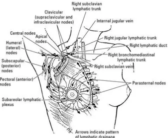
LYMPHATIC DRAINAGE OF THE BREAST1
Lymphatic drainage of the mammary gland is of great importance to
the surgeon, since breast malignancy spreads usually along lymphatics to the
8
FIGURE 2 - REPRESENTING LYMPHATIC DRAINAGE OF THE BREAST
LYMPH NODES
The lymphatic drainage of the breast is through the following group of
nodes,
1. The axillary node groups, mainly the anterior (or pectoral)
group. The posterior (or subscapular),axillary vein (or lateral),
central and apical(or subclavicular ) groups of nodes also
receive lymphatic drainage from the breast tissue either
9
2. The internal mammary nodes also called as the parasternal
nodes lie besides the internal thoracic vessels.
3. Few lymphatics from the breast also communicate with the
supraclavicular , the deltopectoral node, the posterior intercostal
groups and the infradiaphragmatic and subperitoneal lymphatic
plexuses.
The skin of the breast with the exception of nipple and areola is
drained by the superficial lymphatics , these lymphatics travel in a radial
fashion from the centre and ultimately drain into the axillary, internal
thoracic, supraclavicular and the cephalic lymph node groups.
The parenchyma of the breast, the nipple and areola are drained by the deep
lymphatics.
Some interesting points about the lymphatics of the breast are as follows,
1. Axillary group of lymph nodes receives more than 75% of the
lymphatics from the breast tissue and about 20% drain into the
internal mammary node groups; and remaining 5% drain into
the posterior intercostal lymph nodes. The anterior axillary
group of lymph node receives most of the lymphatics from the
10
2. The parasternal nodes receive the lymphatics not only from the
medial region of the breast, but also from the lateral region of
the breast.
3. A lymphatic plexus called the subareolar plexus of Sappy is
present beneath the areola and this plexus drains into the
anterior or pectoral group of lymph nodes.
4. The deep lymphatics of the breast finally reach the apical and
internal mammary group of lymph nodes after traversing
through the pectoral muscle and its fascia.
5. The infradiaphragmatic and subperitoneal plexuses receive
some lymphatics from the lower and medial quadrants of the
breast.
LEVELS OF LYMPH NODES1, 2
The lymphatic groups are allotted levels according to their
relationship to pectoralis minor muscle as follows,
Level 1 nodes: situated lateral to the inferior border of the muscle, which
include anterior, posterior and lateral groups.
Level 2 nodes: located deep to the muscle which consists of central nodes
11
Level 3 nodes: located medial to the superior border of the muscle which
include subclavicular nodes.
PHYSIOLOGY AND DEVELOPMENT OF THE BREAST3, 4, 5
The development of breast starts in the fifth week of intrauterine life
but is completed fully only after the first full term pregnancy. The milk line
which is an ectodermal thickening extending from the axilla to groin is
formed during the 5th week of intrauterine life, in the subsequent weeks the
portion of the milk line in the mammary region forms the mammary crest
and the remainder of the milk line involutes. The primary nipple bud formed
from the ectoderm grows into the mesenchyme and forms about 20
secondary buds, these later on form the lactiferous ducts and their
ramifications.
The breast remains dormant until puberty in the female, at puberty the
hormonal changes lead to its development and fat deposition, but the
development is complete only during pregnancy, when terminal ductal
differentiation occurs and gets ready for milk secretion. Later involution of
the breast occurs, which is pronounced during menopause when the acini
decrease in number and size, the connective tissue replaces the stroma and
12
TUMOURS OF THE BREAST1, 2
Most of the breast problems are benign conditions and about 30 % of
women will seek treatment for a benign breast condition in their lifetime,
pain and lump in the breast are the most common clinical features, so it
becomes the need of the hour to exclude malignancy.
BENIGN BREAST CONDITIONS
Aberrations of Normal Development and Involution (ANDI) is a
terminology used to describe the disturbance in physiology and cytology of
the breast and ranges from normal to disorder to disease.
It consists of essentially 4 features
Formation of Cyst – variable in size and found universally
Fibrosis – elastic and fat tissues are replaced by fibrous tissue with
interspersed chronic inflammatory cells.
Hyperplasia – proliferation of acinar and ductal epithelia with or without
cellular atypical changes.
Papillomatosis – extensive epithelial hyperplasia resulting in papillomatous
changes.
Clinical features
13
Fibroadenoma
Fibrodenoma is the commonest cause of benign breast lump; it is a
non proliferative disorder of breast occurs in the age group of 15 to 25 years
but occasionally seen in much elder women. They have plenty of stroma
where cellular elements are histologically normal, similar to normal lobules
of breast. They are hormonal dependent, fibroadenomas lactate during
pregnancy and involute after menopause, so it is a self limiting condition.
Ultrasonogram of breast will reveal pathognomonic features of
fibroadenaoma. In fibroadenomas of size less than 3 cm excision may be
avoided, Fibrodenomas of size more than 3 cm are best managed by surgical
excision.
Periductal mastitis, duct ectasia and breast cysts are other important
non proliferative disorders.
Fibrocystic disease
Fibrocystic disease can present with lump and or pain it may be
confused with carcinoma breast at times and usually resolves after
14
Breast Macrocysts
These are involution disorders that usually occur in the later stages of
reproductive life. Breast cysts of often multiple, bilateral and can mimic
carcinoma; diagnosis is usually confirmed by ultra sonogram and aspiration.
If the cyst is a complex one or the aspirated fluid is blood stained or residual
mass is present after aspiration we have to suspect malignancy, which is
confirmed by cytology or tissue biopsy.
Intraductal papilloma
It is a proliferative breast disorder arising from major ducts it usually
occurs in premenopausal women usually presents with nipple discharge,
malignant transformation is rare unless there is atypia.
Multiple intraducatal papilloma usually occurs in younger age group
and may undergo malignant transformation.
Phyllodes tumour
It usually occurs in fourth decade of life, cystosarcoma phyllodes tumors
are classified as
1. Benign phyllodes tumours.
2. Borderline phyllodes tumours.
15
These tumors are large and massive at times and the surface of the
tumors are unevenly bosselated, main bulk of the phyllodes tumor are
composed of connective tissues mixture of cystic, solid and gelatinous areas.
Infarction and necrosis at certain sites forms cystic areas.
Leaflike appearance (phyllodes means leaf like ) which is a classical
feature of phyllodes tumour is due to this gross morphological alteration ,
small tumours are treated by wide local excision and large tumours may
need mastectomy , borderline and malignant phyllodes tumours are highly
recurrent.
Breast abscess
The most common organism causing lactational infection is Staph
aureus. Duct ectasia and periductal mastitis are polymicrobial , here a series
of infections results in scarring which may lead to inversion and retraction of
the nipple and palpable mass in the subareolar region , so it is challenging to
exclude malignancy here.
Sclerosing adenosis
Increase in number of small distal ductules with stromal tissue
proliferation produces sclerosing adenosis it is difficult to differentiate from
16
Fat necrosis
Fat necrosis may occur following any trauma or previous surgical
procedure or exposure to radiation, characteristic feature is calcifications
which can be visualized by ultra sonogram.
BREAST CANCER
Breast carcinoma is the commonest site specific carcinoma in women;
it is a leading cause of mortality from malignancy for females in the age
group of 20 to 60 years. 29 % of all newly detected malignancy in female
population are breast cancers, it accounts for 14% of the malignancy related
mortality in females. There is gross variation in the incidence of carcinoma
breast among various countries.
Worldwide, Cyprus and Malta account for the highest age adjusted
mortality for carcinoma breast and Haiti accounts for the lowest age adjusted
mortality. There is increase in incidence in China and East Asia up to 3 to
4% annually. Breast carcinoma burden has well defined differences by
regional lifestyle, geography and ethnic background. Asian and African
17
The highest burden of disease is seen in European and North
American women. Due to absence of effective screening programmes for
earlier detection of malignancy and lack of accessibility to multidisciplinary
treatment programmes underdeveloped countries have disproportionate
mortality risk.
DIAGNOSIS OF A BREAST LUMP
Early detection of breast pathology forms is very important and in this
regard self examination of breast plays a great role. The 2013 NCCN
guidelines states that in addition to frequent breast self examination a
clinical examination of breast by medical personnel is recommended once in
a year.
Usually breast disease presents as a painless lump in the mammary
gland, discharge or erosion of the nipple, puckering or retraction of the skin
over the breast or swelling in the axillary region.
In order to arrive at a clinical diagnosis of breast lump and to detect
whether it is a benign or malignant lesion several modalities are used they
include detailed clinical examination of the breast , imaging tests like
mammography or ultra sonogram and thermography or tissue or cytology
based tests like excision biopsy , core needle biopsy or fine needle aspiration
18
If these tests if used alone the diagnostic yield is not much , hence
nowadays a combination of several tests are used for screening the patients
who have a breast lump for the earlier detection of a malignant condition,
this combination improves the sensitivity and specificity thereby cutting
down time and cost in diagnosis.
In India carcinoma Breast is the most common malignancy in females
and worldwide it is the foremost cause of malignancy related death among
women. Hence the diagnosis of a breast lump assumes utmost importance
both for the clinician and the patient, a dominant breast mass by definition is
a cystic or solid mass that is present in the substance of the breast throughout
the menstrual cycle.
Clinical assessment of breast masses
The Clinician is the person responsible for the diagnosis of a breast
mass, most of the breast lumps are benign, but diagnosis of malignancy
poses a great challenge and in most of the cases a history and clinical
examination however meticulously performed, will be inadequate to arrive
at definite diagnosis.
The breast has a lobular architecture and the identification of smaller
lumps amongst this lobularity is a great challenge and hence adjunctive
19
The imaging studies of breast dates back to as early as 1913 when a
German pathologist Salomon used X-rays to examine the amputated
specimens of the female breast , the demonstration of micro-calcification
and heterogeneity in X-ray in cases of malignancy still holds good6 . This
investigation has stood the test of time and evolved as mammography which
is still being used as a screening test for breast malignancy.
Since mammography involves radiation and studies have proven that
patients subjected to mammography are at a slightly increased risk of
development of malignancy in situ, the use of other economically viable and
safe imaging modalities was considered.
ULTRASONOGRAPHY OF BREAST
The examination of breast tissue by ultrasound was proposed as early
as 1950 but could be put into clinical practice only after the development of
grayscale examination by Swedish scientists and then tissues could be
clearly examined. In recent era the advances in ultrasound technology has
made it into a safe economical and reliable method for the examination of
superficial and moderately deeply situated soft tissue lesions, and the
examination can give more information than differentiating cysts from solid
20
TISSUE DIAGNOSIS
The gold standard for the detection of malignancy remains the
demonstration of malignant cells in the tissue specimen and FNAC and
excision biopsy are used for this.
In 1912 FNAC was used for the examination of cause of lymph node
enlargement and gradually it percolated to other fields for neoplasia
evaluation and in 1962 Ellis and Martin used FNAC for the evaluation of
breast masses7, FNAC interpretation is operator dependent and it requires
years of experience to master this technique and experienced operators can
distinguish benign and malignant lesions with good accuracy.
CLINICAL EXAMINATION
Inspection and palpation of breast masses are important and in order
to become palpable the mass should be large enough to be distinct from the
tissues surrounding it. But it might be detected earlier on imaging
modalities, Since the breast mass is lobular with varying contents of
glandular tissue, adipose tissue and fibrous tissue, the exact identification of
21
But true breast masses stand out from the surrounding tissues with a
different consistency and are asymmetric when both breasts are compared
and have a three dimensional demarcation from surrounding tissues.
Of the breast diseases, benign breast diseases predominate but the fear
that a breast disease creates is enormous and a plan should be devised for the
rapid detection and treatment of malignancy, of this first are history and
physical examination.
A detailed history includes the following points:
Age at presentation
Menstrual history
Family history and reproductive history
History of pregnancy and lactation
Exposure to radiation especially the upper body
22
PHYSICAL EXAMINATION OF BREAST
A private examination cubicle with proper lighting with adequate
warmth and comfort are essential for breast examination, the room should be
properly illuminated and the patient is stripped up to the waist to enable a
complete examination.
General examination
Built and Nourishment
Colour of skin
Mucous membranes
Palms and soles
Inspection of breast
First examine the patient from bed end with arms by side and then
with arms elevated.
The symmetry of breasts and nipples are first assessed
The next points for inspection are
a. Abnormalities of the nipple and areola
b. Prominent vessels and scars
c. Prominence or dimpling of the skin
23
e. Peau d’ orange appearance
f. Nodules over the surface
g. Skin or Nipple Ulceration
Palpation of breast is done in an orderly fashion, the following is the
schema
1. Palpation of both Supraclavicular fossa
2. Breasts:
a. Initially light palpation is done, followed by
b. Deep palpation extending systematically from the areola
and moving concentrically outwards to the axilla
including the axillary tail.
3. Examination of Axilla:
a. Examine the axilla from front and behind
b. The forearm of the patient is supported by the examiner
4. Abdomen is examined for ascites, organomegaly or abnormal
24
Particularly valuable in practice are
Inspection with arms firstly kept by the side, then arms raised.
First light palpation over the breasts followed by deep palpation
in concentric rings, travelling outwards.
Examination of the both axillae performed from front and back
of the patient.
Malignant tumours are usually firm or hard and tend to have
ill-defined margins and attachments to the overlying skin or fascia with
puckering or retraction of the nipple and areola. Benign lesions are well
defined and have clear borders and are mobile without definite attachments
to superficial or deeper tissues.
Solid lesions and cystic lesions can be differentiated by palpation to a
certain extent but the sensitivity of palpation is only around 58 to 66% as
reported by Rosner et al, so significant difference in diagnosis can be present
even among seasoned examiners8 , In a study done by Boyd et al involving
patients with breast masses which had experienced surgeons doing physical
examination the need for biopsy was agreed upon only in 73% of patients
with malignancy.
25
In another study, Somers et al studied certain palpable abnormalities
which were defined as areas of thickening, tenderness without an associated
dominant mass on clinical examination, no suspicious mammography
findings and firm, rubbery, cystic soft mass, needle sensation by FNA. The
malignancy incidence in this group was less than 1%, so it was concluded
that these palpable lesions did not require open biopsy.
Sometimes in the earlier stages of malignancy benign features and
malignant features may overlap and without the use of FNAC or imaging
modality certain malignant lesions may be classified as benign by physical
examination causing a delay in treatment, So in order to arrive at an early
definitive diagnosis an imaging modality is required to complement the
findings of physical examination and for the examination of the opposite
breast and look for multi-centricity. In the contrary a negative imaging
finding in a highly suspicious looking lesion also should be interpreted with
caution.
ULTRASONOGRAPHY OF BREAST
Ultrasound Examination of breast has been in vogue since 1950s but
the resolution and technology was inadequate for a comprehensive
examination in the earlier phases, hence physicians were not able to utilize
26
technique by Korasakoff obviated this difficulty and ultrasound began to be
used in a widespread fashion.
The ultrasound transducers were initially quite crude devices that
operated with low frequencies.30
Low frequency waves have good penetration but the image resolution
is not adequate hence initial ultrasonography had a low sensitivity to
differentiate between tissue types.
Later generation transducers were improved with design changes that
allowed use of a range of frequencies and higher frequency transducers were
used for examination of breast, thyroid and testis.
Even with modern ultrasound technology, ultrasound examination is
operator dependent and adequate training is essential for the correct
interpretation of findings.
Skilled Sonography interpreters follow three golden rules, they are
1. A single image is never used for making an interpretation but a
mental 3 dimensional image is imagined with the acquired
images; superimpose the ultrasonic images mentally to
formulate a 3 dimensional image of the scanned tissues and to
ensure that the displayed feature is concordant with the mental
27
2. Rule out artifacts; don’t always think that all which is displayed
in the image is real.
3. Look for the invisible things, things which are not seen in a
field of imaging can be seen in other planes.
Initially ultrasound could differentiate only between solid and cystic
masses but later generations of Ultrasound devises are capable of
characterizing the density of tissues and can be used to differentiate between
benign, malignant and equivocal masses.70
ULTRASONOGRAPHY OF THE BREAST
First task is to confirm the side of breast, site and position of the
lesion which is to be evaluated; this can be done by referring the clinician’s
notes and the diagrams provided by the examining physician
For Breast USG, a 7.5 to 10 Mhz linear- small parts high definition
transducer is used.
The Region of interest (ROI) is first evaluated and the side and Site
are confirmed to be in concurrence with request given and USG findings are
28
Position of the patient
A pillow is placed under the shoulder of the side to be examined, the
patient is made to lie in an oblique position with the degree of obliquity
depending on the position of the breast, this aims to bring the corresponding
breast to the centre of the examination field, the arm is raised above the
patients head for even distribution of the breast tissue, but not very much as
to cause breast retraction. Better positioning eases examination and provides
clear images.
a. Lesions that are felt better in the upright position may be
scanned in the same position.
b. Confirmation of fluid in cysts can be done by changing from
upright to decubitus position.
The Ultrasound transducer is placed directly over the lesion after
trapping the region of interest with the examiners fingers.
Examination should be done in radial/antiradial planes to avoid
mistaking fat islands as solid masses and determine the relation of the lump
29
Shape, nature of margins and surrounding tissue can be determined by
evaluating the lesion in entirety including the periphery, in multiple planes.
Artifacts can be eliminated by slightly compressing the breast tissue
with transducer which will make the breast tissue to spread evenly over the
chest wall.
The breast is examined from the periphery to centre and finally the
areola and nipple are imaged and the retroareolar tissue is also imaged in
multiple planes by angling the transducer.30
LESION LABELING
The position lesion is labeled on a clock face.
The distance of the lesion from nipple is given in centimeters.
The longest diameter is measured.
Height width ratio of the lesion is obtained.
NORMAL APPEARANCE OF THE BREAST
Knowledge about breast architecture and its normal variations are
30
SKIN
The skin appears as an echogenic layer with a thickness of 3mm or
less by ultrasound and most of the times a hypo echoic central line is seen, if
the skin is diffusely thickened it is difficult to recognize it without
comparing the Region of Interest to the opposite breast or to a normal area
within the same breast. To detect subtle abnormalities a standoff pad may be
used.
SUBCUTANEOUS TISSUE
The subcutaneous fat layer is present beneath the skin as a hypo
echoic layer sandwiched between the skin line and the parenchyma of the
breast. The ligaments of Cooper are seen as curvilinear lines extending from
the breast parenchyma to the superficial fascial layer thereby producing a
scalloped appearance. Cancer breast does not arise in the subcutaneous plane
but it might involve it by direct extension.
Focal increase in echo texture is seen in Malignancy,
31
Diffuse increased reflectivity of breast tissue is seen in edema of any
cause (e.g., Congestive Cardiac failure), diffuse form of Carcinoma breast,
inflammatory breast cancer, inflammatory mastitis, or radiotherapy to breast.
Sebaceous cysts, epidermoid cysts, hemangiomas, and rarely smooth
muscle and fibrous tissue tumors are the indigenious to this plane.
NIPPLE AND AREOLA
The Nipple and areola are imaged after compressing the area slightly
with the transducer and imaging is done in multiple planes angled towards
the subareolar region, application of light pressure with the transducer
prevents air trapping between the skin and the transducer thereby
eliminating artefacts , The lactiferous ducts are visualized in this area and
they are traced into the breast tissue , usually echogenic masses represent
debris collected within, but the presence of a dilated duct along with a mass
lesion may indicate carcinoma , papilloma or other solid lesions.
BREAST PARENCHYMAL LAYER
The breast parenchymal tissue has varying proportions of ductal,
32
uniform, as age advances the breast become more lobular, but age and parity
do not exactly correlate with the ultrasound appearance of breast.
The normal breast can range from almost completely fatty with only a
few ehogenic fibroglandular tissue in one spectrum to presence of more
amount of fibroglandular tissue with little or no fat on the other.
The normal fibroglandular tissue appears to be arranged in multiple
layers parallel to the chest wall without distortion and should not have a
pulled or swirled appearance.
Most benign and malignant lesions are imaged as hypoechoic nodules
in comparison with the paraenchyma and are easily visualized the in
homogenously echogenic breast tissue, which does not deform with
compression. Moreover, the most useful maneuver in imaging of the breast
is to image in multiple planes, which helps to demonstrate the fat island
continuity with other areas of fat.
RETROMAMMARY AREA
It is present deeper to the echogenic plane of glandular tissue, and
visualization by ultrasound is difficult, it is a hypoechoic plane containing
33
LYMPHNODES, ARTERIES, AND VEINS
Lymphnodes are seen within the axillary region as round to oval
nodules with an hilum which is more echogenic and a peripheral rim of less
echogenic tissue.
Benign nodes are large in size but morphology is preserved but
malignant nodes appear as hypoechoiec lobulated nodules. Ultrasound can
only detect gross abnormalities and not minor variations in lymph nodes.
Arteries and veins appear as tubular anechoic structures within the
breast tissue. Arteries have a pulsatile appearance on the examination of the
breast; Colour Doppler ultrasound can clearly demonstrate the vascular
structures.
BREAST MASS CHARACTERIZATION 31
CYSTS
The characterization of breast mass starts with solid and cyst
differentiation, the accuracy of USG approaches 100% for the diagnosis of
34
These are the criteria
a. Oval, lobulated or round shape
b. Anechoic
c. Clearly defined posterior border
d. Increased through and through transmission
e. Surrounding parenchyma shows no alteration.
But in real life situations all cysts do not display these criteria. Cysts
of size up to 5mm can be diagnosed with USG with an accuracy approaching
100%. The common difficulty in the differentiation of cysts from solids is
when internal echoes are present. Internal echoes may be seen in cysts due to
35
FOLLOW UP OF CYSTS 32
SIMPLE CYSTS
36
SOLID MASSES
Solid masses have the following signs by USG
Primary signs – changes produced by the mass per se.
Secondary signs – changes in the surrounding tissues due to the mass.
BENIGN FEATURES OF SOLID MASSES
USG has less specificity in differentiating benign masses from each
other.
There is overlap of findings between fibroadenomas, tubular
adenoma, focal fibrocystic changes and other solid benign nodules.
Features of solid benign lesions are
1. Shape is round or oval with few lobulations.
2. Presence of a thin echogenic pseudocapsule with sharply
defined margins.
3. A depth/width ratio of less than 1.
4. Surrounding tissues clear of disease.
5. Absence of malignant characteristics, by Ultrasound guided
aspiration or biopsy.
37
FIBROADENOMAS
Fibroadenomas are the most common benign solid nodules.
Fibroadenomas are seen in young females and their ultrasound features are
determined by the varying amounts of epithelial and fibrous components.
The features of Fibroadenomas are
a. Enhanced through and through transmission.
b. Posterior attenuation in relation to the fibrous component.
c. Degenerating fibroadenomas have shadowing in USG due to
coarse calcifications.
d. Lactating and juvenile types of fibroadenomas exhibit tubular
structures.
e. Differentiated from fat lobules by their non compressible
nature.
LIPOMAS AND FIBROADENOMYOLIPOMA
1. Lobulated masses with no distortion of surrounding tissues.
38
FAT NECROSIS
Fat necrosis is seen as a hyperechoic nodule with a central lucency.
ULTRASOUND IMAGES
NORMAL BREAST BREAST CYST
39
MALIGNANT FEATURES OF BREAST LESIONS
1. Stellate lesions
2. Circumscribed lesions
3. Presence of diffuse edema
4. Presence of calcifications
STELLATE MASSES
The normal parallel arrangement of soft tissue planes in malignant
tumours is lost and the desmoplastic reaction in and around the lesion pulls
the breast tissues towards the mass resulting in a stellate appearance.
The centre of the lesion is irregular and hypoechoic and there is
peripheral distortion producing a star like appearance and the features are
Normal trabecular architecture is disrupted.
Extension of disease along the plane of the ducts.
Posterior acoustic shadowing is seen.
40
Differential diagnosis of stellate lesions
Carcinoma (commonest)
Radial scar
Sclerosing adenosis
Post operative scarring
INDICATIONS FOR BREAST ULTRASOUND
1. To evaluate a breast lump that is detected by mammography.
2. To confirm the nature and the presence of a lesion.
3. Used as a single imaging method in pregnant or lactating
women who present with breast symptoms and in outpatient
setups.
4. To delineate solid from cystic lesions.
5. Used to guide biopsy /aspiration for pathological diagnosis.
6. Used for localization of lesions in preoperative stages and to aid
41
7. Follow up of post surgical breast tissue.
8. Post breast augmentation surgery follow up to investigate the
contour of prosthesis or to find out extra capsular rupture.
9. Follow up of benign lesions that have not been subjected
biopsy.
10.As a screening test for Carcinoma breast adjunctive to
mammography.
THE ADVANTAGES OF ULTRASONOGRAM
1. Absence of Radiation and its complications.
2. Can evaluate the lesion and describe details of size, shape,
echogenicity and relation to surrounding tissues as against
mammography that gives a result of a density which has less
specificity.
3. Ultrasound guided biopsy was found to be more accurate in
studies than conventional methods and since the biopsy needle
is seen through the lesion it aids in accurate sample and boosts
42
4. In preoperative stages mammography is more time consuming
and the positioning is cumbersome and the results are less
accurate, hence ultrasonography is better for immediate
preoperative guidance.
5. USG gives findings in surgically altered breasts compared to
mammography.
6. USG is superior for the imaging of extra capsular rupture of
prosthesis which cannot be diagnosed by mammography.
7. In follow-up of benign nodules which are not excised,
mammography is associated with risk of radiation and
malignant transformation; hence USG is the investigation of
choice.
LIMITATIONS OF ULTRASOUND
1. Less accurate in larger and dense breasts, but highly sensitive in
compare to mammography.
2. Less accurate in small (< 1cm) lesions.
3. Cannot be used as routine for postoperative cases.
4. Has less sensitivity for the detection of non palpable malignant
43
5. Microcalcifications are not clearly detected as in
mammography.
6. Has high false positive rates compared to mammography, it
classifies many benign lesions as malignant
CYTOPATHOLOGICAL DIAGNOSIS
Fine Needle Aspiration Cytology
The development of the needle aspiration technique for diagnosis was
reported as early as 1847 when Kun described a novel method for diagnosis
of tumors. In 1883 Leydon aspirated and isolated pneumonic organisms
using needle aspiration, later in 1904 cervical lymph nodes were subjected to
needle aspiration by Greig and Gray for the identification of
trypanosomiasis13.
Initially larger bore needles were used for aspiration from a variety of
lesions of lymph nodes; breast, thyroid and prostate, as time passed by
smaller needles were used, in the recent times needles of 1mm or thinner are
44
FNAC did not gain much importance till the 1960s and remained a
controversial topic; only after 1960s it gradually gained the confidence of
surgeons.10, 16
European scientists in the Stockholm Karolinska institute in 1960s
pioneered the use of fine needle aspiration techniques and developed
protocols for diagnosis and interpretation criteria.10, 16
Recent developments in FNAC technique and cytological diagnosis
have propelled its use in multiple sectors and it has developed to be as
accurate as excision biopsy.
The advantages of FNAC are that it is an outpatient procedure, does
not require anesthesia, cost effective and helps adequate preoperative
planning.
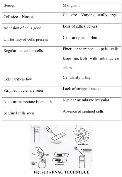
FNAC technique
Definition: FNAC is defined as the study of cells obtained using a
thin bore or fine needle with negative pressure. The needle aspirate
specimen contains a minimal quantity of tissue along with fluid. FNAC can
45
FNAC Personnel
In western countries the surgeon experienced in doing FNAC does the
aspiration and the Pathologist interprets it, in our country both aspiration and
interpretation are done by the pathologists. Adequate skill is necessary for
both sample acquisition and interpretation.18
Equipment required
The equipments required are 1. Needles-23 G disposable needles. 2.
Syringes –Disposable BD plastic regular 10 ml syringes. 3. Slides-2 or 3
labeled clean non contaminated sterile slides for smear preparation. 4.
Alcohol 95% as fixative. 5. Stains- Hematoxylin and Eosin stain, Special
stains as required by pathologists.19
Technique
FNAC is usually a painless procedure, after proper explanation to
patients, the lesion is held firmly in between the fingers of the examiner and
the skin is stretched and made taut, the area is properly sterilized and wiped
clear.68,69
The needle is attached to the syringe with the plunger in fully closed
46
skin overlying the lesion, the needle is inserted as to feel the anterior edge of
the lump and after entering it firm negative pressure is applied to the piston
using a syringe holder or the thumb.
Several passes are made through the lesion in different angles and
rotation of the syringe without withdrawing it fully until a small amount of
fluid is seen in the hub of the syringe. Negative pressure is then released and
needle withdrawn.
If the aspirate is more than expected or hemorrhagic is advisable to
interrupt the procedure and try it from a different angle or do it in another
session. If tissue is not obtained adequately due to needle blockage the
needles can be changed and re aspiration done. Breast lumps are usually
deeper than they appear and sometimes longer needles may be necessary.
After this slides are smeared with aspirate, fixed with 95% alcohol
47
FNAC CYTODIAGNOSTIC CRITERIA 12
There are 4 categories of breast FNAC cytology19, 20, 21
1. Inadequate or Unsatisfactory cytology- Here the cell elements
are inadequate or absent. There has not been a standard criteria
established for a diagnostic FNAC aspirate, but each field
should contain 3 to 6 epithelial cells and should not be obscured
by blood or other cells. Unsatisfactory smears are due to air
drying artifacts or due to minimal cellularity of lesion.
2. No Malignant cells seen –No malignant cells seen, only benign
cells seen. Benign cells of breast are duct epithelium, apocrine
cells, RBCs, adipocytes and degenerative nuclei. The nature of
benign lesion may also be qualified.
3. Malignant cells present- The report is given as malignancy
positive only when cells confirmatory of malignancy are
undoubtedly present and it also should provide information on
the degree of differentiation and the type of malignancy, but it
cannot provide details of invasion of surrounding tissues.
4. Presence of suspicious cells but not diagnostic of malignancy-
48
Differences between benign and malignant cytology22, 23
Benign Malignant
Cell size – Normal Cell size – Varying usually large
Adhesion of cells good Loss of adhesiveness
Uniformity of cells present Cells are pleomorhic
Regular but coarse cells Finer appearance , pale cells,
large nucleoli with intranuclear
edema
Cellularity is low Cellularity is high
Stripped nuclei are seen Lack of stripped nuclei
Nuclear membrane is smooth Nuclear membrane irregular
[image:59.612.114.530.95.699.2]Sentinel cells seen Absence of sentinel cells
49
FNAC - CYTOLOGY IMAGES
BENIGN MILDLY ATYPICAL
MARKEDLY ATYPICAL MALIGNANT
50
HISTOPATHOLOGICAL DIAGNOSIS
Biopsy of breast lump
“Bio” is life and “Opsis” is vision so biopsy means visualization of
the surgically excised specimen both macroscopically and microscopically.
Ernest Henri Besnier a French dermatologist coined the word biopsy
in 1879.Even earlier Rudolph Virchow had explained the fundamentals of
this procedure and now it has become in indispensable tool in surgical
practice9.Biopsy and the Histopathology is the ultimate gold standard truth
for the surgeon, and treatments of lesions either benign or malignant greatly
rely upon histopathological diagnosis.40
In this context excision biopsy of breast lesions and its histopathology
remains gold standard for the diagnosis of malignancy.40. Aspiration
cytology provides cytopathological diagnosis, whereas biopsy provides
histopathological diagnosis10.
Different types of biopsy are available they are
Incision biopsy
51
Trucut biopsy
Intra ductal biopsy
Slide biopsy smears of nipple secretion
Superficial biopsy of lesions of the nipple
Incision biopsy
Incising the lesion and sending a small amount of tissue is preferred
by some surgeons for detection of malignancy, it is usually carried out as a
preliminary procedure before definitive surgery; Incision biopsy and frozen
section have good accuracy for the detection of malignancy except papillary
carcinoma of the breast which may be mistaken for papilloma.
Incision biopsy is usually used to remove a portion of tissue from
large tumours in which needle biopsy is inadequate.
Excision Biopsy
Excision biopsy involves excising the whole tumour in an operating
room under anaesthesia , if performed adequately with good clearance of
52
Excision biopsy and histopathology is the ultimate gold standard for
breast mass evaluation, it is usually done when imaging or tissue
investigations are inconclusive or equivocal.
With advances in FNAC techniques and combination triple
investigation, the use of excision biopsy is on the decline.
Trocar or Trucut biopsy
Small cores of tissue are removed from the breast lesions using small
hollow trephines or trocars , it helps to obtain tissue for histopathology.
This technique was first used by Vim Silverman in 1938 and a good trocar
or trephine biopsy should provide an adequate representational specimen
But the disadvantages are that they provide only a minimal amount of tissue,
it cannot provide details about margins and invasiveness of tumour and they
may miss small lesions.
In recent era Ultrasonographic guided trocar biopsy has improved the
accuracy of this test.
Intraductal biopsy
Dilators, small loops and curettes are used to dilate the ducts and
obtain biopsy specimen from the breast11.
Biopsies may also be obtained from Nipple smears and surface of
53
HISTOPATHOLOGY SLIDES
NORMAL BREAST FIBROADENOMA
LOBULAR CARCINOMA COMEDOCARCINOMA
54
THE MODIFIED TRIPLE TEST
Previously in the early era of diagnosis open biopsy was done for the
detection of malignancy, in mid 1970's a triple test consisting of clinical
examination, mammography and FNAC was introduced which improved
diagnosis of breast cancer, later mammography was substituted by
ultrasonogram of the breast and this was called as the modified triple test, if
these three modalities had a similar correlation then the accuracy of
diagnosis was about 99%. Even in lesions that are not palpable the
diagnostic yield is similar to palpable lesion when the triple test is used.33, 34
Few years ago frozen section after excision was used to confirm the
diagnosis of breast carcinoma but now preoperative detection of malignancy
has been facilitated by using cytological tests such as testing the nipple
discharge or using fine needle aspiration cytology tests from the lumps.
Making a definitive preoperative diagnosis is very vital because it
helps to plan surgical decisions. It can avoid unnecessary radical dissections.
If malignancy is detected by triple test, curative surgery can be planned
earlier thereby avoiding two surgeries, one for biopsy and other for curative
55
In recent days treatment of Carcinoma breast is patient centric and
patient themselves are involved in the process of deciding about the mode of
surgery and this needs earlier planning so the detection of metastasis and the
type of lesion become very important.
Few years ago it was stated in medical literature that every breast
lump should be excised but this has now been emphasized as every breast
lump should be assessed and clarified, so assessment includes the triple test
and its variants, so this test assumes a vital proportion in workup for cancer
breast.66
Origin of triple test
In his 1987 article Hermansen et al used the term triple test in breast
cancer which includes physical examination, mammography and Fine needle
aspiration of breast mass , about 650 women with breast tumors were
studied and the conclusion given was triple test had a accuracy of diagnosis
similar to that of histological examination. 24
In another study done by Hardy et al 143 patient with breast masses
where assessed by several modalities which included mammography,
ultrasound examination of breast , Magnetic resonance imaging of the breast
56
cytology and ultrasound imaging of the breast had the most accurate
diagnosing power for malignant lesions.25
In another study done by Lawrence N Bassett et al about 1016 women
of age 35 years or less presenting with a palpable breast lump were studied
over a eight year period, in this study it was shown that mammography was
less sensitive when compared to sonography in the detection of breast
masses and the benefit of ultrasound was to avoid unwanted biopsy
procedures , so ultrasound of breast was found to be a preferable mode of
examination of younger women with breast lumps, but ultrasound was less
sensitive in detecting very small lumps and could not differentiate benign
from malignant in smaller lesions.26
A study done by Vetto et al included fifty five young women with
palpable breast mass and the value of the Triple test was appreciated, it had a
negative predictive value and a specificity of 100 % for the detection of
malignant lesion in younger women when compared to conventional
excision biopsy, thereby reducing the economic burden on the healthcare
system and the patient.27
In another study done by Purasri et al used four tests instead of three
and included physical examination, FNAC, mammography and
57
and put forth a novel scoring system which had an accuracy of 98% in
detecting malignant lesions.26
In a study done by Hatada et al biopsy specimens of 114 malignant
breast lumps were examined and retrospective analysis of the method of
cytological analysis was made, in this they found out that USG guided
FNAC was more accurate (86%) than plain FNAC (65%) in the diagnosis of
malignant breast lumps, and the results were more pronounced in smaller
lesions of size <2cm.28
In a office based ultrasound study done in 660 women by Heiken et al
brought to light that about 75% of the suspicious lesions detected by USG
were found to be malignant and only 5% of the lesions described as
fibroadenaoma turned out to be malignant, this study shows that USG of the
breast is a valuable screening tool for detection of malignancy.19
In other study, women with age of less than 35 years presenting with a
high risk history, breast symptoms and indeterminate findings on
mammography were studied by Jill S Montrey et al and Ultrasound
examination of breast was found to have accurate diagnostic power for the
58
MATERIALS AND METHODS
TYPE OF STUDY
Prospective observational study.
STUDY CENTRE
Department of General Surgery, Coimbatore Medical College
Hospital, Coimbatore.
PERIOD OF STUDY
From July 2016 to June 2017 – 1 Year.
PATIENT POPULATION
103 consecutive patients presenting to the outpatient and Inpatient
department of the Department of General surgery, Coimbatore Medical
College with complaints of a palpable breast mass were included in this
study.
INCLUSION CRITERIA
1. Female patients with age of > 20 years with palpable Breast
lump
59
EXCLUSION CRITERIA
1. Patients who are below 20 years.
2. Male patients
3.Female patients with advanced disease which makes the
diagnosis obvious
4. Patients not willing for lump excision
The study was conducted after obtaining permission from the
Institutional Ethics Committee. The patients were clearly explained about
the nature of study and its implications and an informed written consent was
obtained from the patients after explaining the procedure in their vernacular
language.
COLLECTION AND ACCUMULATION OF DATA
The patients were enrolled in the study after applying the inclusion
and the exclusion criteria. A detailed History regarding the complaints, the
mode of presentation, site of lump and associated symptoms was obtained, a
complete physical examination and examination of the breast and the mass
was made. Each patient underwent a modified triple test which included a
complete clinical examination, next was the ultrasound examination of the
60
Based on each test the palpable breast lumps were classified as
benign, malignant or inconclusive.
Malignant lesions
These lesions are ill defined. These are hard in consistency with
angulated and abrupt borders and microcalcification seen in Ultrasound
examination.
Benign Lesions
These lesions may be cystic lesions or solid lesions.
Cysts are oval or round with clearly defined margins and they are not
echogenic, these cysts may be simple cysts with through and through
transmission of echoes or may be abscesses with septations and internal
echogenicity.
Solid benign lesions may be fibroadenomas which may be lobulated
with single to several lobulations, rounded or oval in shape and covered by a
pseudo capsule and has uniform homogenous echo pattern in Ultrasound.
Fibrodenosis is a condition in which there is increase in fibrous and
61
may be certain cystic areas with increased echogenicity and the architecture
of the breast is maintained.
Breast Examination included examination of the breast, the axilla on
both sides, both supraclavicular fossa and all lymph node areas were
examined to rule out generalized lymphadenopathy.
Ultrasound examination of breast
Ultrasound examination of the breast was done using a high frequency
linear transducer (7.5 to 10 megahertz) in the department of Radiology,
Coimbatore Medical College Hospital, Coimbatore by an experienced
radiologist well versed with radiological examination of the breast and the
patients were examined in supine position and scanning of the breast was
done horizontally and vertically, Ultrasound examination of both breasts,
axillary region and supraclavicular lymph nodes was also done.
Fine needle aspiration cytology
FNAC was done by a pathologist and aspiration was done using a 22-
23 gauge needle attached to a 10 ml syringe with the patient in supine
position and the ipsilateral upper limb raised beside the head, the lesion was
fixed with one hand and the biopsy needle was inserted into the lump and
62
suction was maintained until aspirate was seen at the hub of the needle , then
suction was released and the needle withdrawn and the material was spread
on three slides, then taken up for cytological examination after fixation and
staining.
The results were reported as benign, malignant or indeterminate.
Histopathological examination
All the patients had some form of surgery based on the result of the
modified triple test, patients with benign lesions had excision biopsy and
malignant lesions had Modified Radical Mastectomy, the surgical specimens
were examined in the pathology department and the results were classified
as benign or malignant.
Analysis and interpretation of data
The particulars in the pro forma were tabulated in Microsoft excel
program and statistical analysis was done using SPSS software system and
appropriate statistical tests were used as necessary and various parameters
were analyzed and result of the modified triple test were analyzed
individually and collectively, finally the result was compared to
63
Fine Needle aspiration Cytology was reported and tabulated as follows:
a. Inadequate material - The aspirate had poor cellular content and
was not sufficient for cell type assessment.
b. Benign cytology – Adequate aspirate with benign appearing
cells indicating the diagnosis of fibrocystic disease of breast or
a Fibroadenoma.
c. Atypical cell cytology – Minimal atypia; which was interpreted
as benign breast lesion.
d. Suspicious cytology - Malignancy – suspect cells with features
of malignancy and non interpretable cytology.
The interpretation of results was done as follows
Repeat cytology
Benign lesion
Malignant lesion
Inconclusive test
The patients with benign and inconclusive lesions underwent an
excision biopsy and the patients with report as malignancy were subjected to
modified radical mastectomy, the excision biopsy and mastectomy
specimens were subjected to histopathology examination and the results