0022-538X/05/$08.00⫹0 doi:10.1128/JVI.79.4.2474–2483.2005
Copyright © 2005, American Society for Microbiology. All Rights Reserved.
Adenovirus Protein VII Functions throughout Early Phase and
Interacts with Cellular Proteins SET and pp32
Yuming Xue,
1† Jeffrey S. Johnson,
1† David A. Ornelles,
2Judy Lieberman,
3and Daniel A. Engel
1*
Department of Microbiology, University of Virginia Health System, Charlottesville, Virginia1; Department of Microbiology andImmunology, Wake Forest University School of Medicine, Winston-Salem, North Carolina2; and CBR Institute for
Biomedical Research and Department of Pediatrics, Harvard Medical School, Boston, Massachusetts3
Received 12 August 2004/Accepted 5 October 2004
Adenovirus protein VII is the major component of the viral nucleoprotein core. It is a highly basic nonspe-cific DNA-binding protein that condenses viral DNA inside the capsid. We have investigated the fate and function of protein VII during infection. “Input” protein VII persisted in the nucleus throughout early phase and the beginning of DNA replication. Chromatin immunoprecipitation revealed that input protein VII remained associated with viral DNA during this period. Two cellular proteins, SET and pp32, also associated with viral DNA during early phase. They are components of two multiprotein complexes, the SET and INHAT complexes, implicated in chromatin-related activities. Protein VII associated with SET and pp32 in vitro and distinct domains of protein VII were responsible for binding to the two proteins. Interestingly, protein VII was found in novel nuclear dot structures as visualized by immunofluorescence. The dots likely represent individual infectious genomes in association with protein VII. They appeared within 30 min after infection and localized in the nucleus with a peak of intensity between 4 and 10 h postinfection. After this, their intensity decreased and they disappeared between 16 and 24 h postinfection. Interestingly, disappearance of the dots required ongoing RNA synthesis but not DNA synthesis. Taken together these data indicate that protein VII has an ongoing role during early phase and the beginning of DNA replication.
The adenovirus nucleoprotein core consists of double-stranded genomic DNA, the highly basic viral proteins VII, V, and(mu), as well as protein IVa2 and the 55-kDa terminal protein (1, 5, 19, 25, 31, 33, 34, 42). Protein VII is the major protein component of the core with an estimated 1,070 copies present per virion (9) and is primarily responsible for estab-lishing viral chromatin structure. It can potently condense DNA in vitro and in vivo and also repress transcription (3, 6, 21, 35). This is consistent with the highly condensed configu-ration of viral chromatin found within the virion. When deliv-ered to the nucleus, the chromatin is silent prior to stimulation by the viral transcriptional activator E1A (13).
We and others have reported that protein VII from infec-tious viral particles enters the nucleus along with viral DNA and remains associated with it, suggesting that the protein VII-DNA complex is the substrate for transcriptional activa-tion by E1A during early phase (8, 15, 18, 21). Moreover we found that protein VII can associate with E1A protein in vitro (21).
In this report we have studied the fate and function of protein VII during the early phase of infection. We demon-strate that virus-derived “input” protein VII persists in the nucleus throughout early phase and the beginning of viral DNA replication, suggesting that it has an ongoing role in gene regulation and perhaps DNA replication. During this period protein VII is found in discrete nuclear dot structures and viral DNA continues to associate with protein VII.
We have also investigated the association of protein VII with cellular proteins SET and pp32. SET, also known as TAF1, has recently been shown to associate with adenovirus chroma-tin during infection (18). SET is homologous to evolutionarily conserved nucleosome assembly proteins (NAPs) found in yeast and higher eukaryotes (2, 14, 24, 27, 39, 41). Moreover, SET satisfies the requirements of a NAP experimentally: when added to reactions containing histones and relaxed circular plasmid, SET mediates the assembly of histones and DNA into a supercoiled structure (2, 14, 23, 28). Interestingly, when SET is incubated with purified adenovirus core, some structural rearrangement occurs. SET can stimulate in vitro DNA repli-cation and transcription reactions containing adenovirus core as the template and can also stimulate transcription from nu-cleosomal templates composed of histones and DNA (28). It also has been shown to trigger decondensation of Xenopus
sperm chromatin (26, 28).
SET has been found by several groups to be a component of at least two distinct multisubunit complexes termed SET and INHAT (2, 4, 10–12, 36, 37). We find that SET and pp32, components of the SET and INHAT complexes, associate with viral chromatin in vivo and bind to protein VII specifically in vitro. These data suggest that SET and pp32 associate with viral chromatin as components of the SET and INHAT com-plexes and, along with protein VII, likely have important roles in viral chromatin function during early phase.
MATERIALS AND METHODS
Cells and viruses.HeLa cells were grown as monolayers in Dulbecco’s mod-ified Eagle medium (DMEM) supplemented with 10% newborn calf serum (NCS), penicillin, and streptomycin (all from GIBCO). Phenotypically wild-type
adenovirus type 5dl309 (22) was propagated in HeLa cells to produce virus
stocks.
* Corresponding author. Mailing address: Department of Microbi-ology, University of Virginia Health System, P.O. Box 800734, Char-lottesville, VA 22908. Phone: (434) 924-8633. Fax: (434) 982-1071. E-mail: [email protected].
† Y.X. and J.S.J. contributed equally to this work.
2474
on November 8, 2019 by guest
http://jvi.asm.org/
Plasmids.pGSTE1A289 and pGSTVII were described previously (21). pGST CAS YXXP, a gift from Amy Bouton, contains the YXXP domain of CAS in pGEX-2TK (7). To produce pGSTVII, pGSTVII 1–57, pGSTVII 45–94, pGSTVII 94–148, and pGSTVII 149–174, inserts were generated by PCR with
adenovirus type 5 DNA. Primers contained 5⬘BamHI and 3⬘EcoRI sites and the
PCR products were cloned into pGEX3X (Amersham Biosciences).
Western blotting.Western blot analysis was performed essentially as described previously (17). Samples were subjected to sodium dodecyl sulfate-polyacrylam-ide gel electrophoresis (SDS-PAGE), transferred to nitrocellulose, and probed with antibodies directed against protein VII (21), E1A (16), the 72-kDa DNA-binding protein (DBP) (32), SET (2), pp32 (2), or Ku86 (a gift from Gary Kupfer). Anti-rabbit horseradish peroxidase (Amersham Pharmacia) or anti-mouse horseradish peroxidase (Amersham Pharmacia) was used as secondary antibody and blots were visualized by enhanced chemiluminescence.
Production of GST fusion proteins.Plasmids were transformed into Esche-richia coliBL21 CodonPlus RP (Stratagene). Cultures (3 ml) were grown
over-night at 37°C in Luria-Bertani broth with ampicillin (100g/ml) and
chloram-phenicol (50g/ml). They were expanded in 500 ml of Luria-Bertani broth to an
optical density at 600 nm of 0.2, the temperature was then reduced to 30°C and
IPTG (isopropyl--D-thiogalactopyranoside) (Gibco BRL) was added to a final
concentration of 0.25 mM. After 3 h, the cells were pelleted by centrifugation and
resuspended in binding buffer (540 mM NaCl, 2.7 mM KCl, 10.15 mM Na2HPO4,
1.75 mM KH2PO4, 10 mM MgCl2, 1% [vol/vol] Triton X-100, 50g of DNase 1
[Sigma] per ml, 30g of phenylmethylsulfonyl fluoride/ml, 1g of aprotinin/ml,
1g of pepstatin A/ml, 10g of leupeptin/ml, pH 7.4). Lysis was by French press
at 18,000 lb/in2at 4°C, and the lysate was cleared by centrifugation in a Sorvall
GSA rotor (5,000 rpm, 15 min, 4°C). To purify GST fusion proteins, 1 ml of a 1:1 slurry of glutathione agarose beads (Sigma) in phosphate-buffered saline (PBS) was added to 10 ml of cleared lysate at 1 mg of protein/ml and incubated for 4 h at 4°C. The bead-bound proteins were washed three times in 15 ml of binding buffer.
GST pull-down assays.To make lysates, HeLa cells were scraped from plates
and placed in lysis/binding buffer (50 mM HEPES, 250 mM NaCl, 10 mM MgCl2,
1 mM CaCl2, 0.2 mM ZnCl2, 0.1% Nonidet P-40, 50g of DNase 1 [Sigma] per
ml, 30g of phenylmethylsulfonyl fluoride/ml, 1g of aprotinin/ml, 1g of
pepstatin A/ml, 10g of leupeptin/ml, pH 7.8) on ice for 90 min. Lysates (250l;
approximately 1.5 mg of total protein/ml) were then cleared by centrifugation in
a Sorvall MC-12V at 12,000 rpm and 4°C for 20 min. Lysate was added to 5g
of bead-bound GST fusion proteins (1:1 slurry in PBS) in 300l of lysis/binding
buffer and incubated 2 h at 4°C while rotating, and the beads were washed four times in 1.4 ml of binding buffer. Bound proteins were separated by SDS-PAGE and analyzed by Western blotting.
Chromatin immunoprecipitation.HeLa cells (4⫻107) were infected with
adenovirus type 5dl309 at a multiplicity of infection (MOI) of 50 and incubated
for 4 h. Cells were recovered, washed, and resuspended in cross-linking buffer
(5 mM HEPES, pH 8.0, 50M EGTA, 10 mM NaCl, 100M EDTA, 1%
form-aldehyde, 1% methanol) for 30 min at room temperature. Glycine (125 mM) was added to stop the reaction, and the cells were pelleted as described above. The cells were washed once in PBS, resuspended in radioimmunoprecipitation buffer, and sonicated twice for 15 s at 4°C. The lysate was then cleared by centrifugation (13,000 rpm in a Sorvall MC12V microcentrifuge, 10 min, 4°C). Antibody or control antiserum was prebound to protein A-Sepharose beads (Sigma), washed two times in PBS, and incubated overnight at 4°C with nuclear lysate. Beads were washed five times with PBS, NaCl was added to a final concentration of 200 mM, and the beads were incubated for 6 h at 65°C to reverse cross-linking. Proteinase
K (50g) and RNase A (5g) were added and the sample was incubated for 2 h
at 37°C followed by extraction two times with phenol-chloroform–isoamyl
alco-hol. DNA was ethanol precipitated overnight at⫺20°C, air dried, and
resus-pended in water. PCR was performed using Deep Vent polymerase (New En-gland Biolabs) under standard conditions for 35 cycles of 1 min at 97°C, 1.5 min at 55°C, and 2 min at 72°C, with adenovirus-specific primers (sequences available on request).
Immunofluorescence.HeLa cells were grown on 22- by 22-mm glass coverslips (Fisher) at 37°C in DMEM supplemented with 10% NCS for 24 h prior to infection. Cells were infected for 1 h as previously described (21). Cells were then washed two times with 37°C PBS to remove unadsorbed virus, followed by incubation at 37°C in DMEM–10% NCS for the indicated times. After incuba-tion, the cells were washed with 37°C PBS and fixed by a 10-min incubation in
100% methanol at⫺20°C followed by air drying.
Cell staining was performed essentially as described previously (29). Cells were
washed two times for 5 min in PBS supplemented with 1.5 mM MgCl2(PBS⫹)
followed by blocking in TBS-BGT blocking solution (25 mM Tris, pH 8.0, 137
mM NaCl, 3 mM KCl, 1.5 mM MgCl2, 2.5% bovine serum albumin, 13 mM
glycine, 0.05% Tween 20, 20% goat serum) for 1 h. Cells were then incubated with affinity-purified anti-protein VII rabbit polyclonal and in some instances anti-DBP mouse monoclonal primary antibodies in TBS-BGT blocking solution for 1.5 h followed by washing four times for 5 min in TBS-BGT (25 mM Tris, pH
8.0, 137 mM NaCl, 3 mM KCl, 1.5 mM MgCl2, 0.5% bovine serum albumin, 13
mM glycine, 0.05% Tween 20). Cells were then incubated with anti-rabbit rho-damine and anti-mouse fluorescein isothiocyanate (FITC) (Boehringer Mann-heim) secondary antibody conjugates in TBS-BGT blocking solution for 45 min under dark conditions, followed by washing four times for 5 min in TBS-BGT
and once in PBS⫹for 1 min. Cells were counterstained with 200 ng of DAPI
(4⬘,6⬘-diamidino-2-phenylindole)/ml in PBS⫹for 8 min under dark conditions
and washed four times for 5 min in PBS⫹. Cells were then dipped in H20 to
remove salt and mounted on glass slides in Vectashield mounting media (Vector Laboratories, Inc.). All steps were at room temperature unless otherwise noted. Slides were examined using a Nikon Eclipse E800 fluorescence microscope and a Princeton Instruments charged-coupled-device camera. Exposures were stan-dardized for each experiment.
RESULTS
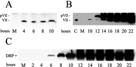
[image:2.585.308.534.69.181.2]Virus-derived protein VII persists during infection.We and others have reported that protein VII from infectious viral particles enters the cell along with viral DNA (8, 15, 18, 21). We wanted to examine the appearance of this input protein VII and also the new production of pre-protein VII and ma-ture protein VII during infection. These studies were expected to reveal when the various forms of protein VII function dur-ing infection. HeLa cells were infected with phenotypically wild-typedl309 at a MOI of 50. This MOI was used to more easily observe amounts of input protein VII. Western blot analysis was performed using our previously described anti-protein VII antibody (21). Previously we showed that 100% of cell-associated protein VII enters the nucleus by 4 h postinfec-tion (21). As shown in Fig. 1A, input protein VII was apparent within 4 h of infection and remained constant through the first 10 h. In other experiments input protein VII could be seen entering the nucleus as early as 1 h postinfection (data not shown; see also Fig. 7). For Fig. 1B, later time points were examined. Production of pre-protein VII began around 12 h postinfection. This is consistent with the fact that the mRNA for pre-protein VII is transcribed from the L2 gene, which is controlled by the major late promoter. Mature protein VII began to accumulate between 12 and 14 h postinfection and both the precursor and mature forms of the protein then ac-cumulated to high levels. To establish the timing of early-phase events, expression DBP was also monitored. DBP is encoded
FIG. 1. Appearance of input protein VII and new synthesis of pre-VII and mature protein pre-VII during infection. HeLa cells were infected for the indicated times and analyzed by Western blot for protein VII (A and B) or the 72-kDa DBP (C). The anti-protein VII antibody recognizes both pre-VII (pVII) and protein VII. M, mock infection; C, control lane containing virus-derived protein VII.
on November 8, 2019 by guest
http://jvi.asm.org/
by the E2A early gene. As shown in Fig. 1C, DBP was first detected 6 h postinfection and accumulated to high levels thereafter. To relate these protein expression results to viral DNA replication, PCR analysis of extracts from infected cells revealed that DNA replication commenced between 6 and 8 h postinfection (data not shown), as has been reported previ-ously (38). These data demonstrate that input protein VII is present in cells during the early phase of infection and that it persists during the beginning of the DNA replication phase. They also show that accumulation of pre-protein VII starts after the onset of DNA replication and that production of mature protein VII commences shortly thereafter.
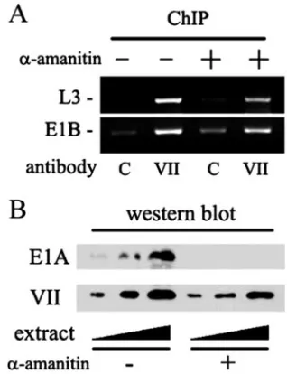
[image:3.585.51.276.66.214.2]Protein VII associates with viral DNA throughout the early phase and also during DNA replication. Recently we used chromatin immunoprecipitation (ChIP) to demonstrate that input protein VII can be found associated with viral DNA at an early-phase time point, 4 h postinfection (21). Furthermore, we observed that the input protein VII was restricted to viral sequences and did not migrate to cellular sequences (21). We next wanted to extend this analysis to determine if the protein VII-DNA association persists through later phases of infec-tion. HeLa cells were again infected withdl309 at a MOI of 50 and subjected to ChIP analysis as described previously. As shown in Fig. 2A, specific association of protein VII with viral DNA was detected throughout a 14-h time course of infection. Figure 2B shows that greater than 7% of the input DNA was immunoprecipitated from an extract of cells infected for 4 h. This is the same as the percentage of protein VII brought down in a parallel immunoprecipitation (data not shown), indicating that the ChIP was very efficient. Since newly synthesized pre-protein VII does not begin to accumulate significantly until around 12 h postinfection, these data demonstrate that input protein VII remains associated with the viral DNA throughout the early phase and also well into the DNA replication phase, which starts between 6 and 8 h postinfection. This suggests a functional role for protein VII throughout early transcription
and possibly DNA replication. In repeated experiments the extent of protein VII-DNA association increased somewhat over time (Fig. 2A and see also Fig. 4A). It is likely that this increase was due in part to an accumulation of newly synthe-sized viral DNA and/or pre-protein VII as the infection pro-ceeds. A similar time course ChIP experiment was recently reported by Haruki et al. (18). However, they reported a de-crease in protein VII-DNA association as infection proceeds. This discrepancy with our results, which may stem from pro-cedural differences, will be addressed in Discussion.
The majority of protein VII is not released from the viral DNA during early phase. Our ChIP data indicate a stable, ongoing association between protein VII and the DNA. How-ever, we considered the possibility that during the first 4 h of infection the establishment of early viral transcription might lead to the loss of a majority of input protein VII from the viral chromatin. We used the potent transcription inhibitor␣ -aman-itin to examine this possibility. If early transcription normally causes a significant release of protein VII from the DNA, then a general inhibition of transcription by␣-amanitin would allow the bulk of protein VII to remain chromatin associated. This would result in an increase in the observed protein VII-DNA association at 4 h. Accordingly, infected cells were treated with 10g of␣-amanitin/ml starting at 1 h postinfection and har-vested for ChIP analysis 3 h later. As shown in Fig. 3A, there was no effect of␣-amanitin on the protein VII-DNA associa-tion. As a control, infected cells treated with␣-amanitin were analyzed by Western blotting for the presence of E1A protein and also for input protein VII (Fig. 3B). Complete inhibition of E1A production occurred, indicating that early transcription was efficiently shut off by␣-amanitin. Since the E1A gene is the first viral gene to be transcribed during infection, this result
FIG. 2. Protein VII association with viral chromatin. (A) HeLa cells were infected for the indicated times and analyzed by ChIP as described in Materials and Methods. Cells were treated with formal-dehyde to cross-link protein to nucleic acid. Cell extracts were pre-pared, sonicated to shear the DNA to roughly 200 bp, and then incu-bated with anti-protein VII antibody (VII) or control preimmune serum (C). Precipitates were heated to reverse the cross-links, depro-teinized, and assayed by PCR with primers specific for the E1B gene. G, genomic virus DNA control. pi, postinfection. (B) ChIP efficiency. Cells were infected for 4 h and analyzed by ChIP.
FIG. 3.␣-Amanitin treatment of infected cells. (A) Cells were in-fected withdl309 for 4 h and treated with 10 g of␣-amanitin/ml starting 1 h postinfection. (A) ChIP analysis was performed as de-scribed in Materials and Methods by using primers representing the L3 and E1B coding regions. C, control preimmune serum; VII, anti-protein VII antiserum. (B) Western blot analysis of E1A and input protein VII. Ramps indicate that increasing amounts of extract were analyzed.
on November 8, 2019 by guest
http://jvi.asm.org/
[image:3.585.340.499.443.650.2]also confirms previous studies demonstrating that early tran-scription was indeed ongoing during the initial 4-h time period in our experiments. As expected there was little or no effect of
␣-amanitin on the presence of input protein VII because this protein is not synthesized during the early phase (Fig. 3B). We conclude that the majority of input protein VII that is bound to viral DNA in the infecting virion remains associated with the DNA during the early phase of infection. These data are fur-ther supported by immunofluorescence experiments (below).
The data presented above strongly suggested that protein VII remains associated with incoming viral DNA at least through the onset of viral DNA replication. To test directly if the presence of protein VII on the DNA is consistent with all steps that precede the onset of viral DNA replication, the DNA synthesis inhibitor hydroxyurea (HU) was employed. Cells were infected and then treated with 3g of HU/ml starting 1 h postinfection. The cells were harvested at 4 and 12 h postin-fection and analyzed by ChIP. As shown in Fig. 4A, there was no effect of HU on protein VII-DNA association at the 4-h time point. This was expected because no DNA replication takes place during this time. In the absence of HU there was a detectable increase in protein VII-DNA association at 12 h postinfection. This was due to the accumulation of newly syn-thesized DNA and possibly newly synsyn-thesized pre-protein VII. In the presence of HU this increase was completely inhibited, as expected. Significantly, the remaining level of protein VII-DNA association at the 12-h time point in the presence of HU was equivalent to that seen in the absence of HU at 4 h postinfection (compare lane 2 to lane 8). At 12 h postinfection, a background signal was reproducibly observed due to nonspe-cific trapping of newly synthesized viral DNA in the immuno-precipitation with preimmune serum (lane 5). This background signal was also inhibited by HU (lane 7).
These data indicate that protein VII remains associated with viral DNA at least up to the point at which DNA replication commences. As a control to confirm that early-phase events were indeed intact in HU-treated cells, the accumulation of the
72-kDa DBP was measured. As shown in Fig. 4B, HU had no effect on production of DBP, demonstrating that early-phase transcription was normal in this experiment. These data dem-onstrate that the presence of protein VII in viral chromatin is consistent with all steps that precede the onset of DNA repli-cation.
Viral chromatin and protein VII associate with SET and pp32.The SET protein has been shown to interact with viral chromatin in vivo and with protein VII in vitro (18). SET is a component of two multiprotein complexes called SET and INHAT (2, 4, 10–12, 36, 37). We wondered if only the SET protein could associate with adenovirus chromatin, as reported earlier (18), or if additional members of the SET and INHAT complexes are also present. To test this we performed a ChIP experiment by using an antibody directed against pp32, which is a component of both the SET and INHAT complexes. As shown in Fig. 5A, at 6 h postinfection pp32 was indeed found associated with adenovirus chromatin. Identical results were obtained for the SET protein, confirming previous data (18).
FIG. 4. (A) ChIP of infected cells treated with HU. Cells were infected withdl309 for the indicated times and treated with 3g of HU/ml starting 1 h post infection (pi). ChIP was performed as de-scribed in Materials and Methods by using primers specific for the L3 gene. C, control preimmune serum; VII, anti-protein VII antibody. (B) Cells were infected for the indicated times and treated with HU as described for panel A. Extracts were prepared and analyzed by SDS-PAGE and Western blotting for DBP.
FIG. 5. Viral chromatin and protein VII association with pp32 and SET. (A) ChIP analysis. Cells were infected withdl309 for 6 h and analyzed by ChIP with anti-pp32 (␣-pp32), anti-SET (␣-SET), or con-trol (C) antibodies. PCR products: MLP, major late promoter; L3, late region 3; E1A, early region 1A. (B) HeLa cell lysates were used in binding reactions with GST-protein VII, GST-E1A289, or the unrelated protein control GST-CAS YXXP (7). Bound proteins were separated by SDS-PAGE and analyzed by Western blotting for SET or pp32. Lower panel, structure of GST-protein VII mutants. Bars indicate regions of protein VII expressed in the mutants. The GST moiety is not illustrated.
on November 8, 2019 by guest
http://jvi.asm.org/
Additionally we asked if cellular pp32 and SET could inter-act with protein VII in a GST pull-down assay. HeLa cell extracts were incubated with agarose-bound GST-protein VII. The beads were washed and then analyzed by SDS-PAGE. As shown in Fig. 5B, GST-protein VII could specifically pull down pp32 and also SET. As a negative control, GST-protein VII was unable to associate with the nuclear DNA-binding protein Ku86 (21; also data not shown). GST-E1A also did not show any association with either SET or pp32.
We mapped the domains of protein VII that associate with SET and pp32. Interestingly, these two proteins bound to ad-jacent domains on protein VII. pp32 bound to the 45–94 do-main and SET associated with the 94–148 dodo-main (Fig. 5B). Since pp32 and SET are components of multiprotein com-plexes we might have expected that they would be pulled down together by either of the protein VII binding domains, but this was not the case. The 45–94 domain associated efficiently only with pp32 and the 94–148 domain associated only with SET. However, the interaction between pp32 and SET is very weak (J. Lieberman, unpublished observation) so this result is not surprising. These data suggest that a multiprotein complex containing SET and pp32 associates with viral chromatin in vivo. Furthermore, they suggest that this association takes place by way of multiple contacts with protein VII. Protein VII can serve to nucleate the formation of a higher-order complex containing SET and pp32.
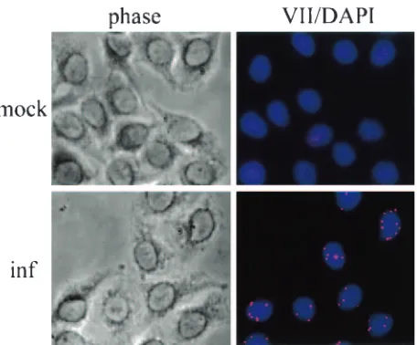
Protein VII localizes as discrete dots in the nucleus.Further evidence for an important role for input protein VII was de-veloped using immunofluorescence. Cells were infected for 4 h at a MOI of 5, followed by methanol fixation and staining with anti-protein VII antibody. As shown in Fig. 6, input protein VII localized as discrete dots within the nucleus. No staining was observed in mock-infected control cells (Fig. 6, top) or when only a fluorescent secondary antibody was used (data not shown). Comparison with phase-contrast images and
DAPI-stained nuclei indicated that at 4 h postinfection, 100% of the protein VII staining was located within the nuclei. These data, taken together with the ChIP and protein expression data presented above, suggest that each dot represents an individual viral genome associated with protein VII. The success of these assays was probably due to the presence of up to 1,000 copies of protein VII per genome and the polymeric nature of the protein VII-DNA complex, which resulted in an intense signal upon binding of antibody.
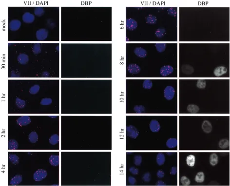
Early-to-late shift in protein VII localization.The localiza-tion of protein VII dots was examined over a time course of infection. As shown in Fig. 7, specific staining was visible as early as 30 min postinfection. At the 30-min and 1-h time points a majority of dots were located outside the nucleus. Comparison with phase-contrast images indicated that at these very early times all protein VII dots were cell associated; none were located outside of the cells (data not shown). A few nucleus-associated dots were present at these times, and these generally showed more-intense staining than those outside the nucleus. There was no staining of mock-infected cells. Nuclear accumulation continued during the next 3 h such that by 4 h postinfection, staining was almost entirely nuclear and the in-tensity of staining was maximal. These data also indicate that 100% of the cells were infected. Considering the ChIP data presented above, these data strongly suggest that there is an accumulation of protein VII-DNA complexes within the nu-cleus during the initial few hours of infection. We also note that the intensity of the dots increased as they accumulated in the nucleus during the initial 4 h of infection. This suggests that there is an unmasking of protein VII epitopes perhaps due to changes in the molecular architecture of the viral chromatin.
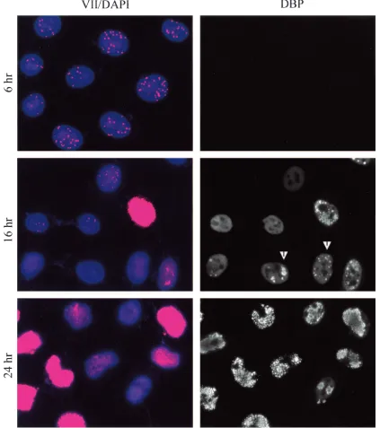
Interestingly, nuclear dots persisted through 14 h postinfec-tion, but their intensity weakened significantly between 8 and 14 h. By 16 h postinfection (Fig. 8) some cells began to exhibit general (diffuse) nuclear protein VII staining. Considering the protein expression data in Fig. 1, it is likely that the general nuclear staining is due to new production of pre-protein VII and mature protein VII and that the dots represent persistent input protein VII in association with input viral DNA. The diffuse nature of the nuclear staining suggests that newly syn-thesized pre-protein VII is dispersed in its localization. This is in sharp contrast to input protein VII, which probably localizes in a condensed form as a component of viral chromatin. At 16 h only a minority of cells contained dots, and their intensity was weak compared with dots in early phase (Fig. 8, compare 16 h to 6 h). By 24 h postinfection there was intense general-ized protein VII staining in a majority of the cells. Because of the intensity of staining in those cells, it was not possible to know if dots were present. Some cells exhibited no general staining and no nuclear dots even though they were clearly infected as judged by the presence of the 72-kDa DNA-binding protein. These data indicate that there was a peak of nuclear dot intensity (input protein VII) between 2 and 10 h postin-fection and that these structures subsequently dissipated, were masked, or lost their association with protein VII. The immu-nofluorescence data for the early time points (through 8 h postinfection) support our conclusion from ChIP assays that the majority of protein VII is not released from the DNA during early phase. They also suggest that there is an
indepen-FIG. 6. Protein VII is in nuclear dots in early phase. HeLa cells were grown on glass coverslips and infected with phenotypically wild-typedl309 for 4 h. Cells were fixed by cold methanol dehydration and probed with rabbit polyclonal anti-protein VII antibody. For visualiza-tion, cells were incubated with rabbit rhodamine secondary anti-body conjugate. Prior to mounting, cells were counterstained with DAPI. Phase, phase-contrast microscopy; mock, uninfected control; inf, infected cells.
on November 8, 2019 by guest
http://jvi.asm.org/
[image:5.585.51.279.66.255.2]dent accumulation of pre-protein VII and mature protein VII at late times that results in general nuclear staining.
Localization of 72-kDa DNA-binding protein.To relate the appearance and localization of protein VII to other events during infection, the cells were costained for the 72-kDa DBP, whose localization has been previously described. Starting at 8 h postinfection individual cells could be seen to express DBP, and their number increased dramatically after this (Fig. 7 and 8). As the infection progressed to 16 h, DBP localized as expected in replication centers, as has been reported (30, 40) (Fig. 8). By 24 h postinfection virtually all cells expressed DBP, which showed punctate staining. As expected, the overall mor-phology of these late infected cells was drastically altered com-pared to uninfected cells (data not shown).
Clearly, cells that showed general DBP staining (e.g., at 12 h postinfection) were further along in the infection cycle than cells that had yet to express DBP. Furthermore, cells in which DBP was localized to replication centers (e.g., at 16 h postin-fection) had progressed further than cells with only general
[image:6.585.56.528.67.449.2]DBP staining. Examination of the 16-h time point revealed a trend: cells with DBP staining showed a decrease in dot inten-sity compared with the 6-h time point, and cells containing prominent replication centers contained no or very few dots. Conversely, cells that had no DBP staining contained more-intense nuclear dots and little if any general protein VII stain-ing (e.g., at 6 h). These data strongly suggest a progression in protein VII localization that correlates with the progress of infection. At very early times (no DBP staining) only dots are present. At later times (general DBP staining or localization to replication centers) there is production of pre-protein VII which results in general nuclear staining and a concomitant decrease in dot intensity due perhaps to release of input pro-tein VII from the viral DNA. This indicates that the cells that express new protein VII are those that have progressed further in the infection. Taken together, these data provide further support for the protein expression and ChIP data presented above. During the early phase (0 to 6 h) and at least partially into the DNA replication phase, input protein VII persists in
FIG. 7. Protein VII dot localization throughout infection. HeLa cells were infected for the indicated times at a MOI of 20. Cells were fixed by cold methanol dehydration and probed with mouse monoclonal anti-DBP and rabbit polyclonal anti-protein VII antibodies. For visualization, cells were probed with anti-mouse FITC and anti-rabbit rhodamine secondary antibody conjugates. Prior to mounting, cells were counterstained with DAPI.
on November 8, 2019 by guest
http://jvi.asm.org/
discrete structures that are likely to include input viral ge-nomes. As the infection progresses, production of pre-protein VII and mature protein VII correlates with other late-phase events. At the same time there is a decrease in dot intensity that may indicate a release of input protein VII from the DNA at late times or a specific reorganization of the viral genome that precludes recognition by the anti-protein VII antibody.
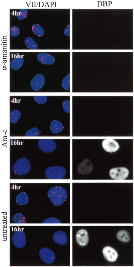
Disappearance of input protein VII dots is due to an active process but does not require ongoing DNA replication. As mentioned above, input protein VII dots decreased
signifi-cantly in number and intensity at late times (Fig. 7 and 8). We considered the possibility that this change was due to loss of protein VII from chromatin during DNA replication. To test this, infected cells were treated with Ara-C, an inhibitor of DNA replication. Infected cells were treated starting 1 h post-infection with 25g of Ara-C/ml and then prepared for im-munofluorescence at 4 and 16 h postinfection. Figure 9 shows that cells treated with Ara-C behaved identically to untreated cells: they contained dots at 4 h postinfection but not at 16 h postinfection. This demonstrates that ongoing DNA
replica-FIG. 8. Protein VII dot localization at late times. HeLa cells were infected for the indicated times at a MOI of 20. Cells were fixed by cold methanol dehydration and probed with mouse monoclonal anti-DBP and rabbit polyclonal anti-protein VII antibodies. For visualization, cells were probed with anti-mouse FITC and anti-rabbit rhodamine secondary antibody conjugates. Prior to mounting, cells were counterstained with DAPI. Arrowheads indicate examples of cells containing replication centers.
on November 8, 2019 by guest
http://jvi.asm.org/
[image:7.585.82.505.72.549.2]tion does not trigger disappearance of the protein VII dots. Identical results were obtained with the DNA synthesis inhib-itor hydroxyurea (data not shown). We also challenged cells with 10g of␣-amanitin/ml to determine the effect of inhibi-tion of RNA synthesis on disappearance of the dots. Interest-ingly,␣-amanitin largely prevented loss of the protein VII dots. This indicates that loss of the dots was not due to passive diffusion of protein VII away from their concentrated centers. Rather, these data argue that an active process was involved in altering the signal of input protein VII in the cell.
DISCUSSION
We have examined the fate and function of adenovirus pro-tein VII during infection. Five lines of investigation support an ongoing role for protein VII throughout early phase and per-haps during DNA replication as well. First, as judged by West-ern blotting, input protein VII persists in the nucleus at least through the beginning of DNA replication. Second, ChIP as-says reveal an extended association between protein VII and viral DNA. Third, protein VII can be found in nuclear dot structures during the early phase and beyond. Fourth, SET and pp32, both of which associate with protein VII in GST pull-down assays, are found in association with viral DNA during early phase. Finally, our previous finding that protein VII can bind to E1A supports a role for protein VII in early transcrip-tional activation (21).
We wondered if the nuclear dots seen at early times might be precursors to previously characterized viral replication cen-ters, which are the major sites of DNA replication. Input viral genomes by definition must be the templates for early viral transcription and replication. Both input adenoviral genomes at early times and adenoviral replication centers at late times have been found adjacent to promyelocytic leukemia nuclear bodies (PML-NBs or ND10) (20), suggesting that adenoviral replication centers develop from input viral genomes. We ob-served no concordance between protein VII dots and viral replication centers as judged by costaining of protein VII and DBP, a marker for replication centers. However, cells that contained visible replication centers had generally lost their dots (Fig. 8). A small minority of cells were observed in which some protein VII dots were visible along with replication cen-ters, though these signals did not colocalize (data not shown). It is possible, however, that protein VII dot structures progress into replication centers after protein VII fluorescence is no longer detectable. It is also possible that, after the earliest rounds of DNA replication, there could be a shift in localiza-tion of DNA replicalocaliza-tion to the replicalocaliza-tion centers. Both hy-potheses are consistent with our data. We showed that the nuclear dots disappear at late times and that their disappear-ance occurs even in the absence of ongoing DNA synthesis. Similarly, Haruki et al. used ChIP to show that a decrease in protein VII-DNA association at late times does not require DNA replication (18). These data indicate that the process of DNA replication does not cause release of protein VII from the DNA. Rather, we find that RNA synthesis is required for disappearance of the dots to occur. Since loss of the dots occurs at late times but not at early times, our data are con-sistent with the idea that late transcription itself could be responsible for this effect. Other possibilities exist however.
For instance, it is conceivable that DBP could displace protein VII from the DNA either in the presence or absence of ongo-ing DNA replication. Alternatively, the observed decrease in intensity could be due to a change in chromatin structure that leads to an altered accessibility of the viral chromatin to the antibody. This possibility is consistent with a stable or increas-ing level of protein VII in association with the viral DNA. Finally, it is possible that at late times the nuclear dot struc-tures experience a weakening in the protein VII-DNA
associ-FIG. 9. Disappearance of nuclear dots requires RNA synthesis but not DNA synthesis. HeLa cells were infected for the indicated times and were treated at 1 h postinfection with either 25g of Ara-C/ml or 10g of␣-amanitin/ml. Cells were fixed by cold methanol dehydration and probed with mouse monoclonal anti-DBP and rabbit polyclonal anti-protein VII antibodies. For visualization, cells were probed with anti-mouse FITC and anti-rabbit rhodamine secondary antibody con-jugates. Prior to mounting, cells were counterstained with DAPI.
on November 8, 2019 by guest
http://jvi.asm.org/
[image:8.585.310.533.70.512.2]ation and that our methanol fixation method results in removal of protein VII. We are currently working to characterize the association between protein VII and viral DNA at late times. The observation that protein VII remains associated with viral DNA during the beginning of the DNA replication phase sug-gests a role for protein VII in this process.
We note a minor discrepancy between our results and those of Haruki et al. In their ChIP experiments the association be-tween protein VII and viral DNA diminished over the course of a 12-h infection (18). In our ChIP experiments we showed an increase in association as the infection proceeded through 12 to 14 h (Fig. 2 and 4). As described earlier, the increase we observed was due to an increase in available DNA due to new DNA replication. This conclusion is based on the fact that the observed increase in protein VII-DNA association was com-pletely inhibited by addition of HU to the infected cells (Fig. 4). The difference between our results and those of Haruki et al. are likely due to our longer cross-linking times used in the ChIP assay, which would result in the detection of both strong and weak DNA-protein interactions. If newly synthesized DNA at late times is not fully condensed and therefore is weakly associated with protein VII, these interactions might be detected only with longer cross-linking times. In our immuno-fluorescence experiments we did observe a decrease in the protein VII signal in nuclear dots as the infection proceeded. We used methanol for fixing the infected cells, which might cause disruption of weak protein VII-DNA interactions at late times, leading to a less-intense signal. On the other hand, our ChIP experiments were performed with formaldehyde cross-linking, which would preserve those weak interactions.
Our data concerning the SET and pp32 proteins clearly suggest that a multiprotein complex, possibly the SET and/or INHAT complex, is functioning at the viral chromatin. The iden-tity, integrity, and function of such a complex will be the sub-ject of additional studies. Both the SET complex and INHAT have been implicated in chromatin metabolism. INHAT can inhibit histone acetyltransferases by binding directly to his-tones and masking their sites of acetylation (36, 37). The SET complex has been shown to mediate granzyme A-dependent apoptosis, in part due to a DNase activity of the complex (2, 10–12). It is interesting to speculate that this complex has additional functions including the regulation of viral chroma-tin. The SET protein, when studied alone by using in vitro ap-proaches, has NAP, chromatin remodeling, and chromosome decondensing activities (2, 26, 28). It remains to be determined how these activities relate to one another biochemically and whether the SET protein acts alone at adenovirus chromatin or in combination with other components of the INHAT and SET complexes. The fact that both SET and pp32 can bind to pro-tein VII, and that distinct domains of propro-tein VII mediate binding to these proteins, suggests that a multiprotein complex containing protein VII, SET, and pp32 is likely to be function-ing durfunction-ing the early stages of adenovirus replication.
ACKNOWLEDGMENTS
This work was supported by NIH Public Health Service grants CA60675 to D.A.E., CA77342 to D.A.O., and AI45587 to J.L.
REFERENCES
1.Amin, M., A. Mirza, and J. Weber.1977. Genetic analysis of adenovirus type 2. VII. Cleavage-modified affinity for DNA of internal virion proteins.
Vi-rology80:83–97.
2.Beresford, P. J., D. Zhang, D. Y. Oh, Z. Fan, E. L. Greer, M. L. Russo, M. Jaju, and J. Lieberman.2001. Granzyme A activates an endoplasmic retic-ulum-associated caspase-independent nuclease to induce single-stranded
DNA nicks. J. Biol. Chem.276:43285–43293.
3.Black, B. C., and M. S. Center.1979. DNA-binding properties of the major
core protein of adenovirus 2. Nucleic Acids Res.6:2339–2353.
4.Brennan, C. M., I. E. Gallouzi, and J. A. Steitz.2000. Protein ligands to HuR
modulate its interaction with target mRNAs in vivo. J. Cell Biol.151:1–14.
5.Brown, D. T., M. Westphal, B. T. Burlingham, U. Winterhoff, and W. Doer-fler.1975. Structure and composition of the adenovirus type 2 core. J. Virol.
16:366–387.
6.Burg, J. L., J. Schweitzer, and E. Daniell.1983. Introduction of superhelical turns into DNA by adenoviral core proteins and chromatin assembly factors.
J. Virol.46:749–755.
7.Burnham, M. R., M. T. Harte, A. Richardson, J. T. Parsons, and A. H. Bouton.1996. The identification of p130cas-binding proteins and their role
in cellular transformation. Oncogene12:2467–2472.
8.Chatterjee, P. K., M. E. Vayda, and S. J. Flint.1986. Adenoviral protein VII packages intracellular viral DNA throughout the early phase of infection.
EMBO J.5:1633–1644.
9.Everitt, E., L. Lutter, and L. Philipson.1975. Structural proteins of viruses. XII. Location and neighbor relationship among proteins of adeno-virion type 2 as revealed by enzymatic iodination, immunoprecipitation and
chemical cross-linking. Virology67:197–208.
10.Fan, Z., P. J. Beresford, D. Y. Oh, D. Zhang, and J. Lieberman.2003. Tumor suppressor NM23-H1 is a granzyme A-activated DNase during CTL-medi-ated apoptosis, and the nucleosome assembly protein SET is its inhibitor.
Cell112:659–672. (Erratum,115:241.)
11.Fan, Z., P. J. Beresford, D. Zhang, and J. Lieberman.2002. HMG2 interacts with the nucleosome assembly protein SET and is a target of the cytotoxic
T-lymphocyte protease granzyme A. Mol. Cell. Biol.22:2810–2820.
12.Fan, Z., P. J. Beresford, D. Zhang, Z. Xu, C. D. Novina, A. Yoshida, Y. Pommier, and J. Lieberman.2003. Cleaving the oxidative repair protein
Ape1 enhances cell death mediated by granzyme A. Nat. Immunol.4:145–
153.
13.Flint, J., and T. Shenk.1997. Viral transactivating proteins. Annu. Rev.
Genet.31:177–212.
14.Fujii-Nakata, T., Y. Ishimi, A. Okuda, and A. Kikuchi.1992. Functional analysis of nucleosome assembly protein, NAP-1. The negatively charged COOH-terminal region is not necessary for the intrinsic assembly activity.
J. Biol. Chem.267:20980–20986.
15.Greber, U. F., M. Suomalainen, R. P. Stidwill, K. Boucke, M. W. Ebersold, and A. Helenius.1997. The role of the nuclear pore complex in adenovirus
DNA entry. EMBO J.16:5998–6007.
16.Harlow, E., B. R. Franza, Jr., and C. Schley.1985. Monoclonal antibodies specific for adenovirus early region 1A proteins: extensive heterogeneity in
early region 1A products. J. Virol.55:533–546.
17.Harlow, E., and D. Lane.1988. Antibodies: a laboratory manual, p. 471–510. Cold Spring Harbor Laboratory, Cold Spring Harbor, N.Y.
18.Haruki, H., B. Gyurcsik, M. Okuwaki, and K. Nagata.2003. Ternary com-plex formation between DNA-adenovirus core protein VII and TAF-Ibeta/
SET, an acidic molecular chaperone. FEBS Lett.555:521–527.
19.Hosokawa, K., and M. T. Sung.1976. Isolation and characterization of an
extremely basic protein from adenovirus type 5. J. Virol.17:924–934.
20.Ishov, A. M., and G. G. Maul.1996. The periphery of nuclear domain 10
(ND10) as site of DNA virus deposition. J. Cell Biol.134:815–826.
21.Johnson, J. S., Y. S. Osheim, Y. Xue, M. R. Emanuel, P. W. Lewis, A. Bankovich, A. L. Beyer, and D. A. Engel. 2004. Adenovirus protein VII condenses DNA, represses transcription, and associates with transcriptional
activator E1A. J. Virol.78:6459–6468.
22.Jones, N., and T. Shenk.1979. Isolation of adenovirus type 5 host range
deletion mutants defective for transformation of rat embryo cells. Cell17:
683–689.
23.Kawase, H., M. Okuwaki, M. Miyaji, R. Ohba, H. Handa, Y. Ishimi, T. Fujii-Nakata, A. Kikuchi, and K. Nagata.1996. NAP-I is a functional ho-mologue of TAF-I that is required for replication and transcription of the
adenovirus genome in a chromatin-like structure. Genes Cells1:1045–1056.
24.Loyola, A., and G. Almouzni.2004. Histone chaperones, a supporting role in
the limelight. Biochim. Biophys. Acta1677:3–11.
25.Maizel, J. V., Jr., D. O. White, and M. D. Scharff.1968. The polypeptides of adenovirus. II. Soluble proteins, cores, top components and the structure of
the virion. Virology36:126–136.
26.Matsumoto, K., K. Nagata, M. Miyaji-Yamaguchi, A. Kikuchi, and M. Tsu-jimoto.1999. Sperm chromatin decondensation by template activating factor
I through direct interaction with basic proteins. Mol. Cell. Biol.19:6940–
6952.
27.Nagata, K., H. Kawase, H. Handa, K. Yano, M. Yamasaki, Y. Ishimi, A.
on November 8, 2019 by guest
http://jvi.asm.org/
Okuda, A. Kikuchi, and K. Matsumoto.1995. Replication factor encoded by a putative oncogene, set, associated with myeloid leukemogenesis. Proc.
Natl. Acad. Sci. USA92:4279–4283.
28.Okuwaki, M., and K. Nagata.1998. Template activating factor-I remodels the chromatin structure and stimulates transcription from the chromatin
template. J. Biol. Chem.273:34511–34518.
29.Ornelles, D. A., and T. Shenk.1991. Localization of the adenovirus early region 1B 55-kilodalton protein during lytic infection: association with nu-clear viral inclusions requires the early region 4 34-kilodalton protein. J.
Vi-rol.65:424–429.
30.Pombo, A., J. Ferreira, E. Bridge, and M. Carmo-Fonseca.1994. Adenovirus replication and transcription sites are spatially separated in the nucleus of
infected cells. EMBO J.13:5075–5085.
31.Prage, L., and U. Pettersson.1971. Structural proteins of adenoviruses. VII. Purification and properties of an arginine-rich core protein from adenovirus
type 2 and type 3. Virology45:364–373.
32.Reich, N. C., P. Sarnow, E. Duprey, and A. J. Levine.1983. Monoclonal antibodies which recognize native and denatured forms of the adenovirus
DNA-binding protein. Virology128:480–484.
33.Rekosh, D. M., W. C. Russell, A. J. Bellet, and A. J. Robinson.1977.
Identification of a protein linked to the ends of adenovirus DNA. Cell11:
283–295.
34.Russell, W. C., W. G. Laver, and P. J. Sanderson.1968. Internal components
of adenovirus. Nature219:1127–1130.
35.Sato, K., and K. Hosokawa.1984. Analysis of the interaction between DNA
and major core protein in adenovirus chromatin by circular dichroism and
ultraviolet light induced cross-linking. J. Biochem.95:1031–1039.
36.Seo, S. B., T. Macfarlan, P. McNamara, R. Hong, Y. Mukai, S. Heo, and D. Chakravarti.2002. Regulation of histone acetylation and transcription by
nuclear protein pp32, a subunit of the INHAT complex. J. Biol. Chem.277:
14005–14010.
37.Seo, S. B., P. McNamara, S. Heo, A. Turner, W. S. Lane, and D. Chakravarti.
2001. Regulation of histone acetylation and transcription by INHAT, a
human cellular complex containing the set oncoprotein. Cell104:119–130.
38.Shenk, T.1996. Adenoviridae: the viruses and their replication, p. 2111–
2148.InB. N. Fields, D. M. Knipe, and P. M. Howley (ed.), Fields virology,
vol. 2. Lippincott-Raven, Philadelphia, Pa.
39.Vaesen, M., S. Barnikol-Watanabe, H. Gotz, L. A. Awni, T. Cole, B. Zim-mermann, H. D. Kratzin, and N. Hilschmann.1994. Purification and char-acterization of two putative HLA class II associated proteins: PHAPI and
PHAPII. Biol. Chem. Hoppe-Seyler375:113–126.
40.Voelkerding, K., and D. F. Klessig.1986. Identification of two nuclear
sub-classes of the adenovirus type 5-encoded DNA-binding protein. J. Virol.60:
353–362.
41.von Lindern, M., S. van Baal, J. Wiegant, A. Raap, A. Hagemeijer, and G. Grosveld.1992. Can, a putative oncogene associated with myeloid
leukemo-genesis, may be activated by fusion of its 3⬘half to different genes:
charac-terization of the set gene. Mol. Cell. Biol.12:3346–3355.
42.Weber, J. M., G. Khittoo, and A. R. Bhatti.1983. Adenovirus core proteins.
Can. J. Microbiol.29:235–241.