0022-538X/05/$08.00
⫹
0
doi:10.1128/JVI.79.14.9270–9284.2005
Copyright © 2005, American Society for Microbiology. All Rights Reserved.
ERV3 and Related Sequences in Humans: Structure and
RNA Expression†
Ann-Catrin Andersson,
1,2Zhihong Yun,
2Go
¨ran O. Sperber,
3Erik Larsson,
1,4and Jonas Blomberg
2*
Department of Pathology,
1Section of Virology, Department of Medical Sciences,
2and Unit of Physiology,
Department of Neuroscience,
3Uppsala University, Uppsala, Sweden, and Department of
Laboratory Medicine, Women and Childrens Health, NTNU,
Trondheim, Norway
4Received 27 September 2004/Accepted 26 March 2005
The ERV3 locus at chromosome 7q11 is a much studied human endogenous retroviral (HERV) sequence,
owing to an
env
open reading frame (ORF) and placental RNA and protein expression. An analysis of the
human genome demonstrated that ERV3 is one of a group of 41 highly related elements (ERV3-like HERVs)
which use proline, isoleucine, or arginine tRNA in their primer binding sites. In addition to elements closely
related to ERV3, the group included the previously known retinoic acid-inducible element, RRHERVI, also
referred to as HERV15, but was separate from the related HERV-E elements. The ERV3-like elements are
defective. The only element with an ORF among
gag
,
pro
,
pol
, and
env
genes was the
env
ORF of the original
ERV3 locus. A search in dbEST revealed ERV3 RNA expression in placenta, skin, carcinoid tumor, and adrenal
glands. Expression was also studied with newly developed real-time quantitative PCRs (QPCR) of ERV3 and
HERV-E(4-1)
env
sequences. Results from a novel histone 3.3 RNA QPCR result served as the expression
control. QPCR results for ERV3 were compatible with previously published results, with a stronger expression
in adrenal gland and placenta than in 15 other human tissues. The expression of the envelope (
env
) of ERV3
at chromosome 7q11 was also studied by using stringent in situ hybridization. Expression was found in corpus
luteum, testis, adrenal gland, Hassal’s bodies in thymus, brown fat, pituitary gland, and epithelium of the lung.
We conclude that ERV3
env
is most strongly expressed in adrenal and sebaceous glands as well as in placenta.
A substantial part of the human genome consists of
retro-elements in which the human endogenous retroviruses
(HERVs) are included. Depending on the definition of a
ret-rovirus, the HERVs occupy between 2 and 8% of the human
genetic makeup (57, 70). Three decades of HERV research has
generated a number of outstanding issues regarding their role
in genomic evolution, physiology, and pathology (10, 44).
Nearly all HERVs known at present integrated up to 100
million years ago. They retain the proviral structure of
exoge-nous retroviruses. Inherited from generation to generation, a
few of the HERVs have open reading frames (ORFs) that
encode known or putative proteins, while others are highly
mutated. Activation of these elements through their long
ter-minal repeats (LTRs) has been observed at the mRNA level in
selected tissues and different human cell lines (reviewed in
references 10 and 44). The main focus has been on HERV
expression in relation to disease, for example, HERV-W and
HERV-H in multiple sclerosis and schizophrenia and
HERV-K in testis tumors (13, 17, 25, 29, 56). Moreover,
down-regulation of ERV3 is reported in choriocarcinoma (25, 29).
The presence of conserved ORFs is circumstantial evidence for
a functional role for HERV-encoded proteins, exemplified by
Rec/cORF, syncytin, and ERV3 Env (11, 45, 49). Furthermore,
it is established that HERV LTRs also control transcription of
cellular genes such as those for phospholipase A2/otoconin
(22, 35, 79) and pleiotrophin (64). In general, HERVs are
expressed as RNA at a low level in most studied tissues (43, 47,
48) but at higher levels in others (67). Although protein
ex-pression is less studied, a few HERV proteins are more or less
well known (49, 78). In a recent study, Scho
¨n et al. reported
that the polymerase gene of HERVs is active in an
organ-specific way due to the tissue organ-specificity of the LTRs (63),
analogous to other promoters. The envelope gene of ERV3 is
highly expressed in certain tissues, like sebaceous and adrenal
glands (3, 31).
ERV3 is one of the most studied HERVs (3–5, 11, 15, 18, 19,
26, 29–31, 41, 51, 59, 60, 78, 86). It is sometimes referred to as
HERV-R. We dislike this term for reasons discussed below.
ERV3 is a class I HERV which clusters with viruses belonging
to the genus
Gammaretrovirus
(10, 77). It integrated 30 to 40
million years ago and is present in humans and other higher
primates, with the exception of gorillas (26). It was originally
thought to be a solitary provirus (47). A few later observations
indicated the existence of a group of ERV3-like sequences (8,
53). Despite this, a detailed description of this group, its
ex-pression, and the extent of ERV3-like sequences is lacking.
This study describes the structure of the original ERV3 locus
at chromosome band 7q11, identifies several novel ERV3-like
sequences in the human genome, and defines an ERV3-like
HERV group. Transcription factor binding sites unique to the
5
⬘
LTR which possibly are involved in ERV3 7q11 expression
were identified. Expressed sequence tag (EST) sequences
probably transcribed from the ERV3 7q11 locus were searched
* Corresponding author. Mailing address: Section of Virology,
De-partment of Medical Sciences, Uppsala University Academic Hospital,
751 85 Uppsala, Sweden. Phone: 46-18-665593. Fax: 46-18-551012.
E-mail: [email protected].
† Supplementary material for this article may be found at http:
//jvi.asm.org/.
9270
on November 8, 2019 by guest
http://jvi.asm.org/
for in different cell types and used to corroborate previously
reported cap, splice, and polyadenylation sites (30). We
devel-oped real-time quantitative PCRs (QPCRs) that quantitatively
detect transcripts from the SU of
env
from HERV-E(4-1) and
ERV3 7q11 in comparison with nonretroviral reference genes.
The QPCR was used to study the tissue-specific pattern of
RNA expression of the
env
genes of ERV3 7q11 and a related
group, HERV-E. We also checked ERV3
env
mRNA
expres-sion at the tissue and cellular level by an in situ hybridization
(ISH) technique.
MATERIALS AND METHODS
Search for ERV3-like sequences.ERV3-like sequences were gathered by two different methods. First, the SU part of Env was used to search for ERV3 7q11-and HERV-E(4-1)-like sequences in the htgs (high-throughput genomic se-quences) and nr (nonredundant) databases at the National Institute of Biotech-nology Information website, using the TBLASTN program (1). The 50 sequences which had scores of at least 200 were included for further work. Proviral se-quences were extracted from the corresponding genomic clone sese-quences by use of the program RetroTector (G. O. Sperber and J. Blomberg, unpublished data). The 11 most highly scoring and nonredundant ERV3 7q11 SU-like sequences and 6 HERV-E SU-like sequences were aligned at the protein and nucleotide levels by use of the program CLUSTAL W (75) (http://bioweb.pasteur.fr/seqanal /interfaces/clustalw.html). Later, an additional ERV3 7q11 SU-like sequence (in BAC AL356804) was found (Table 1). Data from the alignment were used for constructing an unrooted tree using the programs DNAdist, BioNJ, and Draw-tree as implemented at http://bioweb.pasteur.fr/seqanal/interfaces/clustalw.html (21). This tree included human sequences available in March 2003.
When version hg15 (April 2003), the first assembly of the human genome, became available at the UCSC website (http://genome.ucsc.edu), a genomewide search for retroviral sequences was performed. A second selection method, based on Pol similarity, was used to single out ERV3-related sequences among
these sequences. Altogether, 76 suchpol-containing sequences were selected
from the total collection of 3,661pol-containing RetroTector-defined retroviral
sequences. RetroTector is described further below. Their relation to each other and to other gammaretroviral sequences was investigated using Clustal W and a crosswise similarity table. The latter was produced using a program written by J. Blomberg (see below).
EST sequences were searched in dbEST (National Center for Biotechnology Information) during the spring of 2004 by using BLAST (1). EST sequences were selected according to percent identity to the ERV3 7q11 search sequence, as indicated in Results.
Characterization of ERV3-like sequences. The programs RetroTector and RetroTector Shell (G. O. Sperber and J. Blomberg, unpublished data) were used with the April 2003 version (hg15) of the human genome to identify likely retroviral sequences. Most of the ALU sequences were removed prior to the
analysis. The programs identified 3,661 elements with apolgene. These were
characterized and classified by the programs. Briefly, the program function
Ret-rovID searches for LTR-like sequences and conserved retroviral motifs fromgag,
pro,pol, andenvand parses them together into chains by the use of distance
tables and other heuristics. The function ORFID constructs likely protein
se-quences (puteins) from the machine-identifiedgag,pro,pol, andenvgene
can-didates. It uses codon statistics, frequency of stop codons, and alignment to proteins of known retroviruses belonging to the same genus to approximate the original ORF. The Xonid function searches for possible ORFs in stretches not used by ORFID. The RetroTector interpretations were automatically matched with sequences in an annotated list of retroviral sequences from GenBank, using similarity to Pol proteins, and in a list of repetitive elements from Repbase (27), using similarity to the entire nucleotide sequence of the elements. Both similarity searches were performed with word-based BLAST-like algorithms (1) written by J. Blomberg. This initial screening was followed by alignments and clustering
usingpolandenvnucleotides and Pol and Env proteins with the Clustal W 1.83
[image:2.585.42.542.80.236.2]program. Similarity matrices were generated from all alignments. The Kimura and PAM250 score matrices were used for nucleotides and proteins, respectively. Both alignments and similarity matrices were based on the pairwise deletion technique. This minimizes the influence of insertions and deletions in the retro-viral elements. Thus, similarities were not much affected by gaps. The cladogram
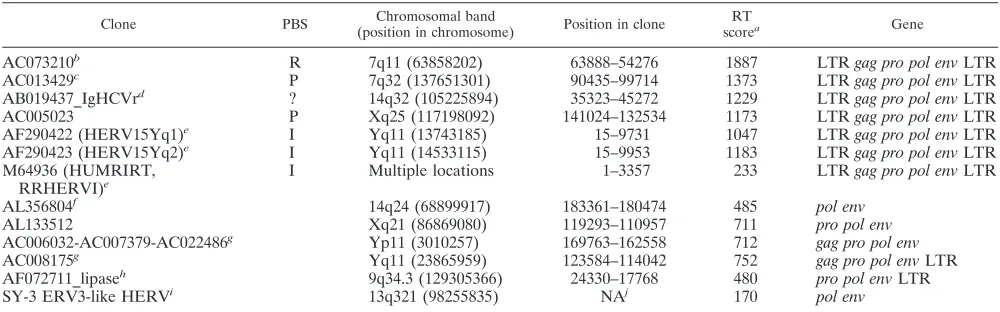
TABLE 1. Selected ERV3
env
-like loci from the human genome version hg15
Clone PBS Chromosomal band
(position in chromosome) Position in clone
RT
scorea Gene
AC073210
bR
7q11 (63858202)
63888–54276
1887
LTR
gag pro pol env
LTR
AC013429
cP
7q32 (137651301)
90435–99714
1373
LTR
gag pro pol env
LTR
AB019437_IgHCVr
d?
14q32 (105225894)
35323–45272
1229
LTR
gag pro pol env
LTR
AC005023
P
Xq25 (117198092)
141024–132534
1173
LTR
gag pro pol env
LTR
AF290422 (HERV15Yq1)
eI
Yq11 (13743185)
15–9731
1047
LTR
gag pro pol env
LTR
AF290423 (HERV15Yq2)
eI
Yq11 (14533115)
15–9953
1183
LTR
gag pro pol env
LTR
M64936 (HUMRIRT,
RRHERVI)
eI
Multiple locations
1–3357
233
LTR
gag pro pol env
LTR
AL356804
f14q24 (68899917)
183361–180474
485
pol env
AL133512
Xq21 (86869080)
119293–110957
711
pro pol env
AC006032-AC007379-AC022486
gYp11 (3010257)
169763–162558
712
gag pro pol env
AC008175
gYq11 (23865959)
123584–114042
752
gag pro pol env
LTR
AF072711_lipase
h9q34.3 (129305366)
24330–17768
480
pro pol env
LTR
SY-3 ERV3-like HERV
i13q321 (98255835)
NA
j170
pol env
aScore for the respective element identified by RetroTector (see Materials and Methods).
bAC073210 (NT_030723) close to humanplk, a Kru¨ppel-like zinc finger gene. Mosaic ERV3-humanplktranscripts are produced through alternative splicing. This
is the ERV3 locus, which gave rise to the ERV3pol-envclone of Cohen et al. (13).
cAC013429 (NT_007680) close to the TIF1 gene (the gene for the transcriptional intermediary factor 1, a multi-zinc finger transcription factor, locus identifier 8805,
positions 524084 to 397969; the ERV-like sequence is at positions 417766 to 427065, between exons 9 and 10 of TIF1, in the opposite direction from TIF1). The protein encoded by this gene mediates transcriptional control by interaction with the activation function 2 (AF2) region of several nuclear receptors, including the estrogen,
retinoic acid, and vitamin D3receptors. It contains three zinc-binding domains. ERV18Y colocalizes with this element.
dThis element resides in the immunoglobulin heavy-chain variable region on chromosome 14.
eThe two HERVI65 integrations on chromosome Yq11 occur close to each other and cause loss of a gene through homologous recombination. There is a striking
concentration of RRHERVI integrations at Yq11. There are also five ERV3-like integrations in the same portion of chromosome Y. HUMRIRT (RRHERVI)
integrations are split up. There is no exact match to HUMRIRT, which is RNA based. There are multiple hits on chromosomes 16p12, Xq24, Yq11(⫽HERVI5y), and
others.
fThis element was detected after the search on which Fig. 2A was based.
gNot only are AC008175 and AC006032 very similar, but there are other sequences in this portion of chromosome Y which have closely matching sequences on
chromosome Y.
hThe ERV3-like element occurs in the 5⬘UTR of a lipase gene on chromosome 9.
iThe SY-3 locus is between exons 5 and 6 of a hypothetical gene (locus 121919).
jNA, not applicable. Clone information for Sy-3 is not available.
on November 8, 2019 by guest
http://jvi.asm.org/
shown below (see Fig. 2B) was based on the guide neighbor-joining tree pro-duced by Clustal. It was processed with TreeView (courtesy of R. Page, Taxon-omy and Systematics, Division of Environmental and Evolutionary Institute of Biomedical and Life Sciences, University of Glasgow; available at http://taxonomy .zoology.gla.ac.uk/rod/rod.html) and tree-modifying programs written by J. Blomberg. The final classification of retroviral elements was consequently based on several independent methods. RetroTector also has an XonID function, which localizes ORFs whose products are longer than 50 amino acids (aa) which
do not overlap the four major ones (gag,pro,pol, andenv). XonID prioritizes
ORFs which begin just after a predicted splice acceptor and end at a predicted splice donor site. It also uses the same codon statistical functions as ORFID. It was applied to the ERV3 locus at 7q11.
Transcription factor binding sites (TFBS) were studied in the 5⬘and 3⬘LTRs,
primarily using ConSite (http://mordor.cgb.ki.se/cgi-bin/CONSITE/consite/). ConSite predictions were based on longer weight matrices than those of other available TFBS prediction methods. This gives a higher statistical significance, and ConSite predictions were therefore used. The vertebrate TFBS subset of the JASPAR database used by ConSite was selected.
RNA samples from diverse tissues and cDNA synthesis.A commercial RNA panel, Human Total RNA Master Panel II (Clontech Laboratories, Palo Alto, CA), was used for cDNA synthesis with subsequent QPCR. Human Total RNA Master Panel II contains RNA from 20 different tissues, most of which consist of pooled RNA from two or more persons. RNA from tissues from the brain cerebellum, whole brain, heart, liver, and lung originated from a single person. Samples derived from a single person of Asian origin, except for the whole brain, which was of Caucasian origin. The sources of pooled RNA varied between 2 and 84 persons, all of Caucasian origin. During the work, it was discovered that three of the RNA samples, from heart, lung, and liver, probably were degraded. They yielded almost no signal with both HERV and housekeeping gene QPCRs. They were therefore excluded from further analysis. The RNA described above was
used as a template for cDNA synthesis with 2g RNA in each reaction mixture.
Synthesis of cDNA was made in a 50-l reaction mixture containing Stratascript
reverse transcription (RT; 1 U/l) (Stratagene, Amsterdam, The Netherlands),
1⫻Stratascript buffer (Stratagene, Amsterdam, The Netherlands), 0.01 M
di-thiothreitol (Promega, Madison, WI), 0.8 mM each deoxynucleoside triphos-phate (Applied Biosystems, PE Europe, The Netherlands), random hexamers
(10.6 ng/l; Amersham Pharmacia Biotech, Uppsala, Sweden), and RNasin (1.6
U/l; Promega, Madison, WI). The cDNA reaction mixture was incubated at
25°C for 10 min, 37°C for 90 min, and 70°C for 15 min and then stored at⫺20°C.
According to the manufacturer, the RNA contained virtually no genomic DNA. However, a control for DNA contamination, a reaction mixture without RT, was made for every RNA sample. The signal was usually negative. In cases where the negative reaction became positive, it was at least 10 times weaker than the RT-positive signal. It was then subtracted from the RT-positive signal. Two microliters of each cDNA was used in each PCR. We prepared five samples in addition to the 16 tissues included in the RNA panel. The samples included three from different placentas (three individuals) and two samples from skeletal mus-cle, one from a single person and one pooled from five different persons. Total RNA was isolated from 30 mg of tissue. The QIAGEN RNA isolation kit was
used according to the manufacturer⬘s recommendations (Merck Eurolab,
Stock-holm, Sweden). Total RNA was DNase treated using a DNA-freekit from
Ambion (Austin, TX), following the protocol from the manufacturer. After DNase treatment, RNA was used for cDNA synthesis, which was performed as
described above in a 50-l reaction mixture. Using this cDNA, 4l was used for
each PCR.
Primer design and QPCR.Primers and reporter-quencher (R-Q) probes were designed with the help of the Primer3 program (http://www.genome.wi.mit.edu
/cgi-bin/primer/primer3). The midpoint temperature (Tm) was calculated by
us-ing the Cybergene program (http://www.cybergene.se/). The primers were syn-thesized by Interactiva (Thermo Hybaid GmbH, Ulm, Germany), and the three
R-Q probes were synthesized by Scandinavian Gene Synthesis (SGS, Ko¨ping,
Sweden). Primer pairs and probes were all high-performance liquid chromatog-raphy purified by the manufacturer. All R-Q probes were labeled with
6-car-boxyfluorescein as a reporter at their 5⬘ends and an internal Dark Quencher
attached to a thymidine in a middle position of the probe sequence (12 to 16 bp
from the 5⬘end). We primarily chose histone 3.3 (M11353) for use as a reference
gene since it is evenly expressed in many cell types, regardless of cell cycle stage (80, 82, 83). Histone 3.3 mRNA has previously been used as a reference in other RNA expression studies (6, 47, 48). Histone 3.3 mRNA is polyadenylated, unlike other histone mRNAs (80). There is room for further studies of its expression in different tissues. The following histone 3.3 primers were based on those originally designed by Mats Lindeskog (Lund University) (48) but were modified by us to
fit new PCR conditions: forward primer 5⬘ CCTCTACTGGAGGGGTGA
AGAA 3⬘(Tm⫽59.4°C) and reverse primer 5⬘TGCCTCCTGCAAAGCACC
GATA 3⬘(Tm⫽59.4°C). The probe, 5⬘CTCTGGAAGCGCAGATCTGTTTT
AAAGTCCT 3⬘, is situated 6 nucleotides (nt) from the reverse primer, with aTm
that is 10°C higher than that of the primer pair (Tm⫽69.7°C). By the use of
dilutions of a clone of an amplimer from a human DNA sample (L. Hu and D. Uzhameckis, unpublished data), the sensitivity for the histone PCR was found to be 1 to 10 target gene equivalents per PCR. The integrity of the plasmid was ascertained by sequencing. Optimization was first made with an iCycler (Bio-Rad Laboratories AB, Sundbyberg, Sweden). Normalization of mRNA expression against cell number, DNA content, total protein, and mRNAs from housekeep-ing genes, like those for actin, GAPDH (glyceraldehyde-3-phosphate dehydro-genase), and histone 3.3, has been used by us or others in numerous studies (50, 62, 76). These genes were selected because of their previously observed wide and approximately even expression levels in many tissues. Besides histone 3.3 RNA, RNAs from three widely used housekeeping genes, i.e., the GAPDH, hypoxan-thine phosphoribosyltransferase 1, and ubiquitin C (UBC) genes (GenBank accession number M26880) (76), were also determined (data not shown). The mean of these three last-mentioned reference RNAs with the Clontech tissue RNA panel was 260 eq per ng of total RNA, with a standard error of the mean of 40.9%, while a histone 3.3 average was 19.5 eq per ng of total RNA, with a standard error of the mean of 15.2%. Thus, histone 3.3 RNA was more evenly expressed. HERV QPCR data were therefore normalized to histone 3.3 RNA expression only. In the following, the word “equivalents” is used instead of “copies” for description of the amount of RNA or DNA detected in QPCR because of probable slight variations in amplification efficiency in samples.
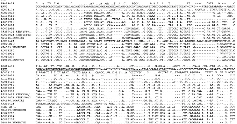
Primer pairs and probes for HERV-E(4-1) and ERV3 7q11 were designed in the SU region where the related HERVs are most divergent and thus should generate specific probes. The chosen primers and probes for HERV-E (HU-MER41, accession no. M10976) and ERV3 (HUMERVA34A, accession no. M12140.1) are marked in the original sequence as shown below (see Fig. 3A and
B, respectively). TheTmfor the ERV3 probe (68.5°C), 5⬘AACTCGCAACTG
TTGGGTTGAGCGGGTCC 3⬘, was 15°C higher than theTms of its primer pair,
namely, forward 5⬘CGCTAGGGGCACGAGTCA 3⬘(Tm⫽53.4°C) and reverse
5⬘AGGCAATTTCTGGTTAACATGCT 3⬘(Tm⫽59.2°C).
TheTmdifference between primers and probe for HERV-E was 9°C. The
sequences were as follows: probe, 5⬘TCACAAACCCCTGACTCATGACAAA
CATATTTA 3⬘(Tm⫽68.4°C); forward primer, 5⬘ATTTGATGCTTGTGCA
GCCATTA 3⬘(Tm⫽59.2°C); and reverse primer, 5⬘TTCTTTTTCCAAGTA
GCCCAAAT 3⬘(Tm⫽58.8°C). Thus, the annealing and extension temperatures
were relatively stringent for both ERV3 and HERV-E SU PCRs. Initially, ERV3 and HERV-E QPCRs were optimized by using human DNA, the above-men-tioned plasmids, and an iCycler iQ multicolor real-time PCR detection system (Bio-Rad Laboratories AB, Sundbyberg, Sweden). The ERV3 clone, php 1.7, is a 1.7-kb HindIII-PstI subclone and was a kind gift from Maurice Cohen. The HERV-E plasmid was a kind gift from Arnold Rabson and Malcolm Martin and contained a 1.1-kb HindIII-BamHI subclone from clone 4-1. All subsequent PCRs were run on a Rotor-Gene real-time PCR cycler (Corbett Research, Corbett Robotics, Eight Mile Plains, QLD, Australia) with the following tem-perature profile: 120 s at 50°C, 600 s at 95°C, and cycling for 15 s at 95°C and 60 s at 54°C. Primer pairs, R-Q probes, and distilled water were added to the twice-concentrated TaqMan Universal PCR Master Mix containing AmpliTaq Gold DNA polymerase, AmpErase uracil N-glycosylase (UNG), deoxynucleoside triphosphates with UTP, Passive reference 1, and optimized buffer components
such as MgCl2(Applied Biosystems, PE Europe, The Netherlands). The
thresh-old for background noise was calculated at cycle 11. We used both plasmid and genomic DNA, isolated from normal human blood, to create standard curves, typically giving a correlation coefficient ranging from 0.970 to 1.000. The
stan-dard curves for the histone 3.3, ERV3 7q11env, and HERV-EenvQPCRs were
essentially straight lines over the range of 106
to 101
target gene equivalents per PCR. Although we describe ERV3-like sequences (Table 1; see Fig. 2), it is likely that under the relatively stringent PCR conditions presented here, most of the amplimers will be derived from the ERV3 7q11 sequence itself. Thus, one haploid genome corresponds to one target gene in the ERV3 QPCR. The number of target genes for the HERV-E QPCR is harder to estimate, due to an
unknown degree of sequence variation in the amplifiable target genes (env).
Judging from a genomewide RetroTector HERV survey (unpublished data), there are approximately 10 copies of SU-containing highly HERV-E(4-1)-related proviruses per haploid genome.
Tissues used for ISH with ERV3 env probe.Normal human tissues were obtained from the Department of Clinical Pathology, Uppsala University Hos-pital. They were treated according to routine clinical procedures, following surgery, as briefly described below. All tissues were investigated by the same pathologist and judged normal and without remarks. With permission, pieces of
on November 8, 2019 by guest
http://jvi.asm.org/
the following organs were taken from a 50-year-old woman who died from a ruptured aortic aneurysm: aorta, breast, brain, liver, lung, esophagus, spleen, skeletal muscle, kidney, thyroid, bladder, and stomach. The other tissues (men-tioned below) were taken from anonymous routine cases, which were judged as histologically normal. Tissues were fixed in 4% phosphate-buffered formalde-hyde and processed for routine histopathology and archived as paraffin blocks. The following tissues were derived from one (not necessarily the same) individ-ual: aorta, bladder, bone marrow, breast, brown fat (from a child), brain (cere-bellum), colon, kidney, liver (from a newborn), esophagus, parathyroid gland, pituitary gland (adeno part), skeletal muscle (striated), and seminal vesicle. The following tissues were derived from two or more individuals: adrenal gland, brain, gastric mucosa, heart, lung and bronchial epithelia, lymph node, ovary
(corpus luteum), pancreas, parotid gland, placenta (from⬎10 individuals),
pros-tate, skin (from⬎10 individuals), testis (slightly atrophic), thymus, thyroid gland,
and uterus (endometrium).
ISH.ISH concerning ERV3 7q11 was performed as described previously (3,
5). Briefly, paraffin-embedded tissues were sectioned (4-m thick) and mounted
on 3-aminopropyltrietoxylsilane-coated slides (Sigma, St. Louis, MO). Sections
were pretreated with 0.2 M HCl for 10 min and permeabilized with 2g/ml
Proteinase K (Merck, Darmstadt, Germany) at 37°C for 15 min prior to hybrid-ization. Hybridization conditions were stringent. Tissue sections were hybridized
with35
S-labeled riboprobes produced as described before (3) according to a Promega standard protocol for in vitro transcription. Hybridization was then
continued overnight at 56°C, and samples were washed in 2⫻standard saline
citrate and 50% formamide prior to treatment with RNase A (Boehringer
Mann-heim, Germany), at 100g/ml, at 37°C for 30 min. Application of NTB2
pho-tographic emulsion (KODAK AB, Ja¨rfa¨lla, Sweden) diluted 1:1 in distilled water
was followed by exposure at 4°C for 2 to 4 weeks. Slides were developed and
counterstained with Mayer⬘s hematoxylin and mounted with Pertex (Histolab
Products AB, Go¨teborg, Sweden). Photos were taken in a bright field with a 40⫻
oil immersion SPLAN objective through a Vanox Zeiss microscope.
Grading of ISH.The grading system used has been previously described (4, 5). The selected tissues were screened by use of a microscope for elevated levels of
silver grain density, representing ERV3envmRNA. A semiquantitative grading
was done using scores ranging from⫺to⫹⫹⫹, and sample slides were
com-pared to their respective positive control slides with sections of placenta and skin. Most tissues found positive were tested at least twice and from more than one individual (see Table 3).
RESULTS
The aim of this study was to bioinformatically define ERV3
7q11 and any ERV3-like sequences and to analyze the
expres-sion of ERV3 7q11 in normal human tissues using ESTs,
real-time PCR, and ISH. The last two techniques were focused on
the
env
gene. As a reference, the expression of the related
HERV-E(4-1)
env
was also studied. ERV3 has earlier been
considered a solitary integration. This is unusual for HERVs,
which tend to occur in multiple copies. The recent expansion of
sequence information made it possible to ascertain whether
ERV3 is a single-copy HERV or if there is a group of
ERV3-like HERVs. This question is answered below (see “ERV3-ERV3-like
sequences based on SU protein sequence”).
Structure of the ERV3-locus on chromosome 7, band q11.
We found that the clone AC073210.8 (Table 1; see Fig. 3A) in
GenBank contains not only the annotated ERV3
pol env
3
⬘
LTR sequence (accession no. M12140.1), submitted by Cohen
et al. (15) but also a 5
⬘
LTR. Its absolute position on
chromo-some 7 is antisense to the assigned chromosomal direction,
with the 5
⬘
LTR starting at position 63858202 (human genome
version hg15, April 2003) (Fig. 1). The LTRs are 91%
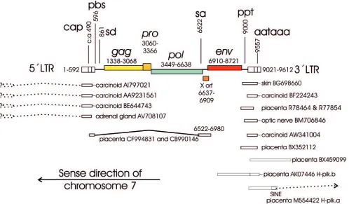
identi-FIG. 1. Overview of the ERV3 locus at chromosome 7q11 as interpreted by the RetroTector computer program (G. O. Sperber and J.
Blomberg, unpublished data) from the human genome version hg15 (April 2003). Positions of selected ESTs and cDNAs and observed or probable
splices (indicated by questions marks) are also shown. Positions are given relative to the first nucleotide of the 5
⬘
LTR. Note that the element is
antisense to the ascribed start of chromosome 7. SINE, short interspersed element. Sequences are identified by GenBank accession numbers.
on November 8, 2019 by guest
http://jvi.asm.org/
[image:4.585.46.544.70.361.2]cal. The provirus has a primer binding site (PBS)
complemen-tary to arginine tRNA, and
gag
,
pro
,
pol
, and
env
sequences
similar to those of other members of genus
Gammaretrovirus
.
The only longer ORFs are in
pro
and
env
. A detailed
interpre-tation done with RetroTector is available as supplemental
ma-terial (see Fig. S1). The distance between the PBS and the
probable start of the
gag
sequences is unusually long (900 bp)
but has no likely ORF, as studied with XonID (Fig. 1). The
predicted Gag polyprotein contains a matrix protein (MA)
with an MGQ myristylation signal and a PPPY motif necessary
for “late” functions, i.e., budding (54, 55, 81). A capsid protein
(CA) with a major homology region (MHR) related to that of
MLV, HERV-H, and HERV-E and a nucleocapsid protein
(NC) with one zinc finger are also present. At the predicted
end of Pol is a GPY/F motif, called IN7 in RetroTector. This
motif occurs in several gammaretroviruses as well as in gypsy
(46) and chromovirus (23) elements, both widespread
retro-transposons. In the latter two, the GPY/F motif may border a
chromodomain. Both of these C-terminal integrase portions
may influence integration specificity by interaction with
DNA-binding proteins (69).
Between the predicted 3
⬘
end of
pol
(chromosome position
63851564) and the 5
⬘
end of
env
(chromosome position
63851292) is a 255-bp (Fig. 1) ORF (corresponding to 85
amino acids) of unknown significance, here referred to as the
ERV3 XORF. It was suggested by the RetroTector XonID
function. Theoretically, it could start at the methionine at nt
6623 (chromosome position 63851579), in frame 1, requiring a
shift to frame 3 at approximately nt 6738 (chromosome
posi-tion 63851464). Alternatively, the putative XORF protein
could have an internal ribosomal entry start after this
frame-shift, or it could represent an unusually long DNA-binding
domain of the ERV3 integrase. The putative ERV3 XORF
protein is histidine rich and has a moderate similarity to a toxin
receptor. It is further analyzed in the supplementary material
(see Fig. S1 in the supplemental material).
According to the RetroTector interpretation, Env starts with
MLGMNMLLITLFLLLPLSMLK, 40 amino acids upstream
of MTKTLLYHTYYECAGTCLGTC, the Env start suggested
by Cohen et al. (15). We favor the first sequence. It includes a
hydrophobic von Heijne signal sequence motif typical of the
amino terminus of retroviral membrane proteins. Other groups
(19, 26) made interpretations similar to ours.
ERV3 7q11 ESTs, splicing, and the LTR structure of the
ERV3 7q11.
A BLAST search with three nucleotide sequences
from three portions of the ERV3 7q11 provirus in the human
EST database yielded 25 to 86 hits which were
⬎
95% identical
to the ERV3 7q11 sequence (Table 2). The hit frequency for a
tissue depends on the number of hits per tissue cDNA library.
Some tissues have many libraries and thus give many hits.
Additionally, the cloning strategy, using 5
⬘
, 3
⬘
, or random
prim-ing, determines the likelihood that a certain query sequence
will find a hit. Thus, this statistic is at best semiquantitative.
However, it demonstrates in which tissues ERV3 is expressed.
Hits for placenta, testis, skin, and brain were especially
fre-quent. Notable were also six ESTs from carcinoid, a multiple
endocrine tumor with a strong genetic dependence. Illustrative
ESTs are depicted together with the ERV3 7q11 provirus (Fig.
1). Four of the carcinoid ESTs were almost identical (98%) to
the 5
⬘
LTR of ERV3 7q11 and clearly different from the 3
⬘
LTR. The simplest explanation for this is that these ESTs are
polyadenylated at the 5
⬘
LTR of ERV3 at 7q11, which is
unusual. It is unlikely that these transcripts were
polyadenyl-ated at single ERV3 LTRs. We did not find any single ERV3
LTRs which were as similar to the transcripts as the ERV3
7q11 5
⬘
LTR is. The start site of these transcripts is not known.
According to this interpretation it should be upstream of
ERV3 7q11.
Many of the ESTs were polyadenylated. Their sequence
revealed the 3
⬘
border of the R region of the ERV3 3
⬘
LTR. It
was situated 15 nt from the end of the polyadenylation signal
(AAATAAAA; which started at nt 537 (chromosome position
63857665) in the 5
⬘
LTR and at nt 9021 (chromosome position
63849181) in the 3
⬘
LTR). This is a typical distance for
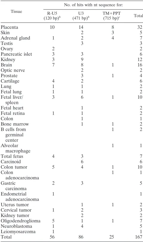
gam-TABLE 2. ESTs detected by a BLAST search with four different
nucleotide sequences
aTissue
No. of hits with nt sequence for: R-U5
(120 bp)b U3
(471 bp)b TM⫹PPT
(715 bp)c Total
Placenta
10
14
8
32
Skin
2
3
5
Adrenal gland
1
2
4
7
Testis
3
3
Ovary
2
2
Pancreatic islet
3
3
6
Kidney
3
9
12
Brain
7
8
1
16
Optic nerve
2
2
Prostate
3
1
4
Cartilage
4
2
6
Lung
1
1
2
Fetal lung
1
1
2
Fetal liver/
spleen
3
6
1
10
Fetal heart
1
2
Fetal retina
1
1
2
Colon
1
1
Bone marrow
1
1
2
B cells from
germinal
center
1
2
Alveolar
macrophage
1
1
Total fetus
4
3
7
Carcinoid
6
6
Colon tumor
5
4
1
10
Colon
adenocarcinoma
1
1
Gastric
carcinoma
2
3
5
Endometrial
adenocarcinoma
1
1
Uterus tumor
1
1
2
Cervical tumor
1
2
3
Kidney tumor
2
2
Oligodendroglioma
5
1
1
7
Neuroblastoma
1
4
5
Leiomyosarcoma
1
1
Total
56
86
25
167
aHits with a score of more than 46 and a percent identity to ERV3 7q11 of
more than 95% are shown. Searches withgagandpolsequences did not yield any
ERV3 7q11 specific hits (not shown).
bAn LTR consists of two unique sequences (U3 and U5) and one repeat
sequence (R).
cTM, transmembrane protein nucleotide sequence; PPT, polypurine tract.
on November 8, 2019 by guest
http://jvi.asm.org/
[image:5.585.300.540.89.494.2]maretroviruses (14). It conforms exactly with the
experimen-tally derived polyadenylation site (29). The 5
⬘
border of R was
harder to verify. Two ESTs (CF994831 and CB990146) which
began close to this site were detected by a BLAST search with
the predicted RU5 of the 5
⬘
LTR. They started with 20
dis-similar non-ERV3 nucleotides. Their ERV3 dis-similarity
com-menced at nt 494 (chromosome position 63857708) of the 5
⬘
LTR, 13 nt downstream of the end of a typical TATAAAA
box. The earlier reported experimentally obtained cap site at nt
498 (30, 51) is 4 nt beyond the probable EST-derived cap site.
The somewhat corrupted start of the ESTs makes it impossible
to determine an exact cap site based on them. The ESTs were
spliced at a predicted canonical splice donor at nt 861
mosome position 63857341) to an acceptor at nt 6522
(chro-mosome position 63851680). These splice sites were exactly the
same as those found earlier (30).
Using EST data, and RetroTector and TFBS predictions, a
map of the ERV3 7q11 5
⬘
and 3
⬘
LTRs was made (see Fig. S5
and S6 in the supplemental material). The 5
⬘
-3
⬘
LTR
noniden-tity of 9% indicates that many of the preintegrational TFBS
should have been damaged by random mutation. If indeed
there was selection for expression of any ERV3 transcripts, it
is likely that TFBS essential for their expression were spared or
created postintegration. Despite the 9% divergence between
the LTRs, the ERV3 7q11
env
ORF, covering 1,812 nt, has
been maintained. Observations of an ERV3 protein by
West-ern blotting and immunohistochemistry with anti-ERV3 sera
(78) also support the existence of an ERV3 Env protein. A
possible selection for production of the ERV3 Env protein in
certain tissues should be mirrored by selection at the 5
⬘
LTR,
and less so at the 3
⬘
LTR, if the 5
⬘
LTR has the dominating
promoter. Comparing the 5
⬘
and 3
⬘
LTRs, a region upstream
of the TATAA box was almost spared of mutation and may be
especially important for ERV3 promoter activity. Its
down-stream adjacent region was more extensively mutated (see Fig.
S5 and S6b in the supplemental material). To better
under-stand any selective forces on the 5
⬘
LTR, we mapped TFBS in
the 5
⬘
and 3
⬘
LTRs. Using ConSite at moderate stringency (10
bits), we determined that TFBS with more than one predicted
occurrence in the 5
⬘
LTR included 12 SOX-5, 5 HFH-2, 4
ROR
␣
1, 3 Sox17, and 3 Thing1/E47 sites. Of these, three
SOX-5, two ROR
␣
1, and three SOX17 sites were unique to
the 5
⬘
LTR relative to the 3
⬘
LTR. For comparison, five
ran-dom control sequences of equal lengths (492 nt) and with
approximately the same ATGC content as that of the 5
⬘
LTR
of ERV3 (27% A, 29% T, 20% G, and 24% C) were also
analyzed. SOX-5 sites occurred 2, 3, 3, 4, and 5 times (average,
3.4), while SOX17 occurred 1, 3, 3, 3, and 4 times (average,
2.8), and ROR
␣
1 occurred 0, 0, 1, 2, and 2 times (average, 1.0)
in each of these five control sequences, respectively. The SOX5
frequency thus deviated most from random prediction (
P
⫽
0.01, Mann-Whitney test) and was the most frequent among
the TFBS unique to the 5
⬘
LTR. The predicted SOX-5 sites (12
in the 5
⬘
LTR and 10 in the 3
⬘
LTR) clustered in the middle of
U3. Although several ERV3-like ESTs mapping to sequences
downstream of the canonical splice donor and upstream of the
canonical splice acceptor were found, none of them probably
came from ERV3 7q11, judging from the degree of nucleotide
identity. A scarcity of full-length mRNAs from the ERV3 7q11
provirus was also noted earlier (30).
Among the more completely recorded mRNAs, only one
encompassing most of ERV3 7q11
env
, the placental H-plk.a,
was found. Most other
env
-containing ESTs encompassed only
the TM domain. A chimeric cDNA clone starting close to the
3
⬘
LTR of ERV3 7q11 (the placental H-plk.b) runs through the
ERV3 7q11 3
⬘
LTR into an adjacent long interspersed element
and is then spliced from an ensuing short interspersed element
into a Kru
¨ppel zinc finger, as described earlier (30).
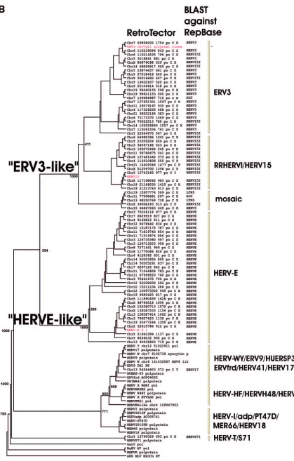
ERV3-like sequences based on SU protein sequence.
ERV3
has been described as a single-copy HERV (47, 78). However,
even if the SU part is one of the most variable in a retrovirus,
many ERV3-like sequences were found when searches for
matches were done at the protein level, using the SU part of
Env. These sequences were subsequently aligned at the
nucle-otide level. A neighbor-joining unrooted tree was then built
(Fig. 2A). This SU-based tree shows the relationship with four
other class 1 HERVs, HUMRIRT (M64936), HERV15yq1
(AF290422), HERV15yq2 (AF290423), and HUMERGPE
(M74509). HERV-E(4-1) formed a cluster together with other
known HERV-E sequences (e.g., HUMERGPE). Close to
ERV3 were sequences from clone AC013429, which have a
proline tRNA as the PBS, and from clones AB019437 and
AC008175, which did not have an identifiable PBS. More
dis-tant from ERV3, among the cluster of ERV3 SU-like proviral
sequences, was HUMRIRT, also known as RRHERVI (28).
The branching pattern was similar to that of the Pol
amino-acid based tree (Fig. 2B; discussed below).
Overall, ERV3 SU-like proviruses with an identifiable PBS
had a PBS complementary to proline, arginine, or isoleucine
tRNA (Table 1). ERV3 itself uses arginine tRNA. The use of
an arginine PBS is not confined to ERV3-like elements (see
below). We therefore could not call the group “HERV-R.”
Delineation of ERV3-like sequences based on similarity in
Pol.
When the RetroTector version 010 and the RetroTector
Shell version 01 programs became available, we selected all
ERV3-like sequences in the human genome version 15 using
an ERV3 Pol protein-based search. Out of 76 retrieved
ERV3-related sequences, 41 ERV3-like sequences based on Pol
sim-ilarity formed a separate cluster at 80% simsim-ilarity or higher,
using the PAM250 score matrix (Fig. 2B; see Fig. S3b and
Table S4a in the supplemental material). Although the 11
ERV3 SU-like elements of Fig. 2A and the 12 elements in
Table 1 had a
pol
gene, several were defective in
pol
and
therefore hard to classify by Pol. However, an alignment
showed that all could be accommodated in the larger
ERV3-like group, which was Pol based (data not shown). Thus,
env
and
pol
genes evolved together in this gammaretroviral group.
In the following, the 41 Pol-based ERV3-like elements are
simply called “ERV3-like.” However, the 80% limit did not
completely separate ERV3-like elements from HERV-E-like
ones, neither in Pol nor in Env (see Tables S4a and 4b,
respec-tively, in the supplemental material). The neighbor-joining tree
derived from the Clustal alignment of the 76 elements (Fig.
2B) also demonstrated the ERV3- and HERV-E-like clusters,
with bootstrap support. The similarity contingency table (see
Table S4b in the supplemental material) showed a somewhat
incomplete separation of ERV3-like and HERV-E-like
ele-ments, possibly indicating recombination and/or a common
evolutionary origin of the groups. As shown in Fig. S3b (see the
supplemental material), the ERV3-like elements consisted of
on November 8, 2019 by guest
http://jvi.asm.org/
23 elements highly similar to ERV3 7q11 in Pol, hereafter
called “ERV3 elements,” and 18 elements highly similar to
RRHERVI, hereafter called “RRHERVI elements.” In the
first group, 13 elements had a recognized
env
gene, while in the
second group, 11 elements had such a gene. Out of the 41
ERV3-like sequences, only one element, the ERV3 locus on
7q11, had an
env
ORF. Among the 76 elements, HERV-E
elements had the most complete
pol
gene, based on the
pres-ence of conserved motifs. The LTR divergpres-ence of the 41
ERV3-like elements was on average 11.4%, with a range of 5.1
to 24.2%. The corresponding figures for the HERV-E
ele-ments shown in Fig. 3B were 15.1% with a range of 7.0 to
30.5%. Some of the more complete ERV3 elements were
an-alyzed in greater depth (Table 1 and Fig. 3B).
PBS tRNA usage: HERV-R in Repbase (27).
Of the 41
ERV3-like elements, 2 used arginine, 2 used proline, and 1
used isoleucine tRNA as a PBS. A weak lysine tRNA-like PBS
was also predicted. The PBS of the remaining elements was not
identified by RetroTector, for unknown reasons. A
RetroTec-tor Shell search indicated that the human genome contains
three ERV3-related elements with an arginine PBS. Two of
them (ERV3 7q11 itself and chrY.3018841) belonged to the 41
ERV3-like elements, while the third (chr19.21399629) was
classified as “HUERSP3-like,” a gammaretroviral sequence
which is more distant from ERV3 than HERV-E (data not
shown).
Altogether, RetroTector detected 72 retroviral elements
with an arginine PBS in the human genome. The great majority
of them, 69, thus fell outside of the ERV3-like group. Of the
69, 3 were HERVRB-like, 5 were HUERSP3-like, 2 were
MER41-like, 2 were HERV19-like, and 2 were HERVFc-like,
based on RepBase nomenclature. The rest have not yet been
classified, but some of them contained stretches similar to the
HERV9 and the little-studied MER41, MER51, and MER66
sequences. This is an example of “PBS promiscuity” (see
Dis-cussion). RepBase (27) contains a sequence called HERV-R.
This HERV-R was 100% identical to the baboon endogenous
retrovirus (74) and therefore is not a human ERV. This is a
further reason to avoid using the term HERV-R.
Structure and localization of ERV3-like elements.
ERV3-like elements were common on chromosome Y. Six elements
were located there, while two occurred on each of
chromo-somes 2, 6, and 19. Chromochromo-somes 14 and X contained one
ERV3-like element each. A complete list of the ERV3-like
elements is available in the supplemental material (Table S2).
Table 1 contains a partial list of the ERV3-like elements.
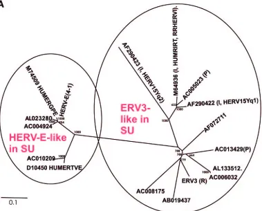
FIG. 2. (A) Dendrogram of ERV3- and HERV-E-like sequences based on the nucleotide alignment of the SU part of
env
, partly shown in Fig.
3. PBS were analyzed by using the RetroTector program (see Materials and Methods) (G. O. Sperber and J. Blomberg, unpublished data).
Sequences with an identifiable PBS are shown in parentheses, giving the amino acid (using the one-letter code) corresponding to parts of the tRNA
sequence. (B) Pol amino acid-based cladogram, derived from the alignment shown in Fig. S3a (see the supplemental material). The
neighbor-joining method was used. For each element, the following RetroTector-derived data are shown: chromosomal position, chain score, PBS usage,
sum of stops and frame shifts, and the most similar RepBase element. The bootstrap value (1,000 replicates) for the branch which divides the
ERV3-like and the HERVH HERV-E-like elements is also shown.
on November 8, 2019 by guest
http://jvi.asm.org/
[image:7.585.104.483.69.374.2]FIG. 2—
Continued
.
on November 8, 2019 by guest
http://jvi.asm.org/
The ERV3-like sequences harbored not only
env
genes but
also other proviral structures (Table 1). The LTRs of HERV-E
contain a promoter known to affect transcription of several
cellular genes. Moreover, HERV-E transcripts have been
seen in both normal and diseased tissues (52, 65). To
re-trieve HERV-E(4-1) Env-encoding sequences, the same
type of TBLASTN search that was initially made with ERV3
Env was also made with HERV-E Env. The results are
shown with the ERV3-like SU sequences in Fig. 3. The
[image:9.585.98.487.79.282.2]nucleotide alignments presented in Fig. 3 illustrate how the
subsequently designed real-time primers and probe fit with
the ERV3-like sequences. As anticipated, most similarities
were observed in the conserved transmembrane region
(TM). Therefore, we chose the SU region of
env
for
design-ing specific primers. As shown in the alignment, point
mu-tations, insertions, and deletions favor a PCR specific for
the ERV3 7q11 locus. A similar narrow specificity was also
expected of the HERV-E(4-1) SU-derived primers and
FIG. 3. Parts of the SU region of the ERV3- and HERV-E-related sequences aligned with the annotated repetitive sequences for HUMRIRT,
HUMERGPE, HERV15Yq1, and HERV15Yq2. QPCR primers are indicated for ERV3 (A) and HERV-E (B) in the alignment by underlining
and probe sequences are indicated by underlining and shading. Double slashes in the alignment in panel B show where 60 nt were omitted in the
figure due to space limitations. Periods denote identity to the master sequence, which is underlined.
on November 8, 2019 by guest
http://jvi.asm.org/
probe. The searches for ERV3-like sequences revealed
in-teresting details regarding the genetic environment at their
integration sites, mentioned in footnotes to Table 1. It is
beyond the scope of this paper to discuss the possible effects
of ERV3-like elements on the surrounding genes.
QPCR results.
In order to study the HERV expression
quantitatively, specific real-time PCRs were developed for
ERV3 and HERV-E. The ERV3 QPCR method had a
sensi-tivity of around 10 haploid human genomes and around 10
equivalents of ERV3 plasmid DNA per PCR. The HERV-E
QPCR method could detect 5 to 10 haploid genomes and
around 10 equivalents of HERV-E(4-1) plasmid DNA. The
specificity for both methods measured as the absence of
cross-amplification from the HERV-E(4-1) and ERV3 plasmids was
100%.
The ERV3 and HERV-E SU QPCRs confirmed previous
data showing high expression in adrenal gland and placenta
(30, 33).
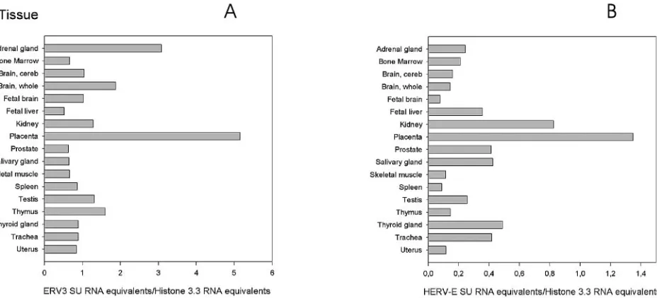
QPCR with ERV3 and HERV-E.
The two graphs showing
the results of QPCR with ERV3 and HERV-E (Fig. 4) reveal
great differences in mRNA expression between organs, but
also between the SU of ERV3 (Fig. 4A) and HERV-E (Fig.
4B). The high expression of ERV3 in the adrenal gland
con-firmed previous ISH results. Other organs with high expression
levels were whole brain (ERV3), placenta (ERV3 and
HERV-E), kidney (ERV3 and HERV-HERV-E), thymus (ERV3), thyroid
gland E), prostate E), and trachea
(HERV-E). However, all organs had some expression of ERV3 and
HERV-E SU.
ISH results with an ERV3 probe.
Based on our previous
Northern blot analysis of ERV3 and RT-PCRs of ERV3 7q11
and HERV-E(4-1) of others, we knew that there was a
differ-ence in expression both between organs and between
individ-uals (4, 66). Since a tissue is composed of several different cell
types, existing in different ratios, we wanted to know which of
the cell types expressed high levels of the retroviral transcript.
This question was addressed by ISH using a 1.7-kb ERV3 7q11
env
probe under high-stringency conditions. The results are
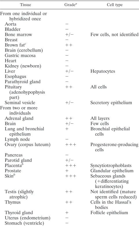
presented in Table 3 and Fig. 5.
Table 3 shows that several tissues have elevated levels of
ERV3 7q11
env
mRNA. In summary, all layers of the adrenal
gland, cells in brown fat, bronchial epithelial cells of lung,
progesterone-producing cells of corpus luteum, all cells in the
adeno part of the pituitary gland, glandular epithelium of the
prostate, sebaceous gland of the skin, cells in testis (not Leydig
cells), Hassal’s bodies of the thymus, and follicle cells of the
thyroid gland contained ERV3 SU mRNA. Adrenal gland,
skin, and placenta (a positive control) have previously been
reported to harbor high concentrations of ERV3 transcripts (3,
31). A selection of tissues containing cells with the highest
levels of detected mRNA are illustrated in Fig. 5, i.e., corpus
luteum, testis, thymus, brown fat, and pituitary gland.
DISCUSSION
This study is a systematic analysis of a class I HERV
ge-nome, ERV3 7q11, sometimes in contrast to the related
HERV-E. Both have been frequently studied before. However,
a systematic analysis using the full information from the human
genome databases and quantitative RNA detection methods
has not been done.
[image:10.585.63.527.69.280.2]ERV3-like sequences and the ERV3 locus.
ERV3 is a rather
typical gammaretrovirus. Most of the structures of MLV are
discernible in it. However, unlike MLV, there is no indication
of a pp12 Gag protein. In addition, a space separates the
predicted
pol
from the predicted
env
. It contains a putative
ORF, here referred to as ERV3 XORF. Theoretically, it could
encode a protein of its own, an extension of the Pol protein, or
FIG. 4. ERV3 (A) and HERV-E (B)
env
mRNA expression detected by QPCR specific for the SU region of HERV-E and ERV3, respectively.
Each value represents a mean of the results for at least two experiments. cDNA from an RNA panel (Clontech) from several human tissues was
analyzed. The values for placenta and skeletal muscle are averages based on samples from a commercial tissue RNA panel and RNA extracted
from local patient samples, as described in Materials and Methods. All values are expressed relative to the histone 3.3 signal of the same sample.
Note that the scale for HERV-E is different from that of ERV3. cereb, cerebellum.
on November 8, 2019 by guest
http://jvi.asm.org/
an unusual leader sequence for the Env protein. The position
between predicted
pol
and
env
is typical of sequences for
reg-ulatory proteins of complex retroviruses. This putative protein
or protein subdomain should be searched for in tissues with
high ERV3 expression using antisera and proteomic
tech-niques. If it is a C-terminal extension of the integrase new to
gammaretroviruses, a participation in chromatin binding (69)
should be investigated.
Mutilated and recombinant HERVs create taxonomic
prob-lems. The somewhat incomplete separation in the similarity
matrices (see Fig. S3a and S3b in the supplemental material)
may be symptoms of recombination. Gene conversion is an
often invoked but rarely proven recombinatory mechanism for
highly related HERVs (71). Nevertheless, the Pol-based
clas-sification used in this paper was to a large extent consistent
with the most similar RepBase (27) sequence, found by a
BLAST algorithm with the nucleotide sequence of the whole
element. The older PBS-based classification (37) was
some-times congruent. For example, the HERV-E group was well
delineated from other HERV groups. A glutamine tRNA PBS
is almost exclusively associated with the HERV-E group (work
with RetroTector; data not shown). Meanwhile, neutral group
names referring to established loci are preferable to minimize
confusion. We therefore chose the name “ERV3-like” for the
group of elements comprising ERV3 and RRHERVI
ele-ments. They share a Pol protein similarity of 80% or higher, as
estimated with the PAM250 matrix. Based on a comparison of
RepeatMasker (A. Smit, unpublished data) output and its
cor-ollary, the HERVd database (53), both of which are based on
RepBase, and RetroTector output, from the human genome
version 15 (April 2003), the 41 ERV3-like elements defined
here were classified either as HERV3 or HERV15 by
Repeat-Masker (J. Blomberg and G. O. Sperber, unpublished data).
The two independent classifications thus were approximately
concordant.
ERV3 7q11 has been described as a single-copy gene,
re-lated to the higher-copy group HERV-E (51). We here show
that it is part of a group of 41 elements. A few of the
ERV3-like sequences were previously described, some of which occur
on the Y chromosome (34). The
env
gene of orthologs of the
human ERV3 7q11 locus had an ORF that was maintained in
several Old World monkeys. However, the locus was absent in
gorillas; hence, it may not perform an exclusive essential
func-tion in primates (26). Single-nucleotide polymorphisms have
been reported in the original ERV3 provirus, where not only
env
but also LTR polymorphic sequences were discovered by
Rasmussen et al. (59, 60). Additionally, de Parseval et al., at
the same time, discovered a single-nucleotide polymorphism in
the carboxy terminus of ERV3 SU, introducing a stop codon in
1% of Caucasians, which would create a truncated Env protein
(18, 19). It is, however, not known if the premature stop in the
carboxy-terminal surface region would preclude a function or
not. Regardless, women with the mutation had normal
preg-nancies (19). In previous studies of ERV3, Lin et al. showed
that when ERV3 Env was expressed in the trophoblastic cell
line BeWo, the trophoblasts differentiated, fused, and
pro-duced beta-human chorionic gonadotropin (41). A similar
ef-fect was attributed to the HERV-W Env protein syncytin (49),
which can fuse cells in placenta, possibly forming the
syncy-tiotrophoblastic layer.
[image:11.585.43.283.80.470.2]Interestingly, some of the ERV3-like sequences were found
to be integrated in the vicinity of genes encoding regulatory
proteins, including an estrogen and retinoic acid receptor
reg-ulatory protein TIF1 (Table 2), with unknown effects.
Al-though TFBS predictions must be judged with caution, the
frequent occurrence (12 hits) of predicted SOX-5 sites in the 5
⬘
LTR of ERV3 requires a comment. Sox (
S
ry-type
high-mobil-ity-group b
ox
) proteins are a subfamily of DNA-binding
pro-teins with a high-mobility-group domain. Sry is the
testis-de-termining factor (68). Sox proteins have functions in sex
determination and neurogenesis (38). SOX-5 is highly
ex-pressed in testis, especially in spermatids (9, 12, 73, 84).
Ex-pression in the germ line is relevant for an endogenous
retro-virus. The other frequently predicted sites were HFH2 and
Thing1. HFH2 (FoxD3; 5 hits in the 5
⬘
LTR) drives the
devel-opment of the neural crest and production of parathyroid
hormone (58), and Thing1 (
Hand1
; 3 hits) is important for
trophoblast transition to syncytiotrophoblast and neural crest
TABLE 3. Tissues used for ISH with ERV3
env
probe
Tissue Gradea Cell type
From one individual or
hybridized once
Aorta
⫺
Bladder
⫺
Bone marrow
⫹
/
⫺
Few cells, not identified
Breast
⫺
Brown fat
c⫹⫹
Brain (cerebellum)
⫺
Gastric mucosa
⫺
Heart
⫺
Kidney (newborn)
⫺
Liver
⫹
/
⫺
Hepatocytes
Esophagus
⫺
Parathyroid gland
⫺
Pituitary
(adenohypophysis
part)
⫹⫹
All cells
Seminal vesicle
⫹
/
⫺
Secretory epithelium
From two or more
individuals
Adrenal gland
⫹⫹
All layers
Brain
⫹
/
⫺
Few cells
Lung and bronchial
epithelium
⫹
Bronchial epithelial
cells
Lymph node
⫺
Ovary (corpus luteum)
⫹⫹⫹
Progesterone-producing
cells
Pancreas
⫺
Parotid gland
⫹
/
⫺
Placenta
b⫹⫹⫹
Syncytiotrophoblasts
Prostate
⫹
Glandular epithelium
Skin
b⫹⫹⫹
Sebaceous glands
(
⫹
differentiating
keratinocytes)
Testis (slightly
atrophic)
⫹⫹
Not identified (mature
sperm cells reduced)
Thymus
⫹⫹
Cells in the Hassal’s
bodies
Thyroid gland
⫹
Follicle epithelium
Uterus (endometrium)
⫺
Stomach (ventricle)
⫺
a
Semiquantitative grading of in situ hybridization results. The positive control was placenta.
b
Tissues tested from more than 10 individuals
c
This tissue was hybridized several times.
on November 8, 2019 by guest
http://jvi.asm.org/
development (16). Carcinoid tumors, here reported to express
ERV3 RNA, have been considered to be a neural crest
deriv-ative (36). ROR
␣
1 (3 hits) is expressed in sebaceous glands
(61) and belongs to the retinoic acid receptor family.
Reti-noids influence lipid synthesis in sebaceous glands (72) and
activate RRHERV-I, which belongs to the ERV3-like
[image:12.585.58.527.68.580.2]HERVs (Table 1) (28). As shown here, ERV3 RNA is
abundant in placenta, testis, and sebaceous glands. It is
tempting to see a pattern of ERV3 expression
correspond-ing to the known activities of these transcription factors.
However, random sequences of similar nucleotide
composi-tions were also predicted to bind some of the same factors.
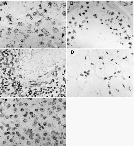
FIG. 5. Hybridization signal obtained with the ERV3 antisense SU probe in five different tissues. (A) Tissue section from a corpus luteum
demonstrating a positive and elevated ERV3 signal in progesterone-producing luteinized follicular cells. (B) Positive signal in yet unidentified cells
obtained from a tissue section from a human, partly atrophic, testis with reduced spermatogenesis. (C) Tissue section from thymus with an elevated
signal in cells in the outer part of a Hassal’s body. (D) Brown fat section showing a high ERV3 message in typical multivacuolated brown fat cells.
(E) Tissue section from a human pituitary gland (adenohypophysis portion), where an elevated ERV3 message is visible in all cell types.
on November 8, 2019 by guest
http://jvi.asm.org/
Their possible involvement in promotion of transcription
from ERV3 at 7q11 should be experimentally verified in
transient-transfection experiments.
Tissue-specific expression of ERV3 and related sequences.
ESTs which likely were encoded at ERV3 7q11 were found in
cDNA libraries from normal tissues like placenta, testis, and
adrenal gland. A reason for the relative scarcity of unspliced
transcripts described previously (30), and in this work, could be
nonsense-mediated decay of the full-length mRNAs, which are
relatively more defective than the
env
mRNAs (39). Some
malignancies, primarily carcinoid and colon tumors, also
con-tained ERV3 cDNA. As mentioned above, carcinoid is a
neu-roendocrine cancer with an inheritable predisposition. The
observation of multiple ERV3 ESTs in two different cDNA
libraries from the neuroendocrine tumor carcinoid should be
followed up.
QPCR showed high levels of ERV3 7q11
env
RNA
expres-sion in adrenal gland, skeletal muscle, brain, placenta, thymus,
and testis. HERV-E(4-1)
env
RNA was present primarily in
placenta, skeletal muscle, kidney, thyroid gland, prostate, and
trachea. Thus, tissues with a function in reproduction as well as
endocrine functions tended to have a higher expression of
ERV3 7q11
env
. The expression was higher than that for
env
of
the related HERV-E(4-1) and had a different tissue profile.
Our ISH studies of ERV3 confirmed previous Northern blot
analyses showing that ERV3 is active in some specific cell
types, such as syncytiotrophoblasts in placenta and cells in
sebaceous glands (3, 30). In addition, high activities were found
in brown fat, corpus luteum, testis, and Hassal’s bodies in
thymus. Several of these tissues are under strong hormonal
influence. The EST and QPCR data also showed high
expres-sion levels in adrenal gland. The possible functions of a
retro-viral protein in these tissues are unknown. In animals, a high
expression of ERVs is common in reproductive tissues like
seminiferous tubules, vesicula seminales, testis, and placenta
(20, 24, 25, 32, 40). The results of EST searches, QPCR, and
ISH from this investigation and previous Northern blot
anal-yses were similar. They cover different aspects of RNA
expres-sion. dbEST covers a broad range of normal and pathological
tissues, but quantification is imperfect. QPCR gives an
approx-imate number of HERV RNA molecules per nanogram of
RNA or per household gene transcript but is sensitive to
mu-tations in primer and probe sequences. ISH identifies which
cell types express the RNA but is not as quantitative as QPCR.
Northern blot analysis describes the molecular weights of the
RNAs but is less quantitative than QPCR. Since ISH was
performed with fixed tissues and PCR was done with fresh
ones, the procedures are not completely comparable. Besides,
it is known that external factors such as ischemia and anoxia
can increase the activities of selected retroelements such as
VL-30 (2). Further, polymorphism and epigenetic events such
as imprinting could lead to both inter- and intraindividual
variation in HERV expression. An interindividual variation in
RNA expression has been shown for several HERVs (7, 42, 48,
85).
The results presented here demonstrate that both ERV3
7q11 and HERV-E(4-1) are expressed in most organs, as
re-ported by Sibata et al. (66). Although an exact comparison
cannot be made, ERV3 expression was higher than HERV-E
expression in several tissues. Similar to the results presented
here, expression of ERV3
env
has been previously reported to
be high in adrenal and sebaceous glands (3, 4) and in placenta
(11, 41), whereas HERV-E
env
expression was found to be
high in placenta (66).
In conclusion, this investigation provides a description of
ERV3 structure, ERV3-like elements in the human genome,
and the degree of ERV3 7q11 and HERV-E(4-1)
env
RNA
expression in normal tissues. Measurements of organ-, cell
type-, and transcript-specific expression by QPCR, ISH, and
previous Northern blot analysis gave concordant results. The
techniques described here will allow further studies of the
pattern of HERV expression in selected model systems and in
diseased persons.
ACKNOWLEDGMENTS
This work was supported by grants (projects 2037-B99-16BB and
4239-B00-02XBB) from the Swedish Cancer Society to Erik Larsson
and Jonas Blomberg, respectively, and by a grant from the Swedish
Scientific Council to Jonas Blomberg (521-2001-6520).
We thank Anna Forsman, Lijuan Hu, Patric Jern, and Dmitrijs
Uzhameckis for valuable assistance.
REFERENCES
1.Altschul, S. F., W. Gish, W. Miller, E. W. Myers, and D. J. Lipman.1990.
Basic local alignment search tool. J. Mol. Biol.215:403–410.
2.Anderson, G. R., D. L. Stoler, and L. A. Scarcello.1989. Retrotransposon-like VL30 elements are efficiently induced in anoxic rat fibroblasts. J. Mol.
Biol.205:765–769.
3.Andersson, A.-C., M. Merza, P. Venables, F. Ponten, J. Sundstro¨m, M. Cohen, and E. Larsson.1996. Elevated levels of the endogenous retrovirus
ERV3 in human sebaceous glands. J. Investig. Dermatol.106:125–128.
4.Andersson, A.-C., A.-C. Svensson, C. Rolny, G. Andersson, and E. Larsson.
1998. Expression of human endogenous retrovirus ERV3 (HERV-R)
mRNA in normal and neoplastic tissues. Int J. Oncol.12:309–313.
5.Andersson, A.-C., P. J. W. Venables, R. R. To¨njes, J. Scherer, L. Eriksson, and E. Larsson.2002. Developmental expression of HERV-R (ERV3) and
HERV-K in human tissue. Virology297:220–225.
6.Andersson, M.-L., M. Lindeskog, P. Medstrand, B. Westley, F. May, and J. Blomberg.1999. Diversity of human endogenous retrovirus class II-like
se-quences. J. Gen. Virol.80:255–260.
7.Andersson, M. L., P. Medstrand, H. Yin, and J. Blomberg.1996. Differential expression of human endogenous retroviral sequences similar to mouse mammary tumor virus in normal peripheral blood mononuclear cells. AIDS
Res. Hum. Retroviruses.12:833–840.
8.Benit, L., P. Dessen, and T. Heidmann.2001. Identification, phylogeny, and evolution of retroviral elements based on their envelope genes. J. Virol.
75:11709–11719.
9.Blaise, R., J. Grober, P. Rouet, G. Tavernier, D. Daegelen, and D. Langin.
1999. Testis expression of hormone-sensitive lipase is conferred by a specific promoter that contains four regions binding testicular nuclear proteins.
J. Biol. Chem.274:9327–9334.
10.Blomberg, J., D. Uzhameckis, and P. Jern.2005. Evolutionary aspects of human endogenous retroviral sequences (HERVs) and disease, p. 204–238.
InE. D. Sverdlov (ed.), Retroviruses and primate genome evolution. Landes
Bioscience, Georgetown, Tex.
11.Boyd, M. T., C. M. Bax, B. E. Bax, D. L. Bloxam, and R. A. Weiss.1993. The human endogenous retrovirus ERV-3 is upregulated in differentiating
pla-cental trophoblast cells. Virology196:905–909.
12.Budde, L. M., C. Wu, C. Tilman, I. Douglas, and S. Ghosh.2002. Regulation
of IkappaBbeta expression in testis. Mol. Biol. Cell.13:4179–4194.
13.Christensen, T., P. Dissing Sørensen, H. Riemann, H. J. Hansen, M. Munch, S. Haahr, and A. Møller-Larsen. 2000. Molecular characterization of HERV-H variants associated with multiple sclerosis. Acta Neurol. Scand.
101:229–238.
14.Coffin, J. M., S. H. Hughes, and H. E. Varmus (ed.).1997. Retroviruses. Cold Spring Harbor Press, Cold Spring Harbor, N.Y.
15.Cohen, M., M. Powers, C. O’Connell, and N. Kato.1985. The nucleotide sequence of the env gene from the human provirus ERV3 and