0022-538X/07/$08.00⫹0 doi:10.1128/JVI.01990-06
Copyright © 2007, American Society for Microbiology. All Rights Reserved.
Long-Lasting Decrease in Viremia in Macaques Chronically Infected
with Simian Immunodeficiency Virus SIVmac251 after
Therapeutic DNA Immunization
䌤
Agneta S. von Gegerfelt,
1Margherita Rosati,
1Candido Alicea,
2Antonio Valentin,
1Patricia Roth,
1Jenifer Bear,
2Genoveffa Franchini,
3Paul S. Albert,
4Norbert Bischofberger,
5Jean D. Boyer,
6David B. Weiner,
6Phillip Markham,
7Zimra R. Israel,
8John H. Eldridge,
8George N. Pavlakis,
1* and Barbara K. Felber
2Human Retrovirus Section1and Human Retrovirus Pathogenesis Section,2Vaccine Branch, Center for Cancer Research, National Cancer Institute at Frederick, Frederick, Maryland 21702; Animal Models and Retroviral Vaccines Section,
Vaccine Branch, Center for Cancer Research,3and Biometric Research Branch,4National Cancer Institute, Bethesda, Maryland 20892; Gilead Sciences, Foster City, California 944045; Department of Pathology and
Laboratory Medicine, University of Pennsylvania School of Medicine, Philadelphia, Pennsylvania 191046; Advanced BioSciences Laboratories, Inc., Kensington, Maryland 208957; and
Wyeth Vaccines Research, Pearl River, New York 109658
Received 12 September 2006/Accepted 20 November 2006
Rhesus macaques chronically infected with highly pathogenic simian immunodeficiency virus (SIV) SIVmac251 were treated with antiretroviral drugs and vaccinated with combinations of DNA vectors expressing SIV antigens. Vaccination during therapy increased cellular immune responses. After the animals were released from therapy, the virus levels of 12 immunized animals were significantly lower (Pⴝ0.001) compared to those of 11 animals treated with only antiretroviral drugs. Vaccinated animals showed a persistent increase in immune responses, thus indicating both a virological and an immunological benefit following DNA therapeutic vaccination. Several animals show a long-lasting decrease in viremia, suggesting that therapeutic vaccination may provide an additional benefit to antiretroviral therapy.
Antiretroviral treatment has changed the prognosis of hu-man immunodeficiency virus (HIV) infection, as patients can remain free of symptoms for extended periods of time. How-ever, continuous antiretroviral treatment is associated with toxicity and the emergence of resistant viral strains. Therapy must be continued indefinitely, since virus replication resumes rapidly upon treatment interruption (5, 22, 23) due to the persistence of HIV in stable reservoirs and also due to contin-uous residual virus replication (3, 4, 34). Thus, additional ap-proaches to control viral propagation are needed. Strengthen-ing the host immune response is a possible strategy in the management of HIV infection (reviewed in reference 11). Few therapeutic vaccine modalities have been tested in animal models and in humans. Substantial control of viremia has been reported in humans and macaques with early HIV/simian im-munodeficiency virus (SIV) infection (9, 13, 30, 37) upon apeutic vaccination or even by a period of antiretroviral ther-apy (ART) alone. Early therther-apy was hypothesized to work by limiting the damage to the immune system. Despite evidence of immunogenicity of therapeutic vaccination, some studies did not show substantial virologic benefit (10, 18). Therapeutic vaccination of persons treated with ART early after infection showed evidence of an induction of immune responses, but the
magnitude and dynamics of virus rebound after therapy dis-continuation was similar in both vaccinated and unvaccinated subjects (18). Results of a randomized placebo control trial (QUEST [10]) of HIV-infected individuals treated during pri-mary infection and therapeutically vaccinated with a vaccinia recombinant vector (ALVAC-HIV) or in addition with inacti-vated virus particles (Remune) showed an induction of HIV-1-specific cellular immunity, although it did not lead to better virological control of HIV-1 after the discontinuation of ART. Compared to those with early infection, results have been even more variable after intervention during chronic infection. Some reports in macaques suggest that immune therapy during chronic infection was only transiently effective in controlling viral load (VL) and boosting immune responses (14, 15, 36). Successful therapeutic dendritic cell vaccination, in the ab-sence of ART, has also been reported; in vitro treatment of macaque and human antigen-presenting cells with antigen, fol-lowed by reinfusion of the cells, resulted in a long-lasting decrease in viral load (16, 17). Several immunotherapy studies suggest that the restoration of the immune system and more efficient immunization procedures may improve virus control (38). A recent study combining ALVAC-HIV with lipopep-tides and interleukin-2 (IL-2) administration showed immuno-logical and viroimmuno-logical benefits after vaccination compared to results with the control group (12).
The use of DNA-based vaccines in therapeutic immuniza-tion is attractive since DNA vaccines can be easily combined with other vaccine modalities and adjuvants, and can be ad-ministered repeatedly, which is an advantage over viral
vector-* Correspondence author. Mailing address: Human Retroviruses Section, Vaccine Branch, Center for Cancer Research, National Can-cer Institute at Frederick, 1050 Boyles Street, Building 535, Room 210, Frederick, MD 21702-1201. Phone: (301) 1475. Fax: (301) 846-7146. E-mail: [email protected].
䌤Published ahead of print on 29 November 2006.
1972
on November 8, 2019 by guest
http://jvi.asm.org/
based vaccines. Improved protocols for prophylactic DNA vac-cination have shown promising results against SIV and SHIV either alone or in combination with other vaccine modalities (reviewed in reference 27). We have previously reported a signif-icant reduction in acute and chronic viremia after SIVmac251 challenge of macaques vaccinated with a combination of SIV
gagand envexpression vectors modified to produce secreted and intracellular forms of SIV antigens (29). Here, we show that SIVmac251-infected rhesus macaques improve control of the virus upon immunization with DNA vectors expressing these SIV antigens during ART treatment. This vaccination induces long-lasting, virus-specific immune responses, result-ing in a significant reduction of high-level viremia after ART termination. Interestingly, DNA vaccination of infected ani-mals promoted a Th1-biased response, with a more prominent induction of cellular immune responses.
MATERIALS AND METHODS
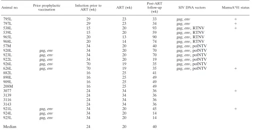
Animals.Indian rhesus macaques (Macaca mulatta) were housed and handled in accordance with the standards of the Association for the Assessment and Accreditation of Laboratory Animal Care International. Screening for 10 major histocompatibility complex class I and II alleles was performed by PCR (D. Watkins, Wisconsin Regional Primate Center). The animals were infected by pathogenic SIVmac251 mucosally, except for animals 795L, 882L, 890L, 909L, and 208M, which were infected by the intravenous route. During the drug treatment period (13 to 34 weeks; median, 20 weeks) (Table 1), animals were
given a combination of three drugs [(R)-9-(2-phosphonylmethoxypropyl)
ade-nine (PMPA), 20 mg/kg of body weight injected subcutaneously once daily; didanosine (ddI), 5 mg/kg injected intravenously once daily; and stavudine, 1.2 mg/kg orally twice daily]. Five of the macaques (animals 920L, 922L, 923L, 926L, and 626L, in the vaccine group) (Table 1) and three in the control group (animals
921L, 924L, and 925L) were prophylactically vaccinated with the same SIVgag
andenvDNA vectors before SIV infection, as part of a previous study (29).
DNA vectors.The plasmids used for DNA vaccination contain the cytomega-lovirus promoter without an intron, the bovine growth hormone polyadenylation
site, and the kanamycin-resistant gene. The RNA (codon)-optimized expression
vectors forgagandenvwere generated by introducing multiple silent point
mutations not affecting the sequence of the encoded proteins, as previously
described for HIV-1gagandenv(20, 31–33) using synthetic DNAs. The secreted
and intracellularly degraded variants of the SIV antigens were generated by the fusion of either IP10-MCP3 (2) or of a beta-catenin-derived peptide (amino acids 18 to 47) (1), respectively, at the N terminus of Gag and Env, as previously described (29). All animals received mixes of DNA vectors producing secreted
and intracellular forms ofgagandenv. In addition, as indicated in Table 1, most
animals received vectors expressing intracellular forms of a fusion protein polNTV consisting of Pol, Nef, Tat, and Vif proteins or alternatively a vector expressing a fusion (RTNV) of Rev-Tat-Nef (ReTaNef [7, 8]) and Vif proteins.
Several point mutations were introduced intopolto inactivate protease (D25A,
T26A, and G27A), reverse transcriptase (D186A and D187A), RNaseH (E478A), and integrase (D64A, D116A, and E152A). In addition, the myristoy-lation signal of Nef was removed. These changes parallel changes we have introduced into HIV expression vectors.
Therapeutic immunization. Highly purified, endotoxin-free DNA plasmid preparations were produced using the QIAGEN kit (Hilden, Germany). Animals
795L and 797L received a total of 6 mg of plasmids expressing nativeenvandgag,
the secreted forms ofenvandgag, and the intracellular degraded forms ofgag(1
mg each). Animals 538L, 539L, 965L, and 968L received a total of 10 mg DNA comprising 2 mg each of the intracellular degraded and secreted forms of SIV
DNAs expressinggagorenvand 2 mg polNTV. These DNAs were prepared at
a concentration of 1 mg/ml in phosphate-buffered saline and injected (0.5 ml per injection) at separate sites intramuscularly. The remaining animals were immu-nized with mixtures of 8 mg of SIV DNA vectors in combination with 2 mg IL-15 DNA (animals 57M, 920L, 923L, and 922L) or IL-12 DNA (animals 926L and 626L), formulated as previously described (6) using 2 mg/ml DNA.
Humoral responses.Antibody production was measured in serial dilutions of plasma by enzyme-linked immunosorbent assay against SIVmac251 lysate spiked with gp120. The plates were analyzed at 450 nm. The binding antibody titers are reported as the reciprocal of the highest positive dilution.
Viral load analysis.SIV RNA copy numbers were determined by nucleic acid sequence-based isothermal amplification assay using SIVmac251-specific prim-ers (28). During ART, an assay with a cutoff value of 20,000 RNA copies/ml was used and values below the cutoff were assigned the value of 10,000. Most of the chronic and post-ART period samples below this cutoff, if available in sufficient quantity, were analyzed by more sensitive assays, either a nucleic acid
sequence-TABLE 1. History and treatment of the animals in the study
Animal no. Prior prophylactic
vaccination
Infection prior to
ART (wk) ART (wk)
Post-ART follow-up (wk)
SIV DNA vectors MamuA*01 status
795L 29 23 33 gag,env ⫹
797L 29 23 34 gag,env ⫹
538L 15 20 93 gag,env, RTNV ⫹
539L 15 20 59 gag,env, RTNV
965L 20 13 90 gag,env, RTNV
968L 20 14 74 gag,env, RTNV
57M 34 20 40 gag,env, polNTV
920L gag,env 34 20 70 gag,env, polNTV
923L gag,env 34 20 70 gag,env, polNTV
922L gag,env 34 20 19 gag,env, polNTV
926L gag,env 70 19 35 gag,env, polNTV
626L gag,env 70 19 35 gag,env, polNTV ⫹
882L 16 25 41
890L 16 25 49
909L 16 25 49
208M 16 25 49
3077 24 34 36 ⫹
3139 24 34 36
3116 24 34 36
3143 24 34 36
921L gag,env 34 20 45 ⫹
924L gag,env 34 20 14
925L gag,env 34 20 14
Median 24 20 40
on November 8, 2019 by guest
http://jvi.asm.org/
[image:2.585.41.550.81.342.2]based isothermal amplification assay having a cutoff value of 2,000 copies/ml (28) or a real-time, quantitative reverse transcription-PCR assay with a cutoff value of 100 RNA copies/ml of plasma (35).
ELISPOT assay.The enzyme-linked immunospot (ELISPOT) assay was
per-formed as previously described (21, 29). Eighty-eight peptides spanninggagp39
and 100 peptides covering gp120env were used as two separate pools. Specific spots to a given peptide pool were calculated by subtracting the cutoff value and adjusted to spot-forming cells per million peripheral blood mononuclear cells. The cutoff value was defined as the average number of spots in the negative control measured in triplicate plus two standard deviations (SD).
Statistical analysis.We compared the median change in average viral load (log transformed, base 10) from the chronic to the post-ART period between vaccinated and control animals using a Wilcoxon rank sum test. Before combin-ing previously vaccinated animals with naı¨ve animals, we examined whether differences between previously vaccinated and naive animals existed by testing for an interaction between vaccination group and prevaccination status in a two-way analysis of variance with an interaction term, with the outcome being the difference in average viral load between the chronic and post-ART periods. Further, we performed analysis of covariance to adjust for any differences in pretreatment viral load between groups.
The animals were assigned randomly into the vaccine and control groups. The
groups had similar values for mean pretreatment log10-transformed viral load
(mean, 5.855; standard deviation, 0.7078; and mean, 5.768; SD, 0.9490 for the vaccine and control groups, respectively). After the exclusion of animals that did not fulfill the study criteria, there was a small difference in the means of the two
groups (log10-transformed mean, 5.606; SD, 0.3977; and mean, 5.970; SD, 0.5889;
for the vaccine and control groups, respectively). Analysis of covariance showed that the decrease in viral load after DNA vaccination remained significant after adjusting for the small difference in pretreatment virus loads. The statistical
analyses performed indicate that the therapeutic DNA vaccination benefit does not depend on the specific inclusion criteria or the differences in the history of the animals in the study.
Analyses of immune responses were conducted with a linear mixed model (26), which allows for testing period effects while accounting for the fact that repeated observations on the same subject may be correlated. We examined whether the mean varied across periods using a conditional F test. Prior to combining naı¨ve and prevaccinated groups, we examined whether the pattern in changes in mean values across periods changed by prevaccination status by also using a condi-tional F test.
Survival curves for high versus low benefit (defined as above or below the median reduction, respectively) were compared using a log-rank test. Because of the small sample sizes (six in each group), a permutation testing procedure was used for
calculating thePvalue. Specifically, the reference distribution under the null
hy-pothesis was generated by randomly permuting high- versus low-benefit status and generating 20,000 chi-square statistics corresponding to the log-rank test. The
re-portedPvalue (0.03) was the proportion of times the generated chi-square values
were greater or equal to the observed chi-square value. AllPvalues were two sided.
All test results withPvalues of⬍0.05 were considered statistically significant.
RESULTS
DNA vaccination lowers virus loads after release from ART.
Twenty-three Indian rhesus macaques were chronically in-fected with pathogenic SIVmac251. The animals were enrolled in smaller groups as they became available from other studies and had been infected for periods ranging from 15 to 70 weeks prior to the start of ART. The animals were treated with a combination of three drugs (PMPA, ddI, and stavudine) for 13 to 34 weeks and subsequently released (Fig. 1). Animals in-cluded in the study had persistent chronic viremia above 104
RNA copies per milliliter of plasma and showed decreased plasma virus load to undetectable levels for at least one-third of the time during ART. Twelve animals were vaccinated in-tramuscularly with DNA during ART, whereas 11 animals were treated with only ART (control group). The DNA vac-cine consisted of a mixture of plasmids expressing modified forms of Gag and Env antigens as well as additional SIV antigens (29). Table 1 summarizes information on the study animals, indicating the length of time of infection (median, 24 weeks), ART treatment (median, 20 weeks), post-ART fol-low-up period (median, 40 weeks), the types of DNA used, and the time and number of immunizations.
The plasma viral loads from the day of infection to the end of the follow-up period for all animals are shown in Fig. 2.
FIG. 1. Therapeutic vaccination of animals chronically infected by SIVmac251. Animals in the vaccine group (DNA⫹ART;n ⫽ 12) received three to four immunizations during therapy and were evalu-ated for several months after ART termination. Animals in the control group (n⫽ 11) received only ART. The average viremia for the 10-week period before ART and for the 13-week period after ART were compared for each animal in the vaccine and control groups.
FIG. 2. Virus load in plasma of all macaques in the study, from infection to the end of follow-up period. Gray bars indicate the period under ART. (A) Twelve animals treated with ART⫹DNA vaccination. (B) Control group treated with only ART. Stars next to animal numbers indicate animals positive for MamuA*01.
on November 8, 2019 by guest
http://jvi.asm.org/
During ART, all animals decreased virus load to below the cutoff value for the assay for at least one-third of the time (Fig. 2A and B). Animals were kept on ART for at least 20 weeks, except for two animals that showed signs of drug toxicity (965L and 968L), for which ART was terminated earlier. The animals shown in Fig. 2A received DNA immunizations as outlined in Table 1, whereas the animals shown in Fig. 2B received only ART. After release from therapy, virus rebounded rapidly in most of the animals. Despite the initial rebounds, viral loads decreased dramatically in many DNA-vaccinated animals a few weeks after ART termination (Fig. 2A). Seven of the 12 vac-cinated animals (920L, 923L, 926L, 626L, 795L, 538L, and 965L) showed significant long-term decreases in the levels of viremia; five of these animals (920L, 923L, 926L, 795L, and 538L) suppressed the virus to levels close to or below the detection level for several months. In contrast, in most of the control animals (Fig. 2B), viral loads returned to levels similar to those prior to therapy. The inability of ART alone to induce long-lasting benefits in viral load is in agreement with the results of other investigators in both macaques and humans, where therapy interruption resulted in virus rebound to levels similar to those prior to ART (5, 22, 23).
For statistical comparisons, we determined the average vire-mia during the 10 weeks immediately preceding ART and during the first 13 weeks after ART termination, for which data were available for all animals in the study. The comparison of the change in average viremia for the individual animals in vaccine and control groups is shown in Fig. 3A. All animals in
the vaccine group showed lower average viremia after ART release compared to that in the chronic phase. The mean differences in the log10-transformed viral load measurements
(mean VL after ART for each animal minus mean VL before ART) were ⫺0.93 for the vaccine group and ⫺0.28 for the control group. These differences were highly statistically sig-nificant (P⫽0.001; Wilcoxon rank sum test) and demonstrate a benefit of DNA therapeutic immunization in macaques chronically infected with SIVmac251.
A previous study has suggested that the benefits of immu-notherapy may be transient (36). To study the long-term effects of DNA vaccination, we performed additional analyses com-paring the differences in viral load using the entire chronic and post-ART periods (Fig. 3B). In this comparison, the mean differences in viral load were⫺1.05 log10for the vaccine group
and⫺0.068 for the control group (P⫽0.0004; Wilcoxon rank sum test). Thus, a bigger difference between vaccinees and controls was found upon considering a longer period after therapy termination. This can be explained by the long pe-riods of low viremia seen in many vaccinated animals after the initial period of virus rebound. As shown in Fig. 2, the majority of the animals in the vaccine group and some animals in the control group had one to three virus rebounds shortly after therapy termination. Thus, the inclusion of a longer follow-up period decreased the value of average vire-mia in the post-ART period.
Five of the animals in the vaccine group (animals 920L, 922L, 923L, 926L, and 626L) (Table 1) and three in the control
FIG. 3. (A) Comparison of average virus load over fixed periods of the 10 weeks preceding and the 13 weeks following ART therapy. Average viremia before and after therapy is shown for the ART⫹DNA group (top) and the ART group (bottom). (B) Comparison of average virus load for the entire chronic period before therapy versus the entire period post-ART. (C) Change in viral load in previously naı¨ve animals after ART⫹DNA (seven animals) or ART (eight animals). These animals did not receive any vaccination prior to enrollment in the present therapeutic vaccination protocol. The difference between the groups is significant (P ⫽0.002; Wilcoxon rank sum test). (D) Survival curves for the two subgroups of DNA-immunized animals. The 12 animals were ranked by the benefit in VL after therapy release using the difference in VL between chronic phase and the 3 months after release value (⬃13 weeks). The animals were separated in high- and low-benefit groups of six, defined as above or below the median reduction in VL. Circle and closed triangle symbols indicate the time of termination of observation for animals in the high- and low-benefit groups, respectively (censored). The difference between the two groups is significant using a log-rank test (P⫽0.03). The dashed line indicates the survival curve of all the animals in the ART⫹DNA group (All Vacc).
on November 8, 2019 by guest
http://jvi.asm.org/
group (animals 921L, 924L, and 925L) were prophylactically vaccinated with the same SIVgagandenvDNA vectors before SIV infection as part of previous studies (29). At the time of ART initiation, these animals had been infected for 34 or 70 weeks and had stable chronic virus load levels (range, 4.8 to 7.1 log10) (Fig. 2). We used a two-way analysis of variance (test for
interaction) to examine whether prophylactic DNA vaccina-tion influences the immunotherapy outcome. This analysis did not reveal a different effect of previous vaccination status (test of interaction was not significant;P⫽0.97), suggesting that the benefit derived from therapeutic immunization is independent of the previous prophylactic vaccination. Therefore, combining
the previously vaccinated animals in the two groups described in this study was appropriate. Alternatively, even if we consider only the animals without any previous prophylactic vaccination (seven vaccinees and eight controls) (Fig. 3C), the difference in viral load between the groups also remains significant. The mean changes in VL were⫺1.10 and⫺0.074 for the vaccine and control groups, respectively, when considering the entire period of follow-up. Using a Wilcoxon rank sum test, the dif-ferences between these groups, which included only naı¨ve chal-lenged animals, were statistically significant (Pvalues of 0.008 and 0.002 for the 13 weeks post-ART period and for the entire post-ART period, respectively). Some therapeutically
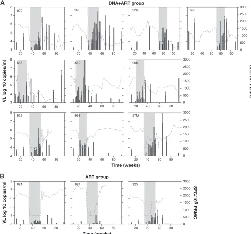
vacci-FIG. 4. ELISPOT analysis for 10 ART⫹DNA animals (A) and 3 ART-only controls (B). Gray and open stacked bars represent ELISPOT values (right scale) forgagand gp120env, respectively, for the indicated dates. Dotted lines indicate log10virus load (left scale). Shaded area indicates the period on ART.
on November 8, 2019 by guest
http://jvi.asm.org/
[image:5.585.43.535.70.530.2]nated animals in this study received, in addition, DNA vectors expressing macaque IL-12 (926L and 626L) or IL-15 (57M, 920L, 922L, and 923L). There was no difference in VL out-come between animals receiving IL-12 or IL-15 compared with those of the other vaccinated animals (P⫽0.53; Wilcoxon rank sum test). This preliminary analysis is based on a comparison of a few animals receiving low levels of cytokine DNA and must await further experiments with more animals for final conclusions on the effects of cytokine DNA inclusion during therapeutic vaccination.
Four animals in the vaccine group (795L, 797L, 538L, and 626L) and two animals in the control group (3077 and 921L) carried the MamuA*01 allele (Table 1), which has been asso-ciated with a milder disease course after challenge with SIV-mac in some studies (19, 24). Analysis of virus loads did not reveal any statistical difference in the decrease of VL for the four MamuA*01 animals compared to the eight others in the vaccine group, using a Wilcoxon rank sum test (P⫽0.81).
In conclusion, this analysis indicates that the decrease in viremia derived from immunotherapy did not depend on pre-vious prophylactic vaccination or the MamuA*01 haplotype. The results also indicate that infected animals with previous histories of prophylactic vaccination may get an independent benefit in terms of viral load reduction upon therapeutic im-munization.
Survival of DNA-vaccinated macaques. The animals that benefited most in virus load reduction also showed increased survivals compared to those of the animals that show less benefit. The vaccinees were ranked according to VL benefit and split in two groups of six macaques. Six of 12 vaccinees had decreases in viremia above the average of 1.05 log10
(high-benefit group). Five of these animals (538L, 926L, 920L, 965L, and 923L) were alive at the end of the study, after 111 to 177 weeks of follow-up post-ART. In contrast, four of the six an-imals with low virological benefit (low-benefit group) died and two were removed from the study after 34 and 74 weeks of observation, respectively. The survival curves for these two groups are shown in Fig. 3D. The survival curve of all 12 vaccinees is also shown. It is interesting that the group of animals that have a strong long-lasting benefit in VL also shows increased survival (P was 0.03 using a log-rank test as described in Materials and Methods).
Immunological analysis. To monitor the effects of DNA immunization on cellular immunity, we measured the fre-quency of antigen-specific, gamma interferon-producing T cells in peripheral blood mononuclear cells by ELISPOT assay after stimulation by overlapping peptide pools for gag and gp120env over time (see Fig. 4 and 5A). Most of the DNA-vaccinated animals showed strong and increasing responses during the vaccination period (Fig. 4). These responses were maintained in several animals (920L, 923L, 926L, and 538L) despite low levels of viremia. Figure 5A shows the peak ELISPOT response to gag and gp120env for 10 vaccinated animals for which serial measurements were available, divided into four periods: chronic phase, ART before vaccination, ART and DNA vaccination (ART⫹DNA), and follow-up after ART termination. The frequency of antigen-specific T cells decreased immediately upon drug treatment, as expected from the decreasing viral load. Interestingly, ELISPOT values in-creased immediately upon vaccination and persisted at high
levels after ART termination. The ELISPOT values forgag
and gp120env were compared during these four periods using a linear mixed model (26). The overall global test of a changing mean peak level over the four periods was highly significant (P⬍0.0001). Increases from chronic to ART⫹DNA period (mean change, 980 SFC/106 cells; P ⫽ 0.003) and from
chronic to post-ART period (mean change, 895;P⫽0.008) were highly significant.
ELISA measurements showed that the animals had high antibody levels against SIV (Fig. 5B), which, in contrast to ELISPOT (Fig. 5A), did not increase during therapeutic vac-cination. After ART termination, the antibody levels increased to higher levels. Application of a linear mixed model as above gave a significant global test of a changing mean over the four periods (P⬍0.001). There was a nonsignificant decrease from the chronic period to the ART period (mean change,⫺0.11;
P⫽0.21), followed by a significant decrease in the ART⫹DNA period (mean change from chronic,⫺0.23;P ⫽0.02), and a significant increase in the post-ART period (mean change from chronic, 0.56;P⫽0.001). Therefore, unlike cellular im-munity, the antibody levels did not increase during therapeutic vaccination, indicating a polarized Th1 response to DNA vac-cination in the infected macaques. Despite this, a significant antibody increase after release from therapy indicates a change in the humoral immune response after vaccination, despite the lower VL after immunotherapy. Three animals in the ART group were also studied in parallel by the same assays. These animals showed a progressive drop in antibody titers during ART and did not show increased immune responses after release.
FIG. 5. Comparisons of cellular and humoral immune responses. (A) Peak ELISPOT responses togagandenvfor 10 vaccinated animals divided into four periods: chronic phase, ART before vaccination, ART and DNA vaccination, and follow-up post-ART. (B) Peak anti-body levels against SIV (reciprocal titers) during the same time peri-ods. Brackets indicate comparisons showing statistical difference.
on November 8, 2019 by guest
http://jvi.asm.org/
DISCUSSION
This study demonstrates that chronically infected macaques with substantial viremia mount more efficient immune re-sponses after ART and DNA vaccination, which results in decreases in viral load by approximately 1 log on average. The animals varied widely in their responses; those showing strong and long-lasting virological benefits may also have prolonged survival. It will be important to understand the reasons for this heterogeneity in order to design better therapy approaches. Several animals in the vaccine group were able to suppress viremia close to the detection limits of the assay. In contrast, ART alone did not result in any significant viral load decrease in these chronically infected animals, which is in agreement with data from several studies on structured therapy interrup-tion in monkeys and humans.
It is interesting that the initial viral rebound upon termina-tion of ART was subsequently suppressed, presumably by the immune system, in the majority of therapeutically immunized macaques. The increase in cellular immune responses mea-sured by ELISPOT in this period agrees with the hypothesis that viral rebound leads to increased cytotoxic T-lymphocyte activity and the elimination of the infected cells. In several animals, we observed maintenance of the high frequency of
gag- andenv-specific T-cell responses in spite of low viral loads. This is in contrast to the expected decrease in the level of immune responses upon a decrease in viremia and suggests that the immune system of the therapeutically immunized an-imals reached a different steady state. This observation is sup-ported by the negative correlation of viral load with ELISPOT values during the post-ART period.
Our analysis focused on the comparison of all DNA-vacci-nated animals to the control group. Small variation in the schedule, DNA vectors, and the inclusion of small amounts of cytokine DNA in some vaccinated animals could not change the basic conclusions. The inclusion of some chronically in-fected but previously prophylactically vaccinated animals in both the control and vaccine groups could be questioned be-cause such animals may have more intact immune systems. Using two-way analysis of variance, we show that there is no evidence of different outcome in the animals with prior pro-phylactic vaccination, suggesting that the benefit derived by ART and therapeutic immunization is independent of previous history. To further address this concern, in a separate analysis presented in Fig. 3C, we considered only naı¨ve animals in-fected by SIV without any history of previous treatments. The basic conclusion of our study, that there is a long-lasting viro-logical benefit in therapeutically vaccinated macaques, is also verified by this analysis. Several studies support the notion that therapeutic immunization may augment immune responses against HIV or SIV, but the beneficial effect on VL decrease and disease progression remains controversial. Our data agree with recent reports indicating a virological benefit after ther-apeutic immunization (12, 16, 17).
It could be argued that animals enrolled in an immunother-apy study like the one presented here are preselected since rapid progressors are excluded. We (unpublished data) and others (9) noted that animals with very high acute viremia not decreasing substantially during the chronic phase respond poorly to an ART regimen (PMPA, emtricitabine, ddI,
stavu-dine). Thus, the animals enrolled represent macaques with immune systems that may be less damaged by the virus during the acute phase. Therefore, one cannot exclude the possibility that individual differences among infected macaques may af-fect the outcome of the study. Although such parameters are still under investigation, the positive and long-lasting benefit in 7 of 12 animals is an encouraging observation which warrants detailed comparisons of animals with differential benefit, so that the factors responsible for better outcomes are discovered. The sustained cellular immune response after therapeutic DNA vaccination may indicate an important clue for addi-tional studies. We have found a better preservation of central memory antigen-specific SIV lymphocytes in animals with long-lasting virologic benefit (our unpublished data).
It has been proposed that decreases of 1.5 to 2 logs in virus load caused by prophylactic vaccination may be important for achieving decreases in transmission and slowing disease devel-opment, providing a useful vaccine, despite the absence of sterilizing immunity (25). Long-lasting reduction in chronic virus loads in already infected animals may also provide a meaningful benefit, as suggested by the survival difference of the high responders versus low responders to therapeutic vac-cination (Fig. 3D). Comparison of strong responders to non-responders may provide ways to identify correlates of protec-tion. In conclusion, DNA vaccination during ART was able to elicit effective antiviral cellular responses in chronically in-fected macaques, resulting in long-term decrease of viremia. Therefore, repeated vaccination during antiretroviral therapy may provide better control of virus propagation, which is as-sociated with better disease control. Unlike viral recombinant vectors expressing HIV/SIV antigens, DNA therapeutic vacci-nation can be delivered repeatedly without focusing the im-mune response to other antigens. In addition, DNA vaccina-tion provides efficient access to the major histocompatibility complex class I pathway and should be considered for further development and optimization. It is anticipated that improve-ments in DNA delivery to the tissues, obtained with techniques such as electroporation and effective doses of molecular adju-vants, can dramatically improve the magnitude of responses to DNA vaccination.
ACKNOWLEDGMENTS
We thank D. Weiss, J. Treece, R. Pal, S. Orndorff, and the staff at Advanced BioScience Laboratories, Kensington, MD, for their expert help. We are grateful to M. Piatak, J. Lifson, for viral load assays and S. Strobel and A. Malyguine for use of the ELISPOT reader. We thank T. Jones for editorial assistance.
This research was supported (in part) by the Intramural Research Program of the NIH, National Cancer Institute, Center for Cancer Research.
REFERENCES
1.Aberle, H., A. Bauer, J. Stappert, A. Kispert, and R. Kemler.1997.-catenin
is a target for the ubiquitin-proteasome pathway. EMBO J.16:3797–3804.
2.Biragyn, A., K. Tani, M. C. Grimm, S. Weeks, and L. W. Kwak.1999. Genetic fusion of chemokines to a self tumor antigen induces protective, T-cell
dependent antitumor immunity. Nat. Biotechnol.17:253–258.
3.Chun, T. W., and A. S. Fauci.1999. Latent reservoirs of HIV: obstacles to the
eradication of virus. Proc. Natl. Acad. Sci. USA96:10958–10961.
4.Chun, T. W., L. Stuyver, S. B. Mizell, L. A. Ehler, J. A. Mican, M. Baseler, A. L. Lloyd, M. A. Nowak, and A. S. Fauci.1997. Presence of an inducible HIV-1 latent reservoir during highly active antiretroviral therapy. Proc. Natl.
Acad. Sci. USA94:13193–13197.
5.Davey, R. T., Jr., N. Bhat, C. Yoder, T. W. Chun, J. A. Metcalf, R. Dewar, V.
on November 8, 2019 by guest
http://jvi.asm.org/
Natarajan, R. A. Lempicki, J. W. Adelsberger, K. D. Miller, J. A. Kovacs, M. A. Polis, R. E. Walker, J. Falloon, H. Masur, D. Gee, M. Baseler, D. S. Dimitrov, A. S. Fauci, and H. C. Lane.1999. HIV-1 and T cell dynamics after interruption of highly active antiretroviral therapy (HAART) in patients with a history of sustained viral suppression. Proc. Natl. Acad. Sci. USA
96:15109–15114.
6.Egan, M. A., S. Y. Chong, S. Megati, D. C. Montefiori, N. F. Rose, J. D. Boyer, M. K. Sidhu, J. Quiroz, M. Rosati, E. B. Schadeck, G. N. Pavlakis, D. B. Weiner, J. K. Rose, Z. R. Israel, S. A. Udem, and J. H. Eldridge.2005. Priming with plasmid DNAs expressing interleukin-12 and simian
immuno-deficiency virusgagenhances the immunogenicity and efficacy of an
exper-imental AIDS vaccine based on recombinant vesicular stomatitis virus. AIDS
Res. Hum. Retrovir.21:629–643.
7.Hel, Z., J. M. Johnson, E. Tryniszewska, W. P. Tsai, R. Harrod, J. Fullen, J. Tartaglia, and G. Franchini.2002. A novel chimeric Rev, Tat, and Nef
(Retanef) antigen as a component of an SIV/HIV vaccine. Vaccine20:3171–
3186.
8.Hel, Z., E. Tryniszewska, W. P. Tsai, J. M. Johnson, R. Harrod, J. Fullen, V. S. Kalyanaraman, J. D. Altman, J. McNally, T. Karpova, B. K. Felber, J. Tartaglia, and G. Franchini.2002. Design and in vivo immunogenicity of a
polyvalent vaccine based on SIVmac regulatory genes. DNA Cell Biol.21:
619–626.
9.Hel, Z., D. Venzon, M. Poudyal, W. P. Tsai, L. Giuliani, R. Woodward, C. Chougnet, G. Shearer, J. D. Altman, D. Watkins, N. Bischofberger, A. Abimiku, P. Markham, J. Tartaglia, and G. Franchini.2000. Viremia con-trol following antiretroviral treatment and therapeutic immunization during
primary SIV251 infection of macaques. Nat. Med.6:1140–1146.
10.Kinloch-de Loes, S., B. Hoen, D. E. Smith, B. Autran, F. C. Lampe, A. N. Phillips, L. E. Goh, J. Andersson, C. Tsoukas, A. Sonnerborg, G. Tambussi, P. M. Girard, M. Bloch, M. Battegay, N. Carter, R. El Habib, G. Theofan, D. A. Cooper, and L. Perrin.2005. Impact of therapeutic immunization on HIV-1 viremia after discontinuation of antiretroviral therapy initiated during
acute infection. J. Infect. Dis.192:607–617.
11.Letvin, N. L., and B. D. Walker.2003. Immunopathogenesis and
immuno-therapy in AIDS virus infections. Nat. Med.9:861–866.
12.Le´vy, Y., H. Gahery-Segard, C. Durier, A. S. Lascaux, C. Goujard, V. Meiffredy, C. Rouzioux, R. E. Habib, M. Beumont-Mauviel, J. G. Guillet, J. F. Delfraissy, and J. P. Aboulker.2005. Immunological and virological efficacy of a therapeutic immunization combined with interleukin-2 in chronically HIV-1 infected
pa-tients. AIDS19:279–286.
13.Lisziewicz, J., E. Rosenberg, J. Lieberman, H. Jessen, L. Lopalco, R. Siliciano, B. Walker, and F. Lori.1999. Control of HIV despite the discontinuation of
antiretroviral therapy. N. Engl. J. Med.340:1683–1684.
14.Lisziewicz, J., J. Trocio, J. Xu, L. Whitman, A. Ryder, N. Bakare, M. G. Lewis, W. Wagner, A. Pistorio, S. Arya, and F. Lori.2005. Control of viral
rebound through therapeutic immunization with DermaVir. AIDS19:35–43.
15.Lori, F., M. G. Lewis, J. Xu, G. Varga, D. E. Zinn, Jr., C. Crabbs, W. Wagner, J. Greenhouse, P. Silvera, J. Yalley-Ogunro, C. Tinelli, and J. Lisziewicz.
2000. Control of SIV rebound through structured treatment interruptions
during early infection. Science290:1591–1593.
16.Lu, W., L. C. Arraes, W. T. Ferreira, and J. M. Andrieu.2004. Therapeutic
dendritic-cell vaccine for chronic HIV-1 infection. Nat. Med.10:1359–1365.
17.Lu, W., X. Wu, Y. Lu, W. Guo, and J. M. Andrieu.2003. Therapeutic
dendritic-cell vaccine for simian AIDS. Nat. Med.9:27–32.
18.Markowitz, M., X. Jin, A. Hurley, V. Simon, B. Ramratnam, M. Louie, G. R. Deschenes, M. Ramanathan, Jr., S. Barsoum, J. Vanderhoeven, T. He, C. Chung, J. Murray, A. S. Perelson, L. Zhang, and D. D. Ho.2002. Discon-tinuation of antiretroviral therapy commenced early during the course of human immunodeficiency virus type 1 infection, with or without adjunctive
vaccination. J. Infect. Dis.186:634–643.
19.Mothe´, B. R., J. Weinfurter, C. Wang, W. Rehrauer, N. Wilson, T. M. Allen, D. B. Allison, and D. I. Watkins.2003. Expression of the major histocom-patibility complex class I molecule Mamu-A*01 is associated with control of
simian immunodeficiency virus SIVmac239 replication. J. Virol.77:2736–
2740.
20.Nasioulas, G., A. S. Zolotukhin, C. Tabernero, L. Solomin, C. P. Cunningham, G. N. Pavlakis, and B. K. Felber.1994. Elements distinct from human immu-nodeficiency virus type 1 splice sites are responsible for the Rev dependence of
envmRNA. J. Virol.68:2986–2993.
21.Newberg, M. H., M. J. Kuroda, W. A. Charini, A. Miura, C. I. Lord, J. E. Schmitz, D. A. Gorgone, M. A. Lifton, K. Kuus-Reichel, and N. L. Letvin.
2002. A simian immunodeficiency virus nef peptide is a dominant cytotoxic T lymphocyte epitope in Indian-origin rhesus monkeys expressing the
com-mon MHC class I allele mamu-A*02. Virology301:365–373.
22.Ortiz, G. M., D. F. Nixon, A. Trkola, J. Binley, X. Jin, S. Bonhoeffer, P. J. Kuebler, S. M. Donahoe, M. A. Demoitie, W. M. Kakimoto, T. Ketas, B. Clas, J. J. Heymann, L. Zhang, Y. Cao, A. Hurley, J. P. Moore, D. D. Ho, and M. Markowitz.1999. HIV-1-specific immune responses in subjects who tempo-rarily contain virus replication after discontinuation of highly active
antiret-roviral therapy. J. Clin. Investig.104:R13–R18.
23.Oxenius, A., D. A. Price, H. F. Gunthard, S. J. Dawson, C. Fagard, L. Perrin, M. Fischer, R. Weber, M. Plana, F. Garcia, B. Hirschel, A. McLean, and R. E. Phillips.2002. Stimulation of HIV-specific cellular immunity by struc-tured treatment interruption fails to enhance viral control in chronic HIV
infection. Proc. Natl. Acad. Sci. USA99:13747–13752.
24.Pal, R., D. Venzon, N. L. Letvin, S. Santra, D. C. Montefiori, N. R. Miller, E. Tryniszewska, M. G. Lewis, T. C. VanCott, V. Hirsch, R. Woodward, A. Gibson, M. Grace, E. Dobratz, P. D. Markham, Z. Hel, J. Nacsa, M. Klein, J. Tartaglia, and G. Franchini.2002. ALVAC-SIV-gag-pol-env-based vacci-nation and macaque major histocompatibility complex class I (A*01) delay
simian immunodeficiency virus SIVmac-induced immunodeficiency. J. Virol.
76:292–302.
25.Picker, L. J., and D. I. Watkins.2005. HIV pathogenesis: the first cut is the
deepest. Nat. Immunol.6:430–432.
26.Pinheiro, J. C., and D. M. Bates. 2000. Mixed-effects models in S and S-PLUS. Springer, NY.
27.Robinson, H. L.2002. New hope for an AIDS vaccine. Nat. Rev. Immunol.
2:239–250.
28.Romano, J. W., R. N. Shurtliff, E. Dobratz, A. Gibson, K. Hickman, P. D. Markham, and R. Pal.2000. Quantitative evaluation of simian
immunode-ficiency virus infection using NASBA technology. J. Virol. Methods86:
61–70.
29.Rosati, M., A. von Gegerfelt, P. Roth, C. Alicea, A. Valentin, M. Robert-Guroff, D. Venzon, D. Montefiori, P. Markham, B. K. Felber, and G. N. Pavlakis.2005. DNA vaccines expressing different forms of SIV antigens
decrease viremia upon SIVmac251 challenge. J. Virol.79:8480–8492.
30.Rosenberg, E. S., M. Altfeld, S. H. Poon, M. N. Phillips, B. M. Wilkes, R. L. Eldridge, G. K. Robbins, R. T. D’Aquila, P. J. Goulder, and B. D. Walker.
2000. Immune control of HIV-1 after early treatment of acute infection.
Nature407:523–526.
31.Schneider, R., M. Campbell, G. Nasioulas, B. K. Felber, and G. N. Pavlakis.
1997. Inactivation of the human immunodeficiency virus type 1 inhibitory elements allows Rev-independent expression of Gag and Gag/protease and
particle formation. J. Virol.71:4892–4903.
32.Schwartz, S., M. Campbell, G. Nasioulas, J. Harrison, B. K. Felber, and G. N. Pavlakis.1992. Mutational inactivation of an inhibitory sequence in
human immunodeficiency virus type 1 results in Rev-independentgag
ex-pression. J. Virol.66:7176–7182.
33.Schwartz, S., B. K. Felber, and G. N. Pavlakis.1992. Distinct RNA
se-quences in thegagregion of human immunodeficiency virus type 1 decrease
RNA stability and inhibit expression in the absence of Rev protein. J. Virol.
66:150–159.
34.Siliciano, J. D., J. Kajdas, D. Finzi, T. C. Quinn, K. Chadwick, J. B. Margolick, C. Kovacs, S. J. Gange, and R. F. Siliciano.2003. Long-term follow-up studies confirm the stability of the latent reservoir for HIV-1 in
resting CD4⫹T cells. Nat. Med.9:727–728.
35.Suryanarayana, K., T. A. Wiltrout, G. M. Vasquez, V. M. Hirsch, and J. D. Lifson.1998. Plasma SIV RNA viral load determination by real-time quan-tification of product generation in reverse transcriptase-polymerase chain
reaction. AIDS Res. Hum. Retrovir.14:183–189.
36.Tryniszewska, E., J. Nacsa, M. G. Lewis, P. Silvera, D. Montefiori, D. Venzon, Z. Hel, R. W. Parks, M. Moniuszko, J. Tartaglia, K. A. Smith, and G. Franchini.2002. Vaccination of macaques with long-standing SIVmac251 infection lowers the viral set point after cessation of antiretroviral therapy.
J. Immunol.169:5347–5357.
37.Tsai, C. C., P. Emau, K. E. Follis, T. W. Beck, R. E. Benveniste, N. Bischof-berger, J. D. Lifson, and W. R. Morton.1998. Effectiveness of
postinocula-tion (R)-9-(2-phosphonylmethoxypropyl) adenine treatment for prevention
of persistent simian immunodeficiency virus SIVmneinfection depends
crit-ically on timing of initiation and duration of treatment. J. Virol.72:4265–
4273.
38.Tubiana, R., G. Carcelain, M. Vray, K. Gourlain, C. Dalban, A. Chermak, C. Rabian, D. Vittecoq, A. Simon, E. Bouvet, R. El Habib, D. Costagliola, V. Calvez, B. Autran, and C. Katlama.2005. Therapeutic immunization with a human immunodeficiency virus (HIV) type 1-recombinant canarypox vac-cine in chronically HIV-infected patients: The Vacciter Study (ANRS 094).
Vaccine23:4292–4301.