EVALUATION OF IN VIVO ANTICANCER ACTIVITY OF ETHANOLIC EXTRACT OF LEAF OF Plumeria acutifolia ON DALTONS ASCITES
LYMPHOMA INDUCED CANCER IN MICE Dissertation submitted to
THE TAMIL NADU DR. M.G.R. MEDICAL UNIVERSITY,
CHENNAI - 32.
In partial fulfillment for the award of the degree of
MASTER OF PHARMACY
IN
PHARMACOLOGY
Submitted by Reg. No.: 261525216
Under the Guidance of
Mr.V.VENKATESWARAN,M.Pharm.,
DEPARTMENT OF PHARMACOLOGY J.K.K. NATTRAJA COLLEGE OF PHARMACY
Komarapalayam – 638183. Tamil Nadu.
CERTIFICATE
This is to certify that the work embodied in this dissertation entitled
"Evaluation of in vivo anticancer activity of ethanolic extract of leaf of plumeria
acutifolia on Daltons ascites lymphoma induced cancer in mice", submitted to
"The Tamilnadu Dr.M.G.R. Medical University", Chennai. in partial fulfillment to the requirement for the award of Degree of Master of Pharmacy in Pharmacology,is a bonafide work carried out by Mr.N.YOGESH, Reg.No-261525216, during the academic year 2016-2017, under my guidance and direct supervision in the department of pharmacology, J.K.K.Nataraja College of Pharmacy, Komarapalayam.
CERTIFICATE
This is to certify that the work embodied in this dissertation entitled
"Evaluation of in vivo anticancer activity of ethanolic extract of leaf of plumeria
acutifolia on Daltons ascites lymphoma induced cancer in mice", submitted to
"The Tamilnadu Dr.M.G.R. Medical University", Chennai. in partial fulfillment to the requirement for the award of Degree of Master of Pharmacy in Pharmacology, is a bonafide work carried out by Mr.N.YOGESH, Reg.No-261525216, during the academic year 2016-2017, under my guidance and direct supervision in the department of pharmacology, J.K.K.Nataraja College of Pharmacy, Komarapalayam.
Place: Komarapalayam, Mr.V.Venkateswaran, M.Pharm., Date: Asst. Professor,
Department of Pharmacology,
CERTIFICATE
This is to certify that the work embodied in this dissertation entitled "Evaluation of in vivo anticancer activity of ethanolic extract of leaf of plumeria
acutifolia on Daltons ascites lymphoma induced cancer in mice", submitted to
"The Tamilnadu Dr.M.G.R. Medical University", Chennai. in partial fulfillment to the requirement for the award of Degree of Master of Pharmacy in Pharmacology, is a bonafide work carried out by Mr.N.YOGESH, Reg.No-261525216, during the academic year 2016-2017, under my guidance and direct supervision in the department of pharmacology, J.K.K.Nataraja College of Pharmacy, Komarapalayam.
Place:Komarapalayam Dr.R.Sambath Kumar,M.pharm.,Ph.D., Date: Principal and Professor,
CERTIFICATE
This is to certify that the work embodied in this dissertation entitled "Evaluation of in vivo anticancer activity of ethanolic extract of leaf of plumeria
acutifolia on Daltons ascites lymphoma induced cancer in mice", submitted to
"The Tamilnadu Dr.M.G.R. Medical University", Chennai. in partial fulfillment to the requirement for the award of Degree of Master of Pharmacy in Pharmacology, is a bonafide work carried out by Mr.N.YOGESH, Reg.No-261525216, during the academic year 2016-2017, under my guidance and direct supervision in the department of pharmacology, J.K.K.Nataraja College of Pharmacy, Komarapalayam.
Place: Komarapalayam Dr.R.Shanmuga sundaram, M.Pharm., Ph.D,
Date: Vice Principal and Professor,
CERTIFICATE
This is to certify that the work embodied in this dissertation entitled
"Evaluation of in vivo anticancer activity of ethanolic extract of leaf of plumeria
acutifolia on Daltons ascites lymphoma induced cancer in mice", submitted to
"The Tamilnadu Dr.M.G.R. Medical University", Chennai. in partial fulfillment to the requirement for the award of Degree of Master of Pharmacy in Pharmacology, is a bonafide work carried out by Mr.N.YOGESH, Reg.No-261525216, during the academic year 2016-2017, under my guidance and direct supervision in the department of pharmacology, J.K.K.Nataraja College of Pharmacy, Komarapalayam.
Dr.R.Sambath,Kumar,M.pharm.,Ph.D.
Principal and Professor, Department of Pharmacology, J.K.K.Nataraja College of Pharmacy.
Mr.V.Venkateswaran,M.Pharm.,
Asst. Professor,
Department of Pharmaceutics, J.K.K.Nataraja College of Pharmacy.
.
Dr.R.Shanmuga Sundaram,M.Pharm.,Ph.D., Vice Principal and Professor,
CERTIFICATE
I hereby declare that the dissertation entitled "Evaluation of in vivo anticancer activity of ethanolic extract of leaf of plumeria acutifolia on Daltons
ascites lymphoma induced cancer in mice", has been carried out under the guidance and supervision of Mr.V.Vvenkateswaran,M.Pharm, Assistant Professor, Department of Pharmacology, J.K.K.Nataraja College of Pharmacy. Komarapalayam, in partial fulfillment of the requirements for the award of degree of Master of Pharmacy in Pharmacology during the academic year 2016-2017.
I further declare that, this work is original and this dissertation has not been submitted previously for the award of any other degree, diploma associate ship and fellowship or any other similar title.
Place: Komarapalayam, N.YOGESH, Date: Reg.No:261525216,
CONTENTS
S. No.
CONTENTS
Page No.
1. INTRODUCTION 1
2 REVIEW OF LITERATURE 20
3 PLANT PROFILE 23
4 AIM AND OBJECTIVE OF THE STUDY 25
5 PLAN OF WORK 26
6 MATERIALS AND METHODS 27
7. RESULTS AND DISCUSSION 60
8. SUMMARY AND CONCLUSION 94
ACKNOWLEDGEMENT
I express my whole hearted thanks to my guide Mr.V.Venkateswaran, M.pharm.,
Assistant Professor, department of pharmacology, for suggesting solution to problems
faced by me and providing in dispensable guidance, tremendous encouragement at each
and every step of this dissertation work. Without his critical advice and deep-rooted
knowledge, this work would not have been a reality.
It is my most pleasant duty to thank our beloved Principal and Professor
Dr.R.Sambath Kumar, M.Pharm., Ph.D., J.K.K.Nataraja College of Pharmacy.
Komarapalayam for ensuring all the facilities were made available to me for the
smooth running of this project.
My sincere thanks to
Dr.R.Shanmuga Sundaram, M.pharm., Ph.D.,
Vice Principal and Professor, head department of pharmacology, for their valuable
suggestions during my project work.
My sincere thanks to
Mr.C.Kannan, M.Pharm.,
Lecturer, Department of
pharmaceutics, Mrs.R.Elavarasi, M.Pharm., Lecturer, Department of pharmacology,
Mrs. Baby Kala M.Pharm., Lecturer ,Deartment of Pharmacology for their valuable
suggestion,ideas and help.
My sincere thanks to
Dr. V.Sekar, M.Pharm, Ph.D., Professor and Head,
Department of Analysis, Dr.I. Caolin Nimila, M.Pharm., Ph.D., Assistant professor,
Mr K.K.Senthil Kumar, M.Pharm., Assistant Professor, Department of
Pharmaceutical Analysis for their valuable suggestions.
My sincere thanks to
Mrs. S.Bhama, M.Pharm., Assistant Professor ,
Deapartment of Pharmaceutics, Mr. C.Kannan. M.Pharm., Assistant Professor,
Deartment of Pharmaceutics for their valuable help during my project.
It is my privilege to express deepest sense of gratitude toward
Dr.
M.Vijayabaskaran, M.Pharm., Professor & Head, Department of Pharmaceutical
Pharmaceutical chemistry, Mrs.S.Gomathi M.Pharm., Lecturer, Department
Chemistry, for their valuable suggestions.
My sincere thanks to Dr. Senthilraja, M.Pharm., Ph.D., Professor and Head,
Department of Pharmacognosy, Dr. Rajkumar, M.Pharm, Ph.D., Prosfessor,
Department of pharmacognosy, for their valuable suggestions during my project work.
My sincere thanks to
Mr.E.Vasantha kumar M.C.A.,Assistant Professor,
Department of Computer, for his valuable help during my project work.
I express my sincere and deep sense of gratitude to the founder, (Late)
Thiru
J.K.K. Nattaraja Chettiar, providing us the historical institution to study
My sincere thanks and respectful regards to our reverent Chairperson
Smt. N.
Sendamaraai,
B.Com, and the Managing Director
Mr.S .Omm Sharravana, B.Com,
LLB., J.K.K.Nattaraja Educational Institutions, Kumarapalayam for their blessings,
encouragement and support at all times.
My glorious acknowledgement to our administrative officer
Dr. K. Sengodan ,
M.B.B.S., for encouraging us in a kind and generous manner to complete this work .
I greatly acknowledge the help rendered by Mrs. K.Rani, Office Superintendent,
Mrs. V.Gandhimathi, M.A., M.L.I.S., Librarian, Mrs. S.Jayakala B.A., B.L.I.S., and
Assistant Librarian.
I owe my thanks to all the technical and non- teaching staff members of the
institute for their precious assistance and help.
Last, but nevertheless, Iam thankful to my colleagues and all my friends for their
co- operation, encouragement and help extended to me throughout my project work.
N.YOGESH,
Department of Pharmacology 1 J.K.K.Nataraja College of Pharmacy 1.INTRODUCTION
MEDICINAL PLANTS
Various plants have been used for medicinal purpose long before prehistoric period. Traditional systems of medicine continue to be widely practiced on many accounts. population rise inadequate supply of drugs prohibitive cost of treatments, side effects of several synthetic drugs and development of resistance to currently used drugs for infection disease have led to increased emphasis on the use of plant materials as a source of medicines for a wide variety of human ailments. Among ancient civilization india has been known to be rich repository of medicinal plants. About 8000 herbal remedies have been codified in AYUSH systems in india. ayurveda, unani, siddha and folk (tribal) medicines are the major systems of indigenous medicines. Among these systems, ayurveda and unani medicines are most developed and widely practised in india.according to WHO around 21000 plant species have the potential for being used as medicinal plants.The economic importance of medicinal plants is much more to countries provide two third of plants used in modern systems of medicine and health care system of rural population depend on indigenous systems of medicine. Treatment with medicinal plants is considered very safety as there is no or minimal side effects. there remedies are in sync with nature.which is biggest advantage. The golden fact is that use of herbal treatments is independent of any age groups and sexes.The most of the drugs thus formulated are of side effects or reactions.this is the reason why medicinal treatment growing in popularity across the globe. Medicinal plants are considered as a rich resources of ingredients which can be used in drug development either pharmacopoeial or synthetic drugs.
WHY PEOPLE WITH CANCER USE NATURAL MEDICINES.
Department of Pharmacology 2 J.K.K.Nataraja College of Pharmacy Although great advancements have been made in the treatment and control of cancer. progression significant deficiencies and room for improvement remain. plants have enormous potential to provide newer drugs and as such are a reservoir of natural chemicals that may provide chemo productive potential against cancer. A number of side effects sometimes occur during chemotherapy. Natural therapies such as the use of plant derived products in cancer treatment may reduce adverse side effects. Currently a few plant products are being used to treat cancer. However a myriad of many plant products exist that have shown very promising anti cancer properties in vitro but rare yet to be evaluated in humans.
Further study is required to determine the efficacy of these plants products in treating cancer in humans
TUMOR1:
Neoplasia literally means the process of "new growth," and a new growth is called a neoplasm. The term tumor was originally applied to the swelling caused by
inflammation. Neoplasms also may induce swellings, but by long precedent, the
non-neoplastic usage of tumor has passed into limbo; thus, the term is now equated with
neoplasm. Oncology (Greek oncos = tumor) is the study of tumors or neoplasms. Cancer
is the common term for all malignant tumors. Although the ancient origins of this term
are somewhat uncertain, it probably derives from the Latin for crab, cancer-presumably
because a cancer "adheres to any part that it seizes upon in an obstinate manner like the
crab."
1. DEFINITIONS:
Solid tumors are defined as abnormal masses of tissue that usually do not
contain cysts or liquid areas. Solid tumors may be benign (not cancerous), or
malignant (cancerous). A number of malignant diseases are often also categorized as
“solid tumors” such as breast cancer, cancer of the pancreas, lung, colon, etc.
Solid tumors can be split into three separate categories, depending on the type of
Department of Pharmacology 3 J.K.K.Nataraja College of Pharmacy Sarcomas: Cancers arising from connective or supporting tissues such as bone
or muscle.
Carcinomas: Cancers arising from the body's glandular cells and epithelial cells, which line the air passages and gastrointestinal tract.
Lymphomas: Cancers of the lymphoid organs such as lymph nodes, spleen, and thymus, which produce and store infection-fighting cells. Lymphoma is
cancer of the lymphatic system, which is part of the immune system.
Lymphoma is a form of cancer that affects the immune system - specifically, it is
a cancer of immune cells called lymphocytes, a type of white blood cell. There are
two broad types of lymphoma and many subtypes.
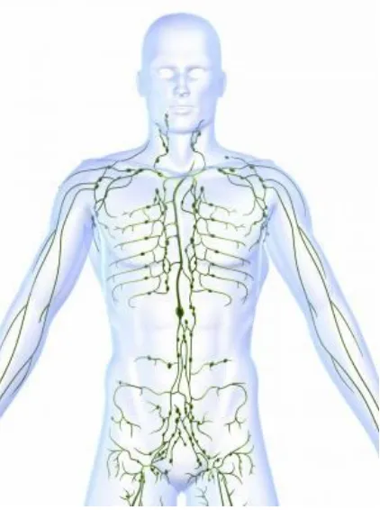
[image:13.612.217.428.282.565.2]
Figure.1 The lymphatic system is a system of vessels that branch back from virtually all our tissues to drain excess fluids and present foreign material to the lymph nodes.
The two types of lymphoma are described as: Hodgkin's or non Hodgkin's.
Lymphoma can occur at any age but is the most common cancer in young people. It is
Department of Pharmacology 4 J.K.K.Nataraja College of Pharmacy
About 90% of lymphomas are the non-Hodgkin's type while about 10%
are Hodgkin's.
Cancer is a group of over 100 diseases, all of which start with the growth of
abnormal cells. Instead of dying in the normal cell life cycle, cancerous cells continue
to divide into new abnormal cells, and grow out of control.
Department of Pharmacology 5 J.K.K.Nataraja College of Pharmacy
Lymphatic cancers are classified by the type of immune cells affected.
In non-Hodgkin's lymphoma, B-cells and T-cells are affected - both being
types of lymphocyte white blood cell with special roles in immunity. In the US, B-cell
lymphomas are much more common than T-cell ones.
In Hodgkin's lymphoma, the cancer cells are usually an abnormal type of B
lymphocyte, named Reed-Sternberg cells. There are many subtypes of Hodgkin's
lymphoma, typed by differences seen under the microscope - but a very high
percentage of cases are classed as "classic" Hodgkin's.
Types of lymphoma
There are many different types of lymphoma depending on the type of
lymphatic cells affected.
Hodgkin's lymphoma can occur at any age, affects more men than women and
the majority will be completely cured.
Hodgkin's lymphoma is diagnosed when a special type of cell, the
Reed-Sternberg cell, is seen under the microscope.
Non-Hodgkin's lymphoma accounts for all the other types of lymphoma.
These can be high grade or low grade and the treatment and prognosis varies.
Factors for Non-Hodgkin's lymphoma
Age - most non-Hodgkin lymphomas are in people 60 years of age and over
Sex - there are different rates of different types of non-Hodgkin's lymphoma across
the sexes
Ethnicity and location - in the US, African-Americans and Asian-Americans are less prone than white Americans, and the disease is more common in developed
nations of the world
Chemicals and radiation - some chemicals used in agriculture have been linked,
as has nuclear radiation exposure
Immune deficiency - for example, caused by HIV infection or in organ
Department of Pharmacology 6 J.K.K.Nataraja College of Pharmacy Autoimmune disease, in which the immune system attacks the body's own cells
Infection - certain viral and bacterial infections increase the risk. The Helicobacter Infection has been implicated, as has the Epstein Barr Virus (the virus that
causes glandular fever)
See the American Cancer Society's page for more detail on risk factors for
non-Hodgkin's lymphoma.
Factors for Hodgkin's lymphoma
Infectious mononucleosis - infection with Epstein-Barr virus
Age - two specific groups are most affected: typically people in their 20s, and people over the age of 55 years
Sex - slightly more common in men
Location - most common in the US, Canada and northern Europe; least common in
Asia
Family - if a sibling has the condition, the risk is slightly higher, and very high if
there is an identical twin
Affluence - people from higher socioeconomic status are at greater risk
HIV infection
See the American Cancer Society's page for more detail on risk factors for
Hodgkin's lymphoma.
Symptoms of lymphoma
The symptoms and signs of lymphoma are very similar to those of simple
illnesses such as viral illnesses and the common cold, and this can cause problems
with delayed diagnosis.
The difference is that the symptoms of lymphoma persist long after the usual run of
a viral infection.
Swelling in the legs or ankles
Cramping and bloating of the abdomen Night sweats and fever
Department of Pharmacology 7 J.K.K.Nataraja College of Pharmacy Chills
Unusual itching Fatigue
Pain or altered sensation Loss of appetite
Unusual tiredness/lack of energy Persistent coughing
Breathlessness Enlarged tonsils Headache.
Treatments and prevention
A number of treatment options are used against lymphoma cancer these are
Radiation therapy
Biologic therapy
Antibody therapy
Stem-cell transplantation
Splenectomy
Steroid treatment
Radioimmunotherapy
Department of Pharmacology 8 J.K.K.Nataraja College of Pharmacy 1.2 ANTIOXIDANTS:
Free radical is a chemical compound which contains an unpaired electron
spinning on the peripheral layer around the nucleus. The family of free radicals
generated from the oxygen is called ROS which cause damage to other molecules
by extracting electrons from them in order to attain stability. ROS are ions,
atoms or molecules that have the ability to oxidize reduced molecules. ROS are
various forms of activated oxygen, which include free radicals such as
superoxide anion radicals (O2.) And hydroxyl radicals (OH), as well as non-free
radicals (H2O2) and singlet oxygen2. In the body, free radicals are derived from two
sources: endogenous sources, e.g. Nutrient metabolism, ageing process etc and
exogenous sources e.g. Tobacco smoke, ionizing radiation, air pollution, organic
solvents, pesticides3.
When oxygen traps single electron, it becomes unstable and thus very
reactive, since it generates harmful chain reactions against many biological
molecules. The extreme toxicity of oxygen is related to its high capability of
generating free radicals and in turn destroying many major biological molecules.
They can attack on lipids and proteins and destroy membranes. ROS can damage
DNA and lead to mutation and chromosomal damage. Oxidized cellular thiols
abstract hydrogen atoms from unsaturated fatty acids to initiate the peroxidation
of membrane lipids4. ROS can attack various substrates in body and contribute to
development of chronic diseases. Exogenous chemicals and endogenous metabolic
processes in human body produce free radicals, especially oxygen derived
radicals, which are capable of oxidizing biomolecules, resulting in cell death.
Superoxide anion radicals increase under stress conditions such as heavy exercise,
certain drugs, infection and various disease states. During normal metabolic
processes, human body generates more than 2 Kg of O2 per year5.
Cells are equipped with different kinds of mechanisms to fight against ROS
and to maintain the redox homeostasis of cell. For example, antioxidant enzymes such
as superoxide dismutase (SOD), catalase (CAT) and glutathione peroxidase
(GPx) play important roles inscavenging the free radicals and preventing cell
Department of Pharmacology 9 J.K.K.Nataraja College of Pharmacy the mechanism of antioxidant protection becomes unbalanced in human body,
antioxidant supplement may be used to help reduce oxidative damage.
[image:19.612.121.514.172.403.2]
Table.1 Some selected antioxidants and their mechanisms of action12:
Antioxidants Mechanisms of action
SOD Dismutation of superoxide to H2O2
CAT Decomposes H2O2 to molecular oxygen
and water
GSH Intracellular reducing agent
Lycopene Trapping of singlet oxygen
Quercetin H2O2 scavenging, one of the potent antioxidant among polyphenols
Department of Pharmacology 11 J.K.K.Nataraja College of Pharmacy 1.3FLAVONOIDS:
Flavonoids are found in higher vascular plants, particularly in the flower,
leaves and bark. They are especially abundant in fruits, grains and nuts,
particularly in the skins.
Beverages consisting of plant extracts (beer, tea, wine, fruit juice) are the
principle source of dietary flavonoid intake. A glass of red wine has ~200
mg of flavonoids.
Typical flavonoid intake ranges from 50 to 800 mg/day, which is roughly 5,
50 and 100 times that of Vitamins C, and E, and carotenoids respectively.
1.3.1 Flavonoids role in cancer13:
Few diseases are feared more than cancer because cancerous diseases, after
cardiovascular disorders and accidents, kill more people before a normal life span has
been reached. Besides, the progress of cancer is often accompanied by great pain and
ugly disfiguration of the body. Yet, in principle, cancer is curable if it is discovered
early and treated with the best current therapeutic methods. However, radical cancer
cure is fraught with considerable life-threatening dangers, loss of organs, pain, and
discomfort. Besides, its treatment is expensive. Hence, it is understandable that many
cancer patients look for and try milder anticancer therapies that offer some promise of
a moderate, long-term life-saving cure. The flavonoids are some of the most
promising anticancer natural products that have been tried. Related synthetic
substances, e.g., flavone acetic acid, have been subjected to Phase I clinical trials
already, and they may soon become adopted into the general repertoire of cytostatic
treatment.
1.3.2 Flavonoids role in anti oxidants:
a. Enhance or mimic antioxidant enzymes.
b. Direct scavenging of ROS.
c. Repair damaged cellular components.
Department of Pharmacology 12 J.K.K.Nataraja College of Pharmacy Flavonoid and related compound are effective in scavenging DPPH radical14,
hydroxyl radical and in metal-chelating capacity. Flavonoids are found to exhibit
numerous biological activities like vasodilatory, anti carcinogenic, anti-inflammatory,
antibacterial, immune-stimulating, antiallergic, and antiviral effects 15.
1.4 CELL LINES16:
If the specified number of cells of inoculated in to sensitive mouse strain,
tumors can be developed rapidly as compared to chemical carcinogen-induced tumors
and time can saved using this model.
1.4.1 METHODS INVOLVED IN CELL LINES:
L-1210 and P-388 cell lines are used. These cell lines are derived from mouse lymphatic leukemia and have 100% growth fraction and tumor implanted with
specified number of L-1210 or P-388 cells can be predicted. The effective drug would
retard the tumor growth and increase the life span of the animal. A drug, which
prolongs the lifespan of the animal by 20%, is taken for subsequent studies involving
testing on other transplantable tumors. Some other cell lines which can be inoculated
to induce tumors are B-16(melanoma), Lewis lung carcinoma and sarcoma-180, etc.
The host mouse strain for above type of cell lines is BDF, except Swiss for
sarcoma-180. P-388 and L-1210 cell lines are inoculated intraperitonially and sarcoma-180 as
subcutaneously. The experiment takes about 10 days for completion
ILS (%) = [(Mean survival of treated group/ Mean survival of control group)-1] x100
For sarcoma-180 tumors, reduction of tumor size (tumor weight) is used to
find out the inhibiting activity of solid tumors as follows.
Department of Pharmacology 13 J.K.K.Nataraja College of Pharmacy Hollow-fiber technique:
Small hollow fibers (tubes 1mm in diameter and 2cm long made of plastic,
polyvinylidene flouride), containing cells from human tumors are inserted underneath
the skin and in the body cavity of the mouse. Each candidate drug is administered at
two dosages and is tested against 12 target tumor cells in different hollow fibers. A
total of about 20 compounds per week are screened by this method. Compounds that
retard the growth of the cells are recommended for the next level of testing. The
average length of this test is four days.
Nude mouse model:
Nude mice have been widely used to test the tumorogenicity of cells or for
testing of anticancer drugs. These mice are immunologically incompetent because of
absence of thymus. They neither show mitotic response in mixed lymphocyte
reaction, nor generate cytotoxic effector cell. Lack of helper T and suppressor T cells
alters the antibody response of the animals to antigen. They do not show contact
sensitivity and do not reject the transplanted material. They are required to be
maintained under strictly sterile conditions and in a warm environment (26-280).
Some other points regarding their use are:
a. Certain tumors like melanomas and colon carcinomas grow very well in nude
mice, whereas prostate carcinoma and most types of leukemia grow very
poorly.
b. Large numbers of cells, usually >106 are required to be inoculated beneath the
skin to get a successful tumor take.
c. Metastases are rarely observed.
Department of Pharmacology 14 J.K.K.Nataraja College of Pharmacy
1.5 INVIVO METHODS FOR SCREENING OF NEW ANTICANCER
MOLECULES16:
Studies that are in vivo (Latin for "within the living"; often not italicized in English) are those in which the effects of various biological entities are tested on whole, living organisms, usually animals, including humans, and plants as opposed to a partial or dead organism.
USE
In vivo testing is necessary for medical and research purposes
The medical field benefits from animal models to test the safety of drugs before they are used on patients
The research field benefits from in vivo testing by validating in vitro findings in vertebrates closest to humans
In vivo testing using animal models of disease help discover new ways of solving complex health problems.
METHODS
Xenograft Spontaneous
Carcinogen-Induced
GEMs (Genetically Engineered Mouse models) Transgenic
Knockout
Regulatable transgenic
Xenograft Models of Cancer
Human cell lines in Immunocompromised mouse Human tumor grafts in Immunocompromised mouse Mouse cell lines in syngeneic host
Department of Pharmacology 15 J.K.K.Nataraja College of Pharmacy Subcutaneous Implantation
Implant cells or tissues under the skin.
Advantages
external monitoring of growth
easy & most commonly used human cells
Disadvantage
suboptimal vascular site poor take rate
can be difficult to find tissue that does not grow into a tumor can be an irritant to the anima.
Spontaneous Models
Some strains of laboratory animals are susceptible to spontaneously developing certain types of tumors.
Advantages
May mimic some types of human diseases Can use to study early disease
Can use for prevention
Includes elements of progression
Disadvantages
Variability of disease progression Large animal numbers needed Long time to develop disease
Department of Pharmacology 16 J.K.K.Nataraja College of Pharmacy Carcinogen-Induced
Animal is treated with a carcinogen to induce cancer.Some laboratory animal Strains are more susceptible.
Examples include:
Lung cancer Skin cancer Bladder cancer Stomach cancer Prostate cancer
Advantages
Mimics initiation steps of some cancer Can study early events
Used to id predisposing conditions Can study prevention
Disadvantages
Health hazard to investigator Variability of disease progression Can require large animal numbers
Penetrance (all animals may not get disease)
Genetically Engineered Mouse (GEM) Models
Simple Transgenic Models
Tissue specific promoter Overexpression of gene
Ectopic expression (inappropriate expression) Oncogene
Growth factor
Department of Pharmacology 17 J.K.K.Nataraja College of Pharmacy Dominant Negative molecules
Cell cycle regulators Endogenous gene is intact.
Advantages
May mimic initiation steps of some cancer Can study early events
Can test genetic lesion that predispose Autochthonous (rising in the tissue of origin) Penetrance usually 100%
Immune system intact Can progress with time
Disadvantages
Variability of disease progression Often requires large animal numbers Initiator may be artificial
Time consuming to characterize & validate Expression influenced by site of integration
May produce chimeric offspring if integrated later in development.
Knockout Models
Approaches:
Homologous recombination
Cre-Lox system to KO in a tissue specific manner
Department of Pharmacology 18 J.K.K.Nataraja College of Pharmacy Inducible Systems
Requires a drug or hormone to induce gene expression Can be used with Cre to obtain a regulated knockout
Examples include:
Tet On/Off (Tetracycline) RU486 (Progesterone)
Ecdysone (Insect steroid hormone)
Ideal Inducible System
Tightly regulated
Not leaky- low basal level
Induce to high level of expression Induce only in presence of inducer Induce nontoxic
Nonphysiological- not affect expression of endogenous genes Reversible expression.
Advantages
Control when gene is expressed Can rescue an embryonic lethal gene
Allows for normal development of the organ
Disadvantages
Requires bi-or trigenics to get tissue specific expression Large breeding program
Expensive
Department of Pharmacology 19 J.K.K.Nataraja College of Pharmacy 1.6 SIGNIFICANCE OF THE PLANTS CHOSEN FOR THE PRESENT STUDY:
The plant Plumeria acutifolia Poir was reported that, it have a rich amount of
Flavanoids18 mainly phenolic compound Gallic acid , Terpenoids, tannins, sterols
(β-sitosterol, β-sitosterol-3β -D- glucoside), triterpenes (nonacosane &
hentriacontane) and saponins (sapogenins)19.
1.7 SIGNIFICANCE OF THE DALTON’S LYMPHOMA ASCITES TUMOR CELL LINES (DAL) CHOSEN FOR THE PRESENT STUDY:
1.7.1 Dalton’s lymphoma ascites tumor cell lines (DAL):
It is a tumor cell line originally grown from a tumor of the thymus.
It is propagated by growing as ascites tumor in mice.
We can induce both ascites tumor and solid tumors using DAL cells.
It is easy to maintain in vivo.
It is not an immunogenic.
1.7.2 Maintenance of cell lines:
Tumor Cell Line and Their Maintenance, Dalton’s lymphoma ascites tumor
cell lines (DLA), originally obtained from Amala Cancer Institute, Thrissur, Kerala
were propagated as transplantable tumors in the peritoneal cavity of the mice were
used for the study. The tumor cell lines were maintained by serial peritoneal cavity i.p
transplantation in mice. Full-grown tumor cell-line were aspirated mouse by injecting
PBS in to peritoneal cavity make cells to suspend in PBS, take that suspended
solution and count the number of cells present in one ml by using tryphan blue
exclusion method and adjust the cell count to 1×10⁶ by using PBS were inject
Department of Pharmacology 20 J.K.K.Nataraja College of Pharmacy
2.REVIEW OF LITERATURE
1. vijaya lakshmi.A, et.al., 2014., reported pharmacognostic phytochemical investigation of the root bark of plant plumeria acutifolia showing qualitative and quantitative phytochemical constituents.
2. Akihiko omata et.al., 1992, evaluate the essential oil of plumeria rubra forma acutifolia. poir woodson cv. Common yellow growing in hawai was extracted by simultaneous distillationand extraction (SDE).the essential oil was analysed with GC and GC-MS and total 74 compund were identified.
3. A.vijayalakshmi et.al., 2011,studied that the Anti anaphylactic and anti inflammatory activities of a bio active alkaloid from the root bark of plumeria acutifolia poir. A dose dependent benificial effect was observed on leakage of evans blue dye in skin changed with antigen and on paw anaphylaxis indused by antiserum .
4. surendra kr sharma, et.al., 2012, assayed Anti microbial potential of plumeria rubra syn plumeria acutifolia bark were sucessive extracts of plumeria rubra syn plumeria acutifolia using petrolium ether (60-70°C) chloroform methanol and water. finally reported as plant having anti microbial action.
5. Jeriel naomi B bacer,et al 2017,reported the chemical investigation of dichloro methane extract of the white flower of plumeria rubra. L.syn plumeria acuminata (W.T.aiton) affored a mixture of lupeol (1) amyrin (2) amyrin (3) in about 8:2:1 ratio.the structure of 1-3 were identified by comparison of their NMR data with those reported in the literature.
6. Aidroo M.hasant, et.al., 1997, were investigated species of genus plumeria have been investigated for iridoids and triterpenoid and some of these have been found to exhibit algicidal, antibacterial cytotoxic and plant grow inhibiting activity.bioassay result of plumeria acutifolia indicate that the ccl4
extract has antimutagenic activity .
Department of Pharmacology 21 J.K.K.Nataraja College of Pharmacy 8. pradeep kumar R,2014, analysed Plumeria acutifolia lactase is used for mouth ulcer were studied in ethonochemical survey on plants used by tribal in chitteri hills.
9. Traditional healing practise in north east india were studied various medicinal plants healing activity.they mentioned their article about plumeria acutifolia have antifertility activity.
10.nylane maria nunes de alencar et.al., 2015, were reported Plumeria rubra (Apocynaceae) is frequently used in folk medicine for the treatment of gastrointestinal disorders,hepatitis and tracheitis among other infirmities. Investigate the gastroprotective potential of a protein fraction isolated from the latex of plumeria rubra (prlb) against ethanol -induced gastric lesions.
11.zahid zaheer et.al., 2010, are studied Plumeria rubra used to treat asthma,ease constipation promote mensturation,venerial disease reduce fever and latex used to soothe irritation. The plant contains phyto constituents like cytotoxic iridoids, fulvoplumerin,oleanane type triterpene.
12.umakant sharma, et al 2011,were analysed the genus plumeria apocynaceae is known as a source of iridoids. is this study chloroform extract prepared from the bark of this plant showed significant anti leishmanial activity
13.Gupta M et.al.,2006, reported that the methanol extract of Plumeriaacuminata
leaves exhibited significant anti-inflammatory activity on the tested
experimental animal models. Administration of EPA (500 mg/kg b.w) and
indomethacin (10 mg/kg b.w) significantly reduced the formation of
granuloma tissue induced by cotton pellet method at a rate of 45.06 and
51.57% respectively30. They also studied the anti oxidant activity of the
Plumeria acuminata leaves1.
14.Amelia P. Guevara et.al., 2012, were studied anti mutagenic activityof the
ethanol extract of the green leaves of Plumeria acuminata Air. The
antimutagens were isolated from the bioactive hexane and carbon
tetrachloride fractions following a bioactivity-directed fractionation scheme
Department of Pharmacology 22 J.K.K.Nataraja College of Pharmacy 15.Rasool S. et.al., 2008, studied the Antimicrobial activities of Plumeria
acutifolia Poir. Ethanolic extract ofstem bark was tested for antimicrobial
activity against Gram-positive bacteria, Gram-negative bacteria and fungi by
Department of Pharmacology 23 J.K.K.Nataraja College of Pharmacy 3. PLANT PROFILE
[image:33.612.185.484.121.331.2]
Figure.4 Plumeria acutifolia Poir 3.1 BOTANICAL CLASSIFICATION:
Kingdom : Plantae
Division : Magnoliophyta
Class : Magnoliopsida
Subclass : Asteridae
Order : Gentianales
Family : Apocynacea
Botanical name : Plumeria acutifolia poir
3.2 COMMON NAMES:
English : Temple tree
Hindi : Gulachin, Golainchi
Telugu : Vaada Ganneru, Deva Ganneru
Department of Pharmacology 24 J.K.K.Nataraja College of Pharmacy 3.3DESCRIPTION:
Small tree, 3 to 7 m high, stem smooth and shining, succulent, with abundant
white latex; easily breaks.
Leaves: crowded at the terminal end of the branch, commonly oblong in
shape, reaching a length of 40 cm and a width of 7 cm.
Flowers: fragrant, the upper portion whitish, while the inner lower portion
yellow, 5 – 6 cm long.
Fruits: linear-oblong or ellipsoid follicles.
3.4 MEDICINAL USES
Decoction of bark is used as purgative.
Used in Prevention of heat stroke: the material may be taken as a cooling tea.
For dysentery, diarrhea during summer season: use 12 to 24 gms of dried
material in decoction.
Arthritis, rheumatism, pruritic skin lesions: Mix the latex (sap) with coconut
oil, warm, and apply to affected area.
Decoction of its bark is used as a counterirritant on the gums for toothache.
The latex mixed with coconut oil is used for itching.
The juice was used as rubefacient in rheumatic pains, and with camphor, is
also used for itching.
A poultice of heated leaves is beneficial for swellings.
Decoction of leaves used for cracks and eruptions of the soles of the feet.
Department of Pharmacology 25 J.K.K.Nataraja College of Pharmacy 4.AIM AND OBJECTIVE OF THE STUDY
The aim of the study is to evaluate the Anti Cancer activity. On the world wide basis cancer is the single largest cause of the death both in man and women.
Therefore the challenging task at this moment is to identify the quick and novel
methods that can identify and develop molecules, which can be of therapeutic value in
human cancer.
India has long tradition of the use of drugs, derived from plant sources.
Nowadays even the allopathic specialists are also started to move into Ayurvedic,
Siddha and Unani system of treatment in western countries, to avoid the untoward
effects of certain allopathic medicines, though they are much potent. This indicates
that there will be a much better scope for the natural plant sources, which will have a
therapeutic value.
The present synthetic cancer drugs produce undesirable side effects and
treatment is cost effective. At the same time plant derived anticancer molecules are
safer and potent. So in this present study, plant source was selected.
The plants selection in the present study was done on basis of it easy
availability and phytochemical constituents to screen their therapeutic potential. The
plant Plumeria acutifolia Poir contain alkaloids, flavonoids, steroids, phenol and other
constituents. Flavonoids and phenol compounds plays main role in free radical
scavenging.
Hence an effort was made to explore the exploited properties of the plants, its
Department of Pharmacology 26 J.K.K.Nataraja College of Pharmacy
5.PLAN OF WORK
Phase I:
5.1. Phytochemical studies:
Collection and authentication of plants.
Extraction of plant material by using a suitable solvent system.
Preliminary phytochemical study for the identification of plant secondary
constituents.
Phase II:
5.2. Pharmacological studies:
Evaluation of anticancer activity of ethanol extract of leaf of plumeria acutifolia
(EPA) against Dalton’s Ascites Lymphoma (DAL) in mice.
Evaluation of antioxidant enzymes and its parameters with special reference to
cancer cell lines.
Histopathological study of mice liver tissue
Phase III:
5.3 Statistical Analysis
The data’s were presented as mean ± SEM and were subjected to statistical
analysis by Dunnett’s test followed by one way ANOVA. P-value less than 0.05 were
Department of Pharmacology 27 J.K.K.Nataraja College of Pharmacy
6.MATERIALS AND METHODS
6.1 MATERIALS
6.1.1 COLLECTION AND AUTHENTICATION OF PLANTS:
The plant materials were collected in the month of July from chinesepet of
Salem district and the plants were authenticated by Dr. G.V.S. MURTHY, scientist F,
the Botanical Survey of India, Coimbatore.
6.1.2 EXTRACTION PROCESS:
The plants leaves were shade dried and made into coarsely powder. The
powder was packed into the soxhlet extractor. The fatty matter was removed by
petroleum ether and the active constituents were extracted by using ethanol until the
powder gets decolorized. The extract was concentrated by distillation and stored for
screening its activity.
6.1.3 ANIMAL USED:
Inbred female Swiss mice of 2 months age, weighing 20 ± 5 g, were purchased
from Govt veterinary college Mannuthi, Thrissur, India, were used for the study. The
mice were obtained from the stock in breed colony, which was maintained by mating
brothers and sisters. They were housed at room temperature of 22◦C under 12 hr
light/12 hr dark cycle in the animal house. Mice were fed with commercial pellet diet
and water ad libitum freely throughout the study. All animal procedures were
performed after approval from the IAEC (institution of animal ethical committee) and
in accordance with the recommendations for the proper care and use of laboratory
animals.
6.1.4 DETERMINATION OF ACUTE ORAL TOXICITY Test Substance Details
Name of the test substance …..extract
Colour of the test substance Dark orange
Department of Pharmacology 28 J.K.K.Nataraja College of Pharmacy Experimental Protocol
Name of the study Acute toxicity
Guideline followed OECD 423 method – acute toxic class
method
Animals Healthy young adult Swiss Albino Mice,
nulliparous, non-pregnant
Body weight 25-30 g
Sex Female
Administration of dose and volume 2000 mg/kg body weight, single dose in 0.5 ml
Number of animals
Two groups each consist of 3 animals. After 24h observation of first group animals, second group animals was dosed and both the group of animals was observed 14 days for the toxicity sign.
Route of administration Oral by using mice oral needle
Vehicle 0.2% propylene glycol
Housing and Feeding Conditions
Room temperature 22o C ± 3o C
Humidity 40 – 60 %
Light 12 h :12h (light : dark cycle)
Feed Standard laboratory animal food pellets
with water ad libitum
Study Period and Observations Parameters
Initial once observation First 30 minutes and periodically 24 h
Special attention First 1–4 h after drug administration
Long term observation Up to 14 days
Direct observation parameters Tremors, convulsions, salivation,
diarrhea, lethargy, sleep and coma.
Department of Pharmacology 29 J.K.K.Nataraja College of Pharmacy The time of death, if any, is recorded. (Complete observations: Annexure I).
After administration of the drug, food is withheld for a further 1 – 2 hours.
Study Procedure
Acute oral toxicity study was performed as per Organization for Economic
Cooperation and Development (OECD) guideline 423 method. The test substance was
administered in a single dose by gavage using specially designed mice oral needle.
Animals are fasted 3 h prior to dosing (food was withheld for 3 h but not water).
Following the period of fasting animals was weighed and test substance was
administered. After the test substance administration, food was with held 2 h in mice.
Animals are observed individually after dosing at least once during the first 30
minutes, periodically during the first 24 hrs, with special attention given during the
first 4 hrs, and daily thereafter, for a total of 14 days. Animals are removed, if any
humanely killed for animals welfare reasons or are found dead. All the observations
are systemically recorded and given in annexure.
[image:39.612.248.399.393.529.2]
Figure 5.Haemocytometer 6.1.5.EXPERIMENTAL DESIGN
DAL-INDUCED ASCITIC ANTITUMOR MODEL (1) a) ADJUST CELL COUNT TO 1×10⁶ cells: Requirements:
a) Ehrlich ascites carcinoma (EAC)
Department of Pharmacology 30 J.K.K.Nataraja College of Pharmacy
c) Haemocytometer
d) Tryphan blue solution (0.4%)
Method:
0.5ml of 0.4% Tryphan blue, 0.3ml of PBS and 0.2ml of cell suspension were
mixed and kept aside for 5min and not more than 15min. From this one drop of
solution was taken on a neubar chamber and a cover slip is placed. This is placed on
Haemocytometer and the viable and non-viable cells were counted under 10X power.
Viable cells doesn’t take colour and these cells appear in white colour on blue
background Non-viable cells(dead cells) take blue colour and give dark blue shading
to the cells, cell count was calculated using formula.
Department of Pharmacology 31 J.K.K.Nataraja College of Pharmacy Figure.6 experimental design
Experimental
design
PA
aganistDaltons
ascites lymphoma
(DAL)
DAL-INDUCED ASCITIC TUMOR IN MICE 1×10⁶Cells/mouse(i.p)
METHOD-I I.Determination of survival time
2.Body weight (each group contain 6 mice)
GROUP-I
1.DAL ONLY
2.GROUP-II
DAL
+5FU20mg/kg
(i.p)
3.GROUP-III
DAL
+PA
100mg/kg(p.o)
4. GROUP-IV
DAL
+PA
200 mg/kg(p.o)
1.
Body weight
analysis
2.
Determination
of mean survival
time (MST)
METHOD-II
1..Determination of Hematological & in
vivo-antioxidant (each group contain 6 mice)
GROUP-I NORMAL MOUSE GROUP-II DAL ONLY GROUP-III DAL+5FU20mg/kg(i.p) GROUP-IV DAL+PA 100mg/kg(p.o) GROUP-V DAL+PA 200 mg/kg(p.o)
2.
Viable and
Non-viable cell count
3. Hematological
Department of Pharmacology 32 J.K.K.Nataraja College of Pharmacy b) DAL-induced ascitic antitumor model:
The anti tumor activity of the EXT was determined by injecting DAL cell
suspension (1×10⁶ cells per mouse) in to the peritoneal cavity of the animals and
treatment was started after 24 hours of the tumor inoculation continued once daily for
14 days and the antitumor efficacy of test sample was compared with that of 5-Fu
(20mg/kg, i.p) and DAL control.
6.2 METHODS
6.2.1 PRELIMINARY PHYTOCHEMICAL ANALYSIS39:
CHEMICAL TESTS:
A) TEST FOR CARBOHYDRATES:
1) Molisch Test: In this a small amount of test extract is treated with -naphthol and concentrated sulphuric acid along the sides of the test tube. Purple colour
or reddish violet color at the junction between two liquids was formed. It
indicates presence of carbohydrates.
2) Fehling’s Test: In this small amount of test extract is treated with Equal quantity of Fehling’s solution A and B is and Heat gently, brick red
precipitate was formed. It indicates presence of carbohydrates.
3) Benedict’s test: To the 5 ml of Benedict’s reagent, added 8 drops of extraction solution. Mixed well, boiling the mixture vigorously for two
minutes and then cool. Red precipitate was formed. It indicates presence of
carbohydrates.
4) Barfoed’s test: To the 5 ml of the Barfoed’s solution added 0.5 ml of extraction solution and mixed well and heated to boiling, red precipitate was
formed. It indicates presence of carbohydrates.
B) TEST FOR ALKALOIDS
Department of Pharmacology 33 J.K.K.Nataraja College of Pharmacy 2) Wagner’s test: To the extract Wagner reagent was added. Reddish brown
precipitate is formed it indicates presence of alkaloids.
3) ) Mayer’s Test: To the extract added 2 ml of Mayer’s reagent. Dull white precipitate was produced. It indicates presence of alkaloids.
4) Hager’s Test: To the extract added 3 ml of Hager’s reagent yellow Precipitate is produced. It indicates presence of alkaloids.
C) TEST FOR STEROIDS AND STEROLS
1) Libermann Burchard test: In this test sample is dissolved in 2 ml of chloroform in a dry test tube. Then added10 drops of acetic anhydride and 2
drops of concentrated sulphuric acid. The solution becomes red, then later it
was not changed to blue and bluish green colour it indicates absence of
steroids and sterols.
2) Salkowski test: In this the test sample was dissolved in chloroform and adds equal volume of conc. sulphuric acid. Bluish red cherry red and purple color is
not formed in chloroform layer, and also green fluorescence was not formed
indicate absence of steroids and sterols.
D) TEST FOR GLYCOSIDES
1) Legal’s test: The extract Sample is dissolved in pyridine sodium nitropruside solution is added to it and made alkaline. Pink red color is produced. Indicate
presence of glycosides.
2) Baljet test: To the extract sample, sodium picrate solution is added. Yellow to orange colour is produced. Indicate presence of glycosides.
3) Borntrager test: Add a few ml of dilute sulphuric acid to the test solution. Boiled, filtered and extract the filtrate with ether or chloroform. Then organic
layer is separated to which ammonia is added, pink, red or violet colour is
produced in organic layer. Indicate presence of glycosides.
4) Killer Killen test: Sample is dissolved in acetic acid containing trace of ferric chloride and transferred to the surface of concentrated sulphuric acid. At the
junction of liquid reddish brown color is produced which gradually becomes
Department of Pharmacology 34 J.K.K.Nataraja College of Pharmacy E) TEST FOR SAPONINS
Foam test: About 1 ml of alcoholic sample is diluted separately with distilled water to 20 ml and shacked in graduated cylinder for 15 minutes. 1 cm layer of foam
was formed it indicates the presence of saponins.
F) TEST FOR FLAVANOIDS
Shinoda test: To the sample magnesium turnings and then concentrated hydrochloric acid is added. Red color is produced. Indicate the presence of flavanoids.
G) TEST FOR TRITERPENOID
In the test tube, 2 or 3 granules of tin was added, and dissolved in a 2ml of
thionyl chloride solution and test solution was added. Pink color is produced which
indicates the presence of triterpenoid.
H) TEST FOR PROTEIN AND AMINO ACID
1) Biuret test: Added 1 ml of 40% sodium hydroxide and 2 drops of 1% copper sulphate to the extracts, a violet colour formed indicates the presence of
proteins.
2) Ninhydrin test: Added 2 drops of freshly prepared 0.2% ninhydrin reagent to the extracts and heated. A blue colour developed indicating the presence of
proteins, peptides or amino acids.
3) Xanthoprotein test: To the extracts, added 20% of sodium hydroxide. Orange colour was formed indicates presence of aromatic amino acid.
6.2.2 DETERMINATION OF BODY WEIGHT AND SURVIVAL TIME Body weight analysis:
All the mice were weighed for every five days, after tumor inoculation.
Average gain in body weight was determined and a % decrease in body weight was
calculated by the formula.
%Decrease in body weight = (Decrease in body weight/initial body weight) ×100
Department of Pharmacology 35 J.K.K.Nataraja College of Pharmacy Mean Survival Time (MST):
After induction every day checks all the groups for mortality & record how
many days the mouse is survived the mean survival time (MST) and percentage
increase in life span (ILS %) was calculated by using the formula.
Mean survival time = [1st Death + Last Death] / 2
ILS (%) = [(Mean survival of treated group/ Mean survival of control group)-1] x100
6.2.3 DETERMINATION OF HEMATOLOGICAL & IN VIVO-ANTIOXIDANT
Viable and non-viable cell count: Requirements:
a) Daltons ascites lymphoma(DAL)
b) Phosphate buffer saline solutions
c) Haemocytometer
d) Tryphan blue solution (0.4%)
Method:
After 14 days treatment animals are slightly anaesthetized with diethyl ether
from the intraperitonial cavity of mice take 0.2ml of cell suspension were mixed with
0.5ml of 0.4% Tryphan blue, 0.3ml of normal saline or PBS and kept aside for 5min
and not more than 15min. From this one drop of solution was taken on a neubar
chamber and a cover slip is placed. This is placed on Haemocytometer and the viable
and non-viable cells were counted under 10X power Viable cells doesn’t take colour
and these cells appear in white colour on blue background Non-viable cells(dead
cells) take blue colour and give dark blue shading to the cells, cell count was
calculated using formula .
Department of Pharmacology 36 J.K.K.Nataraja College of Pharmacy Preparation of Blood Serum and Tissue Samples for The Bio Chemical Studies:
After 14 days treatment the animals were fasted over night and collect the
blood sample and intraperitonial fluid by mild anaesthised the mice with diethyl ether
and then sacrificed by cervical dislocation. Blood was collected for estimation of
RBC, WBC, Hb percentage.
Tissues like liver was removed from the mouse body and tissues were
transferred to ice cooled containers. Wiped thoroughly using blotting paper to remove
blood and other body fluids then they were washed in normal saline, again wiped
desired amounts of dried tissues were used for various biochemical analysis and
histopathology studies .
Estimation of Hematological Parameters: A) Enumeration of white blood cells:2,3
The total white blood cells were enumerated according to the method of John
(1972)
Reagents:
Turk’s fluid (WBC diluting fluid).
Procedure:
Using a white blood cell pipette of haemocytometer, well mixed blood was
drawn up to 0.5 mark and WBC diluting fluid was taken up to mark II. The fluid
blood mixture was shaken and transferred onto the counting chamber. The cells were
allowed to settle to the bottom of the chamber for 2 min. See the fluid does not get
dried.
Using 10X or low power objective the WBC’s were counted uniformly in the
larger corner squares.
Department of Pharmacology 37 J.K.K.Nataraja College of Pharmacy B) Enumeration of red blood cells:
Reagents: RBC diluting fluids
Procedure:
Using a red blood cell pipette of haemocytometer, well mixed blood was
drawn up to 0.5 mark and RBC diluting fluid was taken up to mark II. The fluid blood
mixture was shaken and transferred onto the counting chamber. The cells were
allowed to settle to the bottom of the chamber for 2 min. See the fluid does not get
dried. Using 45X or high power objective the RBC’s were counted uniformly in the
larger corner squares.
The cells were expressed as number of cells x1012/L orx 106 / cu.mm
C) Differential Leukocyte Count:
Differential Leukocyte count was determined by the method of John (1972).
Reagent:
Leishmann’s stain: 150mg of powdered leishmann’s stain was dissolved in
133ml of acetone free methanol.
Procedure:
A blood film stained with leishmann’s stain was examined under oil
immersion and the different types of WBCs were identified. The percentage
distribution of these cells was then determined. Smears were made from anticoagulant
blood specimens and stained with leishmann’s stain. The slides were preserved for
counting the number of lymphocytes and neutrophils, per 100 cells were noted.
Department of Pharmacology 38 J.K.K.Nataraja College of Pharmacy D) Estimation of Hemoglobin:
Sahli’s acid haematin method: Principle:
Haemoglobin is converted into acid haematin by the action of HCl. The acid
haematin solution is further diluted with distilled water until its colour matches with
exactly that of permanent standard of comparator block. The Hb concentration is read
directly from the calibration tube.
Requirements:
HCl solution, sahli’s Hemoglobinometer, pipette, distilled water.
Procedure:
By using pipette add 0.1 N HCl in the Hemoglobinometer up to the lowest
marking. 20µl of blood was drawn up to 20µl in the sahli’s pipette. Adjusted the
blood column carefully without bubbles. Wiped the excess of blood on the sides of
the pipette by using a dry piece of cotton. Blown the blood into the acid solution in
the graduated tube, rinsed the pipette well. Mixed the reaction and allow the mixture
to stand at room temperature of 10 minutes. Diluted the solution with distilled water
by adding few drops of water carefully and by mixing the reaction mixture until the
colour matches the colour in the comparator. The lower meniscus of the fluid was
noted and reading was noted in g/100ml. 4.
6.2.4 SERUM BIO CHEMICAL PARAMETRES
The separated serum was used for estimation of alkaline phosphatase ALP),
Serum glutamic oxaloacetic transaminase SGOT) and serum glutamic pyruvic
transaminase SGPT), serum creatinine and protein by using semi auto analyzer
Photometer 5010 v5+)using standard enzymatic kits procured from Piramal
Department of Pharmacology 39 J.K.K.Nataraja College of Pharmacy ESTIMATION OF SGOT: Serum glutamate oxalo acetate transaminase ( SGOT)
Principle:
L-Aspartate + 2-Oxaloglutarate ASAT L-Glutamate + Oxaloacetate
Oxaloacetate + NADH + H⁺ MDH D-Malate + NAD⁺
Addition of pyridoxal-5 phosphate P-5-P) stabilizes the transaminases and
avoids falsely low values in samples containing insufficient endogenous P-5-P, e.g.
[image:49.612.125.497.310.579.2]From patients with myocardial infraction, liver diseases and intensive care patients.
Table.3 Reagents
Method:
Optimized UV- test according to IFCC International Federation Of Clinical
Chemistry and Laboratory Medicine).
R 1 TRIS pH 7.8 80 mmol/l
L- Aspartate 240 mmol/l
MDH malate dehydrogenase) ≥600 U/l
LDH lactate dehydrogenase) ≥600 U/l
R 2 2-Oxaloglutarate 12 mmol/l
NADH 0.18 mmol/l
Pyridoxal-5-phosphate FS
Good buffer pH 9.6 0.7 mmol/l
Department of Pharmacology 40 J.K.K.Nataraja College of Pharmacy Assay procedure:
a) Mixed 800 µl of reagent-1 with 200 µl of reagent-2 in a 5 ml test tube.
b) To this, added 100 µl of serum.
c) Mixed well and took the reading immediately. Clinical significance:
AST Occurs in all human tissue and is present in large amounts in liver, renal
cardiac and skeletal muscle tissue.
Increases:
increased levels are associated with liver disease or damage, myocardial
infarction, cholecystitis.
Decreases:
decreased levels are observed in patient undergoing renal dialysis and those with vitamin B 6 deficiency.
Reagent composition:
when reconstituted as directed)
Normal Range: < 41 U/l ESTIMATION OF SGPT:
Serum glutamate pyruvate transaminase (SGPT):
Principle:
L-Alanine +2-oxoglutarate ALAT L-Glutamate + Pyruvate
Pyruvate + NADH + H⁺ LDH D-Lactate + NAD⁺
Addition of pyridoxal-5-phosphate P-5-P) stabilizes the transaminases and
P-5-Department of Pharmacology 41 J.K.K.Nataraja College of Pharmacy P,eg.from patients with myocardial infarction, liver diseases and intensive care
patients.
Method:
Kinetic UV test, according to the International federation of clinical chemistry
[image:51.612.160.485.241.482.2]and laboratory medicine IFCC).
Table.4 Reagents
Normal range: < 41 u/l Clinical significance:
ALT is present in high concentration in liver and to a lesser extent in kidney,
heart, skeletal muscle, pancreases, spleen and lungs.
Increases:
Increased levels are generally resulting of primary liver disease such as cirrhosis’s carcinoma, viral or obstructive jaundice.
R 1
TRIS pH 7.5 100 mmol/l
L- Alanine 500 mmol/l
LDH lactate dehydrogenase) ≥ 1200 U/l
R 2
2-Oxoglutarate 15 mmol/l
NADH 0.18 mmol
Pyridoxal-5-phosphate FS
Good buffer pH 9.6 0.7 mmol/l
Department of Pharmacology 42 J.K.K.Nataraja College of Pharmacy Decreases:
decreased levels may be observed in renal dialysis patients and that vitamin
B6 deficiency.
ESTIMATION OF SERUM ALKALINE PHOSPHATASE (ALP):
Alkaline phosphatase (ALP), and hydrolytic enzyme act optimally at an
alkaline pH. They are present in blood in numerous distinct forms which originate
mainly from bone and liver
p-Nitro phenyl phosphate +water ALP Phosphate + p-Nitro phenol
Kinetic photometric test, according to the international Federation of clinical
[image:52.612.173.473.359.600.2]chemistry and laboratory Medicine IFCC)
Table.5 Reagents
Reagent 1 : Concentration
2-Amino-2-methyl-1-propanol
pH 10.4 0.35 mol/l
Magnesium sulphate 2.0 mmol/l
Zinc sulphate 1.0mmol/l
HEDTA 2.0 mmol/l
Reagent 2 : Concentration
p-Nitrophenylphosphate 16.0mmol/l
Assay method:
1. Taken 1000 µl of reagent-1 in a 5 ml test tube
Department of Pharmacology 43 J.K.K.Nataraja College of Pharmacy 3. Add 20 µl of serum and mix well and take reading immediately using a
photometer.
4. Normal range: 53-128 µ/l. Soldin et.al., 1996)
Principle:
Under alkaline condition, colorless p-nitrophenol is converted to 4
nitrophenoxide, which develops a very intense yellow color. Its intensity is