R E S E A R C H
Open Access
VPAC1 receptor expression in peripheral blood
mononuclear cells in a human endotoxemia
model
Angela Storka
1, Bernhard Burian
3, Gerhard Führlinger
2, Breanna Clive
1, Terri Sun
1, Richard Crevenna
4, Andrea Gsur
2,
Wilhelm Mosgöller
2and Michael Wolzt
1*Abstract
Background:Vasoactive intestinal peptide (VIP) exerts immune-modulatory actions mainly via VPAC1 receptor stimulation. VPAC1 may be a treatment target of inflammatory diseases, but little is known about the receptor expression profile in immune-competent cellsin vivo.
Material and methods:20 male healthy subjects received a single intravenous bolus of 2ng/kg body weight Escherichia coli endotoxin (LPS). Receptor status was evaluated in peripherial blood cells before and 3, 6 and 24 h after LPS by FACS analysis and q-PCR. VIP plasma concentrations were measured by ELISA.
Results:Granulocytes accounted for 51% of leukocytes at baseline and 58 ± 37% were positive for VPAC1. The granulocyte population increased 2.6 fold after LPS, and a transient down-regulation of VPAC1 to 28 ± 23% was noted at 3 h (p < 0.001), which returned to baseline at 24 hours. Baseline VPAC1 expression was low in lymphocytes (6.3 ± 3.2%) and monocytes (11 ± 9.6%). In these cells, LPS up-regulated VPAC1 at 6 h (13.2 ± 4.9%, p < 0.001) and 24 h (31.6 ± 20.5%, p = 0.001), respectively. Consistent changes were noted for the VIP-receptors VPAC2 and PAC1. VPAC1, VPAC2 and PAC1 mRNA levels were unchanged in peripheral blood mononuclear cells (PBMC). VIP plasma concentration increased from 0.5 ± 0.3 ng/ml to 0.7 ± 0.4 ng/ml at 6 h after LPS (p < 0.05) and returned to baseline within 24 h.
Conclusion:The time profile of VPAC receptor expression differs in granulocytes, monocytes and lymphocytes after LPS challenge in humans. Changes in circulating VIP concentrations may reflect innate immune responses.
Background
Vasoactive intestinal peptide (VIP) has a broad range of biological actions such as dilatation of smooth muscle cells including broncho- and vasodilation, insulin releasing properties, influence on intestinal motility, and neuro-protective capacities [1-8]. VIP is also known as a potent immunomodulator [9-11]. Down-regulation of tumor necrosis factorα(TNFα) [12], IL-6 [13], IL-8, IL-12 [14], inducible nitric oxide synthase (i-NOS) [15], and en-hanced production of anti-inflammatory cytokines such as IL-10 and IL-1RN [16] have all been described to contri-bute to the direct anti-inflammatory action of VIP.
These functions are mediated via three different G-protein coupled receptors, VPAC1, VPAC2 and PAC1, which are localized on immune-competent cells and on other mesenchymal and endothelial tissues in humans [17-20]. For the anti-inflammatory signaling, VPAC1 has been identified as the most important receptor [21].
Since VIP and VPAC-1 receptor agonists appear to affect synthesis and release of multiple cytokines, they might provide an efficient therapeutic alternative to the use of specific cytokine antibodies or antagonists as im-mune modulatory agents. Previous reports showed that administration of VIP attenuates the deleterious conse-quences of septic shock and has been successfully used in various animal models of rheumatoid arthritis, multiple sclerosis and inflammatory bowel disease [22-26]. How-ever, studies characterizing the expression of VPAC1 receptors on immune-competent cells and their regulation * Correspondence:michael.wolzt@meduniwien.ac.at
1
Department of Clinical Pharmacology, Medical University of Vienna, Währinger Gürtel 18-20, 1090, Vienna, Austria
Full list of author information is available at the end of the article
following immunological stimulation in humans are not available.
This study has therefore addressed VPAC receptor ex-pression profiles in peripheral venous blood following systemic LPS administration in healthy human subjects. Small doses of intravenously administered LPS cause an acute inflammatory response, qualitatively similar to that occurring during the early stages of sepsis. Transient changes in systemic hemodynamics, endothelial function, permeability, pulmonary gas exchange and ventricular function can be induced within 3 hours of intravenous administration of LPS to healthy subjects. The results of this study may help to clarify to which extend the VIP re-ceptors are involved in the systemic inflammatory response in humans.
Material and methods
The study was approved by the Ethics Committee of the Medical University Vienna (EK 725/2007) and conforms to the principles outlined in the Declaration of Helsinki, including current revisions and the Good Clinical Prac-tice guidelines. 20 healthy male subjects aged from 18 to 45 years were included. Following a complete health examination, including physical examination, electrocar-diogram, and laboratory screening subjects received
intravenously 2 ng/kg body weight of E. coli endotoxin
(U.S. Standard Reference Endotoxin; NIH-CC, Bethesda, MD) lipopoysaccharide (LPS) within a period of 3 minutes to induce a systemic inflammatory response. Venous blood samples were collected at baseline, 3 hours, 6 hours and 24 hours after LPS administration for peripheral blood mononuclear cells (PBMC) isolation and VIP serum level quantification. For technical reasons white blood cell count was only available from 10 subjects.
Receptor expression in peripheral venous blood
To examine the expression of VPAC1, VPAC2 and PAC1 on different blood cells, FACS analysis was performed from whole blood. As primary antibodies monoclonal mouse anti VPAC1, mouse anti VPAC2 and mouse anti PAC1 antibodies (abcam Antibody Solutions, Cambridge, UK) were used at 1:200 dilution. Following an incubation period of 20 min at 4°C blood cells were washed with FACS buffer (phosphate buffered saline supplemented with 8 mM EDTA, 0.2% bovine serum albumin and 7.6 mM sodium azide). The secondary antibody (FITC labeled goat anti mouse antibody, Bethyl, Montgomery TX, USA) was allowed to incubate for 20 min at 4°C. The different cell populations were distinguished in the for-ward and side scatter and confirmed by double staining for monocytes or lymphocytes, respectively (Online Annex Additional file 1: Figure S2, Additional file 2: Figure S3, Additional file 3: Figure S4). Monocytes were detected using an anti-CD14 antibody (CD14-RPE,
DAKO, Glostrup, Denmark) and lymphocytes with an anti-CD3 antibody (CD3-PE, Becton Dickinson, San Diego CA, USA). Control stainings were performed with isotype-matched antibodies. Following antibody incubation the cells were washed using FACS buffer and
erythrocytes were lysed in 1.55 M NH4Cl, 100 mM
KHCO3and 1 mM EDTA. As negative control, MOLT-4
cells (ATCC, Middlesex, UK) which are known not to express VPAC-1, were used.
Acquisitation was performed on a Becton Dickinson FACScan flow cytometer (Becton Dickinson, San Diego CA USA) by analysing 100.000 to 300.000 events per sample.
VIP plasma concentration
After blood collection in EDTA buffered tubes, 50 μl
Trasylol (Bayer Health Care Pharmaceuticals, Barmen, Germany) was added and centrifuged at 2000 g for 10 minutes at 4°C immediately to avoid degradation. EDTA plasma was frozen at -80°C. Peptides were extracted from the plasma using C-18 sept colums (containing 200 mg of C18), Buffer A and Buffer B according the protocol (Phoenix Pharmaceuticals Inc., Karlsruhe, Germany) and VIP levels measured using a commercial available enzyme linked immunoassay (Phoenix Pharmaceuticals Inc., Karlsruhe, Germany) following the manufacturer´s protocol within 1 month after sample collection. The enzyme linked immuno-assay quantifies concentrations between 0.12-25 ng/ml.
VIP receptor mRNA in PBMC
Venous peripheral blood mononuclear cells (PBMC) were harvested using Ficoll density gradient centrifugation. Ficoll-Paque plus (GE Healthcare Life Sciences, Chalfont St Giles, GB) was overlayed with 5 ml EDTA blood and centrifuged for 20 minutes at 500 g at room temperature. Cells were then collected, washed twice with phosphate buffered saline (PBS) and harvested for PCR analysis.
Total RNA was isolated from PBMC using the Trizol reagent (Invitrogen, California US) according to the man-ufacturer’s instruction. Integrity of RNA was verified by gel electrophoresis. An aliquot of 2 μg RNA was reverse transcribed into first strand cDNA with RevertAid™ First Strand cDNA Synthesis Kit (Fermentas, Massachusetts, USA) using random hexamer primer in compliance with the standard protocol for PCR amplification. qPCR was carried out using TaqMan® Gene Expression Master Mix (Applied Biosystems, California, USA), TaqMan® Gene Expression Assays (Applied Biosystems, California US)
and 1ng cDNA in a 20μl reaction mixture. The PCR was
Threshold cycle-values (Ct) for the VPAC1 (NM_001 251882.1, NM_001251883.1, NM_001251884.1, NM_0012 51885.1, NM_004624.3), VPAC2 (NM_003382.4) and PAC1 (NM_001099733.1, NM_001117.3) genes and the B2M housekeeping gene were determined in triplicates. Relative
quantification of RNA was calculated by ΔΔ Ct method.
Omission of cDNA was used as negative control.
Statistical analysis
Datasets were described using descriptive statistics as means ± SD. Data distribution was skewed and non-parametric tests were performed accordingly using SPSS® software (IBM, NY, USA). After assessing Friedman-ANOVA analysis the Wilcoxon matched-pairs test was used to compare groups. A p-value of 0.05 was consid-ered statistically significant.
Results
All participants (age: 29 ± 6 years, body mass index:
20.7-25.0 kg/m2) completed the study per protocol. As
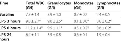
expected, systemic administration of LPS induced mild and transient flu-like symptoms. These included tempo-rary feeling of illness, increase in body temperature, shivering, headache, myalgia, sickness or nausea. Sys-temic blood pressure was slightly reduced and heart rate increased after LPS (Table 1). Following LPS white blood cell count increased significantly at 3 h and 6 h and returned to baseline levels after 24 h (Table 2). Neutrophil counts increased after LPS administration up to 2.6-fold after 6 h and returned to baseline levels after 24 h. After 3 h and 6 h a decrease of monocyte and lymphocyte cell counts by 89% and 31%, and by 75% and 74%, respectively, was seen.
VPAC1 receptor expression
VPAC1 receptors expression was not detectable in MOLT-4 cells (data not shown), which were used as ne-gative controls. The mean fluorescence intensity (MFI) reflecting the absolute receptor signal (receptor expre-ssion) in the different cell populations is summarized in Table 3. Individual values of VPAC-1 receptor expression in the different cell types are shown in Online Additional file 4: Figure S5.
Granulocytes
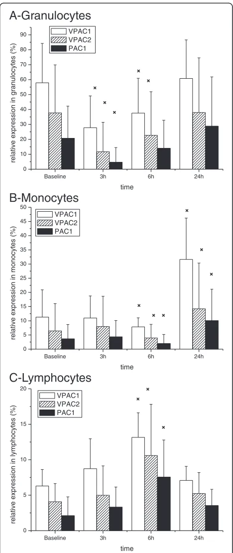
At baseline 58 ± 37% of granulocytes were positive for VPAC1 receptors. Following LPS administration the number of VPAC-1 positive cells was lower after 3 hours and 6 hours compared to baseline (Figure 1A). This was paralleled by decreased MFI values (Table 3).
Monocytes
The VPAC-1 expression profile was different in mono-cytes (Figure 1B). At baseline 11.3 ± 9.6% of monomono-cytes were positive for VPAC1. LPS initially decreased and subsequently increased receptor expression to 7.8 ± 3.2% after 6 hours and to 31.6 ± 20.5% after 24 hours. This pattern was also detectable for MFI values (Table 3).
Lymphocytes
In lymphocytes VPAC-1 receptor expression at baseline was 6.3 ± 3.2% (Figure 1C). LPS increased receptor ex-pression to 8.7 ± 5.9% after 3 hours and to 13.2 ± 4.9% after 6 hours. MFI values were not significantly altered in lymphocytes after LPS (Table 3).
VPAC1 mRNA expression
VPAC1 mRNA expression in PBMC was not changed after LPS administration (data not shown).
VIP plasma levels
VIP plasma concentrations were 0.5 ± 0.3 ng/ml at base-line and increased to 0.6 ± 0.3ng/ml 3 hours and to 0.7 ± 0.4 ng/ml 6 hours after LPS (p < 0.05 versus baseline). 24 hours after LPS VIP plasma concentration was 0.5 ± 0.2 ng/ml.
VPAC2 and PAC1 receptor and mRNA expression
Expression of VPAC2 receptors was substantially lower than that of VPAC1 in all three cell populations under study. A maximum of 5% of monocytes or lymphocytes stained positive for VPAC2 (Figure 1B,1C). Likewise, MFI values were lower compared to VPAC1.
[image:3.595.306.540.101.185.2]After LPS administration the regulation pattern was consistent with that of VPAC1, with a downregulation of VPAC2 and PAC1 in granulocytes and an upregulation
Table 1 Vital signs
Time Mean blood
pressure (mmHg) Heart rate (bpm)
Body
temperature (°C)
Baseline 97 ± 9 73 ± 10 36.1 ± 0.4
LPS 3 hours 92 ± 10* 87 ± 11* 37.1 ± 0.5*
LPS 6 hours 93 ± 10* 86 ± 9* 36.9 ± 0.4*
Mean arterial blood pressure, heart rate and body temperature at baseline and following systemic LPS administration. Data are means ± SD (n = 20). Differences from baseline are indicated (*p < 0.001, Wilcoxon matched-pairs test).
Table 2 White blood cell count
Time Total WBC
(G/l)
Granulocytes (G/l)
Monocytes (G/l)
Lymphocytes (G/l)
Baseline 7.3 ± 1.4 3.9 ± 1.0 0.7 ± 0.2 2.4 ± 0.5
LPS 3 hours 9.8 ± 2.7* 9.0 ± 2.5* 0.1 ± 0.0* 0.6 ± 0.2*
LPS 6 hours 11.2 ± 1.4* 9.9 ± 1.1* 0.5 ± 0.2* 0.6 ± 0.2*
LPS 24 hours
6.4 ± 1.1 3.5 ± 0.8 0.6 ± 0.1 1.9 ± 0.4
[image:3.595.57.291.636.699.2]in monocytes and lymphocytes, respectively (Figure 1). Corresponding changes of MFI values were again detec-table (Table 3). mRNA VPAC2 and PAC1 receptor ex-pression was at the limit of quantification in PBMC and not altered by LPS.
Discussion
The understanding of VPAC 1 receptor expression on immune-competent cells is a crucial step towards the de-velopment of VPAC agonists in human disease. This study demonstrates that in response to systemic low doses of LPS to healthy humans VPAC 1 receptor expression show a dynamic response in granulocytes, monocytes and lym-phocytes. This LPS mediated effect is accompanied by a significant increase in VIP plasma concentrations.
The expression pattern of VPAC1 receptors is heteroge-neous across different leukocyte populations. About 60% of granulocytes express VPAC1 under resting conditions, suggesting that these cells are particularly sensitive to alterations in VIP levels. Following LPS exposure granulocytes rapidly decrease the VPAC1 receptor density on their surface by approximately 50% within 3 hours. It is likely that receptor internalization as described by Langlet et al., [27] is responsible for this decrease in VPAC1 expression.
[image:4.595.305.539.87.642.2]Granulocytes are recruited to infection sites and are of importance in the early microbial clearance [28,29]. This accumulation results in organ damage if unopposed pro-inflammatory signaling persists [30]. Thus, the abi-lity to down-regulate leukocyte receptor activation after
Table 3 Mean fluorescence intensity of VPAC1, VPAC2 and PAC1
Time Granulocytes Monocytes Lymphocytes
A - VPAC1
Baseline 30.5 ± 16.5 19.9 ± 11.7 29.6 ± 11.7
LPS 3 hours 24.3 ± 9.3 * 19.9 ± 20.4 34.1 ± 19.0
LPS 6 hours 21.4 ± 9.9 * 15.3 ± 9 * 32.5 ± 17.3
LPS 24 hours 28.5 ± 16.1 18.2 ± 7.3 31.4 ± 23.7
B - VPAC2
Baseline 19.9 ± 5.7 17.4 ± 7.6 23.5 ± 10.9
LPS 3 hours 17.4 ± 7.5 13.6 ± 6.3 * 20.7 ± 10.8
LPS 6 hours 35.7 ± 86.6 * 16.0 ± 9.2 32.4 ± 41.1
LPS 24 hours 18.3 ± 7.9 19.1 ± 12.7 21.1 ± 8.5 *
C - PAC1
Baseline 17.4 ± 6.2 14.8 ± 5.3 21.6 ± 10.2
LPS 3 hours 16.7 ± 8.6 12.5 ± 10.3 15.7 ± 10.2
LPS 6 hours 21.8 ± 28.1 12.5 ± 9.6 * 17.6 ± 7.2
LPS 24 hours 17.4 ± 6.0 15.4 ± 5.8 15.7 ± 6.2
Mean fluorescence intensity values of VPAC1 (A), VPAC2 (B) and PAC1 (C) in granulocytes, monocytes, and lymphocytes. Values are expressed as mean ± SD (n = 20). Differences from baseline are indicated (*p < 0.005, Wilcoxon matched-pairs test).
C-Lymphocytes
B-Monocytes
A-Granulocytes
Baseline 3h 6h 24h 0
10 20 30 40 50 60 70 80 90
relat
ive expression in granulocyt
es (%)
time VPAC1
VPAC2 PAC1
Baseline 3h 6h 24h 0
5 10 15 20 25 30 35 40 45 50
relative expression in monocytes (%)
time VPAC1
VPAC2 PAC1
Baseline 3h 6h 24h 0
5 10 15 20
relative expression in lymphocytes (%)
time VPAC1
VPAC2 PAC1
Figure 1VIP receptor expression after LPS stimulation.Receptor
expression after systemic LPS administration inA) granulocytes,
[image:4.595.56.292.111.322.2]clearing the offending agent seems to be as essential as cell recruitment per se to prevent excessive tissue injury. The pattern of VPAC1 expression in granulocytes may therefore represent a regulator mechanism for granulo-cyte cell function.
The VPAC1 receptor expression profile was not consis-tent across leukocyte subtypes. Given that leukocyte count was significantly increased by LPS, the absolute reduction in monocyte and lymphocyte count indicates that their VPAC1 receptor - mediated effects presumably contribute to the net action of VIP to a small extent only. In the healthy subjects under study, baseline VPAC1 expression on monocytes and lymphocytes was low but increased
following LPS by 2-3 – fold. Similar results have been
shown in macrophages when stimulated with LPS in vitro [31]. Nevertheless, the upregulation detected in these cells may indicate a refinement in the immunological response to increased VIP concentrations.
The lack of changes in receptor mRNA in PBMC fol-lowing LPS argues against a relevant receptor de novo synthesis in these cells. However, assessment of protein translation may not be indicative of receptor involvement in the immunomodulatory VIP-signalling since receptors are internalized after binding of their ligand VIP on the cell surface [32]. As suggested previoulsy [27], recycling and re-organization of receptors on the cellular surface may be responsible for the observed short-term effect of LPS in leukocytes.
Receptor stimulation is important for the pharmacody-namic action of VIP. In our model of endotoxemia we found a transient increase in VIP plasma concentrations. This is consistent with findings in septic patients, where an acute initial increase in VIP serum levels has been reported [33]. In contrast, decreased VIP serum levels have been described in patients with chronic pulmonary hypertension, where VPAC2 receptors are up-regulated in the pulmonary vasculature [34]. Thus, interpretation of altered VIP serum concentration remains speculative when the receptor expression is unknown.
The factors which influence VIP receptor expression are largely unclear. The present study demonstrates that VPAC1, VPAC2 and PAC1 expression is regulated in leu-kocytes following systemic stimulation with LPS in vivo. In contrast to Lara-Martinez et al, VPAC2 was expressed on monocytes [35]. However, VPAC1 is the predominant receptor in leukocytes. This is consistent with in vitro experiments where LPS is a powerful trigger for VPAC1 receptor regulation [31]. Animal studies have demon-strated that selective activation of VPAC1 is more effective in controlling the immunological answer than VPAC2 agonists [21]. The dynamic receptor regulation pattern observed in this study also favours VPAC1 as principal mediator of the anti-inflammatory action of VIP. Thus se-lective activation of VPAC1 may represent an efficacious
clinical target to elicit anti-inflammatory actions without undesired side effects of simultaneous VPAC2 activation such as vasodilatation.
The present experiments were conducted employingE.
coliendotoxin. It is possible that VPAC1 receptor regula-tion is variably sensitive to challenging agents from differ-ent bacteria, as previously described for cytokines such as TNF-α, INFγor IL-10 [36]. The rapid serum-clearance of LPS indicates that secondary mediators may be involved in the delayed regulation of VPAC1 in PBC [37]. However, systemic LPS administration is more likely to activate immune-competent cells indirectly via systemic pro-inflammatory immunological mechanisms. The anti- in-flammatory effect of VPAC1 agonists in clinical trials will therefore be influenced by the timing of administration and the type of infection and associated mechanisms of the innate immune system.
In summary our data support the concept that VPAC-1 is a promising immunomodulatory target receptor in humans.
Grants
This project was supported and funded by“Medical Scien-tific Fund of the Mayor of the City of Vienna” (Project Number 09055).
Additional files
Additional file 1: Figure S2.Gating and IGg control. The different
populations in PBC, granulocytes, lymphocytes and monocytes, have been gated using forward and side scatter (left). Additionally, isotype controls (upper right) and double stainings (lower right) have been performed. Granulocytes have been gated using forward and side scatter (lower left).
Additional file 2: Figure S3.FACS dot blot of granulocytes.
Granulocytes were gated in the forward and side scatter. The dot blot shows granulocytes stained with VPAC1-FITC at baseline (upper left). Following LPS administration a downregulation in VPAC1 receptor expression can be seen after 3 and 6 hours (upper right and lower left). Baseline levels are re-established after 24 hours (lower right).
Additional file 3: Figure S4.VPAC1 in CD-14 positive cells. Monocytes
were first gated using forward and side scatter. Additional control staining with CD-14PE was performed (A, left side). On the right side (B) CD-14 and VPAC1 positive monocytes are shown at baseline (upper left) and 3 hours (upper right), 6 hours (lower left) and 24 hours (lower right) after LPS. A clear increase in VPAC1 positive cells is seen 24 hours after LPS.
Additional file 4: Figure S5.VPAC1 receptor expression in PBC
(individual values). VPAC1 receptor expression for all subjects and blood cell populations are shown. (A, granulocytes; B, monocytes; C, lymphocytes).
Competing interests
The authors declare that they have no competing interests.
Authors’contribution
Author details 1
Department of Clinical Pharmacology, Medical University of Vienna, Währinger Gürtel 18-20, 1090, Vienna, Austria.2Institute of Cancer Research,
Department of Medicine I, Medical University of Vienna, Währinger Gürtel 18-20, 1090, Vienna, Austria.3Department of Pneumology, Medical University of
Vienna, Währinger Gürtel 18-20, 1090, Vienna, Austria.4Department of Physical Medicine and Rehabilitation, Medical University of Vienna, Währinger Gürtel 18-20, 1090, Vienna, Austria.
Received: 16 October 2012 Accepted: 29 April 2013 Published: 7 May 2013
References
1. Said SI, Mutt V:Polypeptide with broad biological activity: isolation from small intestine.Science1970,169(951):1217–1218.
2. Mutt V, Said SI:Structure of the porcine vasoactive intestinal octacosapeptide. The amino-acid sequence. Use of kallikrein in its determination.Eur J Biochem1974,42(2):581–589.
3. Morice A, Unwin RJ, Sever PS:Vasoactive intestinal peptide causes bronchodilatation and protects against histamine-induced
bronchoconstriction in asthmatic subjects.Lancet1983,2(8361):1225–1227. 4. Huang M, Shirahase H, Rorstad OP:Comparative study of vascular relaxation
and receptor binding by PACAP and VIP.Peptides1993,14(4):755–762. 5. Offen D,et al:Vasoactive intestinal peptide (VIP) prevents neurotoxicity
in neuronal cultures: relevance to neuroprotection in Parkinson's disease.Brain Res2000,854(1–2):257–262.
6. Brenneman DE,et al:Identity of neurotrophic molecules released from astroglia by vasoactive intestinal peptide.Ann N Y Acad Sci1997, 814:167–173.
7. Edwards AV, Bloom SR, Ghatei MA:Pancreatic endocrine responses to the peptides VIP and PACAP in the conscious calf.Exp Physiol1997,82(4):717–727. 8. Kawai K,et al:Pituitary adenylate cyclase activating polypeptide
stimulates insulin release from the isolated perfused rat pancreas.Life Sci 1992,50(4):257–261.
9. Delgado M,et al:VIP and PACAP induce shift to a Th2 response by upregulating B7.2 expression.Ann N Y Acad Sci2000,921:68–78. 10. Gomariz R,et al:Anti-inflammatory actions of VIP/PACAP. Role in
endotoxemia.Ann N Y Acad Sci2000,921:284–288.
11. Leceta J,et al:Receptors and transcriptional factors involved in the anti-inflammatory activity of VIP and PACAP.Ann N Y Acad Sci2000,921:92–102. 12. Delgado M,et al:Vasoactive intestinal peptide and pituitary adenylate
cyclase-activating polypeptide inhibit endotoxin-induced TNF-alpha production by macrophages: in vitro and in vivo studies.J Immunol1999, 162(4):2358–2367.
13. Martinez C,et al:Vasoactive intestinal peptide and pituitary adenylate cyclase-activating polypeptide modulate endotoxin-induced IL-6 production by murine peritoneal macrophages.J Leukoc Biol1998,63(5):591–601. 14. Delgado M,et al:VIP and PACAP inhibit IL-12 production in
LPS-stimulated macrophages. Subsequent effect on IFNgamma synthesis by T cells.J Neuroimmunol1999,96(2):167–181.
15. Delgado M,et al:Vasoactive intestinal peptide and pituitary adenylate cyclase-activating polypeptide prevent inducible nitric oxide synthase transcription in macrophages by inhibiting NF-kappa B and IFN regulatory factor 1 activation.J Immunol1999,162(8):4685–4696. 16. Delgado M,et al:Vasoactive intestinal peptide and pituitary adenylate
cyclase-activating polypeptide enhance IL-10 production by murine macrophages: in vitro and in vivo studies.J Immunol1999,162(3):1707–1716.
17. Sreedharan SP,et al:Cloning and functional expression of a human neuroendocrine vasoactive intestinal peptide receptor.Biochem Biophys Res Commun1993,193(2):546–553.
18. Lutz EM,et al:The VIP2 receptor: molecular characterisation of a cDNA encoding a novel receptor for vasoactive intestinal peptide.FEBS Lett1993,334(1):3–8. 19. Svoboda M,et al:Molecular cloning and functional characterization of a
human VIP receptor from SUP-T1 lymphoblasts.Biochem Biophys Res Commun1994,205(3):1617–1624.
20. Ogi K,et al:Molecular cloning and functional expression of a cDNA encoding a human pituitary adenylate cyclase activating polypeptide receptor.Biochem Biophys Res Commun1993,196(3):1511–1521. 21. Delgado M,et al:Anti-inflammatory properties of the type 1 and type 2
vasoactive intestinal peptide receptors: role in lethal endotoxic shock. Eur J Immunol2000,30(11):3236–3246.
22. Chorny A, Delgado M:Neuropeptides rescue mice from lethal sepsis by down-regulating secretion of the late-acting inflammatory mediator high mobility group box 1.Am J Pathol2008,172(5):1297–1307. 23. Tuncel N, Tore FC:The effect of vasoactive intestinal peptide (VIP) and
inhibition of nitric oxide synthase on survival rate in rats exposed to endotoxin shock.Ann N Y Acad Sci1998,865:586–589.
24. Delgado M,et al:Vasoactive intestinal peptide (VIP) and pituitary adenylate cyclase-activation polypeptide (PACAP) protect mice from lethal endotoxemia through the inhibition of TNF-alpha and IL-6. J Immunol1999,162(2):1200–1205.
25. Cobo M,et al:Mesenchymal stem cells expressing vasoactive intestinal peptide ameliorate symptoms in a model of chronic multiple sclerosis. Cell Transplant2012: . Epub ahead of print.
26. Delgado M,et al:Vasoactive intestinal peptide prevents experimental arthritis by downregulating both autoimmune and inflammatory components of the disease.Nat Med2001,7(5):563–568.
27. Langlet C,et al:Contribution of the carboxyl terminus of the VPAC1 receptor to agonist-induced receptor phosphorylation, internalization, and recycling.J Biol Chem2005,280(30):28034–28043.
28. Baggiolini M,et al:Activation of neutrophil leukocytes: chemoattractant receptors and respiratory burst.FASEB J1993,7(11):1004–1010. 29. Crane MJ, Hokeness-Antonelli KL, Salazar-Mather TP:Regulation of
inflammatory monocyte/macrophage recruitment from the bone marrow during murine cytomegalovirus infection: role for type I interferons in localized induction of CCR2 ligands.J Immunol2009, 183(4):2810–2817.
30. Teng L,et al:Matrix metalloproteinase-9 as new biomarkers of severity in multiple organ dysfunction syndrome caused by trauma and infection. Mol Cell Biochem2012,360(1-2):271–277.
31. Burian B,et al:Vasoactive intestinal peptide (VIP) receptor expression in monocyte-derived macrophages from COPD patients.Peptides2010, 31(4):603–608.
32. Ortner A,et al:VPAC receptor mediated tumor cell targeting by protamine based nanoparticles.J Drug Target2010,18(6):457–467. 33. Brandtzaeg P,et al:Elevated VIP and endotoxin plasma levels in human
gram-negative septic shock.Regul Pept1989,24(1):37–44.
34. Petkov V,et al:Vasoactive intestinal peptide as a new drug for treatment of primary pulmonary hypertension.J Clin Invest2003,111(9):1339–1346. 35. Lara-Marquez M,et al:Selective gene expression and
activation-dependent regulation of vasoactive intestinal peptide receptor type 1 and type 2 in human T cells.J Immunol2001,166(4):2522–2530. 36. Finney SJ,et al:Differences in lipopolysaccharide- and lipoteichoic
acid-induced cytokine/chemokine expression.Intensive Care Med2012, 38(2):324–332.
37. Munford RS, Andersen JM, Dietschy JM:Sites of tissue binding and uptake in vivo of bacterial lipopolysaccharide-high density lipoprotein complexes: studies in the rat and squirrel monkey.J Clin Invest1981, 68(6):1503–1513.
doi:10.1186/1479-5876-11-117
Cite this article as:Storkaet al.:VPAC1 receptor expression in peripheral blood mononuclear cells in a human endotoxemia model.Journal of Translational Medicine201311:117.
Submit your next manuscript to BioMed Central and take full advantage of:
• Convenient online submission
• Thorough peer review
• No space constraints or color figure charges
• Immediate publication on acceptance
• Inclusion in PubMed, CAS, Scopus and Google Scholar
• Research which is freely available for redistribution