REVIEW
A review on the occurrence of companion
vector-borne diseases in pet animals in Latin
America
Ricardo G. Maggi
1*and Friederike Krämer
2Abstract
Companion vector-borne diseases (CVBDs) are an important threat for pet life, but may also have an impact on
human health, due to their often zoonotic character. The importance and awareness of CVBDs continuously increased
during the last years. However, information on their occurrence is often limited in several parts of the world, which
are often especially affected. Latin America (LATAM), a region with large biodiversity, is one of these regions, where
information on CVBDs for pet owners, veterinarians, medical doctors and health workers is often obsolete, limited or
non-existent. In the present review, a comprehensive literature search for CVBDs in companion animals (dogs and
cats) was performed for several countries in Central America (Belize, Caribbean Islands, Costa Rica, Cuba, Dominican
Republic, El Salvador, Guatemala, Honduras, Mexico, Nicaragua, Panama, Puerto Rico) as well as in South America
(Argentina, Bolivia, Brazil, Chile, Colombia, Ecuador, French Guiana, Guyana (British Guyana), Paraguay, Peru, Suriname,
Uruguay, Venezuela) regarding the occurrence of the following parasitic and bacterial diseases: babesiosis, heartworm
disease, subcutaneous dirofilariosis, hepatozoonosis, leishmaniosis, trypanosomosis, anaplasmosis, bartonellosis,
borreliosis, ehrlichiosis, mycoplasmosis and rickettsiosis. An overview on the specific diseases, followed by a short
summary on their occurrence per country is given. Additionally, a tabular listing on positive or non-reported
occur-rence is presented. None of the countries is completely free from CVBDs. The data presented in the review confirm a
wide distribution of the CVBDs in focus in LATAM. This wide occurrence and the fact that most of the CVBDs can have
a quite severe clinical outcome and their diagnostic as well as therapeutic options in the region are often difficult to
access and to afford, demands a strong call for the prevention of pathogen transmission by the use of
ectoparasiti-cidal and anti-feeding products as well as by performing behavioural changes.
Keywords:
Companion vector-borne diseases (CVBDs), Dog, Cat, Occurrence, Vector, Latin America (LATAM),
Prevalence
© The Author(s) 2019. This article is distributed under the terms of the Creative Commons Attribution 4.0 International License (http://creat iveco mmons .org/licen ses/by/4.0/), which permits unrestricted use, distribution, and reproduction in any medium, provided you give appropriate credit to the original author(s) and the source, provide a link to the Creative Commons license, and indicate if changes were made. The Creative Commons Public Domain Dedication waiver (http://creat iveco mmons .org/ publi cdoma in/zero/1.0/) applies to the data made available in this article, unless otherwise stated.
Background
Companion vector-borne diseases (CVBDs) have among
others a major impact on the welfare of pets. They may
also represent a constant risk to humans due to their
zoonotic nature, which emphasizes the importance of
pets as reservoirs.
In Latin America (LATAM), a region with one of the
largest biodiversities in the world, a combination of
fac-tors such as intensification of agricultural practices,
landscape modification, poor ecosystem protection and
potentially slight unstable economics, creates host
popu-lations conducive to the performance and persistence of
parasites and vectors.
This is especially important for CVBDs affecting dogs
and cats as companion animals, as a significant
propor-tion of those (i.e. 52–75%) [
1
,
2
], even though owned
by pet holders, roam freely, besides an exploding
num-ber of stray dogs and cats. In LATAM, the lack of
sensi-tive awareness of animal welfare and disease issues, the
Open Access
*Correspondence: rgmaggi@ncsu.edu
1 Department of Clinical Sciences and the Intracellular Pathogens Research Laboratory, College of Veterinary Medicine, North Carolina State University, Raleigh, NC, USA
restricted economic and technological access to proper
veterinary care, and the absence of responsible pet
own-ership, have created conditions for the emergence and
persistence of many diseases that ultimately will affect
people, livestock, and wildlife [
3
–
10
]. Besides,
socio-economic, demographic and ecological factors, including
globalization, increase in international trade, tourism and
travel, climate change and its effect on vector distribution
in time and space, have also to be reconsidered.
This article summarizes the data of reported
detec-tion (or prevalence when available) of the most
signifi-cant CVBDs affecting companion animals in LATAM in
tabular form and as detailed information per country and
discusses research gaps to be addressed in future
stud-ies. In case of very scarce published data, additionally the
occurrence of the pathogens in potential vectors, wild
canids or felids and in humans is listed, to illustrate the
fact that the pathogen is occurring in a respective region,
even though not officially reported in companion
ani-mals so far. Beforehand a brief introduction on the
dis-eases, usually followed by a short summary or references
for more detailed data on diagnostic methods, treatment
indications and ways of prevention are given.
Generally, for many of the vector-borne diseases
(VBDs) described here, diverse diagnostic tests are
avail-able (microscopic, serological, molecular). Nevertheless,
besides their different performance regarding sensitivity
and specificity in acute and chronic disease, only few are
readily available as diagnostic tools at most clinical
prac-tices in the reported LATAM regions.
Parasitic diseases
Babesiosis
Babesiosis in pet animals in LATAM is mainly caused
by
Babesia vogeli
and
Babesia gibsoni
[
11
–
13
]. The
dis-ease has been reported in many areas especially of
South America, whereas reports from Central America
are scarce so far.
Babesia vogeli
is transmitted directly
via
tick bites [
Rhipicephalus sanguineus
(
sensu lato
)],
whereas
B. gibsoni
in LATAM is expected to be
transmit-ted
via
blood transfer through dog bites, blood
transfu-sions and transplacental supply [
14
–
18
]. Clinical signs,
depending on the species, and further details on
clini-cal and laboratory findings can be found in Irwin [
14
].
As diagnosis microscopy remains the simplest and most
accessible diagnostic test. Different sensitivity
dur-ing the cause of disease may be supported by molecular
methods (see Irwin [
14
] for details). Treatment does not
eliminate the parasite, but only reduces parasitemia and
supports resolution of clinical signs and is summarized
elsewhere [
14
]. Animals diagnosed with
Babesia
spp.
should be considered permanent carriers of the infection.
Due to the missing elimination of the pathogen during
treatment, vaccines have been introduced with variable
efficacy (see Irwin [
14
] for summary). According to the
authors’ knowledge, the vaccines are only available in
Europe, so that prevention of vector exposure in form of
acaricidal treatment is essential especially for LATAM.
Dirofilariosis
Dirofilariosis is caused by
Dirofilaria immitis
,
present-ing as an important disease, causpresent-ing cardiopulmonary
problems and even death in dogs worldwide and
com-monly known as canine heartworm disease, and by
Diro-filaria repens
, a subcutaneous parasite of dogs and cats in
Europe, Africa and Asia.
Canine heartworm disease
Canine heartworm disease has a wide distribution in
LATAM (except Belize, Guatemala, Panama, French
Gui-ana, Chile and Uruguay; for specific data see individual
country sections). The pathogen is transmitted by several
mosquito species. As a mosquito-transmitted disease,
it is more prevalent in tropical and subtropical regions,
due to favorable conditions for mosquito propagation
[
19
–
21
]. Clinical signs vary from nearly asymptomatic to
very severe and are listed elsewhere [
22
–
24
]. Diagnostic
methods include microfilaria testing of blood samples,
ideally after a concentration technique (modified Knott
ʼ
s
test or filtration test), and antigen testing. For details on
different test sensitivities and combinations please see the
guidelines of the American Heartworm Society (AHS)
[
25
]. Treatment against heartworm varies depending on
the severity of the disease and always aims to improve
the clinical condition and to eliminate all life stages of the
heartworms with minimal post-treatment complications.
Prevention by the use of chemoprophylactic drugs is
strongly recommended year-round in endemic areas. For
full recommendations see the guidelines of the Tropical
Council of Companion Animal Parasites (TroCCAP) [
26
]
and the AHS [
25
]. Prevention of vector exposure on the
basis of antifeeding and/or insecticidal treatments and by
the use of mosquito screens etc. and reduction of suitable
breeding sites for mosquitoes support a successful
pre-vention scheme.
Subcutaneous dirofilariosis
in Europe, Africa and Asia, and only single reports with
closely related variants for LATAM exist [
35
,
36
].
Diag-nostic methods usually rely on the detection of
micro-filariae in blood samples as described for
D. immitis
. If
clinically apparent, surgical excision and subsequent
histopathological confirmation is the general treatment
option. From the medical standpoint, here especially
regarding the Old World,
D. repens
is the most frequent
and most widely distributed in comparison to
D. immitis
and other
Dirofilaria
species [
37
] and thus especially of
zoonotic importance. For the New World, different
spe-cies might be involved.
Hepatozoonosis
Hepatozoonosis has been described infrequently in
LATAM, despite high prevalences reported from some
rural areas of Brazil and Costa Rica [
38
–
41
]. Canine
hepatozoonosis is caused by
Hepatozoon canis
, a
proto-zoan transmitted by ingestion of ticks containing mature
H. canis
oocysts. Clinical signs of hepatozoonosis and
laboratory changes can be found in Sherding [
42
] and
Baneth [
43
]. The disease is debilitating and often fatal
if not treated.
Hepatozoon canis
infection is frequently
diagnosed by microscopic detection of intracellular
gamonts in stained blood smears. Antibody detection
and molecular detection
via
PCR are also available; see
Baneth [
43
] for further details. Complete elimination may
frequently not be achievable [
44
]; for details on treatment
see Baneth [
43
]. Prognosis of treated dogs depends on
the parasitaemia. Prevention of vector exposure in form
of ectoparasiticidal treatment is supporting the
protec-tion against
H. canis
.
Leishmaniosis
Leishmaniosis in LATAM is mainly caused by
Leishma-nia infantum
(syn.
Leishmania chagasi
). Other species
(e.g.
Leishmania braziliensis
,
Leishmania amazonensis
)
can also be involved in causing disease. While
L.
infan-tum
is the most important causative agent of canine
visceral leishmaniosis in South America [
45
],
L.
ama-zonensis
has as well been reported causing visceral
leish-maniosis in dogs [
46
], whereas
L. braziliensis
has been
detected in dogs with cutaneous leishmaniosis [
47
].
The parasites are transmitted mainly by sand flies (for
LATAM, species of the genus
Lutzomyia
[
48
,
49
]).
Clini-cal signs can vary from very subtle (asymptomatic) to
very severe. Clinical staging has been deeply elaborated
by LeishVet and published in Solano-Gallego et al. [
50
,
51
] for dogs and in Pennisi et al. [
52
] for cats. The most
useful diagnostic approaches include demonstration of
the parasite DNA in blood or other tissues and detection
of specific serum anti-leishmanial antibodies [
50
,
51
,
53
–
55
], but might not be available in all regions in LATAM.
Direct parasite detection by cytology and further
diag-nostic approaches are described and evaluated in the
LeishVet guidelines for the practical management of
canine leishmaniosis [
51
]. Treatment for leishmaniosis is
controversial in many countries and includes several
anti-leishmanial drugs. Treatment regimens for the different
stages of disease have been published in Solano-Gallego
et al. [
50
,
56
]. In South America, canine leishmaniosis
treatment might often not routinely be performed. The
elimination of seropositive dogs (euthanasia/culling
pro-gram) has been practiced, e.g. in Brazil, even though for
Brazil this control measure has been subject of intense,
ongoing debate, due to ethical reasons and the lack of
sci-entific evidence supporting the effectiveness of this
strat-egy [
57
–
59
]. Meanwhile, a veterinary drug based on oral
miltefosine has been authorized for marketing in Brazil
[
60
]. As
L. infantum
has zoonotic potential, and dogs are
regarded as the main reservoir for this pathogen,
preven-tion is essential from the standpoint of animal welfare as
well as under the aspect of One Health. Besides a reduced
exposure to sand flies based on behavioral codes,
insec-ticidal prophylaxis is strongly recommended. Another
approach to help controlling canine leishmaniosis was
the introduction of a vaccine, which has been licensed in
Brazil in 2014 and which proved to be effective to reduce
the number of canine visceral leishmaniosis cases in
vac-cinated animals [
61
].
Trypanosomosis
Trypanosomosis is a disease of human medical and
veterinary importance caused mainly by
chronic phase [
74
–
81
] and a detailed review [
82
] offer
further information. Regarding dogs, there are few
stud-ies focusing on the diagnosis of
T. cruzi
infection [
83
–
87
]
and even fewer in naturally infected dogs using
recom-binant antigens [
88
]. Different antigens have been tested
by Brasil et al. [
82
] for their suitability in dogs. The drug
of choice for treatment is benznidazole, but nifurtimox
can also be used [
89
]. Symptomatic treatment for heart
failure and arrhythmias is also recommended [
90
].
Pre-vention of disease transmission especially in humans is
among others heavily relying in vector control [
68
]. As
the dog is a major reservoir for human Chagas disease,
vector control should also include the prevention of
dis-ease transmission in dogs.
Bacterial diseases
Anaplasmosis
Anaplasmosis in dogs and cats can be caused by
Ana-plasma phagocytophilum
, causative agent of canine
granulocytic anaplasmosis (CGA), mainly occurring in
temperate zones of the world, and
Anaplasma platys
,
the pathogenic agent of canine cyclic thrombocytopenia,
occurring worldwide with a higher incidence in tropical
and and subtropical areas [
91
]. For LATAM, both
spe-cies have been reported in infections, but mainly with
A.
platys
.
Even though most dogs naturally infected with
A.
phagocytophilum
probably remain healthy, clinical signs
[
92
–
95
] and hematological changes [
94
] have been
reported. In general, infection with
A. platys
may go
along subclinically (e.g. in the USA and Australia), but
distinct clinical abnormalities have also been reported,
besides hematological abnormalities (in Europe and
Israel [
96
,
97
]). A good overview for both pathogens is
given in Sainz et al. [
98
]. In the majority of dogs both
types of anaplasmoses pose a diagnostic challenge and
clinical and hematological abnormalities should be
com-bined with laboratory and diagnostic tests. Microscopic
detection of morulae (intracytoplasmatic inclusions) in
neutrophils (for
A. phagocytophilum
) or platelets (for
A. platys
) in stained blood smears is indicative for an
infection with an intracytoplasmic coccus, but not
dis-tinguishing between
A. phagocytophilum
and other
Ehrli-chia
spp. [
98
], respectively sensitivity appears to be rather
low for
A. platys
[
99
], so that serology and ideally PCR
should also be performed additionally for definitive
diag-nosis. For details on diagnostic interpretation see Sainz
et al. [
98
] and Carrade et al. [
100
]. For treatment of both
pathogen infections doxycycline is effective (see Sainz
et al. [
98
] for a summary on treatment parameters). The
prevention of anaplasmosis in dogs must be focused on
tick control, even though the vector of
A. platys
is still
unknown or unproven. But ticks of various genera (e.g.
Rhipicephalus
,
Dermacentor
and
Ixodes
) have been found
naturally infected by
A. platys
around the world [
101
–
105
]. Regarding
A. phagocytophilum
, tick control is an
essential demand enforced even by the zoonotic
charac-ter of the pathogen.
Bartonellosis
Bartonellosis has been described in dogs and cats
spo-radically in LATAM. The most common species detected
in dogs are
Bartonella henselae
and
Bartonella vinsonii
berkhoffii
, while
B. henselae
and
Bartonella clarridgeiae
are the most commonly detected species in cats [
106
].
Bartonella
species can be transmitted to companion
ani-mals and humans by several insects, including fleas, sand
flies, lice, bed bugs, mites and ticks (e.g. [
107
–
131
]), and
also directly by cat scratches, bites, blood transfusion and
organ transplant (even though the last two have been
mostly reported in humans) (e.g. [
130
,
132
–
150
].
Clini-cal appearance may include a large variety of signs (e.g.
[
143
,
144
,
151
–
170
] and laboratory abnormalities [
165
,
167
,
171
–
173
]. Diagnosis of
Bartonella
infection can be
performed by IFA test, PCR, or blood culture.
Unfortu-nately, their use is mostly restricted to research due to
their limited access (especially in antigen types used for
IFA test). In recent years, DNA amplification after blood
culture pre-enrichment became the gold standard for
diagnosis of
Bartonella
infection [
174
]. Treatment of
bar-tonellosis is very difficult, requiring long term treatment
with a combination of antibiotics (i.e. azithromycin/
minocycline) (e.g. [
175
–
181
]. As the pathogens possess a
zoonotic potential, prevention of pathogen transmission
is essential especially in form of ectoparasite control. This
must include also cats as a major reservoir for
Bartonella
spp.
Lyme borreliosis
diagnosis of borreliosis in dogs is very difficult since
com-patible clinical symptoms with other vector-borne
pathogens are very common. Direct detection methods
(PCR and/or culture) are difficult and of little practical
relevance as the organisms are rarely detected in body
fluids [
199
–
201
]. Regarding serological diagnosis,
detec-tion of specific antibodies does not necessarily correlate
with the presence of clinical disease [
189
]. The method
of choice for serological diagnosis is a two-tiered
labo-ratory test [
202
], consisting of an enzyme-linked
immu-nosorbent assay (ELISA) and immunoblotting (Western
blotting); for more detailed information see also Krupka
& Straubinger [
189
]. Furthermore, a commercial ELISA
based on C6 peptide is also widely used for serodiagnosis
(see Krupka & Straubinger [
189
] for additional
informa-tion and further literature). Treatment of LB should be
initiated as early as possible [
189
]. Whether dogs (or cats)
should be treated when specific antibodies are detected
in the absence of clinical signs is controversial [
203
–
205
].
Treatment is recommended for a period of 28 to 30
days, and the most commonly used drug is doxycycline.
For further information on treatment regimens etc.,
see Krupka & Straubinger [
189
]. Again, prevention of
pathogen transmission by ectoparasiticidal control is an
essential aspect, especially also because of the zoonotic
potential of the pathogens.
Ehrlichiosis
Ehrlichiosis in dogs and cats has been reported in
LATAM. The causative agents are
Ehrlichia canis
(responsible for canine monocytic ehrlichiosis [CME]),
Ehrlichia chaffeensis
and
Ehrlichia ewingii
, with ticks
as the transmitting vectors [
206
–
208
]. Clinical signs of
CME are very similar to the ones presented in
granulo-cytic anaplasmosis and partly also occur in cats.
Ehrlichia
ewingii
infection is also reported to go along with
clini-cal signs in dogs, but none in cats, whereas
E. chaffeensis
infection usually presents mildly or subclinically unless
present in co-infection, and again with no reported signs
in cats. For more details on CME see Sainz et al. [
98
]
and on all three pathogens see Allison & Little [
209
].
Detection of
E. canis
morulae (an aggregate of
E. canis
organisms) in a blood smear, ideally a buffy coat smear,
is indicative, but rather rare in clinical cases [
210
].
Fur-ther diagnostic tests, such as serology or molecular
tech-niques (PCR) must be performed. CME can be diagnosed
with IFA test or ELISA [
211
–
213
]. A fourfold increase
in IgG antibodies over time has been suggested to be
taken as evidence of an ongoing infection [
213
], as well
as the combination of serology and PCR has been
recom-mended for diagnosis of infection [
214
]. Nevertheless,
use of some of these test systems might not be available
for whole of LATAM. Additionally, rapid serological tests
are available; for more detailed information on
diagnos-tics see also Sainz et al. [
98
] and Allison & Little [
209
].
Doxycycline is considered the treatment of choice for
rickettsial infections [
100
,
215
,
216
], thus also for
ehrli-chiosis; for details on the treatment regimen see among
others Allison & Little [
209
] and Sainz et al. [
98
]. Again,
avoidance of tick exposure and prevention of
transmis-sion by use of ectoparasiticidal compounds are essential.
This is of vital importance as the mentioned pathogens
may have zoonotic character (Venezuela [
217
], LATAM
[
218
–
223
]).
Hemotropic mycoplasmosis
Hemotropic mycoplasmosis (formerly known as
hemo-bartonellosis) has rarely been reported in LATAM.
The disease in dogs is caused mainly by
Mycoplasma
haemocanis
and
Mycoplasma haematoparvum
. In cats,
the disease can be caused by single- or co-infections
with
Mycoplasma haemofelis
,
Mycoplasma
haemominu-tum
and
Mycoplasma turicensis
. Blood transfusions have
been reported as a source of infections (e.g. [
224
,
225
]),
but blood-sucking arthropods are likely to be involved
in the transmission as well [
226
–
231
]. Generally,
lit-tle is known on the ecology and form of transmission
of these bacteria. Clinical signs may vary and are listed
elsewhere [
232
,
233
]. Specific conventional and
quantita-tive real-time PCR systems have been introduced and are
now considered the gold standard [
234
–
239
]. Treatment
is performed depending on the severity of the infection.
Antibiotics such as doxycycline or tetracycline should be
effective, but consistent clearance of infection was not
seen with a range of antibiotics [
233
]; for more details on
treatment see among others Messick [
233
] and Willi et al.
[
240
]. As with all potentially vector-transmitted
patho-gens, prevention in form of vector control is essential.
Rickettsiosis
[
243
]. Infection of dogs and cats with
Rickettsia
species
is often subclinical, inapparent, but may also result in
severe disease (especially in the case of
R. rickettsii
) [
244
],
potentially being even fatal [
245
]. For an overview on the
different
Rickettsia
species see also Nicholson et al. [
215
]
and Allison & Little [
209
]. Diagnosis of rickettsial
patho-gens is usually achieved by PCR assays, serological assays
or response to treatment in most clinical cases. When
PCR is not practical or available, serology, and here
par-ticularly documentation of seroconversion in an acutely
ill individual, should be used. For detailed information
on the different diagnostic approaches in
Rickettsia
spp.
see also Allison & Little [
209
]. The antibiotic treatment
of choice is doxycycline [
215
,
246
]. Prompt treatment is
critical as delays can result in fatality [
209
]. Besides the
clinical effect of some
Rickettsia
species in dogs, dogs
are important sentinels of infection and disease (e.g. in
R. conorii
) [
247
,
248
]. They are also expected to play an
important role as biological hosts of the ticks and serve
to increase the infected tick population in close
associa-tion with human habitaassocia-tion (again for
R. conorii
) [
215
].
Thus, ectoparasitic control is essential also under the
zoonotic aspect and the concept of One Health.
At the end of the presentations of the relevant VBDs
we want to remark that veterinarians should be aware
of synergistic effects and clinically relevant
immunosup-pression in co-infected animals [
249
] as well as an altered
clinical appearance in co-infected animals, potentially
making diagnosis more difficult and probably leading to
a more serious disease outcome [
250
]. This is relevant for
the whole LATAM region as exposure to several
patho-gens seems possible.
Country files
Subsequently a listing of occurrence of the pathogens
respectively of corresponding seroprevalence data in
LATAM by country in alphabetical order follows, based
on an actual literature search. Additionally, all described
data are summarized in Table
1
.
Argentina
Parasitic diseases
As in many countries in LATAM, the most common
par-asitic diseases reported in Argentina are trypanosomosis
(responsible for Chagas disease in humans), dirofilariosis
and leishmaniosis.
Babesiosis due to
B. vogeli
has been described in three
dogs from Buenos Aires [
12
,
251
] and detected in 10%
(2/21) and 6.8% (3/41) of shelter dogs from Córdoba and
Santa Fé, respectively, by molecular methods [
252
]. Large
piroplasms have furthermore been detected in 0.2% of
tested animals in a large canine survey with more than
16,000 dogs [
12
,
251
].
Babesia vogeli
was also detected
in cat fleas (
Ctenocephalides felis
) collected from shelter
dogs in Córdoba and Santa Fé (R. Maggi, unpublished
data). Interestingly,
Babesia
was not detected in any of 48
free ranging Pampas gray foxes (
Lycalopex gymnocercus
)
from Rio Negro that showed high prevalence for
hepato-zoonosis [
253
].
Dirofilariosis caused by
D. immitis
has been reported in
Buenos Aires [
254
–
256
] and Mendoza [
257
].
Epidemio-logical studies in Argentina suggest that the prevalence of
dirofilariosis in dogs is highly variable, showing a
signifi-cantly heterogeneous temporal and spatial distribution
[
254
–
256
,
258
,
259
]. In Buenos Aires, screening of 19,298
blood samples from 65 localities showed prevalence
val-ues of 1.63% by microhematocrit tube technique, 3.65%
by modified Knott
ʼ
s test, and 14.41% by antigen test
[
255
].
Hepatozoonosis has been reported in dogs (infected
with
H. canis
) from Buenos Aires [
251
,
260
], and in up to
50% of 48 blood samples from free ranging Pampas gray
foxes (
L. gymnocercus
) from Rio Negro (infected with
Hepatozoon
sp.) [
253
,
261
].
Hepatozoon
sp. infection has
further been described in single canine cases in the
Bue-nos Aires region [
262
]. No prevalence studies are
avail-able up to date.
For leishmaniosis, only few records are available
regarding the overall prevalence in Argentina.
Leish-mania braziliensis
and
L. infantum
have been
associ-ated with canine leishmaniosis in several provinces of
the country, including Entre Rios, Santa Fé, Misiones,
Chaco, Salta and Santiago del Estero [
263
–
270
]. Reports
from Misiones, which represents one of the areas with
highest endemicity for the disease in Argentina, indicate
prevalences as high as 57% in dogs (43.6% seropositive
and 47.3% positive by PCR) [
266
]. In other provinces, i.e.
Salta, a significant seroprevalence (13.0–27.4%) has also
been reported [
263
,
268
].
Trypanosomiasis is one of the most important endemic
VBDs in Argentina. Serological surveys in the northern
rural regions have shown prevalences in dogs ranging
between 23–84%; while seroprevalence in cats has been
reported at 28.7% [
83
,
263
,
271
–
277
]. In hyperendemic
regions, such as Chaco, molecular prevalence as high as
53% has been reported in dogs [
278
].
Bacterial diseases
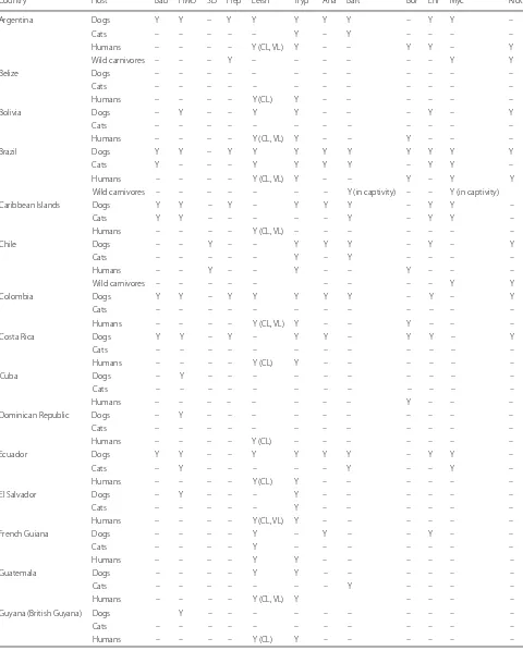
Table 1
Tabular overview on the occurrence of CVBDs in dogs, cats, humans and wild carnivores in LATAM based on an actual
literature search (partly only based on seroprevalence data; single case reports included; questionable cross-reactivities neglected)
Country Host Bab HWD SD Hep Leisha Trypb Ana Bart Bor Ehr Myc Rick
Argentina Dogs Y Y – Y Y Y Y Y – Y Y –
Cats – – – – – Y – Y – – – –
Humans – – – – Y (CL, VL) Y – – Y Y – Y
Wild carnivores – – – Y – – – – – – Y Y
Belize Dogs – – – – – – – – – – – –
Cats – – – – – – – – – – – –
Humans – – – – Y (CL) Y – – – – – –
Bolivia Dogs – Y – – Y Y – – – Y – Y
Cats – – – – – – – – – – – –
Humans – – – – Y (CL, VL) Y – – Y – – –
Brazil Dogs Y Y – Y Y Y Y Y Y Y Y Y
Cats Y – – – Y Y Y Y – Y Y –
Humans – – – – Y (CL, VL) Y – – Y – Y Y
Wild carnivores – – – – – – – Y (in captivity) – – Y (in captivity)
Caribbean Islands Dogs Y Y – Y – Y Y Y – Y Y –
Cats Y Y – – – – – Y – Y Y –
Humans – – – – Y (CL, VL) – – – – – – –
Chile Dogs – – Y – – Y Y Y – Y – Y
Cats – – – – – Y – Y – – – –
Humans – – Y – – Y – – Y – – –
Wild carnivores – – – – – – – – – Y Y
Colombia Dogs Y Y – Y Y Y Y Y – Y – Y
Cats – – – – – – – – – – – –
Humans – – – – Y (CL, VL) Y – – Y – – –
Costa Rica Dogs Y Y – Y – Y Y – Y Y – Y
Cats – – – – – – – – – – – –
Humans – – – – Y (CL) Y – – – – – –
Cuba Dogs – Y – – – – – – – – – –
Cats – – – – – – – – – – – –
Humans – – – – – – – – Y – – –
Dominican Republic Dogs – Y – – – – – – – – – –
Cats – – – – – – – – – – – –
Humans – – – – Y (CL) – – – – – – –
Ecuador Dogs Y Y – – Y Y Y Y – Y Y –
Cats – Y – – – – – Y – – Y –
Humans – – – – Y (CL) Y – – – – – –
El Salvador Dogs – Y – – – Y – – – – – –
Cats – – – – – Y – – – – – –
Humans – – – – Y (CL, VL) Y – – – – – –
French Guiana Dogs – – – – Y – Y – – Y – –
Cats – – – – Y – – – – – – –
Humans – – – – Y Y – – – – – –
Guatemala Dogs – – – – Y Y – – – – – –
Cats – – – – – – – Y – – – –
Humans – – – – Y (CL, VL) Y – – – –
Guyana (British Guyana) Dogs Y – – – – – – – – – –
Cats – – – – – – – – – – – –
shelter dogs in Córdoba and Santa Fé (R. Maggi,
unpub-lished data).
Bartonellosis due to
B. vinsonii berkhoffii
has been
detected in dogs with endocarditis in Buenos Aires (R.
Maggi, unpublished data).
Bartonella
infection has been
detected at a molecular prevalence of 3% in shelter dogs
from Córdoba (close homology to
B. tribocorum
), and
from Santa Fé (
B. clarridgeiae
).
Bartonella clarridgeiae
has also been detected in cat fleas (
C. felis
) collected from
shelter dogs in Córdoba and Santa Fé (R. Maggi,
unpub-lished data). Additionally,
B. henselae
and
B. clarridgeiae
have been detected at a molecular prevalence of 17.8% in
cats from Buenos Aires [
282
].
Lyme borreliosis in dogs or cats in Argentina has not
been reported yet. Nevertheless, the detection of
B.
burg-dorferi
(
s.l
.) infecting ticks in northern provinces [
184
], as
well as the detection of antibodies against
B. burgdorferi
in farm workers has been reported [
283
].
Ehrlichiosis due to
E. canis
has been reported at a
molecular prevalence in 7% of sick dogs from Buenos
Aires [
251
]. No data are available on detection or
preva-lence of
Ehrlichia
spp. infecting dogs from other
prov-inces, although
E. canis
was detected in
R. sanguineus
(
s.l
.) ticks from Formosa Province [
281
].
Ehrlichia
chaf-feensis
has been found at a prevalence of 14% in
peo-ple from Jujuy [
221
] and detected in
A. parvum
ticks
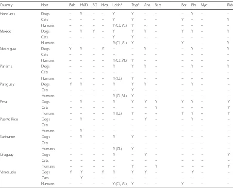
a In case of human visceral and cutaneous leishmaniosis, if not cited otherwise within the additional files, status for humans is according to the WHO webpages [708, 709]b In case of human trypanosomosis, if not cited otherwise within the additional files, status for humans is according to the WHO webpage [710]
Abbreviations: Y, yes (reported); –, not reported; Bab, babesiosis; HWD, heartworm disease; SD, subcutaneous dirofilariosis; Hep, hepatozoonosis; Leish, leishmaniosis; Tryp, trypanosomosis; Ana, anaplasmosis; Bart, bartonellosis; Bor, borreliosis; Ehr, ehrlichiosis; Myc, mycoplasmosis; Rick, rickettsiosis; CL, cutaneous leishmaniosis; VL, visceral leishmaniosis
Table 1 (continued)
Country Host Bab HWD SD Hep Leisha Trypb Ana Bart Bor Ehr Myc Rick
Honduras Dogs – Y – – Y Y – – – Y – –
Cats – – – – Y Y – – Y – – Y
Humans – – – – Y (CL, VL) Y – – – – – –
Mexico Dogs – Y Y – Y Y Y – Y Y – Y
Cats – – – – Y Y – – – – – –
Humans – – – – Y (CL, VL) Y – – Y – – Y
Nicaragua Dogs Y Y – Y – – Y – – Y – Y
Cats – – – – – – – – – – – –
Humans – – – – Y (CL, VL) Y – – – – – –
Panama Dogs – – – – Y Y Y – – Y – Y
Cats – – – – – – – – – – – –
Humans – – – – Y (CL) Y – – – – – –
Paraguay Dogs Y Y – – Y Y Y – – Y – –
Cats – – – – – Y – – – – – –
Humans – – – – Y (CL, VL) Y – – – – – –
Peru Dogs – Y – – Y Y Y Y Y Y – Y
Cats – – – – – – – Y – – – Y
Humans – – – – Y (CL) Y – – Y Y – Y
Puerto Rico Dogs – Y – – – – Y – – Y – –
Cats – – – – – – – – – – –
Humans – Y – – – – – – – – – –
Suriname Dogs – Y – – Y Y – – – – – –
Cats – – – – – – – – – – – –
Humans – – – – Y (CL) Y – – – – – –
Uruguay Dogs – – – – Y – Y – – – – Y
Cats – – – – – – – – – – – –
Humans – – – – – Y – Y – – – Y
Venezuela Dogs Y Y – Y Y Y Y – – Y – –
Cats – Y – – – – – – – – – –
[image:8.595.57.537.98.485.2]