RESEARCH
Poly(ADP-ribose) polymerase as a novel
regulator of 17
β
-estradiol-induced cell
growth through a control of the estrogen
receptor/IGF-1 receptor/PDZK1 axis
Hogyoung Kim
1, Abdelmetalab Tarhuni
1, Zakaria Y Abd Elmageed
2and A Hamid Boulares
1*Abstract
Background: We and others have extensively investigated the role of PARP-1 in cell growth and demise in response to pathophysiological cues. Most of the clinical trials on PARP inhibitors are targeting primarily estrogen receptor (ER) negative cancers with BRCA-deficiency. It is surprising that the role of the enzyme has yet to be investigated in ER-mediated cell growth. It is noteworthy that ER is expressed in the majority of breast cancers. We recently showed that the scaffolding protein PDZK1 is critical for 17β-estradiol (E2)-induced growth of breast cancer cells. We demonstrated
that E2-induced PDZK1 expression is indirectly regulated by ER and requires IGF-1 receptor (IGF-1R).
Methods: The breast cancer cell lines MCF-7 and BT474 were used as ER(+) cell culture models. Thieno[2,3-c]isoqui-nolin-5-one (TIQ-A) and olaparib (AZD2281) were used as potent inhibitors of PARP. PARP-1 knockdown by shRNA was used to show specificity of the effects to PARP-1.
Results: In this study, we aimed to determine the effect of PARP inhibition on estrogen-induced growth of breast cancer cells and examine whether the potential effect is linked to PDZK1 and IGF-1R expression. Our results show that PARP inhibition pharmacologically by TIQ-A or olaparib or by PARP-1 knockdown blocked E2-dependent growth of MCF-7 cells. Such inhibitory effect was also observed in olaparib-treated BT474 cells. The effect of PARP inhibition on cell growth coincided with an efficient reduction in E2-induced PDZK1 expression. This effect was accompanied by a similar decrease in the cell cycle protein cyclin D1. PARP appeared to regulate E2-induced PDZK1 at the mRNA level. Such regulation may be linked to a modulation of IGF-1R as PARP inhibition pharmacologically or by PARP-1 knock-down efficiently reduced E2-induced expression of the receptor at the protein and mRNA levels.
Conclusions: Overall, our results show for the first time that PARP regulates E2-mediated cell growth by controlling the ER/IGF-1R/PDZK1 axis. These findings suggest that the relationship between ER, PDZK1, and IGF-1R may be per-turbed by blocking PARP function and that PARP inhibitors may be considered in clinical trials on ER(+) cancers. Keywords: PARP, 17β-estradiol, Gene regulation, Cell growth, Breast cancer, PDZK1, IGF-1R, MCF-7 cells
© 2015 Kim et al. This article is distributed under the terms of the Creative Commons Attribution 4.0 International License (http:// creativecommons.org/licenses/by/4.0/), which permits unrestricted use, distribution, and reproduction in any medium, provided you give appropriate credit to the original author(s) and the source, provide a link to the Creative Commons license, and indicate if changes were made. The Creative Commons Public Domain Dedication waiver (http://creativecommons.org/publicdomain/ zero/1.0/) applies to the data made available in this article, unless otherwise stated.
Background
It is well established that estrogen receptor (ER) is expressed in the majority of breast cancers and are responsive to standard therapy with tamoxifen as the
leading drug [1]. Despite the success of this therapy,
reduction of breast cancer recurrence is only by 50% and
an even lower reduction in mortality rate (~30%) [1, 2].
Accordingly, finding new therapeutic targets is urgently needed. Achieving this goal requires the identification of new players in the complex process of the disease. We have recently shown that PDZK1, a scaffold protein, plays an important role in estrogen-induced growth of breast cancer cells and demonstrated a strong correla-tion between the expression of the protein and human
Open Access
*Correspondence: hboulr@lsuhsc.edu
breast malignancy [3]. We also reported that PDZK1
gene expression is not a direct product of ER stimula-tion; rather, it requires the expression and function of
IGF-1 receptor (IGF-1R) [3]. PDZK1 appears to harbor
oncogenic activity and promote cell growth by enhanc-ing EGFR-stimulated MEK/ERK1/2 signalenhanc-ing and
IGF-induced Akt phosphorylation [4]. Interestingly, PDZK1
plays this important role through stabilization of the
integrity of Akt, Her2/Neu, and EGFR [4]. The
co-chaper-one Cdc37 appears to play an important role in
PDZK1-mediated stability of Akt [4]. These aforementioned
findings demonstrated a novel relationship between PDZK1, Akt, Her2/Neu, EGFR and Cdc37 in breast can-cer unraveling a new axis that can be targeted therapeuti-cally to reduce the burden of human breast cancer.
Poly (ADP-ribose) polymerase (PARP)-1, a member of the PARP family of proteins, has initially been described as a DNA repair enzyme playing primarily as a regulatory protein controlling traffic of DNA repair proteins during
base excision repair [5, 6]. A prominent function of this
enzyme is in cell death both as an effector and as a
sub-strate to some of the caspases [7]. We demonstrated many
years ago that cleavage of PARP-1 is critical for the nor-mal progression of the apoptotic process and that inter-ference with such cleavage enhances cell death and may
even cause a switch to necrosis [7, 8]. Increasing evidence
from our laboratory and many others demonstrate an important role for this enzyme in tissue injury associated with oxidative stress and inflammation including asthma
and atherosclerosis [8–13]. PARP-1 is thought to
partici-pate in inflammation by regulating the expression of sev-eral inflammatory factors including adhesion molecules,
TNF-α, interleukins, and inducible nitric oxide synthase
(iNOS) most of which are controlled by NF-κB (4). PARP
inhibitors have shown great potential against breast and ovarian cancers especially those with BRCA mutations
[14]. The combination of PARP inhibitors with DNA
dam-aging chemotherapeutic drugs have shown to induce the specific demise of BRCA-deficient cancer cells leading to a synthetic lethality phenotype while sparing the life of
normal cells [15]. Many clinical trials have demonstrated
efficacy of PARP inhibitors and their potential as
thera-peutic strategy that can be utilized in the clinic [14, 15].
However, the focus on BRCA-deficient breast cancer pre-vented the examination of the effects of PARP inhibitors on ER positive breast cancer cells and, as a result, may be reducing the full therapeutic potential of these drugs.
In the present study we wished to determine the effect of PARP inhibition pharmacologically or PARP-1 knock-down on estrogen-induced growth of the ER positive breast cancer cell line MCF-7 and BT474 and to examine whether the potential effect was related to a modulation
of E2-induced PDZK1 expression.
Methods Materials
DMEM, penicillin, streptomycin, and fetal bovine serum (FBS) were purchased from Invitrogen (Camarillo, CA, USA). Charcoal/dextran-treated FBS (CDSS) was
pur-chased from Hyclone (Logan, Utah, USA); 17β-Estradiol
(E2), and the PARP-1 inhibitor TIQ-A were from
Sigma-Aldrich (St. Louis, MO, USA); olaparib (AZD2281) was from Selleckchem (Pittsburgh, PA, USA); IGF-1R inhibi-tor AG1024 was from Calbiochem (San Diego, CA, USA); PARP-1 and PDZK-1 shRNA expressing lentiviral vectors and the control virus were from Santa Cruz Biotechnol-ogy (Santa Cruz, CA, USA). Unless otherwise indicated, all other drugs were purchased from Sigma-Aldrich.
Cell culture, cell proliferation, cell survival, transfection, immunoblot analysis, and RT‑PCR
The ER positive breast Cancer cell line MCF-7 was obtained from ATCC (Manassas, VA, USA). The second ER positive breast cancer cell line BT474 was originally obtained from ATCC but provided by Dr. Wanguo Liu (LSUHSC). The two cell lines were cultured according to ATCC instructions. These cell lines are authenticated by ATCC using short tandem repeat (STR) profiling. This PCR-based approach permits the authentication of human cell lines with high resolution down to the individual donor. Upon receipt from ATCC, the morphology was confirmed by microscopy and population-doubling times were deter-mined using the trypan blue dye exclusion method. After cells reached 70% confluence, complete DMEM medium was changed to DMEM supplemented with 5% CDSS
followed by E2 (1 nM) treatment for the indicated time
periods. Cells were also treated with TIQ-A, olaparib, or AG1024. Cell proliferation was measured by MTT assay
after 2 days of treatment as previously described [3]. In
some experiments, cells were transduced with a lentiviral vector encoding control shRNA or shRNA targeting human PARP-1 or PDZK1 (Santa Cruz Biotechnology) accord-ing to the manufacturer’s instructions. Cells were selected with puromycin dihydrochloride (Santa Cruz Biotechnol-ogy) and treated as described above. After the treatments, cells were collected and subjected to total RNA or protein preparation. Isolated RNA was reverse-transcribed and the resulting cDNA was subjected to conventional or quantita-tive PCR with primer sets purchased from IDT (San Jose,
CA, USA) specific to human PDZK1, IGF-1R and β-actin
as described [3]. Protein extracts were subjected to
Statistical analysis
Data are presented as mean ± SEM from at least three
sep-arate experiments. Comparisons between multiple groups were performed with one-way ANOVA with Bonferroni’s test using GraphPad software, Version 5 (La Jolla, CA,
USA). Statistical significance was considered at p < 0.05.
Results and discussion
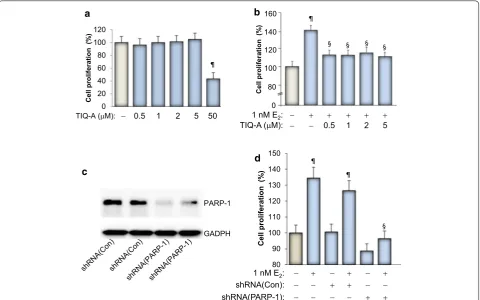
[image:3.595.59.540.337.637.2]PARP inhibition blocks E2‑dependent growth of MCF‑7 cells
Figure 1a shows that PARP inhibition with the specific
inhibitor TIQ-A exerted no effect on the growth of the breast cancer cell line MCF-7 except at a very high
concen-tration of 50 μM. This result is consistent with our previous
report which concluded that the growth inhibition caused by the high concentrations of PARP inhibitors is most likely associated with toxicity of the drugs rather than an effect
related to PARP inhibition [16, 17]. However, it is now
established that PARP inhibition causes cell death when
combined with BRCA deficiency [18]. It is rather surprising
that the role of PARP in estrogen-mediated growth of breast cancer cells has not been investigated. We thus wished to examine whether PARP-1 plays a role in growth
mediated by E2. Figure 1b shows that E2 treatment, as
expected, induced ~40% increase in growth of MCF-7 cells, which is typical as reported by our laboratory and others
[3, 19]. PARP inhibition by TIQ-A blocked E2-induced
growth of MCF-7 cells even at a concentration as low as
0.5 μM. The specificity of this effect was confirmed using
a PARP-1 shRNA-mediated knockdown approach
(Fig-ure 1c, d). These results clearly show that PARP plays a role
in E2-mediated cell growth and that its inhibition can be
regarded as a means to block abnormal proliferation of ER positive breast cancer cells. Such inhibition was achieved with rather low concentrations of the PARP inhibitor. Our results show that in addition to the ability of PARP inhibi-tor to promote a synthetic lethality phenotype in
BRCA-deficient cells [18], it can also reduce E2-dependent growth
in ER positive tumor cells.
1 nM E2:
TIQ-A ( M): 0.5 1 2 5
TIQ-A ( M): 0.5 1 2 5 50
Cell
proliferation
(%
)
a b
0 20 40 60 80 100 120
Cell
proliferation
(%
)
80 100 120 140 160
0
Cell proliferation
(%
)
80 90 100 110 120 130 140 150
1 nM E2:
shRNA(PARP-1): shRNA(Con):
d
¶
¶
¶ ¶
§ § § §
§ PARP-1
GADPH
c
Figure 1 Effect of PARP inhibition on E2-stimulated growth of MCF-7 cells. a MCF-7 cells were treated for 48 h in the absence or presence of increasing concentrations of TIQ-A. Cell viability was assessed using a MTT assay. ¶Difference from viability values of cells that did not receive TIQ-A; p < 0.05. b MCF-7 cells were stimulated with 1 nM E2 for 48 h in the absence or presence of the indicated TIQ-A concentrations. Cell viability was then assessed using a MTT assay. ¶Difference from viability values of cells that did not receive TIQ-A; p < 0.05. §Difference from viability values of cells that were treated with E2 alone; p < 0.05. c MCF-7 cells were transduced with a viral vector encoding control shRNA or a shRNA targeting human PARP-1. Protein extracts were prepared and subjected to immunoblot analysis with antibodies against PARP-1 or GAPDH. d Cells expressing control or PARP-1-targeting shRNA were treated with E2 for 48 h after which viability was assessed as described above. ¶Difference from respective untreated controls; § difference from E
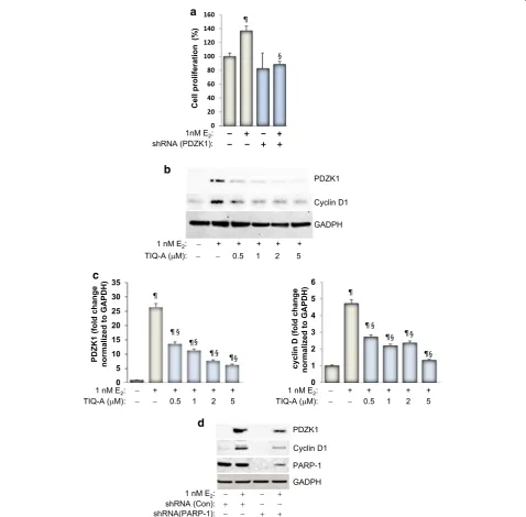
PARP inhibition‑associated reduction in E2‑induced growth of MCF‑7 cells is linked to a reduction in PDZK1 expression with a concomitant decrease in cyclin D1
We recently showed in two detailed studies that
E2-mediated cell growth can be governed by PDZK1 [4].
We thus speculated that PARP-1 may be involved in ER-mediated growth of MCF-7 cells potentially through a
control of PDZK1 expression. As reported previously [3]
and as shown in Figure 2a, knockdown of PDZK1 blocks
growth of MCF-7 cells in response to stimulation with
Cyclin D1
GADPH PDZK1
1 nM E2:
TIQ-A ( M):
+ +
1 + 0.5
+ 2
+ 5
b
GADPH PDZK1
PARP-1 Cyclin D1
d
1 nM E2:
shRNA(PARP-1):shRNA (Con):
0 20 40 60 80 100 120 140 160
1nM E2:
shRNA (PDZK1):
Cell proliferation
(%
)
a
¶
§
0 1 2 3 4 5 6
1 nM E2:
TIQ-A ( M): + 0.5 + + 1 + 2 + 5
cyclin D (fold change normalized to GAPDH)
§
§ § § ¶
0 5 10 15 20 25 30 35
1 nM E2:
TIQ-A ( M): + 0.5 + + 1 + 2 + 5
§ §
§ § ¶
PDZK1
(fold change
normalized to GAPDH)
¶
¶ ¶ ¶ ¶
¶
¶ ¶
[image:4.595.59.538.168.638.2]c
Figure 2 Effect of PARP inhibition on E2-stimulated expression of PDZK1 and cyclin D1. a MCF-7 cells were transduced with a viral vector encoding control shRNA or a shRNA targeting human PDZK1. Cells were then treated with E2 for 48 h after which cell viability was assessed. ¶Difference from untreated controls; §difference from E
1 nM of E2. We next wished to examine whether PARP-1
enzymatic activity or protein expression influences E2
-induced PDZK1 expression. To this end, MCF-7 cells
were exposed to E2 for 48 h in the absence or presence
of increasing concentrations of TIQ-A. Immunoblot analysis of collected cell extracts showed that PARP inhibition was very efficient in reducing expression of
PDZK1 (Figure 2b, c). The results also show that partial
reduction of PDZK1 by 0.5 μM TIQ-A was sufficient to
completely block E2-induced growth of MCF-7 cells (as
shown in Figure 1b). These results are consistent with
our previous observation that partial knockdown of E2
-induced PDZK1 was sufficient to completely block E2
-induced growth of MCF-7 cells [3]. The involvement of
PDZK1 in growth of MCF-7 cells appears to be linked to expression of cyclin D1 as knockdown of PDZK1
pre-vents E2-induced expression of the cell cycle protein [3,
4]. Although it is unlikely that PARP is always required
for the expression of cyclin D1, it was recently reported that the expression of the enzyme as well as its activity
are required for cyclin D1 expression in response to
ectopically-expressed Krüppel-like factor 8 (KLF8) [20].
An additional link between PARP-1 and cyclin D1 is
the relationship of both proteins to NF-κB. We and
oth-ers have shown that PARP-1 plays an important role in
NF-κB function [21, 22]. Furthermore, cyclin D1
expres-sion can be controlled by NF-κB [23]. We next
exam-ined whether the role of PARP-1 in PDZK1 expression
[image:5.595.60.539.352.668.2]influences expression of cyclin D1 upon E2 exposure.
Figure 2d shows that PARP inhibition with TIQ-A
reduced cyclin D1 expression in a manner similar to that exerted on PDZK1. Additionally, knockdown of PARP-1 markedly reduced expression of PDZK1 and cyclin D1
showing the specificity of the effect (Figure 2d). It is
noteworthy that an increase in PDZK1 expression in
response to E2 treatment did not change the levels of
PARP-1 (Figure 2d). This is consistent with our
pub-lished results showing that ectopic PDZK1 expression
in the absence of E2 exposure does not affect expression
of PARP-1 [4]. Although these results do not prove that
1 nM E2:
TIQ-A( M): 1 2
-Actin PDZK1
1 nM E2:
shRNA (PARP-1): shRNA (PDZK1):
-Actin PDZK1
a
c
0 2 4 6 8 10 12
PDZK1
mRNA
(Fold change)
1 nM E2:
TIQ-A( M): 1 2
b
¶
§ ¶ § ¶
Figure 3 Effect of PARP inhibition on PDZK1 mRNA in E2-stimulated MCF-7 cells. MCF-7 cells were treated with E2 for 24 h in the absence or pres-ence of 1 or 2 μM TIQ-A. Total RNA was prepared and subjected to reverse transcription followed by conventional (a) or quantitative (b) PCR with primers specific to human PDZK1 or β-actin. ¶Difference from untreated controls; §difference from E
E2 exposure does not affect the activity of PARP, we can,
however, conclude that it does not change the expression levels of this DNA repair enzyme.
PARP regulates E2‑induced PDZK1 at the mRNA level We have shown previously that PARP-1 may control expression of a number of proteins by regulating their expression at the mRNA level or by influencing their sta-bility. For instance, PARP-1 regulates expression of sev-eral inflammatory proteins including adhesion molecules,
iNOS, TNF-α, IL-1β, IL-5, and IL-13 [12, 24, 25]. PARP-1
may also regulate proteins at the level of their integrity. Indeed, PARP-1 influences the fate of STAT-6 upon IL-4 or allergen exposure by preventing the degradation of
the transcription factor by calpains [26]. We therefore
wished to determine whether PARP-1 regulates
expres-sion of PDZK1 at the level of mRNA. Figure 3a shows that
PARP inhibition was very effective in blocking E2
-medi-ated increase in PDZK1 transcript. The effect of the PARP
inhibitor on expression of E2-induced PDZK1 mRNA was
confirmed by quantitative RT-PCR (Figure 3b).
Consist-ent with these results, PARP-1 knockdown was equally
efficient in blocking E2-mediated increase in PDZK1
mRNA (Figure 3c). These results suggest that PARP-1 may
regulate PDZK1 expression at the level of mRNA. How-ever, it is premature to conclude that it influences its gene transcription. Given the lack of evidence for the control of PDZK1 by proteolysis, it is unlikely that PARP-1 regulates the protein by influencing its integrity.
PARP‑1 regulates E2‑mediated growth of MCF‑7 cells and PDZK1 expression by controlling the expression of IGF‑1R
We reported that PDZK1 expression was indirectly
regu-lated by ER-α activation, requiring IGF-1R expression
and function [3]. Blocking the activity of IGF-1R by the
specific inhibitor AG1024 blocked E2-induced PDZK1 in
MCF-7 cells (Figure 4a). We thus speculated that PARP-1
may be regulating PDZK1 expression through IGF-1R. Indeed, PARP-1 inhibition with TIQ-A reduced
expres-sion of E2-induced IGF-1R even at the lowest
concentra-tion (Figure 4b). Figure 4c shows that PARP inhibition
efficiently blocked E2-induced expression of IGF-1R
mRNA in MCF-7 cells. PARP-1 knockdown almost
com-pletely blocked the expression of E2-induced IGF-1R
(Fig-ure 4d) confirming the results attained using the PARP
inhibitor and demonstrating specificity of the effect.
PARP inhibition by olaparib (AZD2281) blocks E2‑mediated growth of MCF‑7 and BT474 cells and inhibits expression of PDZK1 and IGF‑1R
An important step in proving the validity of targeting
PARP to control growth of ER(+) breast cancer cells is
to demonstrate that PARP inhibitors other than TIQ-A can achieve the same effect and to show that such effect is observed in breast cancer cells other than MCF-7 cells. Olaparib (AZD2281), a potent PARP inhibitor, is currently used in a large number of clinical trials
target-ing BRCA(−) breast and ovarian cancers [15]. We thus
a
IGF-1R
1 nM E2:
TIQ-A ( M): 0.5 1 2 5 GAPDH b
PDZK1
IGF-1R PARP-1
GADPH 1 nM E2:
shRNA(PARP-1): shRNA (Con): d
PDZK1
GADPH
1 nM E2:
AG1024 ( M): 1 2 2
IGF-1R
GADPH
1 nM E2:
[image:6.595.305.539.88.450.2]TIQ-A ( M): 0.5 1 2 5 c
Figure 4 PARP inhibition reduces E2-induced IGF-1R. a MCF-7 cells were treated with E2 for 48 h in the absence or presence of 1 or 2 μM
of the IGF-1R inhibitor AG1024 after which protein extracts were prepared and subjected to immunoblot analysis with antibodies to PDZK1 or GAPDH. b Cells were treated with E2 for 48 h in the absence or presence of increasing concentrations of TIQ-A. Protein extracts were prepared and subjected to immunoblot analysis with antibod-ies to IGF-1R or GAPDH. c MCF-7 cells were treated with E2 for 24 h in the absence or presence of 0.5, 1, 2 or 5 μM TIQ-A. Total RNA was
prepared and subjected to reverse transcription followed by conven-tional PCR with primers specific to human IGF-1R or β-actin. d MCF-7
tested the effect of different concentrations of olaparib
on growth of MCF-7 cells. Figure 5a shows that
olapa-rib at the 5 μM concentration partially but significantly
reduced E2-induced cell growth; the 10 μM
concentra-tion of the drug completely blocked growth of the treated
cells. Figure 5b shows that olaparib was very effective in
blocking growth of BT474 cells, another ER(+) cell line
[27].
We next examined whether growth inhibition of MCF-7 cells by olaparib correlated with a decrease in
PDZK1, cyclin D1 and IGF-1R. Figure 5c clearly shows
that olaparib treatment markedly blocked
expres-sion of E2-induced PDZK1 in a manner similar to that
achieved by TIQ-A. Such inhibition of PDZK1 expres-sion occurred concomitantly with a reduction in cyclin
D1 and IGF-1R upon exposure to E2 in MCF-7 cells.
Fig-ure 5d shows that PARP inhibition reduces PDZK1 and
IGF-1R at the level of mRNA. Overall, these results are consistent with those attained using TIQ-A as a PARP
inhibitor and demonstrate that the ability of PARP
inhibi-tion to block E2-induced growth is not limited to TIQ-A
or MCF-7 cells but may be extended to other ER(+)
breast cancer cells.
Conclusions
It is well recognized that breast and ovarian cancer het-erogeneity continues to represent a major obstacle in tailoring precise and efficient therapies with minimal toxicities. Much effort is needed to identify key deter-mining factors that control the growth of cancer cells. Accordingly, insights on the role of new factors or iden-tification of new functions for already established players may provide us with new directions and strategies with which cancer can be retarded or blocked. Collectively, our results suggest that PARP may play an important role
in E2-induced cell growth by regulating the expression of
PDZK1 through a control of the stimulated expression of IGF-1R. To our knowledge, our results are the first in
1 nM E2: Olaparib ( M):
+ + 10 +
5 5 10
PDZK1
IGF-1R
GADPH
Cell proliferation
(%
) ¶
§
§
1 nM E2: + + 5 + 1
+ 10
80 100 120 140 160
0
b a
c
¶
¶
Cell proliferation (%
)
90 100 110 120 130
0
¶
§ §
1 nM E2: Olaparib ( M):
+ + 10 + 5 Olaparib ( M):
0 0.2 0.4 0.6 0.8 1 1.2 1.4
1 nM E2: 5 M Olaparib:
mRNA
(fold
change)
§ §
PDZK1 IGF-1R
d
[image:7.595.58.539.341.649.2]Cyclin D1
Figure 5 Olaparib treatment blocks E2-mediated growth of MCF-7 and BT474 cells and inhibits expression of PDZK1, IGF-1R, and cyclin D1. MCF-7 (a) or BT474 (b) cells were stimulated with 1 nM E2 for 48 h in the absence or presence of the indicated olaparib concentrations. Cell viability was then assessed using a MTT assay. ¶Difference from viability values of cells that did not receive olaparib; p < 0.05. §Difference from viability values of cells that were treated with E2 alone; p < 0.05. c MCF-7 cells were treated with E2 for 48 h in the absence or presence of increasing concentrations of olaparib after which protein extracts were prepared and subjected to immunoblot analysis with antibodies to PDZK1, cyclin D1, IGF-1R, or GAPDH. d MCF-7 cells were treated with E2 for 24 h in the presence of 5 μM olaparib. Total RNA was prepared and subjected to reverse transcription fol-lowed by quantitative PCR with primers specific to human PDZK1, IGF-1R or β-actin. §Difference from E
showing a connection between PARP and ER-stimulated growth and that such trait may be considered in the cur-rent effort in establishing PARP inhibitors in the treat-ment of breast and ovarian cancers. We have shown a strong relationship between PDZK1 and IGF-1R expres-sion in human breast cancer and that PDZK1 may be a
determinant in breast tumorigenesis [3, 4]. The
molecu-lar link between PDZK1 and IGF-1R was supported by a significant correlation between protein and mRNA levels of the two factors in two independent cohorts of human
breast cancer tissues [3].
The current results suggest that the relationship between ER, PDZK1, and IGF-1R may be an appeal-ing axis to be targeted by PARP inhibitors. It is note-worthy that a great deal of effort is being spent focusing primarily on triple negative breast and ovarian cancers with a BRCA deficiency to specifically achieve synthetic
lethality of cancer cells with the BRCA mutation [28].
Although the presented results do not provide a com-plete understanding of the mechanism by which PARP regulates PDZK1 or IGF-1R, they support the hypothesis that PARP inhibitors, including olaparib, may be useful
for treatment of ER(+) and estrogen-dependent cancers.
Obviously, more work is needed to validate such hypoth-esis and unravel the molecular mechanisms underlying the cross-talk between the different factors.
Abbreviations
PARP: poly(ADP-ribose) polymerase; ER: estrogen receptor; IGF-1R: IGF-1 recep-tor; TIQ-A: thieno[2,3-c]isoquinolin-5-one.
Authors’ contributions
KH conducted most of the experiments, participated in data analysis, and assisted in the writing of the manuscript. AT conducted some of the experi-ments and participated in data analysis. AZ participated in the planning of the experiments, participated in data analysis and the writing of the manuscript. AHB conceived the study, participated in data analysis, provided the funding for the study and wrote the manuscript. All authors read and approved the final manuscript.
Author details
1 The Stanley Scott Cancer Center, Louisiana State University Health Sciences Center, 1700 Tulane Ave, New Orleans, LA 70112, USA. 2 Tulane University Cancer Center, New Orleans, LA 70112, USA.
Acknowledgements
We would like to thank the members of the Boulares’ lab for proofreading the manuscript. This work was supported, in part, by Grant RSG-116608 from the American Cancer Society and Grant HL072889 from the NIH as well as funds from the Louisiana Cancer Research Consortium (New Orleans, LA) to H. Boulares.
Compliance with ethical guidelines
Competing interests
The authors declare that they have no competing interests.
Received: 10 January 2015 Accepted: 29 June 2015
References
1. Early Breast Cancer Trialists’ Collaborative G, Godwin J, Gray R, Clarke M, Cutter D, Darby S, McGale P, Pan HC, Taylor C et al (2011) Relevance of breast cancer hormone receptors and other factors to the efficacy of adjuvant tamoxifen: patient-level meta-analysis of randomised trials. Lancet 378:771–784
2. Mehta A, Tripathy D (2014) Co-targeting estrogen receptor and HER2 pathways in breast cancer. Breast 23:2–9
3. Kim H, Abd Elmageed ZY, Ju J, Naura AS, Abdel-Mageed AB, Varughese S, Paul D, Alahari S, Catling A, Kim JG, Boulares AH (2013) PDZK1 is a novel factor in breast cancer that is indirectly regulated by estrogen through IGF-1R and promotes estrogen-mediated growth. Mol Med 19:253–262 4. Kim H, Abd Elmageed ZY, Davis C, El-Bahrawy AH, Naura AS, Ekaidi I,
Abdel-Mageed AB, Boulares AH (2014) Correlation between PDZK1, Cdc37, Akt and breast cancer malignancy: the role of PDZK1 in cell growth through Akt stabilization by increasing and interacting with Cdc37. Mol Med 20:270–279
5. Shall S (1992) Poly ADP-ribosylation reaction. Jpn J Cancer Res 83 6. Shall S, de Murcia G (2000) Poly(ADP-ribose) polymerase-1: what have we
learned from the deficient mouse model? Mutat Res 460:1–15
7. Boulares AH, Yakovlev AG, Ivanova V, Stoica BA, Wang G, Iyer S, Smulson M (1999) Role of poly(ADP-ribose) polymerase (PARP) cleavage in apoptosis. Caspase 3-resistant PARP mutant increases rates of apoptosis in trans-fected cells. J Biol Chem 274:22932–22940
8. Hans CP, Zerfaoui M, Naura AS, Catling A, Boulares AH (2008) Differential effects of PARP inhibition on vascular cell survival and ACAT-1 expression favouring atherosclerotic plaque stability. Cardiovasc Res 78:429–439 9. Virag L, Szabo C (2002) The therapeutic potential of poly(ADP-Ribose)
polymerase inhibitors. Pharmacol Rev 54:375–429
10. Pacher P, Szabo C (2008) Role of the peroxynitrite-poly(ADP-ribose) poly-merase pathway in human disease. Am J Pathol 173:2–13
11. Hans CP, Zerfaoui M, Naura AS, Troxclair D, Strong JP, Matrougui K, Bou-lares AH (2009) Thieno[2,3-c]isoquinolin-5-one, a potent poly(ADP-ribose) polymerase inhibitor, promotes atherosclerotic plaque regression in high-fat diet-fed apolipoprotein E-deficient mice: effects on inflamma-tory markers and lipid content. J Pharmacol Exp Ther 329:150–158 12. Oumouna-Benachour K, Hans CP, Suzuki Y, Naura A, Datta R, Belmadani
S, Fallon K, Woods C, Boulares AH (2007) Poly(ADP-ribose) polymerase inhibition reduces atherosclerotic plaque size and promotes factors of plaque stability in apolipoprotein E-deficient mice: effects on mac-rophage recruitment, nuclear factor-kappaB nuclear translocation, and foam cell death. Circulation 115:2442–2450
13. Mukhopadhyay P, Horvath B, Kechrid M, Tanchian G, Rajesh M, Naura AS, Boulares AH, Pacher P (2011) Poly(ADP-ribose) polymerase-1 is a key mediator of cisplatin-induced kidney inflammation and injury. Free Radic Biol Med 51:1774–1788
14. Curtin NJ, Szabo C (2013) Therapeutic applications of PARP inhibitors: anticancer therapy and beyond. Mol Aspects Med 34:1217–1256 15. Jelinic P, Levine DA (2014) New insights into PARP inhibitors’ effect on
cell cycle and homology-directed DNA damage repair. Mol Cancer Ther 13:1645–1654
16. Inbar-Rozensal D, Castiel A, Visochek L, Castel D, Dantzer F, Izraeli S, Cohen-Armon M (2009) A selective eradication of human nonhereditary breast cancer cells by phenanthridine-derived polyADP-ribose polymer-ase inhibitors. Breast Cancer Res 11:R78
17. Kim H, Naura AS, Errami Y, Ju J, Boulares AH (2011) Cordycepin blocks lung injury-associated inflammation and promotes BRCA1-deficient breast cancer cell killing by effectively inhibiting PARP. Mol Med 18. Bryant HE, Schultz N, Thomas HD, Parker KM, Flower D, Lopez E, Kyle S,
Meuth M, Curtin NJ, Helleday T (2005) Specific killing of BRCA2-deficient tumours with inhibitors of poly(ADP-ribose) polymerase. Nature 434:913–917
19. Ptak A, Gut P, Blachuta M, Rak A, Gregoraszczuk EL (2009) Direct inhibition of ERK1/2 phosphorylation as a possible mechanism for the antipro-liferative action of 3,4-diOH-PCB3 in the MCF-7 cell line. Toxicol Lett 190:187–192
21. Zerfaoui M, Suzuki Y, Naura AS, Hans CP, Nichols C, Boulares AH (2008) Nuclear translocation of p65 NF-kappaB is sufficient for VCAM-1, but not ICAM-1, expression in TNF-stimulated smooth muscle cells: dif-ferential requirement for PARP-1 expression and interaction. Cell Signal 20:186–194
22. Zerfaoui M, Errami Y, Naura AS, Suzuki Y, Kim H, Ju J, Liu T, Hans CP, Kim JG, Abd Elmageed ZY et al (2010) Poly(ADP-ribose) polymerase-1 is a determining factor in Crm1-mediated nuclear export and retention of p65 NF-kappa B upon TLR4 stimulation. J Immunol 185:1894–1902 23. Hinz M, Krappmann D, Eichten A, Heder A, Scheidereit C, Strauss M (1999)
NF-kappaB function in growth control: regulation of cyclin D1 expression and G0/G1-to-S-phase transition. Mol Cell Biol 19:2690–2698
24. Oumouna M, Datta R, Oumouna-Benachour K, Suzuki Y, Hans C, Mat-thews K, Fallon K, Boulares H (2006) Poly(ADP-ribose) polymerase-1 inhibition prevents eosinophil recruitment by modulating Th2 cytokines
in a murine model of allergic airway inflammation: a potential specific effect on IL-5. J Immunol 177:6489–6496
25. Naura AS, Datta R, Hans CP, Zerfaoui M, Rezk BM, Errami Y, Oumouna M, Matrougui K, Boulares AH (2009) Reciprocal regulation of iNOS and PARP-1 during allergen-induced eosinophilia. Eur Respir J 33:252–262 26. Datta R, Naura AS, Zerfaoui M, Errami Y, Oumouna M, Kim H, Ju J, Ronchi
VP, Haas AL, Boulares AH (2011) PARP-1 deficiency blocks IL-5 expression through calpain-dependent degradation of STAT-6 in a murine asthma model. Allergy 66:853–861
27. Holliday DL, Speirs V (2011) Choosing the right cell line for breast cancer research. Breast Cancer Res 13:215
28. Sonnenblick A, de Azambuja E, Azim HA Jr, Piccart M (2015) An update on PARP inhibitors-moving to the adjuvant setting. Nat Rev Clin Oncol 12:27–41
Submit your next manuscript to BioMed Central and take full advantage of:
• Convenient online submission
• Thorough peer review
• No space constraints or color figure charges
• Immediate publication on acceptance
• Inclusion in PubMed, CAS, Scopus and Google Scholar
• Research which is freely available for redistribution