The pathology of kidney diseases in sheep : a thesis presented in partial fulfilment (30 %) of the requirements for the degree of Master of Philosophy in Veterinary Pathology at Massey University
12
0
0
Full text
(2) THE PATHOLOGY OF KIDNEY DISEASES IN SHEEP. A t,hesis presented in partial fulfilment. (3Cf/o). of the. requirements for the degree of l\laster of Philosophy. in VP.tcrinary Pathology at T·1asse y University.. Pavlos G-eorgiou Toum.azos. 1981.
(3) ii. ACKNOWLEDGEMENTS. I would l ike cor dially to thank Dr A.C. J ohns tone , for his c ontj.nuous and untiring guida�1ee, a ss i sta n ce throughout this study.. a dvic e ,. my supervisor,. encouragemei:t and. Spec ia l thanl�s are a lso clue to. 1-Tr. G. V.. Peter s en for his advice and assist a n c e in tbe stat i s t i c a l a na lys i s of data and for his arrangements for visits at Longburn freezing >·rorks . JI'Ia.ny th anks ar·e expr e s sed to the veterinary and meat in s pe c tion st<;.ff of the 1:-or-thvlick' s f reez�_ ng works at L ong burn, for t:he i r co:Llaboration. duT.Lng. .. :·llection peri od.. Professor B.\if. MankteloH, Dr M.R. A l ley and Dr R.D. Jcl.J.y gELJe helpf1}.1 advice during tbe w-c . iting and preparation. of this thRsis.. I am grateful for te chnical a s sis t a n ce provided by I.Jrs P I : .. and her. ·. .. Slack. "team" for the prep ara ti on of tissues for histopathological. examination; Hr T.G.. Law and tne staff of the };asse y University }':cintc;:y. for the pr eparat i o n of the photographs and 1·qJrod.u.ction of the figures. present e d in this thesis. I a l s o extend my sinc e re st gra t:•... /3. to many other fri ends. frcg. this and other D e pa rtment s of the Ji'ncu.lty of Yeteri:nary Science who v:erc a lw ays keenly receptive to my enquiries and request s and have directly or ind i rec tly helped me in my work. I would like to sinc ere ly thank my wife Vaso for typing tl:is thesl.s and the p.::l.tience,. understa ndin g and sacrifices :Jade by her and our. children Hiranda and Nestoras du:ring preparation of the manusc:cipt.. This inves ti ,:':a tion -vms partly suppor te d by a C ommor.vre a l th Scholal·ship. I am most gratefull to the administrators of this Gran t , especially the secretary Miss D.L.. Anderson ..
(4) iii. ABSTRACT. Renal diseases in sheep form a di verse spectrum of pathology and an extensive literature revievr of spontaneously occurring and e':per i mentally induced diseases of the 3hecp of this t he s i s survey. of. to. pr o v i de a c ompa r is on vri th. ki dneys jn slaughter-house �illed. the l esion s h. s e ep. of results of this survey form the major p ar t provide s information on sheep. uhich. these. . The. 1980.. at the. The p r evalen ce. 13,988. Borth-v;ick'. of. re na. signif icant variat i on ( p ( 0. 05) in the. l. sheep a. t otal. of. vre re ca t e gor i z ed into s even. le si on in each.. In. a. p resentation. The abnormal kidneys. sheep sl ughter e d over a. a. s. free z ing \lOrks,. disease. vras 3 . 1 8. prevalence of. 830 dise11sed ki dne ys. Longburr1. :per cen� and. lesions. ";as. of sheep ex&11ined.. l•rere founrl. groups according to the major. s ome kidneys. in. to large populati ons of. found beh1eE:n the various lines, chains and da ily totals. From t he se. found. thesis and. this. is only s pa rsely re po r t e d elsevrhere.. consecuti ve five day per i o d. no. of. diseases relative. under study were obtained from 444- of. in January. presented in Chapter l. �Kidney is. ancl. these. pathologic&.l. addi b onB-1 m nor lesions vrere present,. i. ma king a total of 1212 macroscopic le sio n s identified.. White finding in and. 179. spots and strealr..s . c on G t i tuted the major. 188. kidneys;. respectively;. Abscesses,. pale,. red and. gross p8.thological. brovm discolourati on in 174,. 120. scars in 107; cysts in 37 and nodules in 25 kidneys.. neoplasms and focal space occupying lesions of uncertain. aetiology 1.vere included under the category of nodule.. Pieces of tissue selected from. 181 kidneys. to represent the vario·LtS. lesions seen at gross examination were examined histologically. were identified,. These. recorded and graded accor ding to the anatomical location,. pattern of distribution, tissue changes and degree of severity.. The main histopathological feature of the white spotted kidneys vras.
(5) iv. chronic, mainly multifocal inflammation of the cortical interstitium. Similar. but. r a dially disposed inflammatory lesions with marked fibrosis. occurred in the. scarred kidneys .. pattern of these lesions suggested. The. a haematogenous di::;tribution of a. pathog en. the scarred k idneys were. the result of ascending inflammation. or infarctive. processes.. Kidneys vli. nephrosis. probably. discolouration showed mild to. th pale. of the cortical. emosi derin deposition .. t o areas of scarring.. moderately. severe. epithelial cells� while kidneys vlith brovm. discolouration shovred corticotubular ha. in the spotted kidney.s vJhile. In some. intracyt oplasmic. kidneys. and intralumenal. haemosiderosis wa::: restricted. Red discoloured kidneys. shm·red. patchy. or d iffuse. congestion.. Cystic lesions were either retention. parasitic or. caused by blockage of tubules .. the. In the. result of urinary latter,. tha block�ge. ,._,.as either congenital or associate d with chronic inflammation.. With the exception of. were chronic. nephrosis. in nature and for most of. diagnosis vras not esta blished.. Echinococcus. and conges-tion all the. granulosus hy. them. In fact,. datid cysts. lesions. a definitive aetiological. in only those lesions containing. could such. a. diagnosis. be. made.. Additional studies are indicated for the provision of further information on. (a). the prevalence of renal diseases in different. gevgraphical locations, and. (c). ( b). variation of disease types from area to area. the causes of the lesions identified from this type of. investigation..
(6) V. TABLE OF CONTENTS Page ACKNOWLEDGEMENTS. iii. ABSTRACT LIST OF ILLUSTRATIONS CHAPI'ER l:. CHAPTER 2:. ii. Review o f the liter atur e Introduction ( I) Diseases of the glomerulus l. Spontaneous glomerulonephropathies 2. Experimental autoinrnune glo merulo neph:ritis' ( II) Diseases of renal tubules l. Inflammatory 2. Degenerative ( III ) Diseases o f the renal pelvis ( IV ) Renal Neoplasms ( v ) Renal Cysts ( VI ) Uro lithiasi:::: A survey of diseased sheep kidneys Introduction Mater ials and Ne tho ds Results Gr o ss Pathology l. Abnormal shape 2. Increased size 3 . Reduced 8ize 4 . Disco louration 5 . Spots and streaks 6. Scars 7. Cysts 8. Nodules Histopathology l. Discolouration 2. Spots and str ealm 3 . Scar s 4. Cysts 5 . Nodules Discussion Conclusion. vi l l. 2. �. 7 10 10 15 24 25 25 27 30 30 30 31 34 37 37 37 37 38 38 38 39 40 41 42 45 47 49 52 60. BIBLIOGHAPHY. 62. APPENDICES. 72.
(7) vi LIST OF ILLUSTRATIONS Follovling Figure 2.1. Figure 2.2. Figure 2.3. Figure 2 .4. Figure 2 .5. Figure 2 .6. Histogram showing the distribution in weight of 8)0 l.i< dne y s collected from 444 sheep 1·ri th renal l esio ns .. Hi st ogra m shovTing the gross pathological fe at ur es of 12 12 lesions recorded in 830 diseased k idneys . Misshapen kidney (No. 23 4). a) Capsular surface shmving dumbbell sha pe caused by seve r e s carring in .central area. Less extensive sc a rr ing is pr esen t over the c a psular surface of the poles. b ) The cut surface of the same k idn ey shovring the scar:J:'ed tissue extending from capsule to pelvis.. Pale shrunken kidney ( No. 30·t) The reduct i on in size is due to di f fus e scarring of the renal parenchyma. The incised surface of this kidney is shovm. 34. 34. 37. •. in f igure 2 .14 .. 37. region.. 37. Pale Slwllen kidney (No . 99). No te the pallor of the outer c ortex c ontrasting with the c ong es ted corticowodullary Pa tch:y red discolouration of kidneys ( No. 5 ) due t o c onge stio n. The change is less severe and. res tricted t o t1w right po le in the ki dney on the righ t .. 37. Figure 2 . 7. Diffuse red discolov.ra cion of a kidney ( No. 2 5 4) due to conge sti on .. Yl. Figure 2.8. Diffuse dark brovm discolouration of a kidney ( No. 201) due to haemosiderosis.. 37. Figure 2.9a Dark brown discolouration of kidneys ( No.l33 ) distr ibuted in scarred and depressed areas of renal cortex. The pale raised areas are the r emaining functional kidney tissue. Figure 2.9 b. Cut section of kidneys showing radial brown str eaking of scar tissue extending from capsular surface to medulla.. 38. 38. rJage.
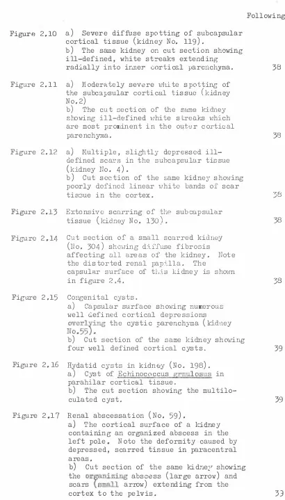
(8) vii Following page Figure 2.10. Figure 2.1 1. Figure 2.12. a) Severe diffuse spotting of subcapsular cortical tissue ( kidney No. 119 ) . b) The same kidney on cut section showing ill-defined, white streaks extending radially into iriller cortical parenchyma.. 38. a) llioderEt.tely severe vrhi te spotting of the subcapsular c ortical tissue ( kidney No.2) b) The cut section of t he same ki dne y showing ill-defined v1hi t e streaks vrhich are most prominent in the outer cortical paren chyma .. 38. a ) M ult ip le , slightly depr e s se d ill defined s cars in the subcap sular tissue ( kidney No . 4) . b) Cut section of the s a me kidney showing poorly defined linear vrhite hands of scar tis3ue in the cortex.. Figure 2.13. Extensive scarring of the subca psular tissue (kidney No. 130).. Figure 2.14- Cut secti on of a sma l l scarred kidney ( no. 304) shovr in g diffuse fibr osis affecting all areas o f the kidney. Note the d i sto rt ed renal papilla. The capsular surface of trLis kidney is shovm in figure 2.4. Figure 2.1 5. Figure 2.16. Figure 2. 17. Congenital cysts. a) Capsular surface showing numerous well d efine d cortical depressions overl r ing the cystic parench:yma ( J:..idney. )8. 38. 38. No.5 5 ) . b) Cut secti on of the same kidney shovring four well defined c ortical cysts.. 39. Hydatid cysts in kidney ( No. 198) . a) Cyst of Ec hi no co ccu s grnulosus in parahilar cortical tissue. b) The cut section shov;ing the multilo culated cyst.. 39. Renal abscessation ( No. 5 9 ) . a ) The cortical surface of a ki��ey containing an organized abscess in the left pole. Note the deformity caused by depressed, scarred tissue in paracentral areas. b) Cut section of the same kidne�r showing the or� anizing abscess ( large arrovr ) and scars \ small arrow) extending from the cortex to the pelvis.. 33.
(9) viii Following page Figure 2.18. Renal abscessation. a). ( No. 200) .. The kidney conLa:i.ns an e n ca ps u l ate d. caseous abscess bnlging from the capsular surface.. b). Figure. 2.19. The. cut section of the. carcinoma (Ho. 237) . o of the left pole of the kidney by a renal co.J:cinoma. b) Cut sectior-1 of the kidney shmving the s l ightly encaps-..11a·�ccl lobulated tumour. Renal a. ). Di s t � t i on. with extensive. hae m or rhage .. Figure. 2.20. areas. Renal lymphoma. of. and. necrosis. 39. (lTo. 236).. a) Di s t orti on of a kidney by a lymphoma occupyin& the l(�f L polar >;tnd hilar zones.. b). The. incised. surf:>.ce. focal twnour mnssec in. Figure 2.21. sho1·1ing. the. additional tissue.. cor tical. Figur�. 2.22. 2 . 23. the. l e s i on upon sectionJ.ng.. 40. Chronic parasitic noJule (kidl1cy No. 20). a) Appearance on capsular surface. On i n c ision the.� uell defined cortical b) nodule was hard and contained gritty mate rial.. 40. Hassive renal infarct. kidney on left. ( no. 2 49 ) . 'rhe. l. is swo le n ,. haemorrhagic and necrotic.. thrombus. is. p1·esent in the. diffusely An orga ni z i ng renal vein.. The right kidney is normal. l<'igure. 2 .24.. Nep h ros i s .. Moderately severe. de ge nera tive. pyknotic nuclei tubules contain. Figure 2.26. Figure 2 .27. Ne phros is .. t. � ca t ere. 40. d. changes in epithelial cells. of proximal convoluted. Fi.gure 2.25. 39. Nodule of undctcr:r;.Lr_ed cause ( k idney No. 12 ) . a) A fib r ous no:iclle elei.Tated slightly above the cap::;ular surface. b) Note the concentric lamination of. Figure. 39. sam8 kidney.. t ub ule s .. and cell loss. hyaline. Lesions. severe than those. in. Note dil ate d casts. HE x 3 2 0 41. The. gran ula r. ar e similar but less figure 2 .24. HE x 4 00. 41. Haemosiderosis. "ifuole mount" section of kidney stained by Perls' method for iron showing extensive cortical accumulation of r�emosiderin ( x 7).. 42. Haemosiderosis. Extensive deposition of haemosiderin in cytoplasm of epithelial cells of proximal convoluted tubules ( arrovm). Granular casts are present in the tubular lumena. HE x 320. 42.
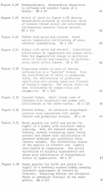
(10) ix Follmring page Figure. 2.28. Haemosiderosis.. Haemosiderin deposition. in inflamed and scarred tis sue of a. HE x. kidney.. 42. 50. Detail o f inset in figure. 2.28. sho-,ring. haemosiclerin granules in epith8lial cells of tubules. ( broad. ) and interstitial ( narrow arrovm ) .. arrow. inflammatory reaction. HE Figure. 2.30. X. 150. Kidney vii th spots and streaks.. 2.31. Focal. mainly l ymp ocyti c infiltration of renal. h. HE x. cortical interstitium. Figure. 42. 125. Kidney vri th spots and streak s .. 45 Interstitial. infiltration by lymphocytes and pl asma cells.. Note the degenerative change in epit he li al cells of tubule s and formation of proteina. ceous casts 1·1i thin lumena.. Figure. 2 . 32. 320. HE x. 45. Glomerulus within an area of c·hronic inflammation in a "spotted" kidney.. the prolife ra t ion of cells tufts,. Note. in glomerular. the obliteration of glomerular. capillari es an d �rinary space and fibrosis. of Bomn.:ln 1 s capsule.. The entire field has. been infiltrated by plasma cells and phagocytes.. Figure. 2.33. HE x. Scarred kidney.. Small linear zone of. fibrosis v!ith lymphocyte and plasma cell. infiltr ation in the outer cortex. Figure. 2.34. 4-5. 32 0. Scarred J:r.idney.. HEx. 125. 45. An extensive iveclgeshaped. cortical scar associated 'lri th marked. tubular and glomerular atrophy . Figure. 2 . 35. Renal papilla right. ). left. ). and pelvis. 50. 4'7. ( on. of a kidney vli th extensive radial. scarring. tubules, arrows. ( on. HE x. ). Note the reduced numbers of several containing casts. ( broad. and others with calcification of. basement membrane and epithelial cells. ( narrovT. arrows. ).. The interstitial ti s sue. of the papilla i s fibrotic and infiltrated by lymphocytes.. lightly. The pelvic. epithelium is slightly hyperplastic and shows mild infiltration of subepithelial. HE. ti ssue by lymphocytes. Figure. 2 . 36. Renal papilla right. ). ( on. left. ). x 50. and pelvis. 47. ( on. of a severely scarred kidney showing. extensive replacement of tubules by fibrosis.. Remaining tubules are atrophied.. There i s extensive fibrosis of the renal pelvis.. HE x. 125. 47.
(11) X. Following page Figure 2.37. Figure 2.38. Degeneration of pelvic and papillary epithelia associated with fibrosis, lymphocytic and plasmacytic infiltrations. HE X320. 47. Renal cortex of a scarred kidney showing tubular atrophy, dilatation of tubules 1vith accumulation of highly eo sino :philic material in lumena (thyroidiza tion ) , interstitial fibrosis and infiltration by lymphocytes and plasma cells. T1v0 atrophied glomeruli are present narrow arrmvs . HE x 125. 47. Glomeruli in area of inflamm�tion and s carring sho-vring gradation in the severity of basement membrane thickening due to membranous deposition. Note the segmental lesion in one glomerulus (arrow). PAS x 125. 47. Glomerulus in an area of chronic inflamma tion shmving severe sclerosis of Bo1:nnan' s capsule 1vi th collagenous adhesion arroi·r to the adjacent glorneru.lar tuft. HE x 320. 47. associated -vrith qhronic of lymphocytes and plasma cells in adjacent renal tiDsue. HE X 320. 47. (. ). Figure 2.39. Figure 2.40. (. Figure 2.4-l. Renal cyst. ). (c). inflammatory exudate. Figure 2.42. Figure 2.43. (c). Renal cyst vrith no inflammatory changes in the adjacent renal parenchyma. HE x 125 Echinococcus granulosu: cyst shm·Iing laminated cuticle adjacent to the collagenous tissue in the second layer of the cyst. Figure 2.44. Figure 2.45. Figure 2.46. 48. (cc).. (Le). HEx320. 48. Carcinoma. Acinar and tubuloacinar arrangement of neoplastic cells supported by a slight reticu�ar stroma. The cells are polyhedral to ovoid vrith abundant, slightly vacuolated, eosinophilic cytoplasm. RE X320. 49. Carcinoma. A more densely, cellular area of tumour to that shovrn in figure 2.44 showing greater cellular pleomorphlsm. HE X320. 49. Carcinoma showing v1hite arrow and representative of into lobules. HE. 49. (. ). dystrophic calcification a band of fibrous tissue that dividing the tumour x320.
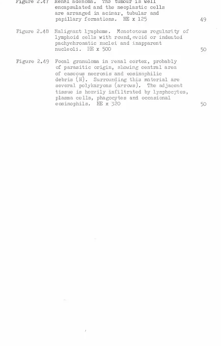
(12) xi. Following page Figure 2 .47 Rene.l a denoma . The tumou:r is well encapsulated and the neoplastic cells are arranged in acinar, tubular and papillary f ormations. HE x 125. 49. Figure 2. 4 8 Nal igPant l�'Tllphoma. Monotonous regularity of lymphoid cells with round,ov0id or indented pachychromatic nuclei and inapparent nucle oli . llli x 500. 50. Figure 2.49 Focal granuloma in renal cortex, probably of parasitic origin, shoHing central area of case ous necrosis and eosinophilic debris (N). Surrounding this material are several polykaryons ( arrovis ) . The adjacent tissue is heavily infiltrated by lymphocytes, plasma cells, phago cyte s and occasional eosinophils. HE x 320. 50.
(13)
Figure

Related documents
The work is not aimed at measuring how effective your local polytechnic is, but rather to see what measures different community groups, including potential students, use when
Figure 5.23 The Keteira core site - continuous coupled logistic growth model.. Figure 5.24 The Keteira core site - discrete logistic growth
In this paper we trace the early history of relativistic su- pertasks, as well as the subsequent discussions of Malament-Hogarth spacetimes as physically-reasonable models
Nitri fi cation and sedimentary denitri fi cation occurred near the river mouth, nitri fi cation prevailed further offshore under the plume, and fi nally, phytoplankton
2 Equity Market Contagion during the Global Financial Crisis: Evidence from the World’s Eight Largest Economies 10..
The use of sodium polyacrylate in concrete as a super absorbent polymer has promising potential to increase numerous concrete properties, including concrete
Radio and TV broadcast music related programmes which provide opportunities not just. to listeners but also to the
21 Department of Neurosurgery, Tangdu Hospital, The Second Affiliated hospital of the Fourth Military Medical University, 1 Xinsi Road, Xian, Shanxi Province 710038, People ’ s
