PREVALENCE OF ORAL HEALTH COMPLICATIONS
IN TYPE 2 DIABETES MELLITUS PATIENTS:
A DESCRIPTIVE STUDY
DISSERTATION
Submitted to The Tamil Nadu Dr. M.G.R Medical University
in partial fulfillment of the requirement for the degree of
MASTER OF DENTAL SURGERY
BRANCH IX
ORAL MEDICINE AND RADIOLOGY
I extend my profound gratitude to my guide Dr. Tatu Joy E MDS, Professor & Head of the Department of Oral Medicine and Radiology for his invaluable guidance, co-operation, constant encouragement and immense patience with me in every step of this endeavor. I thank him for the trust he had in me that made me complete this work and also I consider it as a great opportunity to do my postgraduate programme under his guidance.
I am thankful to my co-guide Dr. Shashi Kiran M., Reader, Department of Oral Medicine & Radiology for his constant enthusiasm, strive for perfection, and patience he showed me for writing this study.
I take this opportunity to express my sincere gratitude to my teachers Dr. Eugenia Sherubin, Associate Professor, Dr. Raghupathy and Dr. Redwin Dhas,Senior Lecturers for the constant encouragement throughout the course of this study.
I would like to acknowledge the help & support given by Dr. Velayuthan Nair, Chairman and Dr. Rema V Nair Director Sree Mookambika Institute of Medical Sciences, for academic support and facilities to carry out my dissertation work. I am thankful to the Administrative Officer Mr J.S Prasad for his timely involvement and encouragement throughout the course.
Dr. Aravind, Dr. Lekshmi, Dr. Divya and Dr. Kartheesan for helping me to get
through difficult times and for all emotional support and caring.
I would like to thank Mr. Sarath Babu for helping me with my statistical work.
Words are less to express my deep gratitude and love to my husband Lt Cdr(Retd) Naveen Nair, my lovely daughter Chitrangada Nair, my parents
Mrs Jameela Kumari, Mr Madhu C K, my in-laws Mrs Radha Devi and
Mr Suchindan and my uncle Mr Anil Kumar and Gopakumar for their constant tireless support and encouragement that they give me throughout my life.
I am extremely thankful to my friends Dr Jejo Mathew, Dr Dhanya Suresh, Dr Ansu Jobin andJenine Joseph for their prayers and constant support.
Sl. No TITLE PAGE NO
1
List of abbreviations
i
2
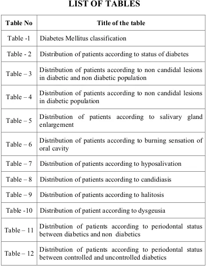
List of tables
iii
3
List of graphs
iv
4
List of color plates
v
5
List of annexure
vi
6
Abstract
vii
7
Introduction
1
8
Aim and objectives
3
9
Review of literature
4
10
Materials and method
35
11
Results and observations
39
12
Discussion
41
13
Summary and Conclusion
48
14
Bibliography
xxx
iii
Table No
Title of the table
Table -1
Diabetes Mellitus classification
Table - 2
Distribution of patients according to status of diabetes
Table – 3
Distribution of patients according to non candidal lesions
in diabetic and non diabetic population
Table – 4
Distribution of patients according to non candidal lesions
in diabetic population
Table – 5
Distribution of patients according to salivary gland
enlargement
Table – 6
Distribution of patients according to burning sensation of
oral cavity
Table – 7
Distribution of patients according to hyposalivation
Table – 8
Distribution of patients according to candidiasis
Table – 9 Distribution of patients according to halitosis
Table -10 Distribution of patient according to dysgeusia
Table – 11
Distribution of patients according to periodontal status
between diabetics and non diabetics
iv
Graph No TITLE OF THE GRAPH
Graph - 1
Distribution of patients according to status of Diabetes
Graph - 2
Distribution of patients according to gender
Graph - 3
Distribution of patients according to diabetic medication
Graph - 4
Distribution of patients according to burning sensation of
oral cavity
Graph - 5
Distribution of patients according to hyposalivation
Graph - 6
Distribution of patients according to candidiasis
Graph-7
Distribution of patients according to halitosis
Graph-8
Distribution of patients according to dysgeusia
Graph-9
Distribution of patients according to periodontal status
between diabetics and non diabetics
Graph-10
Distribution of patients according to periodontal status
v
Color Plate No
Title of color plate
CP - 1
Oral
manifestations
of
diabetes
and
their
mechanisms and interrelationships
CP - 2
Armamentarium 1
CP - 3
Armamentarium 2
CP - 4
Fibroma
CP- 5
Biopsy
CP6 & CP7
Salivary Gland enlargement
CP8 Traumatic
ulcer
CP9 Denture
Stomatitis
CP10 Periodontitis
CP11
Caries and Missing Teeth
CP12 Fissured
Tongue
CP13 Geographic
tongue
CP14 Xerostomia
CP15
Lichen planus buccal mucosa
CP16
Candidiasis of tongue and pigmentation
vi
No Title
Appendix -1
Certificate from Institutional Research Committee
Certificate from Institutional Human Ethics
Committee
Appendix - 2
Patient information sheath
¾
English
¾
Malayalam
¾
Tamil
Appendix - 3
Patient consent form
¾
English
¾
Malayalam
¾
Tamil
Abstract
Background of the study:
Diabetes Mellitus is often associated with a number of medical complications as a result of the metabolic changes taking place systemically. There is considerable evidence it is also associated with oral health complications including gingivitis, periodontitis, xerostomia, oral candidiasis, dental caries, ulcers lichen planus, burning mouth syndrome and an altered taste sensation .
Aim:
To study various types of oral health complications in Type 2 diabetes mellitus patient visiting a dental college in Kanyakumari District.
Materials & Methods:
A comparative cross-sectional study to determine the common oral complications prevalent in diabetics and non-diabetics was carried out in the outpatient department and the study sample consisted of 127 diabetic patients and 127 non diabetic patients. The oral health status was assessed clinically for each patient and recorded. The data was analysed by Statistical Package for Social Sciences (SPSS 16.0). Chi square test was applied to find the statistical significance between the groups.
Results:
The most frequent manifestation observed in diabetic patient was periodontitis followed by oral candidiasis, oral burning sensation, altered taste and hyposalivation. Most common mucosal disorders observed is geographic tongue followed by hyperpigmentation. The result correlated well with other studies.
Abstract
Conclusion:
It is concluded that the oral cavity exhibits the first sign of an undiagnosed or uncontrolled diabetes, hence oral health care providers must be well aware of signs and symptoms to refer such to a physician for further investigation as well as manage and treat the oral health complications.
ͳ
The earliest description of diabetes was documented in the writings of Hindu scholars as long as in 1500 BC. They had already described “a mysterious disease causing thirst, enormous urine output, and wasting away of the body with flies and ants attracted to the urine of people.” The term diabetes was probably coined by Apollonius of Memphis around 250 BC, which literally meant “to go through” or siphon as the disease drained more fluid than a person could consume. Later on, the Latin word “mellitus” was added by Thomas Willis because it made the urine sweet. In 1776 Dobson first mentioned the presence of increased sugar content in urine and blood as a cause of this sweetness. An important milestone in the history of diabetes is the establishment of the role of the liver in glycogenesis, and the concept that diabetes is due to excessive glucose production which was stated by Claude Bernard in 1857. The role of the pancreas in pathogenesis of diabetes was discovered by Mering and Minkowski in 1889. This discovery formed the basis of insulin isolation and clinical use of it by Banting and Best in 1921. Trials were conducted to prepare an orally administrated hypoglycemic agent which resulted in sucess by first marketing of tolbutamide and carbutamide in 1955.1
ʹ
In 2014 the global prevalence of diabetes was estimated to be 387 million people with Type 2 DM making up about 90% of the cases. This represents 8.3% of the adult population, with equal rates in both women and men. In 2012, an estimated 1.5 million deaths were directly caused by diabetes.8 More than 80% of diabetes deaths occur in low- and middle-income countries.8 WHO projects that diabetes will be the 7th leading cause of death in 2030.9 The number of diabetic individuals is
increasing globally because of population growth, ageing, increasing prevalence of obesity, urbanization ( stress and lifestyle).
The two most common forms of diabetes are Type 1 diabetes (diminished production of insulin) and Type 2 diabetes (impaired response to insulin and b-cell dysfunction). Both lead to hyperglycemia, excessive urine production, compensatory thirst, increased fluid intake, blurred vision, unexplained weight loss, lethargy, and changes in energy metabolism. A third type , Gestational diabetes mellitus (GDM) which is defined as any degree of glucose intolerance with onset or first recognition during pregnancy. Approximately 7% of all pregnancies are complicated by GDM, resulting in more than 200,000 cases annually.10
AIM AND
͵
AIMS
To study various types of oral health complications in Type 2 diabetes mellitus patient visiting a dental college in Kanya Kumari District.
OBJECTIVES
1) To evaluate the stomatology changes in diabetics as compared to non diabetics controls.
Ͷ The objective of this review is to provide an overview of diabetes in terms
of its classification, risk factors, pathophysiology, diagnosis and management. The
common oral complications observed in diabetics are discussed as well as the role
of dentists in diagnosis and management of oral complications in a diabetic patient
is also reviewed here.
Diabetes is a clinical syndrome caused by a relative or absolute insulin
deficiency which causes irregularities of carbohydrate, protein and lipid
metabolisms.13 It is pernicious in nature and has strong and well established
assosiations with numerous co-morbid conditions. Its most important feature is
hyperglycaemia that can result from decreased insulin production or insulin
dysfunction or lack of insulin receptor responsiveness at target organs.6,14,3
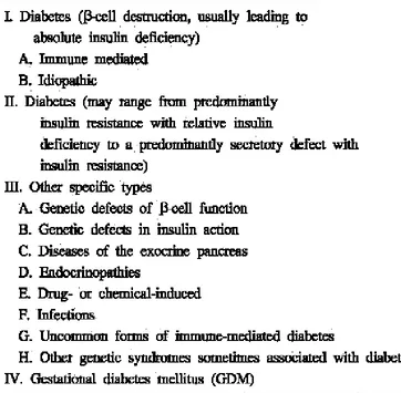
CLASSIFICATION OF DIABETES
The classification of Diabetes Mellitus is based on pathogenic processes
which leads to absolute or relative insulin deficiency, leading to hyperglycaemia, a
important feature of diabetes.15 It is grouped broadly into three categories based on
its signs and symptoms, namely Type 1, Type 2 and gestational diabetes that occur
in pregnant women.16 Type 1 usually has its early onset in childhood and
adolescence and is only prevalent among 5-10% of total diabetic population.15 The
term Type 1 diabetes has replaced what previously known as insulin dependent
diabetes mellitus (IDDM), juvenile-onset diabetes mellitus (JODM) and early onset
diabetes.4 The previous names were used because Type 1 diabetics needed insulin
for life and usually the disease occurred before 30 years of age. The new
ͷ provide a functional and working classification of diabetes that reflects the current
knowledge about the disease rather than a classification based on treatment
methodology.4
The American Diabetes Association recommends that Type 1 DM is to be
further divided into Type 1A and Type 1B.4 Type 1A is caused by cell-mediated
autoimmune destruction of the beta cells of the pancreas while Type 1B refers to
non immune mediated diabetes (NIMD) with severe insulin deficiency. Type 1B is
mainly found in of Asian or African population. The characteristic features of this
type are very similar to those of Type 2 DM which are ketoacidosis and absence of
autoimmune markers.4,17
Type 1 diabetes is a slowly progressive T-cell-mediated autoimmune disease,
although its has ab abrupt onset.17,18 This type of diabetes occurs in very young
individuals less than 30 years, who are mostly adolescent or children.In Type 1
diabetes, the beta cells of the Islets of Langerhans in the pancreas are destroyed and
are unable to produce insulin3,16 which results in a cascade of metabolic reactions
that manifests as complications associated with diabetes. Hyperglycaemia,
accompanied by the features of diabetes occurs only when 70-90% of the beta cells
have been destroyed and together with familial studies has served as proof that this
Type of diabetes has a very slow-onset.18
Insulin is pivotal for glucose metabolism and the human body cannot
function properly in its absence, as it is unable to transport glucose to the cells where
it is required. It facilitates the entry of glucose that is absorbed by blood into the
given to all Type 1 diabetic patients to prevent the build-up of glucose in the tissue
fluids and blood stream.3
Type 1 diabetes most oftenly predisposes patients to a condition known as
diabetic ketoacidosis (DKA). DKA occurs due to poor insulin control and it also
interferes with bone coupling and healing.20 When glucose cannot enter the
bloodstream, fat metabolism occurs through lipolysis and is glycerol and free fatty
acids are released as end products. The glycerol is converted to glucose and the fatty
acids into ketones which then accumulate in the body fluids. A slowly evolving
variant of Type 1 diabetes is known as latent autoimmune diabetes in adults
(LADA). This Type can be detected by specific auto antibodies called glutamic acid
decarboxylase (GAD) the GAD65Ab being specifically associated with LADA in
middle-aged people.18 This subgroup is often masked as Type 2 diabetes until
evidence of autoimmune activity against pancreatic beta cells is discovered.
Type 2 is the most common form of diabetes 21,3,5 and is refered to
non-insulin dependant diabetes mellitus. It occurs as a result of a reduced responsiveness
to insulin or insulin resistance of the target organs leading to a cascade of
complications which includes various systemic complications like retinopathy,
vascular degeneration, neuropathy and nephropathy. It is well accepted fact that
there is excessive production of glucose in the liver accompanied by under
utilization of glucose in the skeletal muscle results from resistance to the actions of
insulin .18This Type has its onset in people aged 40 years and above.15
In both types of DM, the vascular system where the exchange of oxygen,
two ways: either it can be damaged due to atheromatous deposits accumulating in
the lumen of the blood vessels or may develop a thickening of the basement
membrane which reduces the functionality of leukocytes. The reduction in the
activity of leukocytes leads to decreased polymorphoneucleocytes’ (PMNs) killing
ability making the diabetic patient more vulnerable to severe infection than a
non-diabetic patient.19,20
The accompanying hyperglycemia causes advanced glycation end products
(AGEs) and the release of glycoheamoglobin (HgbA1c) that also contributes to the
thickening of the basement membrane of the blood vessels. Unlike hemoglobin, the
glycoheamoglobin is less adept when it comes to transportation of oxygen. Other
pathological mechanisms associated to elevated hyperglyceamia are activation of the
sorbitol pathway, damaging effects of oxidative stress and altered lipid
metabolism.22
Diabetes Mellitus affects the entire body. Tissues affected are those rich in
blood vessels like the kidney, retina and nerves23 because of which complications
such as neuropathy ,renal disease , retinopathy, peripheral vascular disorders and
coronary disease occurs.19 These complications are associated with long-term
fuctional and biochemical abnormalities which occurs in poorly controlled diabetes
and often leads to premature increased morbidity and mortality. This occur as a
local response to the generalized vascular damage due to vascular permeability.18 At
present the annual mortality rate in adult diabetic patients is double that of
non-diabetic adults because of these associated complications . Myocardial infarction is
ͺ women have a significantly higher risk of morbidity and mortality as a result of
diabetes associated complications.24
Gestational diabetes is defined as either onset or first recognition of glucose
intolerance during pregnancy in a woman that has not had this condition before.16,25
The onset is during the third trimester.14 It is a pernicious condition and is
responsible for perinatal morbidity and mortality as it is closely linked to
pre-eclampsia, caesarian delivery, premature rupture of membranes and preterm
delivery. 25 Its pathophysiology resembles that of Type 2 diabetes as it is also
ͻ
Table 1
Classification of Diabetes Mellitus
26Risk Factor
Risk factors for diabetes can be broadly classified into modifiable and
non-modifiable. The non-modifiable factors includes genetic predisposition, increasing
age and ethnicity.27 Diabetes Mellitus is associated with familial history , although
ͳͲ Modifiable risk factors includes obesity, sedentary lifestyles, hypertension,
smoking and increased cholesterol levels. Changing lifestyles with little or no
exercise and a high fat diet lacking in fibre significantly contributes to obesity.27
Obesity (with BMI >30) is a major risk factor for Type 2 diabetes as it plays a very
significant role in the patho-physiology of diabetes. Obesity is an integral part of
metabolic syndrome X and it predisposes to Type 2 diabetes.3 Knowledge of risk
factors is essential for non pharmacological management of diabetes. Diabetes
often enhances the effects of other major cardiovascular disorder risk factors like
smoking, hypertension and hyperlipidaemia.18 Hence diabetic patients should
receive some form of dietary and lifestyle counselling, and should be strongly
discouraged from smoking and unhealthy eating habits.
Other modifiable risk factors particular for diabetes mellitus are impaired
glucose tolerance (IGT) and impaired fasting glucose (IFG). Both these conditions
are referred to as pre-diabetes. The factors refers to a state that exists between
diabetes and normoglycemia.3 People with this condition appear to have worse
cardiovascular prognosis especially those with IGT.27 To diagnose pre-diabetes, a
glucose tolerance test (GTT) has to be done. First, the blood glucose levels are
measured after the patient has been fasting from the previous night (nearly 8 hrs).
The patient is then given an oral dose of glucose and the test is done minutes to
hours there after. A fasting glucose value of 100-125mg/dl as well as a post-glucose
challenge of 140-199mg/dl is used to define IFG and IGT respectively.27 If a patient
is non-diabetic the blood glucose level rises moderately and returns to normal ranges
ͳͳ remain elevated for hours after the oral dose of glucose. The reasons for the
prolonged hyperglycaemia could either be attributed to a lack of insulin release by
the pancreas or impaired target tissue response to insulin or both.11 They are the
important risk factors for developing diabetes in the future because prediabetes
phase represents an active and destructive phase where there is progressive beta cell
deterioration and insulin resistance occurs.
Pathogenesis of Type 2 Diabetes Mellitus
Type 2 diabetes mellitus is preceded by resistance to insulin and the target
tissues have a decreased response to the normal levels of circulating insulin.15
Patients with Type 2 diabetes have a slow-onset of relative insulin deficiency . This
results in more insulin required by the target tissues.
The genetic predisposition for Type 2 diabetes is much stronger than for
Type 1 diabetes.15 It was observed that genes involved in carbohydrate, lipid and
amino acid metabolism pathways, glycan of biosynthesis, metabolism of cofactors
and vitamin pathways, ubiquitin mediated proteolysis, signal transduction pathways,
neuroactive ligand-receptor interactions, nervous system pathways and
neurodegenerative disorder pathways are upregulated in obesity compared to health
subjects. It was also identified that genes involved in signal transduction, regulation
of actin cytoskeleton, complement and coagulation cascades were upregulated in
subjects with Type 2 diabetes.28
In these patients hyperglycaemia develops gradually over a long period and
ͳʹ glomerular filtrate) for glucose rises, so that osmotic symptoms (polyuria and
polydypsia) are usually mild.18 Type 2 DM is also associated with dyslipidaemia.
Obesity and Type 2 diabetes are characterized by chronic oxidative stress and
inflammatory stress.29
Establishing the diagnosis of diabetes
Diagnosing Diabetes Mellitus is the realm of the physician4 as oral health
care providers are not qualified to make a diagnosis.12 But, it is still important for
dentists to understand how the diagnosis is reached since they have a significant role
to play in identifying the persons at risk or those who may have undiagnosed DM.
The American Diabetes Association supports the screening of all those who are at
risk for diabetes and all those above age of 45.27
Several tests can be employed in order to diagnose DM but the main
methods are fasting venous plasma glucose levels and oral glucose tolerance tests.
Generally, a fasting glucose test is used as a way of screening patients but it remains
limited since it cannot detect all forms of diabetes. A fasting glucose of 126 mg /dl
or more is used to identify a diabetic patient.27 A post oral glucose challenge value
of 200 mg/ dl or more can also identify a diabetic patient. When the above tests
aredone and a patient is identified with diabetes, the next step is to do an oral
glucose tolerance test (OGTT) for those who had impaired fasting glucose or in a
high-risk individual with a normal fasting glucose. In the past, oral glucose tolerance
test used to give false positive results as a result of stress-induced adrenaline release
which impairs the response to glucose loading hence this test is not as popular.3 For
ͳ͵ after ingesting a 75g load of glucose. Should two abnormal readings be taken on
different days then it’s undeniably a case of diabetes.27
Glycated haemoglobin provides an accurate and objective measure of
glycaemic control over a period of 2 months .It is used to assess level of glycaemic
control, but is not sufficiently sensitive to make a diagnosis of diabetes. The
non-enzymatic covalent attachment of glucose to Hb (glycation) increases the amount in
the HbA1c fraction relative to non glycated adult. The rate of formation of HbA1c is
directly proportionally to the blood glucose concentration ; a rise in 1% of HbA1c
corresponds to an increase of 2 mmol/l in blood glucose. HbA1c concentration
reflects blood glucose over the erythrocyte lifespan which is 120 days, but is
affected more by recent events. HbA1c estimates may be diminished in anemia and
pregnancy and may be difficult to interpret in uremia and haemoglobinopathy. In
clinical practice it is measured once or twice yearly to assess glycaemic control and
provides an index of risk for developing diabetic complications.18
MANAGEMENT
Primary aim in management of diabetes mellitus is to keep blood glucose
levels as close to normal as possible in order to prevent microvascular and
macrovascular complications of diabetes.Although no total cure exists for diabetes,
the disease and its complications can be prevented, delayed and managed by
identifying risk factors and detecting the condition at an early stage.30 Diabetic
complications and progression there of is much slower with good glycemic control
and also effective treatment of hypertension, irrespective of the type of therapy
ͳͶ ranging from diet modification and lifestyle changes, oral antidiabetic drugs to
insulin.18
Oral hypoglycemic medications depend on functioning pancreatic beta
cells to stimulate insulin secretion and therefore, are used to treat many patients
with type 2 diabetes.3 Insulin is required for patients with type 1 diabetes, as well as
for patients with type 2 diabetes who do not respond to oral therapy alone or in
combination.15 Diet and physical exercise also constitute a necessary component of
therapy for patients with both type 1 and type 2 diabetes.
Oral physicians should be familiar with the medications used for
diabetes.12,31 Oral hypoglycemic agents include sulfonylureas (which enhance
insulin secretion), biguanides (which reduce hepatic glucose production),
alpha-glucosidase inhibitors (which delay glucose adsorption) and thiazolidinediones
(which enhance insulin sensitivity). Insulin is available in short-acting (1 to 1
1/2hours), regular-acting (four to six hours),intermediate-acting (eight to 12 hours)
and long-acting (24-36 hours) formulations. Insulin pumps provide a continuous
burst of insulin to help control serum glucose levels.
Home glucose monitoring is recommended several times daily to help
patients to monitor glycemic levels. Many tools are available to help people with
diabetes, including home-based urine and blood tests and glucometers. Patients must
undergo regular examinations by physicians to monitor triglyceride, fasting glucose
and HbA1c levels. Oral Health care providers should always document their
patients’ most recent home-based glucose and laboratory test results, and monitor
ͳͷ supervision is a critical component of diabetes management, and oral physicians can
assist in this endeavour. Risk factors for impaired nutritional intake include
gingivitis and periodontitis, oral microbial infections, poorly fitting or lack of
removable prostheses, dysphagia and salivary dysfunction. A realistic nutritional
plan that includes regular oral hygiene and requisite dental treatment can help
ͳ
DIABETES AND THE ORAL HEALTH STATUS
The oral complications of uncontrolled DM may include, but are not
essentially limited to infections , poor healing, increased incidence and severity of
caries, candidiasis, gingivitis and periodontal disease, periapical abscesses,
xerostomia, altered taste and burning mouth. The oral complications are most likely
due to excessive dehydration (polyuria), the altered response to infection, the
microvascular changes and possibly the increased glucose concentrations in saliva.
Careful evaluation of glycaemic control is critical in determining the risk assessment
for progression to the oral complications from diabetes. Diabetes causes changes in
periodontal tissues, oral mucosa, salivary gland function ,oral neural functions and
increased risk of caries . Changes in oral soft tissues in addition to periodontium can
ͳ
Hyposalivation and Salivary gland changes
The oral manifestations of diabetes in the salivary glands include
sialoadenosis or non inflammatory, non-neoplastic enlargement of the parotid
salivary glands,36,37 decreased salivary flow rates and changes in salivary
composition. The enlargements are caused by gradual accumulation of fat in the
glands, hypertrophy of the acini or secreting units, and, eventually, impaired
glandular secretion.34 These structural changes may be the result of alteration in
autonomic neuroregulation of the glands and atrophy of the myoepithelial cells that
facilitate secretion. Xerostomia, or sensation of dry mouth is commonly reported
complication in diabetics which is due to decrease in flow of saliva because of
autonomic nerve dysfunction or microvascular changes that diminish the ability of
salivary glands to respond to neural or hormonal stimulation.38,39 Other causes
include medication used in treatment and dehydration.40
Mucosal Disorders
Oral mucosal lesions commonly noticed in diabetics include candidiasis,
lichen planus, lichenoid mucositis which are attributed to chronic salivary
hypofunction and to generalised immune dysfunction seen in such patients.41,42
Tongue Abnormalities
Complete or patchy atrophy of tongue papilla results in the appearance of a
bald tongue is more commonly found in diabetics.42 Focal areas of atrophy may
indicate an infection with candida organisms while generalized atrophy of papilla of
ͳͺ noted is fissuring of tongue , the smooth texture of the dorsum is interrupted with
one or more fissures that are predominantly aligned along length of the tongue. The
fissuring is due to chronic hyposalivation that alters the environment in the oral
cavity such that slow healing soft tissues are more easily traumatized in diabetics.41
Another observation noted in the tongue of diabetics is median rhomboid glossitis
ssen in the dorsal aspect in the midline region which is usually smooth and flat . It is
a recognised manifestation of chronic candidiasis.41 Another condition of the tongue
that is more common in diabetics is geographic tongue, characterized as
inflammation and is associated with similar symptoms of pain , itching and burning
of the mucosa.44
Oral Candidiasis
The combination of a decresed flow rate and immune dysfunction greatly
increases the risk of opportunistic fungal diseases like oral candidiasis and combined
with increased salivary glucose levels which promotes the growth of Candida.42
Typically patients complaints of a burning sensation of mucosa , some patients can
be asymptomatic too. White area on mucosal surface that can be wiped off leaving a
reddened bleeding surface is acute pseudomembraneous candidiasis. Denture
stomatitis is diffuse redness of mucosa occurring under upper dentures in edentulous
patients. This is considered to be a form of candidiasis where organism actually
infects the porous denture acrylic and causing contact hypersensitivity inflammation
of the adjacent mucosa.45 Angular chelitis is redness or fissures at the corners of the
mouth involving the junction of the mucosa and skin and may also represent a form
ͳͻ
Oral lichen Planus and Lichenoid Drug reaction
It is a
chronic subepithelial inflammatory disorder that results in acharacteristic lacey or patch-like white pattern over reddened mucosa which has got
increased prevalence in diabetics.44 Similar mucosal changes called lichenoid drug
reactions occur as an adverse side effect to medications that diabetic patients are
commonly prescribed.46 Lichen planus or lichenoid reactions may be symptomatic
with pain, burning sensation, and sensitivity to acidic foods.35
Burning Sensation of the Oral Cavity
Burning sensation is a frequent complaint in diabetic patients, making
diabetes the systemic condition most frequently associated with this symptom. a
neuropathic basis is supported by observations that the burning sensations in
diabetic patients are frequently accompanied by changes in taste (dysgeusia) or other
sensory distortions.47 Patients with peripheral diabetic neuropathy are more likely to
have burning sensations in oral tissues than those without peripheral neuropathy.
Also altered taste has been attributed to early manifestation of diabetic
neuropathy.48,49
Dental Caries
The literature presents no consistent pattern regarding the relationship of
dental caries and diabetes though a reduction in salivary flow has been reported in
ʹͲ
Periodontitis
Diabetes has been unequivocally confirmed as a major risk factor for
periodontitis. The risk is increased by roughly three times in diabetics when
compared to non-diabetics. There has recently been much emphasis on the two way
relationship between diabetes and periodontitis meaning not only is diabetes a risk
factor for periodontitis but also periodontitis have negative effect on glycaemic
control. The effect of periodontitis on diabetes mellitus is believed to result from the
nature of the inflammatory response in the periodontal tissues.
There are many theories which propose factors such as advanced glycation
end products, changes in collagen statue, and altered immune function that causes
impaired polymorphonuclear leukocyte function which may facilitate bacterial
persistence in the tissue and the accumulation of advanced glycation end products,
which results from prolonged and chronic hyperglycaemia and increased secretion
of pro-inflammatory cytokines such as tumour necrosis factor- and prostaglandin
E-2. The increase in collagenase activity together with the reduction in collagen
synthesis will adversely influence collagen metabolism. This would result in
compromised wound healing as well as periodontal tissue destruction.51
Considering the fact that several oral health issues are linked to diabetes
mellitus (as mentioned above) several studies have been conducted to identify such
oral health complications as proposed and know to be affected by diabetes mellitus
ʹͳ A study was conducted to compare the frequency and severity of Oral
Candida colonization in 60 patients with insulin dependent diabetes mellitus(IDDM)
admitted to a low intensity care diabetes unit with those in age and sex matched
controls by taking swabs from tongue and buccal mucosa. They concluded that in
IDDM there is a predisposition to oral candidiasis and is independent of glucose
control.52
A study was conducted to find out the various oral manifestations in sample
of 70 diabetic patients, divided into controlled and uncontrolled patients. Medical
history and stomatological data were recorded and diabetic patients were matched to
uncontrolled patients. The main symptoms that researchers observed were
hyposalivation, taste alterations and burning mouth, with the main sign being parotid
enlargement. The lesions observed were candidiasis of the erythematous type and
proliferative lesions both associated to the use of total prosthesis. No pathognomic
lesions or alterations could be observed in relation to the disease. The frequency of
carriers of Candida albicans and also the lesions observed could be compared to
normal patients also using total denture.53
A study was done to investigate oral disorders and to compare the findings
with the occurrence of neuropathy in type 2 diabetes mellitus. Mucosal diseases,
tooth loss, and temporomandibular joint dysfunction were examined in 45 patients
with long-term type 2 diabetes mellitus and in 77 control subjects. The occurrence of
neuropathy was evaluated by neurophysiologic tests. Researchers concluded that
diabetic neuropathy was found to be associated with tooth loss and
ʹʹ A study with the aim of studying oral health in patients with type 2 diabetes
was carried out in 102 randomly sampled diabetic patients and 102 age and gender
matched non-diabetic subjects from the same geographical area. Oral conditions
were examined clinically and radiographically.They found out thatdiabetic patients
suffered from xerostomia to a significantly higher degree than non-diabetic controls
did. Diabetic subjects also showed a greater requirement of periodontal treatment,
caries prevention and prosthetic rehabilitation. Patients with longer duration of
diabetes had more manifest caries lesions as had those on insulin treatment when
compared with patients on oral/diet or combined treatment. They concluded that
individuals with type 2 diabetes in some oral conditions exhibited poorer health.54
A study was conducted to determine the prevalence and characteristics of
oral soft tissue diseases identified during a comprehensive oral evaluation of 405
adult subjects with diabetes and 268 control subjects without diabetes. Subjects with
diabetes had significantly higher prevalence for fissured tongue, irritation fibroma,
and traumatic ulcers. They also found out that there were no differences found
between the subjects with diabetes and the control subjects for lichen planus,
gingival hyperplasia, or salivary gland disease. Hence they concluded that oral soft
tissue lesions were seen more frequently in subjects with insulin-dependent diabetes
than in the control subjects.41
A study was conducted to compare the prevalence of candidiasis in 405
subjects with IDDM and 268 non diabetic control subjects. Subjects with IDDM
were found to have clinical manifestations of candidiasis, including median
ʹ͵ use of antimicrobials, immunosuppressants, or drugs with xerostomic side effects
was not related to the presence of Candida. Study concluded that the presence of
Candida pseudohyphae was significantly associated with cigarette smoking, use of
dentures, and poor glycemic control.42
A study was conducted to explore the effect of periodontal therapy on
glycemic control in 36 persons with type 2 diabetes mellitus (DM) who received
therapy for adult periodontitis during an 18-month period. Another 36-person
control group was randomly selected from the same population of persons with type
2 DM and did not receive periodontal treatment.. During the nine-month observation
period, there was a improvement in glycemic control in the treatment group and it
was concluded that periodontal therapy was associated with improved glycemic
control in persons with type 2 DM.55
A study was conducted to assess the prevalence of DM in 62 OLP patients
and also to investigate the existence of clinical and pathological differences between
OLP patients with or without DM. The variables studied for each patient were age,
sex, clinical presentation, extension of the lesions, location of the lesions, number of
locations, Candida albicans colonization, and density of subepithelial inflammatory
infiltrate. Researchers concluded that OLP cases were associated to type 2 DM and
also were related to an impaired fasting glucose (IFG). No significant differences
could be observed in terms of clinical and pathological features between diabetic
and non-diabetic OLP patients.56
A stomato-oncological study was carried out on 200 diabetic patients and
ʹͶ groups: inflammatory lesions, benign tumors, and precancerous lesions. A
retrospective diabetes screening of 610 inpatients with histologically confirmed oral
malignancies was also performed. The control group comprised 574 complaint- and
tumor-free adults. Fasting blood glucose levels were determined in both groups, and
the tumor location was registered in the cancer patients. Researchers found benign
tumors in 14.5% and precancerous lesions in 8% of diabetic patients. In the control
group these values were significantly lower. The proportion of oral cavity lesions
was higher among diabetic patients compared with that of the control patients. In the
oral cancer patient group, diabetes was present in 14.6% and an elevated blood
glucose level in 9.7%. These values are significantly higher than those for the
tumor-free control group. The gingival and labial tumor location was significantly
more frequent among diabetic cancer patients than in the nondiabetic group .The
combination of diabetes and smoking means a higher risk for oral precancerous
lesions and malignancies.It was concluded that diabetes may be a risk factor for oral
premalignancies and tumors.57
A study was conducted to investigate the effect of improved periodontal
health on metabolic control in type 2 diabetes mellitus (DM) patients. Fourty-four
patients with type 2 DM were selected. Subjects were randomly assigned into two
groups.Plaque index (PI), gingival index (GI), probing pocket depth (PPD), clinical
attachment levels (CALs), gingival recession (GR) and bleeding on probing (BOP)
were recorded at baseline at 1st and 3rd months.Fasting plasma glucose (FPG),
2hours post-prandial glucose (PPG), glycated haemoglobin (HbA1c), total
ʹͷ albumin were analysed at baseline, 3 months following the periodontal therapy. The
treatment group received full-mouth scaling and root planing whereas the control
group received no periodontal treatment. A statistically significant effect could be
demonstrated for PI, GI, PPD, CAL and BOP for the treatment group. HbA1c levels
in the treatment group decreased significantly whereas the control group showed a
slight but insignificant increase for this parameter.Hence it was concluded that
non-surgical periodontal treatment is associated with improved glycaemic control in type
2 patients and could be undertaken along with the standard measures for the diabetic
patient care.58
A study was conducted to assess oral signs, symptoms and oral lesions its
type and prevalence, in diabetic patients with end stage renal disease(ESRD). A total
of 229 individuals were examined .They were divided into two groups ESRD DM
on dialysis, and non-ESRD DM . Known DM evolution time, dialysis treatment type
and duration, and laboratory results were recorded. An oral exam was performed,
searching for signs, symptoms and ESRD-associated oral lesions. Signs and
symptoms which are of higher prevalence in DM patients with ESRD on dialysis
are uremic breath, unpleasant taste and xerostomia being the most frequent ones.
The most frequent OL were dry, fissured lips , saburral tongue and candidiasis . No
difference was found in candidiasis prevalence between groups. Candidiasis was
found associated to xerostomia and smooth tongue only in group of patient on
dialysis .Researchers concluded that ESRD DM patients had a significantly higher
ʹ A study consisting of 371 adult T1DM subjects and 261 control subjects was
carried out to assess the prevalence and predictor factors of burning mouth
syndrome BMS. BMS or related discomforts occurred slightly more frequently in
T1DM patients than in the control group. Symptomatic T1DM subjects were more
likely to be female who had also developed peripheral neuropathy. These findings
and other similarities between BMS and diabetic peripheral neuropathy suggests
that a neuropathic process may be an underlying source of BMS in some patients
who have no apparent oral abnormality.60
A study was conducted to evaluate the prevalence of superficial lesions in
the oral cavity mucosa in diabetic patients. 30 diabetes mellitus patients were
selected .Thirteen different types of mucosal alterations were diagnosed. Tongue
varicose veins and candidiasis were the most prevalent. Such alterations can be
associated with the fact that these conditions are commonly found in senile patients
and are also associated with prolonged wear of dentures. Xerostomia was diagnosed
in only 3.33% patient, disagreeing with most of the studies observed in the literature.
They have concluded that most of the diabetic patients presented at least one type of
oral mucosa lesion or alteration.61
A study was done to compare diabetic and non diabetic subjects wearing
complete dentures with regard to salivary flow, salivary buffering capacity, denture
retention, and oral mucosal lesions in sixty subjects, 30 with and 30 without a
diagnosis of diabetes, were matched for gender, race, and age. Salivary flow,
salivary buffering capacity, glycemia, blood pressure, presence of mucosal lesions,
ʹ uncontrolled diet, alcohol consumption, and smoking) reported by the subjects, were
evaluated.It was concluded that no significant differences were observed in salivary
flow, denture retention, or oral lesions in diabetic and nondiabetic subjects.62
A study was done to investigate the prevalence of xerostomia (feeling of
mouth dryness), hyposalivation (the reduction of saliva), and oral microbiota in Thai
patients with type 2 DM. One hundred and fifty-four ambulatory patients with type 2
DM and 50 non-diabetic control subjects were interviewed for symptoms of
xerostomia. The medical records of these subjects were reviewed for pertinent
medical history and laboratory investigations regarding their diabetic control. Oral
examination and measurement of hyposalivation using a modified Schirmer test
(MST) were performed. The presence of oral microbial flora was investigated using
a modified dip-slide test. Results: The prevalence of xerostomia was found to be
62% in patients with type 2 DM compared with 36% in the nondiabetic control
group. The prevalence of hyposalivation was 46% in the patient group, whereas only
28% of the control group had hyposalivation. Patients with hyposalivation had
significantly higher numbers of mutans streptococci, lactobacillus spp., and candida
spp. in the saliva compared with those without hyposalivation. It was concluded that
xerostomia and hyposalivation were prevalent in patients with type 2 DM and were
associated with higher numbers of oral pathogens in the saliva.63
A study was done to evaluate the prospective associations between type 2
diabetes mellitus (T2DM) and the risk of periodontitis and tooth losswhere
researchers examined 35,247 male participants were followed from 1986 to 2006.
ʹͺ were collected at baseline and biennially through mailed questionnaires. Results
showed that men with T2DM has increased risk of periodontitis compared to those
without, when adjusted for age, race, smoking, BMI, fruit and vegetable intake,
physical activity, alcohol consumption and dental profession. It was concluded that
type 2 diabetes mellitus was associated with a significantly greater risk of
self-reported periodontitis.64
A study was done to investigate the oral manifestations in type 2 diabetes
mellitus (DM) and to establish an association between oral manifestations and
associated microvascular and macrovascular complications. 50 cases of DM were
selected who had oral complications. The control group comprised 50 age- and
sex-matched diabetic patients without any oral complications. It was found that oral
manifestations in DM included periodontal disease in 34%, oral candidiasis in 24%,
tooth loss in 24%, oral mucosal ulcers in 22%, taste impairment in 20%, xerostomia
and salivary gland hypofunction in 14%, dental caries in 24%, and burning mouth
sensation in 10% cases. Fasting and postprandial blood glucose levels were
significantly higher among cases. Neuropathy, retinopathy, nephropathy,
cardiovascular disease, dyslipidemia, and sepsis were which were found to be
significant too. It was concluded that several oral complications are seen among
diabetics and association of oral markers in DM and microvascular complications
suggests that there is a significant association between the two.65
A study was conducted to compare the clinical and subjective oral health
indicators of type 2 diabetic patients (154 diabetes and 303 healthy subjects) with age
ʹͻ oral health indicators that discriminate between well controlled and poorly controlled
type 2 diabetes mellitus patients as well as between patients with long and short term
duration. The results showed that chronic periodontitis,tooth mobility, furcation
involvement and oral impacts on daily performance were more prevalent among type
2 diabetes patients when compared to their non diabetic controls.66
A study was conducted in 51 diabetes patients to determine the prevalence of
oral mucosal lesions in patients with diabetes mellitus (both type 1 and type 2
included). The result of the study shows a high prevalence of oral mucosal lesions
in diabetes patients. Lesions are mostly associated with diabetes type 2 and lip and
tongue were most common locations.67
A study was carried out in 395 type 2 diabetes patients and 405 healthy
individuals to explore association between oral mucosal alterations and type 2
diabetes mellitus. The result showed that the prevalence of oral mucosal lesions was
higher in type 2 diabetic than non diabetics and also provides evidence that diabetes
has a negative influence on oral health. They also did not find any association
between type 2 diabetes mellitus and potentially malignant disorders.68
A study was conducted by comparing 60 diabetic patients with 60 healthy
subjects for evaluating various oral health complications associated with Diabetes
Mellitus. The results showed that periodontitis followed by hyposalivation, taste
dysfunction, halitosis, lichen planus were the common manifestations. It was
concluded that oral manifestations in uncontrolled diabetes are more severe and
intense monitoring of prevention as well as early treatment is necessary in both
͵Ͳ
MANAGEMENT OF ORAL HEALTH COMPLICATIONS
Xerostomia
Main objective in management of dryness of oral cavity is to encourage
salivary stimulation to keep the mouth moist, prevent caries and candida
infection.The use of saliva substitutes and stimulants is recommended. Patient can
also be encouraged to chew sugarless gum to stimulate saliva production.12
Candidiasis
Topical and systemic agents are available for treatment of oral candidiasis.
Clotrimazole troches are available and is given as 10mg troche 4 times/day for 2
weeks. Systemic medications commonly used are: Fluconazole 100mg/day for 2
weeks Ketaconazole 200mg/day for 2 weeks Itraconazole 200mg/day for 2
weeks.12
Burning Mouth Disorder
Commonly associated with dryness and candidal infection , management
includes treatment of these conditions along with improved glycemic control.
Symptoms of burning mouth have been found in undiagnosed cases of Type 2
diabetes, most of which have been resolved after medical diagnosis and subsequent
treatment, directed at improving glycaemic control. Benzodiazepines, tricyclic anti
depressants and anti convulsants can be given in low doses to relieve the symptoms
͵ͳ
Gingivitis and Periodontitis
The principle behind treatment of gingivitis is to reduce inflammation by
eliminating plaque and calculus through scaling and polishing as untreated gingivitis
progress to periodontitis. The use of a mouthwash is also recommended. Although
there are many mouthwashes available, the efficacy of chlorhexidine has been
shown to be very good. This drug has been shown to be the most effective
antiplaque and antigingivitis chemotherapeutic agent available.
Periodontal treatment is an essential requirement in a diabetic patient.12 The
therapeutic goal in periodontal disease is to alter or eliminate the origin of the
microbes and all contributing factors so as to prevent disease progression and to
prevent recurrent periodontitis . Patients with diabetes should receive regular
scaling so as to remove plaque and calculus deposits. Oral hygiene practices like
brushing 2 times/day and flossing should be reinforced by the dentist.
The use of antibiotics is recommended and drugs often used are:
Amoxicillin 500mg three times a day for 5 days for patients who are not allergic to
penicillin Erythromycin 500mg three times a day for 5 days for patients who are
allergic to penicillin. Each of the above-mentioned drugs must be accompanied by
any of the following: Metronidazole 400mg three times a day for 5 days or
Clindamycin 600mg three times a day for 5 days or Clavulanic acid and Amoxicillin
625mg two times a day for 5 days. These drugs target a broad spectrum of bacteria
͵ʹ Periodontal surgical procedures are performed on patients with advanced
periodontitis. Surgical treatment of periodontitis involves removal of inflamed
tissues to reduce the damage to the alveolar bone around the infected area and this
has an added advantage since it enhances accessibility to areas where root planning
and scaling could not be reached to remove plaque and calculus.16
Dental caries
Dental caries is treated according to the severity of the lesion. If left
untreated the caries will progress and eventually lead to tooth loss. It is important to
remember the association of caries with hyposalivation since they may well be the
contributing factor to dental caries in a diabetic. Caries is a process. In its early
stages, tooth decay can be stopped. Topical treatments such as fluoride-containing
mouth rinses and fluoride compounds, gels, aqueous solutions and dentrifices can
prevent occurrence as well treat initial caries.70 Dentists should also reinforce oral
hygiene practices and dietary counselling to the patient so that the patient can brush,
floss and choose less cariogenic foods, all of which will enhance caries prevention.
Once a lesion has cavitated, especially if dentin is involved, remineralization
is much more difficult and a dental restoration is usually indicated operative
treatment.Restorative materials include dental amalgam, composite resin, porcelain.
In certain cases, endodontic therapy may be necessary for the restoration of a tooth.
Endodontic therapy, also known as a root canal treatment, is recommended if the
pulp in a tooth dies from infection by decay-causing bacteria or from trauma. During
a root canal, the pulp of the tooth, including the nerve and vascular tissues, is
͵͵ endodontic files to clean and shape them, and they are then usually filled with a
rubber-like material called gutta percha. The tooth is filled and a crown can be
placed. An extraction can also serve as treatment for dental caries. The removal of
the decayed tooth is performed if the tooth is too far destroyed from the decay
process to effectively restore the tooth.
Altered taste sensation
This condition has a strong correlation for Candida and hyposalivation, a
positive outcome is achieved by treating the accompanying fungal infection.
Improvements in altered taste sensation may occur when metabolic control is
established or when hyposalivation and associated candidiasis are controlled.17
Management of emergencies in diabetic patients in Dental surgery
Dentists should be well aware about hypoglycaemia, a condition that is
highly dangerous as it may lead to the patient loosing consciousness.The classical
signs and symptoms of hypoglycaemia include sweating, tremors, confusion,
agitation, anxiety, dizziness, tingling or numbness and tachycardia. If this condition
is suspected it can be confirmed by taking a glucometer reading and the patient must
be given 15g of glucose orally. In the event that a patient is unable to take this
glucose orally, an intravenous line should be set up and 25-50ml of 50% dextrose
solution should be administered.15 A subcutaneous injection of 1mg of glucagon
should be injected in case it is not possible to set up the IV line. After the treatment,
͵Ͷ As a precaution, the patient must be observed for 30-60 minutes after recovery and
the blood glucose levels can then be rechecked using the glucometer .
It is not uncommon for marked hyperglycaemia patients to present with the
exact symptoms as described for hypoglycaemia. Again, the important step is to
confirm the glucose level with a glucometer. However, should a glucometer not be
available it is safer to treat this as hypoglycaemia because the extra dose of glucose
given will not have a significant or detrimental effect on the hyperglycaemia, but if
the patient was not treated as hypoglycaemia, he could suffer life threatening
͵ͷ
SOURCE OF DATA
This study was carried out in the Department of Oral Medicine and Radiology, Sree Mookambika Institute of Dental Sciences, Kulasekaram, KanyaKumari district, Tamil Nadu.
METHOD OF SELECTION OF DATA
1. Sample Size
• Total number of subjects :254
• Total no of diabetics : 127
• Total no of non diabetics: 127
2. Selection of Cases
. Inclusion criteria:
• Being diagnosed with Type 2 Diabetes Mellitus for more than 1 year and attending a diabetic clinic.
• With Fasting and Post Prandial Blood sugar values
Exclusion criteria:
• patients not willing for study • smoking and tobacco chewing • type 1 diabetes
• other systemic complications like hypertension, thyroid disorders etc. • pregnancy
• current acute illness
͵
3. Selection of Control Group
Control group : age, gender matched non diabetic healthy individuals visting our college for other treatment
PARAMETERS TO BE STUDIED :
a) Dental status
b) Periodontal status
c) Xerostomia/ dryness of oral cavity d) Oral burning/ oral dysesthesia e) Dysgeusia
f) Candidiasis
g) Non candidal/ other mucosal lesions h) Sialadenitis/ salivary gland enlargement
MATERIALS REQUIRED
Kidney Tray
Mouth mirror
Straight Probe
Explorer
Mouth Mask
Diagnostic Gloves
Gauze pad and Cotton roll
Wooden spatula/ Ice cream stick
Glass slides
͵ Fixative spray
BP Handle , no 15 blade , needle holder, sutures
10% Formalin solution
Calibrated beaker and sugar free chewing gum
PROCEDURE IN DETAIL
Complete study is explained to the patients and healthy volunteers and written consent is taken in a prefilled form. The study comprises of two stages :
Stage one involves data collection through questionnaire and recording fasting and post prandial blood sugar values of each patient visiting the department.
Stage two involves detailed intraoral examination and recording the details in questionnaire followed by necessary investigation.
Dental status of all patients is assessed by DMFT (decayed, missing, filled ) index followed by periodontal status is assessed using CPITN (Community Periodontal Index of Treatment Needs) index.71
Dryness of oral cavity or hyposalivation is diagnosed by asking the patient to chew on sugar free gum base for 5 minutes without swallowing (roughly 45 strokes per minute) and spit into calibrated cup after 5 mins.
Burning mouth sensation is assessed using DN4 questionnaire (Douleur Neuropathique en 4 questions).72
͵ͺ
Bilateral enlargement of the parotid salivary glands if observed is noted. Ulceration if noticed is examined for size, shape, location and is noted. Patient is also questioned about altered taste using NCI (National Cancer Institute) grading.
Any other mucosal observations (swelling, growth) are also recorded after detailed intraoral examination of each patient. Biopsy is taken whenever detailed investigation is required. Tissue is placed in 10% formalin and send for histopathological examination to Department of Oral Pathology.
͵ͻ
The present study was undertaken to find out common oral health complications in Type 2 Diabetes Mellitus patients in Kanyakumari District, followed by comparison of oral health status between diabetic and non diabetic patients as well severity in stomatological changes observed between controlled and uncontrolled diabetics.
Statistical analysis:
The data was analysed by Statistical Package for Social Sciences (SPSS 16.0). Chi square test was applied to find the statistical significance between the groups. P value less than 0.05 (p<0.05) considered statistically significant at 95% confidence interval.
Results show comparison of various oral manifestations observed in diabetics and nondiabetics. Comparison was also done in controlled group and uncontrolled group. Table 2 and graph 1 shows distribution of patients according to status of diabetes. Graph 2 shows distribution ofpatients according to gender. Graph 3 shows bar diagram based on medication. Table 3 and Table 4 shows distribution of patients based on various non candidal lesions observed in diabetic and non diabetics and also in controlled and uncontrolled diabetic patients. Table 5,6,7,8,9, 10, 11, 12 and graphs 4.5,6,7,8,9,10 shows distribution of patients according to salivary gland enlargement, burning sensation of oral cavity, candidiasis, halitosis, dysgeusia and periodontitis respectively.
ͶͲ
Ͷͳ
Great physician Sir William Osler quoted “Mouth is the mirror to the body”. I would like to go one step further and state that mouth mirrors what body wants to show.
Oral physicians can play a pivotal role in diagnosing the undiagnosed cases of this most common and debilitating endocrine disease on the basis of the oral signs and symptoms. The study of oral health abnormalities in diabetic patient is important due to need of greater knowledge as well conflicting results in regards to the prevalence of oral abnormalities in literature as well as the fact that diabetes is a world wide health problem.
The present study was undertaken to determine the oral health status in type 2 diabetes mellitus patients and also to compare the variations in the stomatological changes in controlled and uncontrolled diabetes. This study was conducted in the Department Of Oral Medicine and Radiology, Sree Mookambika Institute of Dental Sciences, Kulasekaram, KanyaKumari district, Tamil Nadu. The total sample size was 254. This included 2 groups, 127 type 2 DM patients and 127 healthy individuals. Samples were selected based on our preformatted inclusion and exclusion criteria.
Ͷʹ
In this study the most important health complication noticed in both controlled and uncontrolled diabetics was Periodontitis. The prevalence of periodontal disease was found to be 79% .It was found that uncontrolled and long duration diabetic patients presented with severe manifestations of periodontitis like recession, furcation involvement ,mobility and pus exudation from periodontal pockets when compared to their controlled diabetic counterparts. The present study is consistent with study by Brian et al among 2273 diabetics, the prevalence of periodontal disease was high , incidence of which being 2-6 fold higher.73 A study was done by Tsai et al74 in which it was proved that patients with poorly controlled diabetes had a significantly higher prevalence of severe periodontitis when compared to those without diabetes. The level of glycaemic control is of key importance in determining increased risk. Maike et al75 from his study suggested that incidence and severity of periodontitis are influenced by presence or absence of DM as well as severity of hyperglycemia. Improving glycemic control is likely to reduce risk and severity of periodontitis. Periodontal therapy in diabetics showed marked improvement in glycemic control which is clinically significant in management of diabetes.
Ͷ͵
A study done in edentulous type 2 diabetics by Bobkowska and Ziolkiewicz76 has shown that nearly 57 % had denture stomatitis which is a form of candidiasis when compared to control group. Another study done by Bartholomew and Rodu77 in insulin dependent diabetes patients states that there is predisposition to oral candidiasis and is independent of glycemic control. But the studies done by Knight and Fletcher 78states that increased salivary glucose levels promote overall growth of candida in diabetics. The results of our study is consistent with this observation in the sense that the number of patients affected by candidiasis is higher in diabetics. Angular chelitis (fissuring of the corners of mouth) was noticed in 10% of diabetic population examined which also represents a form of candidiasis .This condition is of higher prevalence in diabetics than non diabetics as were in our study also. Guggenheimer et al 42also reported a high prevalence of angular cheilitis associated with the presence of Candida.
Next observed oral health complications in our study were altered taste and oral burning sensation which were prevalent among 73% of diabetic patients examined. Taste alteration was observed by most of the diabetic patients but they were not aware that condition is a complication of long standing diabetes and medication. Taste alteration is more common in uncontrolled diabetics and in our study taste alteration was noticed in 67% in uncontrolled diabetics. Altered taste sensation was noticed in the studies conducted by Sidharta et al79 which back up our study results.
ͶͶ
examined. Collin et al 48 states that the burning sensations in diabetics are frequently accompanied by changes in taste or other sensory distortions. In our study oral mucosal disorders like candidiasis, lichen planus,dryness of oral cavity also contributed oral burning sensations.
The relationship between dental caries and diabetes is complex. Even though it is stated in literature that patients with diabetes are susceptible