Copyright © 2002, American Society for Microbiology. All Rights Reserved.
Epstein-Barr Virus-Induced Changes in B-Lymphocyte
Gene Expression
Kara L. Carter,† Ellen Cahir-McFarland, and Elliott Kieff*
Departments of Medicine and Microbiology and Molecular Genetics, Harvard Medical School, and The
Channing Laboratory, Brigham and Women’s Hospital, Boston, Massachusetts 02115
Received 14 February 2002/Accepted 15 July 2002
To elucidate the mechanisms by which Epstein-Barr virus (EBV) latency III gene expression transforms
primary B lymphocytes to lymphoblastoid cell lines (LCLs), the associated alterations in cell gene expression
were assessed by using 4,146 cellular cDNAs arrayed on nitrocellulose filters and real-time reverse
transcrip-tion-PCR (RT-PCR). A total of 1,405 of the 4,146 cDNAs were detected using cDNA probes from poly(A)
ⴙRNA
of IB4 LCLs, a non-EBV-infected Burkitt’s lymphoma (BL) cell line, BL41, or EBV latency III-converted BL41
cells (BL41EBV). Thirty-eight RNAs were consistently twofold more abundant in the IB4 LCL and BL41EBV
than in BL41 by microarray analysis. Ten of these are known to be EBV induced. A total of 23 of 28 newly
identified EBV-induced genes were confirmed by real-time RT-PCR. In addition, nine newly identified genes
and CD10 were EBV repressed. These EBV-regulated genes encode proteins involved in signal transduction,
transcription, protein biosynthesis and degradation, and cell motility, shape, or adhesion. Seven of seven newly
identified EBV-induced RNAs were more abundant in newly established LCLs than in resting B lymphocytes.
Surveys of eight promoters of newly identified genes implicate NF-
B or PU.1 as potentially important
mediators of EBV-induced effects through LMP1 or EBNA2, respectively. Thus, examination of the
transcrip-tional effects of EBV infection can elucidate the molecular mechanisms by which EBV latency III alters B
lymphocytes.
In primary Epstein-Barr virus (EBV) infection, a substantial
fraction of peripheral blood B lymphocytes are latently
in-fected and express virus-encoded proteins that are capable of
causing continuous B-cell proliferation. The EBV-encoded
proteins engender an unusually vigorous and large T-cell
im-mune response which eliminates most of the virus-infected
cells. In severely T-cell immune-compromised or otherwise
susceptible people, the EBV-infected B lymphocytes can
ma-lignantly proliferate (reviewed in reference 90).
The EBV gene products that are expressed in primary
lym-phocyte infection and that stimulate cell proliferation include
six nuclear antigen proteins (EBNAs), two latent infection
integral membrane proteins (LMPs), two small RNAs
(EBERs), and BamA rightward transcripts (BARTs) of
uncer-tain function (reviewed in reference 59). This complex
infec-tion, termed latency III, is characteristic of EBV gene
expres-sion in EBV-transformed lymphoblastoid cell lines (LCLs) and
many lymphoproliferations that occur in immune-deficient
hu-mans (23, 95).
Many of the EBV effects on cell growth and survival are
likely to be mediated by EBV-induced changes in cell gene
transcription. Five of the EBNAs can alter cell gene
transcrip-tion through their direct or indirect interactranscrip-tion with the
cellu-lar DNA sequence-specific transcription factors RBP-j
/CBF1,
PU.1, and AUF1 (35, 42, 52, 53, 65, 72, 91, 113, 119, 123).
Furthermore, EBV LMP1 is similar to a constitutively
acti-vated CD40 receptor and activates NF-
B, Jun/Fos, and AP1
cellular transcription factors (27, 28, 30, 33, 36, 40, 45, 50, 60,
66, 81, 83). Moreover, EBV LMP2 is similar to a constitutively
activated B-cell antigen receptor (BCR) in causing a stable
small increase in BCR tyrosine kinase signaling and in
desen-sitizing the cell to BCR-type signal transduction (12, 80). Four
of the EBNAs that interact with cellular transcription factors
and LMP1 are critical for EBV effects on cell growth and
survival, while EBNA3B, LMP2, EBERs, and BARTs are not
(19, 37, 49, 56, 68, 73, 76, 92, 102, 108, 109).
In the experiments reported here, cDNA arrays were used to
investigate the effects of EBV latency III infection on cell gene
transcription. cDNA arrays are particularly useful in
charac-terizing changes in expression of large numbers of cellular
genes (3, 4, 48, 51, 57, 74, 97, 100). We compared gene
expres-sion in BL41, an EBV-negative Burkitt’s lymphoma (BL) cell
line, with gene expression in two latency III-expressing cell
lines—BL41 infected in vitro with EBV (BL41EBV) and an
LCL (IB4) (18, 62). In contrast to a comparison of LCLs with
resting B lymphocytes, which would identify a large number of
cell genes whose expression is up-regulated at various points in
the cell cycle, comparison with a non-EBV-infected BL cell
line will identify genes that are specifically up-regulated by
EBV latency III proteins. However, EBV-regulated genes
whose transcription is inherent to the BL phenotype, such as
c-
myc
, may escape detection.
MATERIALS AND METHODS
Cell lines. BL41 and BL30 are EBV-negative B-lymphoma cell lines with c-myc translocations and p53 point mutations (18, 28a). BL41EBV and BL30EBV are the respective BL lines infected in vitro with the prototypic EBV
* Corresponding author. Mailing address: The Channing
Labora-tory, Brigham and Women’s Hospital, 181 Longwood Ave., Boston,
MA 02115. Phone: (617) 525-4252. Fax: (617) 525-4257. E-mail:
ekieff@rics.bwh.harvard.edu.
† Present address: Praecis Pharmaceuticals Incorporated, Waltham,
MA 02451.
10427
on November 8, 2019 by guest
http://jvi.asm.org/
strain B95-8 (13, 29). The LCL used, IB4, is an EBV-transformed B-lympho-blastoid cell line with four integrated copies of the genome (41, 46). All cell lines were maintained in RPMI (Gibco) plus 10% fetal calf serum (HyClone).
Preparation of RNA.RNAs were extracted from cells maintained in log-phase growth. Cells were seeded at 200,000 cells per ml, 24 h prior to RNA preparation. RNA was prepared using RNAzol (Tel-test) according to the manufacturer’s instructions. RNA was resuspended in diethyl pyrocarbonate-treated water, ali-quoted, and stored at⫺80°C. Poly(A)⫹RNA was purified using Oligotex
(Qia-gen) according to the manufacturer’s directions.
Filter hybridizations.Probes were generated by labeling first-strand cDNA from 5g of poly(A)⫹RNA. Probes were primed with oligo(dT) and extended
at 42°C with 1 mM concentrations of deoxynucleoside triphosphates minus dCTP (Gibco), 60Ci of [33P]dCTP (NEN), 10 mM dithiothreitol (DTT), 1 U of
RNasin (Promega), and first-strand buffer (50 mM Tris-HCl [pH 8.3], 75 mM KCl, 3 mM MgCl2 [Gibco]), and 800 U of Superscript II reverse transcriptase (Gibco). RNA was digested from the probes with RNase H (Gibco) followed by 250 mM NaOH (Sigma) at 65°C. Probes were separated from unincorporated nucleotides by using ProbeQuant G-50 Micro columns (Amersham Pharmacia). All probes were analyzed by polyacrylamide gel electrophoresis and autoradiog-raphy to ensure that probes of greater than 300 bp had been generated. Probes were heated to 95°C for 5 min and then hybridized with Human Named Gene-Filters (Research Genetics) in Microhyb hybridization solution (Research Ge-netics) plus 25g of poly(dA) (Research Genetics) and 5g of heat-denatured Cot-1 DNA (Gibco). Hybridizations were carried out at 42°C for 16 to 24 h. Filters were washed twice with 2⫻SSC (1⫻SSC is 0.15 M NaCl plus 0.015 M sodium citrate) and 1% sodium dodecyl sulfate (SDS) and once with 0.5⫻SSC and 1% SDS at 55°C. Filters were exposed to phosphorimager cassettes and analyzed with a Phosphorimager SI (Molecular Dynamics).
Analysis of filter data.Scanned phosphorimages were analyzed using P-scan 1.1 software from the National Institutes of Health (http://abs.cit.nih.gov/pscan). Cluster analysis and profile images were generated using the software Cluster and Treeview from the Eisen lab (http://rana.lbl.gov/EisenSoftware.htm) (26).
Real-time RT-PCR.Reverse transcription (RT) was carried out using 400 ng of total RNA and the TaqMan reverse transcription reagents (PE Applied Biosystems) in a 50-l reaction mixture. Four microliters of the RT reaction mixture was used for real-time PCR using the SYBR green PCR core reagents (PE Applied Biosystems) and sequence-specific primers. The primers used were interferon-inducible 9-27 forward 5⬘TCAACATCCACAGCGAGACC3⬘and re-verse 5⬘GAAGAGGGTGTTGAACAGGGAC3⬘, cystatin B forward 5⬘GAGCG TGCACTTGTGATCCTAA3⬘and reverse 5⬘GCCCCTTCCACCCCAA3⬘, eno-lase 2 forward 5⬘TAGTTCACCCCCTGAGATCCC3⬘and reverse 5⬘CAGCGG AGCAGGTCAATCA3⬘, YMP forward 5⬘CTATGCCACCGGCCTCTG3⬘and reverse 5⬘AGATCAAGGCGCCAGTAAACA3⬘, BASP1 (NAP-22) forward 5⬘C CCAAGCCGGTGGAGG3⬘and reverse 5⬘TGTCCTTGTCACTCTTTCACGG 3⬘, NK4 forward 5⬘AGAAAGAGATGGATTACGGTGCC3⬘and reverse 5⬘TC GGTTGCGGGATCCTC3⬘, PAC-1 forward 5⬘AGGCTTCATTGACTGGGTG AA3⬘ and reverse 5⬘AGATACCCGCCTGGCAGTG3⬘, phosphoglycerate kinase forward 5⬘GCTGGCTGGATGGGCTT3⬘and reverse 5⬘TTAGCCCGA GTGACAGCCTC3⬘, TRAP delta forward 5⬘ATTTCCATCATCCCGCCTCT3⬘ and reverse 5⬘AGGGCCCGTTCCAAGTG3⬘, interferon-inducible IP30 forward 5⬘CGGAGTGTTTGCTTCGAGTGT3⬘and reverse 5⬘AGTTCCCACTCGCCT TCCAT3⬘, ATPase subunit M9.2 forward 5⬘TACCATGTTGGTGACCTGTTC AG3⬘and reverse 5⬘GGGTTGAGTTGGGCCAGAA3⬘, BRF1 forward 5⬘TCC GGCCCATGTCCG3⬘ and reverse 5⬘CGAGAGAGAATCCTGAGGGCT3⬘, DNAS1L3 forward 5⬘CCCCAAGAAGGCCTGGAA3⬘and reverse 5⬘TTGGTC CCCGATCAGCC3⬘, HCK forward 5⬘AGCGCCAACTGCTGGCT3⬘and re-verse 5⬘TCTCGCTATCCCGGATCATG3⬘, HPK1 forward 5⬘GGGATCCCAG ATGCAGACTG3⬘ and reverse 5⬘TCTCCATTCCTCGGAGCTTC3⬘, heat shock factor 1 forward 5⬘AACAGAAAGTCGTCAACAAGCTCAT3⬘and re-verse 5⬘CTTTCTCTTCACCCCCAGGAT3⬘, human transcription factor for-ward 5⬘GGAGGGCAACTTATCAGGCATG3⬘and reverse 5⬘TCGGACACTT CCCTTTCTGC3⬘, initiation factor 4B forward 5⬘CCAATTGACCGTTCCATC CT3⬘ and reverse 5⬘GGTCGATATTGGGTTCCCG3⬘, interferon-inducible 1-8U forward 5⬘ATCCACATCCGCAGCGAG3⬘and reverse 5⬘AGGGTGTTG AACAGGGACCA3⬘, galectin 9 forward 5⬘TTACCCAGACAGTCATCCACA CA3⬘and reverse 5⬘GGGATGGCGGGAGTAGAGAA3⬘, interferon-inducible 1-8D forward 5⬘ACCGCCAAGTGCCTGAAC3⬘and reverse 5⬘GGATGATGA CGAGCAGAATGG3⬘, aldolase A forward 5⬘CACATCTACCTGGAAGGCA CC3⬘and reverse 5⬘CTTCTGAGTGCAAGCATGGC3⬘, cathepsin C forward 5⬘TTTCTATGGAGGCTGCAATGAA3⬘and reverse 5⬘AGCAACTGCCATG GGCC3⬘, FYN forward 5⬘ACAAGGTGCAAAGTTCCCCA3⬘and reverse 5⬘T GAACCTCCCGTACAGGGC3⬘, interferon-induced microtubule aggregating protein forward 5⬘CGTTACAGCCCTGCATTTGA3⬘and reverse 5⬘ATTGCG
GCACACACCAGTACAG3⬘, 40S ribosomal protein S8 forward 5⬘CATCTCT CGGGACAACTGGC3⬘ and reverse 5⬘TCTTGTGGTAGGGCTTTCTCTTG 3⬘, hepatocyte nuclear factor dimerization factor forward 5⬘TTGCTGTGGGA TGTGCCA3⬘and reverse 5⬘GAAGAGAGTGGTGCAGGGAAAA3⬘, alpha 1 interferon forward 5⬘CCGAACTCTACCAGCAGCTGA3⬘and reverse 5⬘GGA GTTTCTCCCACCCTCTCC3⬘, beta interferon forward 5⬘CTCCGAAACTGA AGATCTCCTAGC3⬘and reverse 5⬘TGCTGGTTGAAGAATGCTTGA3⬘, and gamma interferon forward 5⬘GCAGCTAAAACAGGGAAGCG3⬘and reverse 5⬘GGACAACCATTACTGGGATGCT3⬘. PCRs were cycled in a GeneAmp 5700 sequence detection system and analyzed with GeneAmp 5700 SDS software (version 1.1; PE Applied Biosystems). Cycle times were analyzed at a reading of 0.2 fluorescence units. All reactions were done in duplicate. Cycle times that varied by more than 1.0 between the duplicates were discarded. The duplicate cycle times were then averaged and the cycle time of p100 was subtracted from them for a normalized value. The normalized values for BL41EBV or IB4 were subtracted from values for BL to determine a normalized cycle time difference for the appearance of the transcript. For the comparisons of resting B lympho-cytes to LCLs and BL30 to BL30EBV, no normalization to p100 was performed because p100 mRNA levels varied substantially in these comparisons, whereas it does not vary in BL41, BL41EBV, and IB4. Rather, equal masses of total RNA from multiple RNA preparations were used, yielding consistent results.
Promoter analysis.Five hundred base pairs 5⬘of the transcriptional start site were identified using TRASER (http://genome-www6.stanford.edu/cgi-bin/ Traser/traser) and analyzed using AliBaba2.1 and the Transfac database (http:// www.gene-regulation.com).
Establishment of LCLs and purification of resting B cells.Peripheral blood mononuclear cells (PBMC) from normal donors were infected with the B95-8 strain of EBV in the presence of 0.5g of cyclosporine A/ml. Total RNA was extracted from bulk cultures at 3 months (LCL1) and 2 months (LCL2) postin-fection. Resting B cells were purified from 108PBMC by negative selection using
a human B-lymphocyte prep column (Caltag) according to the manufacturer’s instructions.
RESULTS
Effect of EBV latency III infection on cell gene expression.
Research Genetics gf211 filters that have 4,146 partially
redun-dant IMAGE cDNA array elements, 178 total genomic control
spots, and 1,436 no-DNA control spots were successively
hy-bridized to poly(A)
⫹RNA-directed first-strand cDNA probes
from EBV-negative BL41 cells, from BL41 stably converted to
latency III by EBV infection (BL41EBV), and from IB4 LCLs.
BL41EBV and IB4 cells express EBNAs and LMP1 that are
characteristic of latency III EBV infection. Poly (A)
⫹RNA
was used in these experiments because on average 1,352
cDNAs were detectable at twofold over background with
poly(A)
⫹RNA-directed probes, whereas only 930 cDNAs
were detected with total RNA-directed probes (data not
shown). Signal intensities were analyzed by P-SCAN 1.1
soft-ware, which normalizes each cDNA signal to the average signal
intensity for all array elements on the filter. Normalized
inten-sities were then used to compare hybridizations of different
probes to the same filter.
Array elements were considered to have detected RNA if
their hybridized radioactivity was at least twice the mean
back-ground with probes from BL41, BL41EBV, or IB4 RNAs. Of
the 1,436 no-DNA control spots, only 2 to 3% were greater
than two times the mean on any filter, whereas of 4,146 cDNA
array elements, 34% (1,405) gave a signal that was at least
twice the background with repeat hybridizations using probes
from BL41, BL41EBV, or IB4 poly(A)
⫹RNA.
In Fig. 1A, each column represents an individual filter and
each row represents a particular array element. The data are
presented as the ratio of normalized intensities for each
gene with BL41EBV or IB4 cells divided by the normalized
intensities with non-EBV-infected BL41 cells on the
on November 8, 2019 by guest
http://jvi.asm.org/
cal filter. The ratios for BL41EBV to BL41 and IB4 to BL41
were ordered simultaneously using the Cluster
self-organiz-ing map (SOM) function (26). A strong red or green signal
indicates fourfold higher or lower abundance, respectively,
than in BL41. Black is indicative of no difference from the
level in uninfected BL41 cells. Many genes were affected by
EBV infection, as evidenced by the red or green.
The bars labeled a or b at the right of the SOM in Fig. 1A
FIG. 1. (A) Effect of latency III EBV infection on cellular gene expression. Vertical columns represent hybridization to a single filter; a row represents
a single cDNA array element. Values are displayed as fold changes (normalized counts detected from
33P-labeled first-strand cDNAs generated from
BL41EBV or IB4 divided by those from BL41). Red bar, genes highly expressed in both BL41EBV and IB4. Green bar, genes suppressed by EBV in
BL41EBV and IB4. Group a genes are highly expressed in IB4 but unaffected by EBV in BL41; group b genes are highly induced by EBV in BL41EBV
but not highly expressed in IB4; and group c genes are unaffected by EBV in BL41 but expressed at a low level in IB4; group d genes are repressed by
EBV in BL41EBV and are unaffected in IB4. (B) Intersection of genes induced by EBV twofold or greater in both BL41EBV and IB4, including known
and newly identified genes. (C) Intersection of genes repressed by EBV twofold or greater in both BL41EBV and IB4. Scales are indicated.
on November 8, 2019 by guest
http://jvi.asm.org/
demarcate RNAs that are expressed at a higher level in IB4
(group a) or BL41EBV (group b) relative to BL41 but not in
both BL41EBV and IB4, whereas the bars labeled c or d
demarcate RNAs that are expressed at a lower level in IB4
(group c) or BL41EBV (group d) relative to BL41 but not in
both BL41EBV and IB4. These seeming inconsistencies may
be due to constitutive c-
myc
expression or some other
domi-nant transcription factor effect in the BL cells that may obscure
the effect of EBV on these genes in the BL41 background or
may alter expression in BL41 relative to IB4. Less likely, some
RNAs may be differentially expressed in BL41EBV or IB4
because BL41EBV lacks EBER expression and IB4 lacks
EBNA3B expression. However, EBERS and EBNA3B are not
critical for B-lymphocyte conversion to LCLs (102, 108).
In-terestingly, a number of genes in group a have related
func-tions, such as cyclophilin C, cyclophilin B, and
FKBP-associ-ated protein 48 or coronin-1 and p40phox.
The red bar to the left of the SOM in Fig. 1A demarcates
195 RNAs that are expressed at least twofold higher in at least
two of three hybridizations with BL41EBV and IB4 cDNAs,
whereas the green bar indicates 344 RNAs that are expressed
at least twofold lower in at least two of three hybridizations
with BL41EBV and IB4 cDNAs. The former are likely to be
EBV latency III-induced genes, whereas the latter are likely to
be EBV latency III-repressed genes. These genes were further
investigated.
EBV-induced genes.
Of the 195 putative RNAs indicated
with a red bar, 151 in BL41EBV and 58 in IB4 were at least
twofold more abundant than in BL41 in three of three
hybrid-izations. Thirty-eight unique genes encoded the RNAs that
were twofold more abundant in all experiments with both
BL41EBV and IB4 cDNAs (Fig. 1B). Included in the 38 are 10
previously known EBV-induced genes, such as major
histo-compatibility antigens, vimentin, A20, TRAF1, CD48, and
FAS, all of which are LMP1 induced and NF-
B up-regulated
(6, 8, 24, 63, 66, 120). Neither A1/Bfl1 nor I
B
␣
were among
the 38 because they were more than twofold induced in only
five of six filters. ICAM1 was not among the 38 because it was
not detected. Of the 28 new candidate EBV-induced genes,
NK4 and PAC-1 may have been anticipated to be EBV
in-duced because they were known to be NF-
B regulated (70,
99), and EBV LMP1 strongly up-regulates NF-
B.
Of the genes newly identified as EBV induced, four are
known to be interferon (IFN) induced and one, Tyk2, is
im-portant in IFN signaling (5, 69, 112). Since IFNs are not among
the arrayed cDNAs, real-time RT-PCR was used to quantify
steady-state IFN mRNA levels. Although IFN-
␣
1, -

, and -
␥
were two- to fourfold more abundant in LCLs than in BL41
cells, IFN-
␥
was unaffected by EBV infection in BL41EBV,
and IFN-
␣
and IFN-

were suppressed two- to fourfold. Thus,
increased expression of IFN-regulated genes in BL41EBV is
not likely to be due to IFN induction (data not shown).
EBV-repressed genes.
Although 344 array elements
clus-tered as EBV repressed (Fig. 1A), most changed less than
twofold. Only 29 array elements were less abundant in all three
BL41EBV RNA hybridizations, 38 were less abundant in all
three IB4, and 10 genes were less abundant in both BL41EBV
and IB4 cells (Fig. 1C). The 10 less abundant genes included
the gene for CD10; CD10 is known to be repressed by latency
III EBV infection in BL cells (25, 115). For the remaining
genes, there is no indication of a common pathway that may
impact their promoters. However, 14-3-3 epsilon and protein
kinase A (PKA) regulatory subunit 2 beta may be functionally
related since 14-3-3 proteins can affect the subcellular
local-ization of PKA-phosphorylated proteins (84).
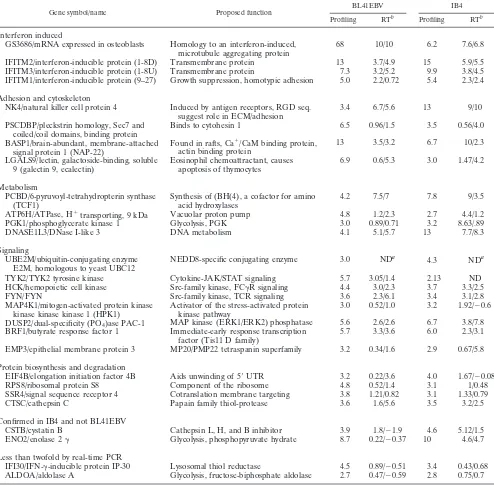
RT-PCR validation of EBV-induced genes.
Expression of
EBV-induced genes was validated by real-time RT-PCR with
p100 RNA as an internal control; p100 RNA abundance was
the same in BL41, BL41EBV, and IB4 by Northern blotting
(data not shown). Titration of either first-strand cDNA or
plasmid DNA for p100 showed that one cycle change
corre-lates with an approximate twofold difference in RNA
abun-dance (data not shown). For most genes the fold change on
profiling correlated with the cycle time change on real-time
RT-PCR, but the magnitude of change by real-time RT-PCR
was generally greater. GS3686, for example, was 68-fold more
abundant in BL41EBV profiling, and the RT-PCR signal
ap-peared 10 cycles earlier with BL41EBV RNA, indicative of a
1,000-fold change. Of 26 genes tested, 13 had replicated
changes of at least one cycle for both BL41EBV and IB4
compared to BL, indicating a more than twofold induction
(Table 1). Eleven others had positive changes in most
experi-ments, confirming EBV induction. ENO2 failed to confirm in
BL41EBV but was 12-fold more abundant in IB4 than in BL
cells. IFI30 and ALDOA were four- and threefold increased by
array analysis but were minimally increased by RT-PCR. One
gene, hUBC12, was incorrectly annotated as HSP1. Real time
RT-PCR failed to show EBV induction of HSP1 (data not
shown), and hUBC12 has not yet been tested.
EBV-induced genes in LCLs versus primary B lymphocytes.
To investigate the relevance of the newly identified
EBV-in-duced gene set to EBV latency III effects in primary
B-lym-phocyte conversion to LCLs, the abundances of seven induced
RNAs were compared by real time RT-PCR using RNAs from
recently established LCLs and from primary B lymphocytes.
GS3686, ENO2, DNASE1L3, TYK2, HCK, FYN, and
MAP4K1(HPK) RNAs were greater than sixfold more
abun-dant in LCLs than in resting B lymphocytes (Table 2). Thus,
most of the newly identified EBV-induced genes are likely to
be relevant to the effects of latency III EBV infection of
pri-mary B lymphocytes.
EBV-induced genes in BL30EBV versus BL30.
Various BL
cell lines differ in their response to EBV latency III infection;
similarities and some differences have been previously noted
between BL41 and BL30 (13, 14). In real-time RT-PCR assays,
GS3686, ENO2, DNASE1L3, and TYK2 were at least 1.7-fold
more abundant in BL30EBV than in BL30, whereas HCK,
FYN, or MAP4K1 were not more abundant in BL30EBV
versus BL30 (Table 2). BL30EBV and BL41EBV cell lines are
similar in EBV latency III protein expression; however, the
EBER status of BL30EBV is unknown (data not shown).
Likely, differences between BL41 and BL30 cells result in
dis-parate effects of EBV latency III on cellular gene expression.
DISCUSSION
EBV infects resting B lymphocytes and growth transforms
them by altering cellular gene expression through the
expres-sion of the essential viral proteins EBNA2, EBNALP,
EBNA3A, EBNA3C, EBNA1, and LMP1 (reviewed in
on November 8, 2019 by guest
http://jvi.asm.org/
ence 59). Since most of the latency III proteins that are
re-quired for LCL outgrowth impact transcriptional pathways,
changes in cell gene transcription are likely to be critical to
EBV effects on cell growth and survival. Indeed, EBV reverse
genetic analyses identify the transcriptional effector domains of
EBNA2 and LMP1 as critical domains for B-lymphocyte
con-version to LCLs (49, 50, 119). EBNA2, EBNA3A, EBNA3B,
and EBNA3C regulate transcription at least in part through
interactions with RBP-J
/CBF1, a transcription factor that
me-diates Notch receptor effects (35, 42, 52, 53, 65, 72, 91, 113,
119, 123). LMP1 is a tumor necrosis factor receptor analogue
which constitutively activates NF-
B, JNK, and p38 signaling
pathways, thereby altering cell gene transcription (27, 28, 30,
33, 36, 40, 45, 50, 60, 66, 81, 83).
[image:5.587.46.540.86.571.2]These experiments are the first broad survey of the potential
effects of latency III EBV infection on 4,146 cDNAs of known
or imputed function. Thus, we were able to identify
EBV-regulated genes and compare the transcriptional profile of
latency III-infected cells to those profiles established for other
B-cell malignancies.
TABLE 1. Assay of EBV-induced genes by real-time RT-PCR
Gene symbol/name Proposed function BL41EBV IB4
Profiling RTb Profiling RTb
Interferon induced
GS3686/mRNA expressed in osteoblasts
Homology to an interferon-induced,
microtubule aggregating protein
68
10/10
6.2
7.6/6.8
IFITM2/interferon-inducible protein (1-8D)
Transmembrane protein
13
3.7/4.9
15
5.9/5.5
IFITM3/interferon-inducible protein (1-8U)
Transmembrane protein
7.3
3.2/5.2
9.9
3.8/4.5
IFITM1/interferon-inducible protein (9–27)
Growth suppression, homotypic adhesion
5.0
2.2/0.72
5.4
2.3/2.4
Adhesion and cytoskeleton
NK4/natural killer cell protein 4
Induced by antigen receptors, RGD seq.
suggest role in ECM/adhesion
3.4
6.7/5.6
13
9/10
PSCDBP/pleckstrin homology, Sec7 and
coiled/coil domains, binding protein
Binds to cytohesin 1
6.5
0.96/1.5
3.5
0.56/4.0
BASP1/brain-abundant, membrane-attached
signal protein 1 (NAP-22)
Found in rafts, Ca
⫹
/CaM binding protein,
actin binding protein
13
3.5/3.2
6.7
10/2.3
LGALS9/lectin, galactoside-binding, soluble
9 (galectin 9, ecalectin)
Eosinophil chemoattractant, causes
apoptosis of thymocytes
6.9
0.6/5.3
3.0
1.47/4.2
Metabolism
PCBD/6-pyruvoyl-tetrahydropterin synthase
(TCF1)
Synthesis of (BH(4), a cofactor for amino
acid hydroxylases
4.2
7.5/7
7.8
9/3.5
ATP6H/ATPase, H
⫹transporting, 9 kDa
Vacuolar proton pump
4.8
1.2/2.3
2.7
4.4/1.2
PGK1/phosphoglycerate kinase 1
Glycolysis, PGK
3.0
0.89/0.71
3.2
8.63/.89
DNASE1L3/DNase I-like 3
DNA metabolism
4.1
5.1/5.7
13
7.7/8.3
Signaling
UBE2M/ubiquitin-conjugating enzyme
E2M, homologous to yeast UBC12
NEDD8-specific conjugating enzyme
3.0
ND
a
4.3
ND
aTYK2/TYK2 tyrosine kinase
Cytokine-JAK/STAT signaling
5.7
3.05/1.4
2.13
ND
HCK/hemopoietic cell kinase
Src-family kinase, FC␥R signaling
4.4
3.0/2.3
3.7
3.3/2.5
FYN/FYN
Src-family kinase, TCR signaling
3.6
2.3/6.1
3.4
3.1/2.8
MAP4K1/mitogen-activated protein kinase
kinase kinase kinase 1 (HPK1)
Activator of the stress-activated protein
kinase pathway
3.0
0.52/1.0
3.2
1.92/⫺0.6
DUSP2/dual-specificity (PO
4)ase PAC-1
MAP kinase (ERK1/ERK2) phosphatase
5.6
2.6/2.6
6.7
3.8/7.8
BRF1/butyrate response factor 1
Immediate-early response transcription
factor (Tis11 D family)
5.7
3.3/3.6
6.0
2.3/3.1
EMP3/epithelial membrane protein 3
MP20/PMP22 tetraspanin superfamily
3.2
0.34/1.6
2.9
0.67/5.8
Protein biosynthesis and degradation
EIF4B/elongation initiation factor 4B
Aids unwinding of 5⬘
UTR
3.2
0.22/3.6
4.0
1.67/⫺0.08
RPS8/ribosomal protein S8
Component of the ribosome
4.8
0.52/1.4
3.1
1/0.48
SSR4/signal sequence receptor 4
Cotranslation membrane targeting
3.8
1.21/0.82
3.1
1.33/0.79
CTSC/cathepsin C
Papain family thiol-protease
3.6
1.6/5.6
3.5
3.2/2.5
Confirmed in IB4 and not BL41EBV
CSTB/cystatin B
Cathepsin L, H, and B inhibitor
3.9
1.8/⫺1.9
4.6
5.12/1.5
ENO2/enolase 2
␥
Glycolysis, phosphopyruvate hydrate
8.7
0.22/⫺0.37
10
4.6/4.7
Less than twofold by real-time PCR
IFI30/IFN-␥-inducible protein IP-30
Lysosomal thiol reductase
4.5
0.89/⫺0.51
3.4
0.43/0.68
ALDOA/aldolase A
Glycolysis, fructose-biphosphate aldolase
2.7
0.47/⫺0.59
2.8
0.75/0.7
aND, not done due to errors in annotation of the RG filters.
bProfiling results are fold changes as in Fig. 1. RT results are the change in cycle number at which a given gene reaches the threshold fluorescence in BL41EBV
or IB4 minus that for BL in two independent experiments. One cycle change is approximately twofold.
on November 8, 2019 by guest
http://jvi.asm.org/
Of the 38 RNAs that are up-regulated by latency III EBV
infection in BL41EBV, 11 were known to be up-regulated by
EBV. Except for

-actin, all are induced by LMP1 through
NF-
B activation (6, 8, 24, 63, 66, 120). Based on the key role
of LMP1 activation of NF-
B in 10 of the 11 previously known
induced genes, many of the 24 newly identified
EBV-induced genes are likely to be up-regulated by LMP1. Indeed,
NK4 and PAC-1 are known to be NF-
B responsive and are
therefore likely to be LMP1 induced (70, 99). Aside from
NF-
B, LMP1 can effect transcription through AP-1 and SP1
activation (60, 104). EBNA2 is also a strong activator of cell
gene expression, and its effects are potentiated by EBNALP
and EBNA3C (38, 72, 85). EBNA2 can up-regulate CD21 and
CD23 with LMP1, and it also up-regulates c-
myc
and c-
fgr
(2,
20, 54, 64, 115, 116).
HCK and FYN, two other Src family tyrosine kinases (89,
98, 125), have now been found to be induced by latency III
EBV infection (Table 1) and may also be EBNA2
up-regu-lated. HCK and FYN are up-regulated fourfold in BL41EBV
and IB4 cells compared to BL41 cells and were induced
ap-proximately eightfold in newly established LCLs relative to
resting B cells. Up-regulation of specific Src family kinases may
affect signal transduction from receptor tyrosine kinases, from
receptors which bind Src family tyrosine kinases through their
SH2 or SH3 domains, and from GPCRs that stimulate G
␣
s
and G
␣
I (1, 9, 96). The Src family kinases can further alter cell
transcription, growth, or survival through multiple signal
cas-cades, including the mitogen-activated protein (MAP) kinase
pathway. We also identified two regulators of the MAP kinase
cascade that are EBV induced. MAP4K1 is expressed at
three-fold higher levels in BL41EBV and in IB4 cells relative to
BL41, and PAC-1 is expressed at sixfold higher levels.
MAP4K1 interacts with BLNK and a novel SLP-76-related
molecule, CLNK/MIST, to couple BCR signaling to NF-
B
activation, interleukin-2 promoter activity, and MAP kinase
activation (44, 58, 118, 121). PAC-1 dephosphorylates ERK1
and ERK2, and this may prevent desensitization as a
conse-quence of continuous signaling from latency III EBV infection
(94, 117).
Four of the identified EBV-induced genes are also IFN
inducible. Latency III EBV infection has been associated with
direct IFN-

induction as well as with cytokine-mediated IFN
induction (5, 55, 105). However, IFN-
␣
1, -

1, and -
␥
1 RNAs
were not up-regulated in BL41EBV. IFN-
␥
alpha receptor and
Tyk2 were up-regulated in BL41EBV and IB4 and could
in-crease IFN signaling. Latency III EBV infection may
up-reg-ulate an IFN that would not have been detected with the
probes for IFN-
␣
1, -

1, or -
␥
1, or the IFN effects may be
augmented by signaling from another EBV-inducible cytokine
and receptor pathway. Cytomegalovirus, another human
her-pesvirus, is known to induce IFN response genes immediately
following infection of cells in the absence of IFN induction
(124).
Five EBV-induced genes, including the IFN-induced gene
GS3686, encode proteins that are involved in cell adhesion or
structure. LMP1 up-regulates expression of vimentin
interme-diate filaments, a p55 actin bundling protein, MARCKS,
LFA-1, LFA-3, and ICAM-1 (10, 13, 47, 101, 106, 107, 114–
116). GS3686, the most highly EBV-induced mRNA, encodes
a putative microtubule aggregating protein (43, 103). PSCDBP
binds to cytoadhesin 1 (61), and BASP1 is an N-terminally
myristolated, Ca
⫹and calmodulin binding protein found in
plasma membrane microdomains. BASP1 colocalizes with
MARCKS, another EBV-induced protein (7), and is also a
PKC substrate (82, 88). Natural killer cell protein 4 may have
a role in cell adhesion (21), whereas galectin 9 has been
im-plicated in eosinophil chemotaxis (77).
Ten other EBV-induced genes are constituents of metabolic
pathways, including protein biosynthesis and degradation,
PCBD/6-pyruvoly-tetrahydropterin synthase, vacuolar ATPase
subunit H, phosphoglycerate kinase 1 (PGK1), enolase 2,
DNase I-like elongation and initiation factor 4B, ribosomal
protein S8, signal sequence receptor 4, cystatin B, and
cathep-sin C (11, 17, 22, 31, 39, 67, 75, 78, 79, 86, 93, 111, 122).
Up-regulation of these RNAs may enhance the ability of
la-tency III-infected cells to rapidly alter location, metabolism, or
proliferation.
Analysis of potential transcriptional regulatory sites near
promoters of these new EBV-induced genes may identify new
signaling pathways impacted by latency III EBV infection.
Pro-grams associated with the Transcription Factor database and
the completed human genomic sequences were used to identify
putative transcription factor binding sites 500 bases upstream
of the Tyk2, PGK1, PCBD, MAP4K1, hUBC12, GS3686, and
ENO2 promoters. All except for Tyk2 have predicted NF-
B
sites and are therefore likely to be LMP1 responsive. The
Tyk2, PGK1, GS3686, and ENO2 promoters have predicted
PU.1 sites that may enable EBNA2 responsiveness. These
pro-moters also have predicted AP-1, AP-2, and SP1 binding sites
that can be affected by LMP1 (28, 104). Thus, most of the
genes identified in this study are likely to be impacted by
pathways already known to be EBV induced.
Of the 10 RNAs that were down-regulated twofold or more
in BL41EBV and IB4, CD10 is known to be down-regulated by
LMP1 (34, 71, 115). CD10 is a neutral endopeptidase that is
down-regulated in Alzheimer’s disease patients. Potentially of
interest is the recent observation that B cells of Alzheimer’s
disease patients transform more rapidly to LCLs following in
vitro infection with EBV than do cells from age-matched
con-trols (87).
[image:6.587.42.283.93.186.2]BL41EBV and IB4 are good model cell lines for the
iden-tification of EBV-induced genes, since EBV has robust effects
in BL41. Furthermore, IB4 cell growth is still dependent on
EBNA2 interactions with RBP-J
(A. Cooper, submitted for
TABLE 2. Quantitation of EBV-induced genes in resting B
lymphocytes, LCLs, BL30, and BL30EBV by real time RT-PCR
Gene symbol Ctin
resting Ba Ctin LCLa ⌬b (BL30EBVAvg⫺⌬BL30)c
GS3686
28.59, 28.18 21.89, 22.75 6.06
8.80
ENO2
29.69, 28.58 24.28, 24.62 4.69
0.83
DNASE1L3 27.51, 28.19 25.05, 25.14 2.75
1.82
TYK2
32.30, 32.36 28.49, 28.50 3.83
0.73
HCK
31.16, 29.74 26.94, 27.13 3.42
0.22
FYN
29.48, 29.48 27.02, 26.14 2.90
⫺0.815
MAP4K1
33.14, 31.47 28.55, 28.36 3.85
⫺0.17
aResults (mean cycle at threshold [C
t]) from two experiments, performed in
triplicate, with two different B-lymphocyte preparations and LCLs, respectively.
bLCL average⫺resting B average.
cAverage of three experiments performed in duplicate.
on November 8, 2019 by guest
http://jvi.asm.org/
publication). In fact, of the genes tested, all showed EBV
induction in the newly established LCLs compared to resting B
cells, validating our model. HCK, FYN, and MAP4K1, was not
confirmed as being EBV induced in the comparison of
BL30EBV to BL30, whereas GS3686, ENO2, DNASE1L3, and
TYK2 were. This likely reflects a difference in the basal gene
expression between BL41 and BL30, possibly due to variations
in accumulated mutations. BL41 and BL30 have c-
myc
trans-locations and p53 mutations; their BCL-6 status is unknown.
In studies of diffuse large B-cell lymphomas, some genes are
abundant in activated antigen receptor-stimulated B cells and
scarce in germinal center (tonsil) B cells. These genes
consti-tute an activated B-cell signature. In contrast, genes that are
expressed at a high level in tonsils and at a low level in
acti-vated B cells constitute a germinal center signature (4). Five
activated and 15 germinal center B-cell signature cDNAs were
detected on the Research Genetics gf211 filters (Fig. 2). Two
of five activation signature genes, CD44 and c-FLIP, were
up-regulated in at least five of six hybridizations with
BL41EBV and IB4 RNAs. Three of five activated signature
genes, c-
myc
, phorbol myristate acetate (PMA)-induced 1, and
dCMP deaminase, were unaffected by EBV infection. c-
myc
is
constitutively expressed at a high level in BL cells and would
not be expected to change in these studies (110). PMA-induced
1 and dCMP deaminase are transiently induced by antigen
receptor signaling, with expression returning to resting levels at
48 h (4). Thus, the effect of EBV infection on activated and
germinal center genes is difficult to predict.
EBV did not consistently up-regulate any germinal center
signature gene. Rather, the only significant effect of EBV
in-fection on germinal center signature genes was
down-regula-tion. RGS13, Spi-B, and CD10 were expressed at a low level in
BL41EBV and in IB4 cells, consistent with active inhibition by
latency III EBV infection. BCL-6 was less abundant in LCLs
compared to BL, as was expected since LMP1 expression can
down-regulate BCL-6 (15, 16). However, BCL-6 levels were
unaffected in BL41EBV, probably due to BL-associated
mu-tations in the BCL-6 promoter (32). Overall, latency III
EBV-infected BL41EBV and IB4 cells have increased expression of
the activation markers CD44 and c-FLIP and decreased
ex-pression of the germinal center markers RGS13, Spi-B, and
CD10 (Fig. 2).
In this study, we have focused on genes that are robustly
induced and highly expressed in latency III EBV-infected cells.
Relaxation of these criteria to include genes that are
up-reg-ulated twofold in more than half of the repeated hybridizations
expands the set of EBV-induced genes to 80 cDNAs, which
include almost all previously identified EBV-induced genes.
Further studies are necessary to characterize gene expression
in EBV-associated malignancies that express latency I or
la-tency II. These future studies, and our present one, may enable
the identification of EBV signatures associated with different
types of latency that can lead to therapeutic targets for the
treatment of EBV-associated diseases.
ACKNOWLEDGMENTS
K. L. Carter and E. Cahir-McFarland contributed equally to this
work.
We thank Peter Munson for providing the PSCAN 1.1 software and
for assisting us in its use; Emily Klotz for providing guidance with
protocols; Danielle Rizzo for technical help; and Jena Giltnane, Louis
Staudt, and the Kieff lab members for helpful discussions.
K.L.C., E.C.-M, and E.K. were supported by Public Health Service
grants CA47006, CA85180, CA87661, CA75646, and CA76727.
E.C.-M is a Special Fellow of the Leukemia and Lymphoma Society.
K. L. Carter and E. Cahir-McFarland contributed equally to this
work.
REFERENCES
1.Abu-Ghazaleh, R., J. Kabir, H. Jia, M. Lobo, and I. Zachary.2001. Src mediates stimulation by vascular endothelial growth factor of the phosphor-ylation of focal adhesion kinase at tyrosine 861, and migration and anti-apoptosis in endothelial cells. Biochem. J.360:255–264.
2.Alfieri, C., M. Birkenbach, and E. Kieff.1991. Early events in Epstein-Barr virus infection of human B lymphocytes. Virology181:595–608. 3.Alizadeh, A., M. Eisen, R. E. Davis, C. Ma, H. Sabet, T. Tran, J. I. Powell,
L. Yang, G. E. Marti, D. T. Moore, J. R. Hudson, Jr., W. C. Chan, T. Greiner, D. Weisenburger, J. O. Armitage, I. Lossos, R. Levy, D. Botstein, P. O. Brown, and L. M. Staudt.1999. The lymphochip: a specialized cDNA microarray for the genomic-scale analysis of gene expression in normal and malignant lymphocytes. Cold Spring Harbor Symp. Quant. Biol.64:71–78. 4.Alizadeh, A. A., M. B. Eisen, R. E. Davis, C. Ma, I. S. Lossos, A. Rosenwald, J. C. Boldrick, H. Sabet, T. Tran, X. Yu, J. I. Powell, L. Yang, G. E. Marti, T. Moore, J. Hudson, Jr., L. Lu, D. B. Lewis, R. Tibshirani, G. Sherlock, W. C. Chan, T. C. Greiner, D. D. Weisenburger, J. O. Armitage, R. Warnke, L. M. Staudt, et al.2000. Distinct types of diffuse large B-cell lymphoma identified by gene expression profiling. Nature403:503–511.
5.Andersson, U., O. Martinez-Maza, J. Andersson, S. Britton, H. Gadler, M. De Ley, and S. Modrow.1984. Secretion of gamma-interferon at the cellular level. Induction by Epstein-Barr virus. Scand. J. Immunol.20:425–432. 6.Baldwin, A. S., Jr., and P. A. Sharp.1987. Binding of a nuclear factor to a
regulatory sequence in the promoter of the mouse H-2Kb class I major histocompatibility gene. Mol. Cell. Biol.7:305–313.
7.Birkenbach, M., K. Josefsen, R. Yalamanchili, G. Lenoir, and E. Kieff.
1993. Epstein-Barr virus-induced genes: first lymphocyte-specific G protein-coupled peptide receptors. J. Virol.67:2209–2220.
8.Birkenbach, M., D. Liebowitz, F. Wang, J. Sample, and E. Kieff.1989. Epstein-Barr virus latent infection membrane protein increases vimentin expression in human B-cell lines. J. Virol.63:4079–4084.
9.Bisotto, S., and E. D. Fixman.2001. Src-family tyrosine kinases, phospho-inositide 3-kinase and Gab1 regulate extracellular signal-regulated kinase 1 activation induced by the type A endothelin-1 G-protein-coupled receptor. Biochem. J.360:77–85.
[image:7.587.70.255.69.288.2]10.Bonnefoy, J. Y., T. Defrance, C. Peronne, C. Menetrier, F. Rousset, J. Pene, J. E. De Vries, and J. Banchereau.1988. Human recombinant interleukin 4 induces normal B cells to produce soluble CD23/IgE-binding factor anal-ogous to that spontaneously released by lymphoblastoid B cell lines. Eur. J. Immunol.18:117–122.
FIG. 2. Effect of latency III EBV infection on activated and
ger-minal center signature genes. Fold changes are displayed as described
in the legend for Fig. 1.
on November 8, 2019 by guest
http://jvi.asm.org/
11.Brenner, V., G. Nyakatura, A. Rosenthal, and M. Platzer.1997. Genomic organization of two novel genes on human Xq28: compact head to head arrangement of IDH gamma and TRAP delta is conserved in rat and mouse. Genomics44:8–14.
12.Caldwell, R. G., J. B. Wilson, S. J. Anderson, and R. Longnecker.1998. Epstein-Barr virus LMP2A drives B cell development and survival in the absence of normal B cell receptor signals. Immunity9:405–411. 13.Calender, A., M. Billaud, J. P. Aubry, J. Banchereau, M. Vuillaume, and
G. M. Lenoir.1987. Epstein-Barr virus (EBV) induces expression of B-cell activation markers on in vitro infection of EBV-negative B-lymphoma cells. Proc. Natl. Acad. Sci. USA84:8060–8064.
14.Calender, A., M. Cordier, M. Billaud, and G. M. Lenoir.1990. Modulation of cellular gene expression in B lymphoma cells following in vitro infection by Epstein-Barr virus (EBV). Int. J. Cancer46:658–663.
15.Carbone, A., G. Gaidano, A. Gloghini, L. M. Larocca, D. Capello, V. Can-zonieri, A. Antinori, U. Tirelli, B. Falini, and R. Dalla-Favera.1998. Dif-ferential expression of BCL-6, CD138/syndecan-1, and Epstein-Barr virus-encoded latent membrane protein-1 identifies distinct histogenetic subsets of acquired immunodeficiency syndrome-related non-Hodgkin’s lympho-mas. Blood91:747–755.
16.Carbone, A., G. Gaidano, A. Gloghini, C. Pastore, G. Saglio, U. Tirelli, R. Dalla-Favera, and B. Falini.1997. BCL-6 protein expression in AIDS-related non-Hodgkin’s lymphomas: inverse relationship with Epstein-Barr virus-encoded latent membrane protein-1 expression. Am. J. Pathol.150:
155–165.
17.Citron, B. A., S. Kaufman, S. Milstien, E. W. Naylor, C. L. Greene, and M. D. Davis.1993. Mutation in the 4a-carbinolamine dehydratase gene leads to mild hyperphenylalaninemia with defective cofactor metabolism. Am. J. Hum. Genet.53:768–774.
18.Cohen, J. H., E. Fischer, M. D. Kazatchkine, G. M. Lenoir, C. Lefevre-Delvincourt, and J. P. Revillard.1987. Expression of CR1 and CR2 com-plement receptors following Epstein-Barr virus infection of Burkitt’s lym-phoma cell lines. Scand. J. Immunol.25:587–598.
19.Cohen, J. I., F. Wang, J. Mannick, and E. Kieff.1989. Epstein-Barr virus nuclear protein 2 is a key determinant of lymphocyte transformation. Proc. Natl. Acad. Sci. USA86:9558–9562.
20.Cordier, M., A. Calender, M. Billaud, U. Zimber, G. Rousselet, O. Pavlish, J. Banchereau, T. Tursz, G. Bornkamm, and G. M. Lenoir.1990. Stable transfection of Epstein-Barr virus (EBV) nuclear antigen 2 in lymphoma cells containing the EBV P3HR1 genome induces expression of B-cell activation molecules CD21 and CD23. J. Virol.64:1002–1013.
21.Dahl, C. A., R. P. Schall, H. L. He, and J. S. Cairns.1992. Identification of a novel gene expressed in activated natural killer cells and T cells. J. Im-munol.148:597–603.
22.Davies, B., and M. Fried.1993. The structure of the human intron-contain-ing S8 ribosomal protein gene and determination of its chromosomal loca-tion at 1p32-p34.1. Genomics15:68–75.
23.Delecluse, H. J., E. Kremmer, J. P. Rouault, C. Cour, G. Bornkamm, and F. Berger.1995. The expression of Epstein-Barr virus latent proteins is related to the pathological features of post-transplant lymphoproliferative disorders. Am. J. Pathol.146:1113–1120.
24.Devergne, O., E. Cahir-McFarland, G. Mosialos, K. M. Izumi, C. F. Ware, and E. Kieff.1998. Role of the TRAF binding site and NF-B activation in Epstein-Barr virus latent membrane protein 1-induced cell gene expression. J. Virol.72:7900–7908.
25.Ehlin-Henriksson, B., A. Manneborg-Sandlund, and G. Klein.1987. Ex-pression of B-cell-specific markers in different Burkitt lymphoma sub-groups. Int. J. Cancer39:211–218.
26.Eisen, M. B., P. T. Spellman, P. O. Brown, and D. Botstein.1998. Cluster analysis and display of genome-wide expression patterns. Proc. Natl. Acad. Sci. USA95:14863–14868.
27.Eliopoulos, A. G., N. J. Gallagher, S. M. Blake, C. W. Dawson, and L. S. Young.1999. Activation of the p38 mitogen-activated protein kinase path-way by Epstein-Barr virus-encoded latent membrane protein 1 coregulates interleukin-6 and interleukin-8 production. J. Biol. Chem.
274:16085–16096.
28.Eliopoulos, A. G., and L. S. Young.1998. Activation of the cJun N-terminal kinase (JNK) pathway by the Epstein-Barr virus-encoded latent membrane protein 1 (LMP1). Oncogene16:1731–1742.
28a.Farrell, P. J., G. J. Allan, F. Shanahan, K. H. Vousden, and T. Crook.1991. p53 is frequently mutated in Burkitt’s lymphoma cell lines. EMBO J.10:
2879–2887.
29.Favrot, M. C., O. Maritaz, T. Suzuki, M. Cooper, I. Philip, T. Philip, and G. Lenoir.1986. EBV-negative and -positive Burkitt cell lines variably express receptors for B-cell activation and differentiation. Int. J. Cancer
38:901–906.
30.Floettmann, J. E., and M. Rowe.1997. Epstein-Barr virus latent membrane protein-1 (LMP1) C-terminus activation region 2 (CTAR2) maps to the far C terminus and requires oligomerisation for NF-B activation. Oncogene
15:1851–1858.
31.Freemont, P. S., B. Dunbar, and L. A. Fothergill-Gilmore.1988. The
com-plete amino acid sequence of human skeletal-muscle fructose-bisphosphate aldolase. Biochem. J.249:779–788.
32.Gaidano, G., A. Carbone, and R. Dalla-Favera.1998. Genetic basis of acquired immunodeficiency syndrome-related lymphomagenesis. J. Natl. Cancer Inst. Monogr.23:95–100.
33.Gires, O., U. Zimber-Strobl, R. Gonnella, M. Ueffing, G. Marschall, R. Zeidler, D. Pich, and W. Hammerschmidt.1997. Latent membrane protein 1 of Epstein-Barr virus mimics a constitutively active receptor molecule. EMBO J.16:6131–6140.
34.Gregory, C. D., C. F. Edwards, A. Milner, J. Wiels, M. Lipinski, M. Rowe, T. Tursz, and A. B. Rickinson.1988. Isolation of a normal B cell subset with a Burkitt-like phenotype and transformation in vitro with Epstein-Barr virus. Int. J. Cancer42:213–220.
35.Grossman, S. R., E. Johannsen, X. Tong, R. Yalamanchili, and E. Kieff.
1994. The Epstein-Barr virus nuclear antigen 2 transactivator is directed to response elements by the Jrecombination signal binding protein. Proc. Natl. Acad. Sci. USA91:7568–7572.
36.Hammarskjold, M. L., and M. C. Simurda.1992. Epstein-Barr virus latent membrane protein transactivates the human immunodeficiency virus type 1 long terminal repeat through induction of NF-B activity. J. Virol.66:6496– 6501.
37.Hammerschmidt, W., and B. Sugden.1989. Genetic analysis of immortal-izing functions of Epstein-Barr virus in human B lymphocytes. Nature
340:393–397.
38.Harada, S., and E. Kieff. 1997. Epstein-Barr virus nuclear protein LP stimulates EBNA-2 acidic domain-mediated transcriptional activation. J. Virol.71:6611–6618.
39.Hart, T. C., P. S. Hart, D. W. Bowden, M. D. Michalec, S. A. Callison, S. J. Walker, Y. Zhang, and E. Firatli.1999. Mutations of the cathepsin C gene are responsible for Papillon-Lefevre syndrome. J. Med. Genet.36:881–887. 40.Hatzivassiliou, E., W. E. Miller, N. Raab-Traub, E. Kieff, and G. Mosialos.
1998. A fusion of the EBV latent membrane protein-1 (LMP1) transmem-brane domains to the CD40 cytoplasmic domain is similar to LMP1 in constitutive activation of epidermal growth factor receptor expression, nu-clear factor-kappa B, and stress-activated protein kinase. J. Immunol.160:
1116–1121.
41.Henderson, A., S. Ripley, M. Heller, and E. Kieff.1983. Chromosome site for Epstein-Barr virus DNA in a Burkitt tumor cell line and in lymphocytes growth-transformed in vitro. Proc. Natl. Acad. Sci. USA80:1987–1991. 42.Henkel, T., P. D. Ling, S. D. Hayward, and M. G. Peterson.1994. Mediation
of Epstein-Barr virus EBNA2 transactivation by recombination signal-bind-ing protein J. Science265:92–95.
43.Honda, Y., J. Kondo, T. Maeda, Y. Yoshiyama, E. Yamada, Y. K. Shimizu, T. Shikata, and Y. Ono.1990. Isolation and purification of a non-A, non-B hepatitis-associated microtubular aggregates protein. J. Gen. Virol.71:
1999–2004.
44.Hu, M. C., W. R. Qiu, X. Wang, C. F. Meyer, and T. H. Tan.1996. Human HPK1, a novel human hematopoietic progenitor kinase that activates the JNK/SAPK kinase cascade. Genes Dev.10:2251–2264.
45.Huen, D. S., S. A. Henderson, D. Croom-Carter, and M. Rowe.1995. The Epstein-Barr virus latent membrane protein-1 (LMP1) mediates activation of NF-B and cell surface phenotype via two effector regions in its carboxy-terminal cytoplasmic domain. Oncogene10:549–560.
46.Hurley, E. A., L. D. Klaman, S. Agger, J. B. Lawrence, and D. A. Thorley-Lawson.1991. The prototypical Epstein-Barr virus-transformed lympho-blastoid cell line IB4 is an unusual variant containing integrated but no episomal viral DNA. J. Virol.65:3958–3963.
47.Hurley, E. A., and D. A. Thorley-Lawson.1988. B cell activation and the establishment of Epstein-Barr virus latency. J. Exp. Med.168:2059–2075. 48.Iyer, V. R., M. B. Eisen, D. T. Ross, G. Schuler, T. Moore, J. C. Lee, J. M.
Trent, L. M. Staudt, J. Hudson, Jr., M. S. Boguski, D. Lashkari, D. Shalon, D. Botstein, and P. O. Brown.1999. The transcriptional program in the response of human fibroblasts to serum. Science283:83–87.
49.Izumi, K. M., K. M. Kaye, and E. D. Kieff.1997. The Epstein-Barr virus LMP1 amino acid sequence that engages tumor necrosis factor receptor associated factors is critical for primary B lymphocyte growth transforma-tion. Proc. Natl. Acad. Sci. USA94:1447–1452.
50.Izumi, K. M., and E. D. Kieff.1997. The Epstein-Barr virus oncogene product latent membrane protein 1 engages the tumor necrosis factor receptor-associated death domain protein to mediate B lymphocyte growth transformation and activate NF-B. Proc. Natl. Acad. Sci. USA94:12592– 12597.
51.Johannes, G., M. S. Carter, M. B. Eisen, P. O. Brown, and P. Sarnow.1999. Identification of eukaryotic mRNAs that are translated at reduced cap binding complex eIF4F concentrations using a cDNA microarray. Proc. Natl. Acad. Sci. USA96:13118–13123.
52.Johannsen, E., E. Koh, G. Mosialos, X. Tong, E. Kieff, and S. R. Grossman.
1995. Epstein-Barr virus nuclear protein 2 transactivation of the latent membrane protein 1 promoter is mediated by Jand PU.1. J. Virol.69:
253–262.
53.Johannsen, E., C. L. Miller, S. R. Grossman, and E. Kieff.1996. EBNA-2
on November 8, 2019 by guest
http://jvi.asm.org/
and EBNA-3C extensively and mutually exclusively associate with RBPJin Epstein-Barr virus-transformed B lymphocytes. J. Virol.70:4179–4183. 54.Kaiser, C., G. Laux, D. Eick, N. Jochner, G. W. Bornkamm, and B.
Kemp-kes.1999. The proto-oncogene c-mycis a direct target gene of Epstein-Barr virus nuclear antigen 2. J. Virol.73:4481–4484.
55.Kanda, K., B. Kempkes, G. W. Bornkamm, A. von Gabain, and T. Decker.
1999. The Epstein-Barr virus nuclear antigen 2 (EBNA2), a protein re-quired for B lymphocyte immortalization, induces the synthesis of type I interferon in Burkitt’s lymphoma cell lines. Biol. Chem.380:213–221. 56.Kaye, K. M., K. M. Izumi, and E. Kieff.1993. Epstein-Barr virus latent
membrane protein 1 is essential for B-lymphocyte growth transformation. Proc. Natl. Acad. Sci. USA90:9150–9154.
57.Khan, J., M. L. Bittner, L. H. Saal, U. Teichmann, D. O. Azorsa, G. C. Gooden, W. J. Pavan, J. M. Trent, and P. S. Meltzer.1999. cDNA microar-rays detect activation of a myogenic transcription program by the PAX3-FKHR fusion oncogene. Proc. Natl. Acad. Sci. USA96:13264–13269. 58.Kiefer, F., L. A. Tibbles, M. Anafi, A. Janssen, B. W. Zanke, N. Lassam, T.
Pawson, J. R. Woodgett, and N. N. Iscove.1996. HPK1, a hematopoietic protein kinase activating the SAPK/JNK pathway. EMBO J.15:7013–7025. 59.Kieff, E., and A. B. Rickinson.2001. Epstein-Barr virus and its replication, p. 2511–2574.InD. Knipe, P. Howley, D. Griffin, M. Martin, R. Lamb, B. Roizman, and S. Straus (ed.), Fields virology, 4th ed., vol. 2. Lippincott-Raven, Philadelphia, Pa.
60.Kieser, A., E. Kilger, O. Gires, M. Ueffing, W. Kolch, and W. Hammer-schmidt.1997. Epstein-Barr virus latent membrane protein-1 triggers AP-1 activity via the c-Jun N-terminal kinase cascade. EMBO J.16:6478–6485. 61.Kim, H. S. 1999. Assignment of the human B3–1 gene (PSCDBP) to
chromosome 2 band q11.2 by radiation hybrid mapping. Cytogenet. Cell Genet.84:95.
62.King, W., A. L. Thomas-Powell, N. Raab-Traub, M. Hawke, and E. Kieff.
1980. Epstein-Barr virus RNA. V. Viral RNA in a restringently infected, growth-transformed cell line. J. Virol.36:506–518.
63.Klaman, L. D., and D. A. Thorley-Lawson.1995. Characterization of the CD48 gene demonstrates a positive element that is specific to Epstein-Barr virus-immortalized B-cell lines and contains an essential NF-B site. J. Vi-rol.69:871–881.
64.Knutson, J. C.1990. The level of c-fgrRNA is increased by EBNA-2, an Epstein-Barr virus gene required for B-cell immortalization. J. Virol.64:
2530–2536.
65.Krauer, K. G., N. Kienzle, D. B. Young, and T. B. Sculley.1996. Epstein-Barr nuclear antigen-3 and -4 interact with RBP-2N, a major isoform of RBP-Jin B lymphocytes. Virology226:346–353.
66.Laherty, C. D., H. M. Hu, A. W. Opipari, F. Wang, and V. M. Dixit.1992. The Epstein-Barr virus LMP1 gene product induces A20 zinc finger protein expression by activating nuclear factor kappa B. J. Biol. Chem.267:24157– 24160.
67.Lay, A. J., X. M. Jiang, O. Kisker, E. Flynn, A. Underwood, R. Condron, and P. J. Hogg.2000. Phosphoglycerate kinase acts in tumour angiogenesis as a disulphide reductase. Nature408:869–873.
68.Lee, M. A., M. E. Diamond, and J. L. Yates.1999. Genetic evidence that EBNA-1 is needed for efficient, stable latent infection by Epstein-Barr virus. J. Virol.73:2974–2982.
69.Lewin, A. R., L. E. Reid, M. McMahon, G. R. Stark, and I. M. Kerr.1991. Molecular analysis of a human interferon-inducible gene family. Eur. J. Biochem.199:417–423.
70.Li, J., G. W. Peet, D. Balzarano, X. Li, P. Massa, R. W. Barton, and K. B. Marcu.2001. Novel NEMO/IB kinase and NF-B target genes at the pre-B to immature B cell transition. J. Biol. Chem.276:18579–18590. 71.Liebowitz, D., J. Mannick, K. Takada, and E. Kieff.1992. Phenotypes of
Epstein-Barr virus LMP1 deletion mutants indicate transmembrane and amino-terminal cytoplasmic domains necessary for effects in B-lymphoma cells. J. Virol.66:4612–4616.
72.Lin, J., E. Johannsen, E. Robertson, and E. Kieff.2002. Epstein-Barr virus nuclear antigen 3C putative repression domain mediates coactivation of the LMP1 promoter with EBNA-2. J. Virol.76:232–242.
73.Longnecker, R., C. L. Miller, X. Q. Miao, B. Tomkinson, and E. Kieff.1993. The last seven transmembrane and carboxy-terminal cytoplasmic domains of Epstein-Barr virus latent membrane protein 2 (LMP2) are dispensable for lymphocyte infection and growth transformation in vitro. J. Virol.67:
2006–2013.
74.Lu, Y. J., D. Williamson, J. Clark, R. Wang, N. Tiffin, L. Skelton, T. Gordon, R. Williams, B. Allan, A. Jackman, C. Cooper, K. Pritchard-Jones, and J. Shipley.2001. Comparative expressed sequence hybridization to chromosomes for tumor classification and identification of genomic regions of differential gene expression. Proc. Natl. Acad. Sci. USA98:9197–9202. 75.Ludwig, J., S. Kerscher, U. Brandt, K. Pfeiffer, F. Getlawi, D. K. Apps, and H. Schagger.1998. Identification and characterization of a novel 9.2-kDa membrane sector-associated protein of vacuolar proton-ATPase from chro-maffin granules. J. Biol. Chem.273:10939–10947.
76.Mannick, J. B., J. I. Cohen, M. Birkenbach, A. Marchini, and E. Kieff.1991. The Epstein-Barr virus nuclear protein encoded by the leader of the EBNA RNAs is important in B-lymphocyte transformation. J. Virol.65:6826–6837.
77.Matsumoto, R., H. Matsumoto, M. Seki, M. Hata, Y. Asano, S. Kanegasaki, R. L. Stevens, and M. Hirashima.1998. Human ecalectin, a variant of human galectin-9, is a novel eosinophil chemoattractant produced by T lymphocytes. J. Biol. Chem.273:16976–16984.
78.Mendel, D. B., P. A. Khavari, P. B. Conley, M. K. Graves, L. P. Hansen, A. Admon, and G. R. Crabtree. 1991. Characterization of a cofactor that regulates dimerization of a mammalian homeodomain protein. Science
254:1762–1767.
79.Milburn, S. C., J. W. Hershey, M. V. Davies, K. Kelleher, and R. J. Kauf-man.1990. Cloning and expression of eukaryotic initiation factor 4B cDNA: sequence determination identifies a common RNA recognition motif. EMBO J.9:2783–2790.
80.Miller, C. L., A. L. Burkhardt, J. H. Lee, B. Stealey, R. Longnecker, J. B. Bolen, and E. Kieff.1995. Integral membrane protein 2 of Epstein-Barr virus regulates reactivation from latency through dominant negative effects on protein-tyrosine kinases. Immunity2:155–166.
81.Mitchell, T., and B. Sugden.1995. Stimulation of NF-B-mediated tran-scription by mutant derivatives of the latent membrane protein of Epstein-Barr virus. J. Virol.69:2968–2976.
82.Mosevitsky, M. I., J. P. Capony, G. Skladchikova, V. A. Novitskaya, A. Plekhanov, and V. V. Zakharov.1997. The BASP1 family of myristoylated proteins abundant in axonal termini. Primary structure analysis and physico-chemical properties. Biochimie79:373–384.
83.Mosialos, G., M. Birkenbach, R. Yalamanchili, T. VanArsdale, C. Ware, and E. Kieff.1995. The Epstein-Barr virus transforming protein LMP1 engages signaling proteins for the tumor necrosis factor receptor family. Cell80:389–399.
84.Muslin, A. J., J. W. Tanner, P. M. Allen, and A. S. Shaw.1996. Interaction of 14–3–3 with signaling proteins is mediated by the recognition of phos-phoserine. Cell84:889–897.
85.Nitsche, F., A. Bell, and A. Rickinson. 1997. Epstein-Barr virus leader protein enhances EBNA-2-mediated transactivation of latent membrane protein 1 expression: a role for the W1W2 repeat domain. J. Virol.71:
6619–6628.
86.Oliva, D., L. Cali, S. Feo, and A. Giallongo.1991. Complete structure of the human gene encoding neuron-specific enolase. Genomics10:157–165. 87.Ounanian, A., B. Guilbert, and J. M. Seigneurin.1992. Characteristics of
Epstein-Barr virus transformed B cell lines from patients with Alzheimer’s disease and age-matched controls. Mech. Ageing Dev.63:105–116. 88.Park, S., Y. I. Kim, B. Kim, C. Seong, Y. Oh, K. Baek, and J. Yoon.1998.
Characterization of bovine and human cDNAs encoding NAP-22 (22 kDa neuronal tissue-enriched acidic protein) homologs. Mol. Cells8:471–477. 89.Quintrell, N., R. Lebo, H. Varmus, J. M. Bishop, M. J. Pettenati, M. M. Le
Beau, M. O. Diaz, and J. D. Rowley.1987. Identification of a human gene (HCK) that encodes a protein-tyrosine kinase and is expressed in hemo-poietic cells. Mol. Cell. Biol.7:2267–2275.
90.Rickinson, A., and E. Kieff.2001. Epstein-Barr virus, p. 2575–2628.InD. Knipe, P. Howley, D. Griffin, M. Martin, R. Lamb, B. Roizman, and S. Straus (ed.), Fields virology, 4th ed., vol. 2. Lippincott Williams and Wilkins, Philadelphia, Pa.
91.Robertson, E. S., S. Grossman, E. Johannsen, C. Miller, J. Lin, B. Tomkin-son, and E. Kieff.1995. Epstein-Barr virus nuclear protein 3C modulates transcription through interaction with the sequence-specific DNA-binding protein J. J. Virol.69:3108–3116.
92.Robertson, E. S., B. Tomkinson, and E. Kieff.1994. An Epstein-Barr virus with a 58-kilobase-pair deletion that includes BARF0 transforms B lym-phocytes in vitro. J. Virol.68:1449–1458.
93.Rodriguez, A. M., D. Rodin, H. Nomura, C. C. Morton, S. Weremowicz, and M. C. Schneider.1997. Identification, localization, and expression of two novel human genes similar to deoxyribonuclease I. Genomics42:507–513. 94.Rohan, P. J., P. Davis, C. A. Moskaluk, M. Kearns, H. Krutzsch, U. Siebenlist, and K. Kelly.1993. PAC-1: a mitogen-induced nuclear protein tyrosine phosphatase. Science259:1763–1766.
95.Rowe, M., A. L. Lear, D. Croom-Carter, A. H. Davies, and A. B. Rickinson.
1992. Three pathways of Epstein-Barr virus gene activation from EBNA1-positive latency in B lymphocytes. J. Virol.66:122–131.
96.Sasaki, A., H. Yasukawa, A. Suzuki, S. Kamizono, T. Syoda, I. Kinjyo, M. Sasaki, J. A. Johnston, and A. Yoshimura.1999. Cytokine-inducible SH2 protein-3 (CIS3/SOCS3) inhibits Janus tyrosine kinase by binding through the N-terminal kinase inhibitory region as well as SH2 domain. Genes Cells
4:339–351.
97.Schena, M., D. Shalon, R. W. Davis, and P. O. Brown.1995. Quantitative monitoring of gene expression patterns with a complementary DNA mi-croarray. Science270:467–470.
98.Semba, K., M. Nishizawa, N. Miyajima, M. C. Yoshida, J. Sukegawa, Y. Yamanashi, M. Sasaki, T. Yamamoto, and K. Toyoshima.1986. yes-related protooncogene, syn, belongs to the protein-tyrosine kinase family. Proc. Natl. Acad. Sci. USA83:5459–5463.
99.Shaffer, A. L., A. Rosenwald, E. M. Hurt, J. M. Giltnane, L. T. Lam, O. K. Pickeral, and L. M. Staudt. 2001. Signatures of the immune response. Immunity15:375–385.
100.Spellman, P. T., G. Sherlock, M. Q. Zhang, V. R. Iyer, K. Anders, M. B.
on November 8, 2019 by guest
http://jvi.asm.org/
Eisen, P. O. Brown, D. Botstein, and B. Futcher.1998. Comprehensive identification of cell cycle-regulated genes of the yeast Saccharomyces cer-evisiae by microarray hybridization. Mol. Biol. Cell9:3273–3297. 101.Sugden, B., and S. Metzenberg.1983. Characterization of an antigen whose
cell surface expression is induced by infection with Epstein-Barr virus. J. Virol.46:800–807.
102.Swaminathan, S., B. Tomkinson, and E. Kieff.1991. Recombinant Epstein-Barr virus with small RNA (EBER) genes deleted transforms lymphocytes and replicates in vitro. Proc. Natl. Acad. Sci. USA88:1546–1550. 103.Takahashi, K., N. Kitamura, T. Shibui, M. Kamizono, R. Matsui, Y.
Yo-shiyama, T. Maeda, J. Kondo, Y. Honda, E. Yamada, et al.1990. Cloning, sequencing and expression in Escherichia coli of cDNA for a non-A, non-B hepatitis-associated microtubular aggregates protein. J. Gen. Virol.71:
2005–2011.
104.Takeshita, H., T. Yoshizaki, W. E. Miller, H. Sato, M. Furukawa, J. S. Pagano, and N. Raab-Traub.1999. Matrix metalloproteinase 9 expression is induced by Epstein-Barr virus latent membrane protein 1 C-terminal activation regions 1 and 2. J. Virol.73:5548–5555.
105.Tang, H., T. Matthes, N. Carballido-Perrig, R. H. Zubler, and V. Kindler.
1993. Differential induction of T cell cytokine mRNA in Epstein-Barr virus-transformed B cell clones: constitutive and inducible expression of interleukin-4 mRNA. Eur. J. Immunol.23:899–903.
106.Thorley-Lawson, D. A., L. M. Nadler, A. K. Bhan, and R. T. Schooley.1985. BLAST-2 [EBVCS], an early cell surface marker of human B cell activation, is superinduced by Epstein Barr virus. J. Immunol.134:3007–3012. 107.Thorley-Lawson, D. A., R. T. Schooley, A. K. Bhan, and L. M. Nadler.1982.
Epstein-Barr virus superinduces a new human B cell differentiation antigen (B-LAST 1) expressed on transformed lymphoblasts. Cell30:415–425. 108.Tomkinson, B., and E. Kieff.1992. Use of second-site homologous
recom-bination to demonstrate that Epstein-Barr virus nuclear protein 3B is not important for lymphocyte infection or growth transformation in vitro. J. Vi-rol.66:2893–2903.
109.Tomkinson, B., E. Robertson, and E. Kieff.1993. Epstein-Barr virus nuclear proteins EBNA-3A and EBNA-3C are essential for B-lymphocyte growth transformation. J. Virol.67:2014–2025.
110.Torsteinsdottir, S., M. L. Andersson, J. Avila-Carino, B. Ehlin-Henriksson, M. G. Masucci, G. Klein, and E. Klein.1989. Reversion of tumorigenicity and decreased agarose clonability after EBV conversion of an IgH/myc translocation-carrying BL line. Int. J. Cancer.43:273–278.
111.Turk, V., and W. Bode.1991. The cystatins: protein inhibitors of cysteine proteinases. FEBS Lett.285:213–219.
112.Velazquez, L., M. Fellous, G. R. Stark, and S. Pellegrini.1992. A protein tyrosine kinase in the interferon alpha/beta signaling pathway. Cell70:313– 322.
113.Waltzer, L., F. Logeat, C. Brou, A. Israel, A. Sergeant, and E. Manet.1994. The human Jrecombination signal sequence binding protein (RBP-J)
targets the Epstein-Barr virus EBNA2 protein to its DNA responsive ele-ments. EMBO J.13:5633–5638.
114.Wang, D., D. Liebowitz, F. Wang, C. Gregory, A. Rickinson, R. Larson, T. Springer, and E. Kieff.1988. Epstein-Barr virus latent infection membrane protein alters the human B-lymphocyte phenotype: deletion of the amino terminus abolishes activity. J. Virol.62:4173–4184.
115.Wang, F., C. Gregory, C. Sample, M. Rowe, D. Liebowitz, R. Murray, A. Rickinson, and E. Kieff.1990. Epstein-Barr virus latent membrane protein (LMP1) and nuclear proteins 2 and 3C are effectors of phenotypic changes in B lymphocytes: EBNA-2 and LMP1 cooperatively induce CD23. J. Virol.
64:2309–2318.
116.Wang, F., C. D. Gregory, M. Rowe, A. B. Rickinson, D. Wang, M. Birken-bach, H. Kikutani, T. Kishimoto, and E. Kieff.1987. Epstein-Barr virus nuclear antigen 2 specifically induces expression of the B-cell activation antigen CD23. Proc. Natl. Acad. Sci. USA84:3452–3456.
117.Ward, Y., S. Gupta, P. Jensen, M. Wartmann, R. J. Davis, and K. Kelly.
1994. Control of MAP kinase activation by the mitogen-induced threonine/ tyrosine phosphatase PAC1. Nature367:651–654.
118.Yablonski, D., and A. Weiss.2001. Mechanisms of signaling by the hema-topoietic-specific adaptor proteins, SLP-76 and LAT and their B cell coun-terpart, BLNK/SLP-65. Adv. Immunol.79:93–128.
119.Yalamanchili, R., X. Tong, S. Grossman, E. Johannsen, G. Mosialos, and E. Kieff.1994. Genetic and biochemical evidence that EBNA 2 interaction with a 63-kDa cellular GTG-binding protein is essential for B lymphocyte growth transformation by EBV. Virology204:634–641.
120.Yokoyama, S., D. Staunton, R. Fisher, M. Amiot, J. J. Fortin, and L. D. Thorley.1991. Expression of the Blast-1 activation/adhesion molecule and its identification as CD48. J. Immunol.146:2192–2200.
121.Yu, J., C. Riou, D. Davidson, R. Minhas, J. D. Robson, M. Julius, R. Arnold, F. Kiefer, and A. Veillette.2001. Synergistic regulation of immu-noreceptor signaling by SLP-76-related adaptor Clnk and serine/threonine protein kinase HPK-1. Mol. Cell. Biol.21:6102–6112.
122.Zeng, Z., D. Parmelee, H. Hyaw, T. A. Coleman, K. Su, J. Zhang, R. Gentz, S. Ruben, C. Rosen, and Y. Li.1997. Cloning and characterization of a novel human DNase. Biochem. Biophys. Res. Commun.231:499–504. 123.Zhao, B., D. R. Marshall, and C. E. Sample.1996. A conserved domain of
the Epstein-Barr virus nuclear antigens 3A and 3C binds to a discrete domain of J. J. Virol.70:4228–4236.
124.Zhu, H., J. P. Cong, and T. Shenk.1997. Use of differential display analysis to assess the effect of human cytomegalovirus infection on the accumulation of cellular RNAs: induction of interferon-responsive RNAs. Proc. Natl. Acad. Sci. USA94:13985–13990.
125.Ziegler, S. F., J. D. Marth, D. B. Lewis, and R. M. Perlmutter.1987. Novel protein-tyrosine kinase gene (hck) preferentially expressed in cells of he-matopoietic origin. Mol. Cell. Biol.7:2276–2285.
on November 8, 2019 by guest
http://jvi.asm.org/