Copyright © 2003, American Society for Microbiology. All Rights Reserved.
Human T-Cell Lymphotropic Virus Type 1 p12
I
Enhances
Interleukin-2 Production during T-Cell Activation
Wei Ding,
1† Seung-Jae Kim,
1Amrithraj M. Nair,
1Bindhu Michael,
1Kathleen Boris-Lawrie,
1,2,3Adam Tripp,
4Gerold Feuer,
4and Michael D. Lairmore
1,2,3*
Center for Retrovirus Research and Department of Veterinary Biosciences,1Department of Molecular Virology, Immunology, and
Medical Genetics,2and Comprehensive Cancer Center, The Arthur G. James Cancer Hospital and Solove Research Institute,3
The Ohio State University, Columbus, Ohio 43210, and Department of Microbiology and Immunology,
State University of New York, Syracuse, New York 132104
Received 7 November 2002/Accepted 15 July 2003
Human T-cell lymphotropic virus type 1 (HTLV-1) causes adult T-cell leukemia/lymphoma (ATLL) and a variety of lymphoproliferative disorders. The early virus-cell interactions that determine a productive infection remain unclear. However, it is well recognized that T-cell activation is required for effective retroviral inte-gration into the host cell genome and subsequent viral replication. The HTLV-1 pX open reading frame I encoding protein, p12I, is critical for the virus to establish persistent infection in vivo and for infection in
quiescent primary lymphocytes in vitro. p12I localizes in the endoplasmic reticulum (ER) and cis-Golgi
apparatus, increases intracellular calcium and activates nuclear factor of activated T cells (NFAT)-mediated transcription. To clarify the function of p12I, we tested the production of IL-2 from Jurkat T cells and
peripheral blood mononuclear cells (PBMC) expressing p12I. Lentiviral vector expressed p12Iin Jurkat T cells
enhanced interleukin-2 (IL-2) production in a calcium pathway-dependent manner during T-cell receptor (TCR) stimulation. Expression of p12I also induced higher NFAT-mediated reporter gene activities during
TCR stimulation in Jurkat T cells. In contrast, p12 expression in PBMC elicited increased IL-2 production in the presence of phorbal ester stimulation, but not during TCR stimulation. Finally, the requirement of ER localization for p12I-mediated NFAT activation was demonstrated and two positive regions and two negative
regions in p12Iwere identified for the activation of this transcription factor by using p12Itruncation mutants.
These results are the first to indicate that HTLV-1, an etiologic agent associated with lymphoproliferative dis-eases, uses a conserved accessory protein to induce T-cell activation, an antecedent to efficient viral infection.
Human T-cell lymphotropic virus type-1 (HTLV-1) infects approximately 15 to 25 million people worldwide (19). The vi-ral infection causes adult T-cell leukemia/lymphoma (ATLL), an aggressive lymphoproliferative disease, and is associated with HTLV-1-associated myelopathy/tropical spastic parapare-sis, as well as a variety of lymphocyte-mediated disorders (3, 14, 21, 44, 49). Although a minority (1 to 5%) of infected subjects develop these diseases, the virus persists in all infected individuals (37). HTLV-1 immortalizes and eventually trans-forms human primary peripheral blood T cells in vitro after long-term culture. This transformation process has been exten-sively investigated but remains to be incompletely understood. Activated T cells, however, are more susceptible to HTLV-1 infection compared to quiescent T cells (32). Therefore, T-cell activation appears necessary for the virus to efficiently establish infection.
The underlying mechanisms of early HTLV-1 infection and details of virus-mediated T-cell activation in vivo remain elu-sive. Recent studies indicate an important role of the HTLV-1
accessory protein p12Iin viral infection and T-cell activation.
p12Iis produced throughout the viral infection, as indicated by
the production of mRNA of open reading frame (ORF) I in HTLV-1-infected cells derived from ATLL patients and asymptomatic carriers (4–7, 17, 25). Cytotoxic T lymphocytes isolated from HTLV-1-infected patients with HTLV-1-associ-ated myelopathy/tropical spastic paraparesis or ATLL, as well as HLA-A2 asymptomatic carriers, recognize peptides
rep-resenting regions of p12I(42). Moreover, antibodies from
nat-urally infected subjects and experimentally infected rabbits
recognize p12I (10). Although initial studies reported that
HTLV-1 ORF I was dispensable for viral infection in vitro (11, 45), selective ablation of ORF I from a HTLV-1 proviral clone dramatically reduced viral infectivity in vivo (9). In support of this finding, ORF I deletion also reduced the viral infectivity in quiescent peripheral blood mononuclear cells (PBMC) in the absence of interleukin-2 (IL-2) and mitogen stimulation (1). The ability of the mutated virus to infect PBMC was restored when mitogen was added to the culture system. These findings indicate a critical role of p12Ifor viral infection in quiescent T lymphocytes, suggesting the potential function of the viral pro-tein in T-cell activation.
Sequence analysis of the HTLV-1 p12Iprotein reveals two
putative transmembrane regions and four putative proline-rich SH3-binding domains (14), implying the possible interactions
with cell signaling pathways. p12Ihas been demonstrated to
associate with the p16 subunit of the vacuolar hydrogen ATPase and has weak oncogenic properties (15). In addition,
this viral protein binds the IL-2 receptor (IL-2R)  and ␥
chains and enhances Stat5 DNA-binding activity (35, 40). This
* Corresponding author. Mailing address: Center for Retrovirus Re-search and Department of Veterinary Biosciences, The Ohio State University, 1925 Coffey Rd., Columbus, OH 43210-1093. Phone: (614) 292-4489. Fax: (614) 292-6473. E-mail: Lairmore.1@osu.edu.
† Present address: Department of Hematology and Oncology, Col-lege of Medicine, The Ohio State University, Columbus, OH 43210.
11027
on November 8, 2019 by guest
http://jvi.asm.org/
interaction may be responsible for the reduced IL-2
require-ment for PBMC expressing p12Ito proliferate. However, the
mechanism of Stat5 activation mediated by p12I is yet to be
determined. p12Ilocalizes in the endoplasmic reticulum (ER)
and cis-Golgi compartment and associates with an ER luminal protein, calreticulin, which regulates calcium homeostasis in a
variety of cell types (13, 24). Furthermore, expression of p12I
in Jurkat T cells elevates basal intracellular calcium (12) and selectively activates in a calcium dependent manner nuclear factor of activated T cells (NFAT), a downstream transcription factor in T lymphocytes (2). As a T-cell growth factor, IL-2 is upregulated, in part, by NFAT activation and promotes T-cell proliferation.
In the present study, we report that p12Iexpression via a
lentiviral system enhances IL-2 production in T lymphocytes in a calcium pathway-dependent manner. Elevated NFAT
tran-scriptional activities in T lymphocytes expressing p12Isuggest
that IL-2 production is a result of increased NFAT activation. Moreover, two positive and two negative regions were
identi-fied in p12Ithat regulate the NFAT activation by using
trun-cation mutants of p12I. Our data indicate the critical function
of this viral accessory protein in T-cell activation and early events of the viral infection.
MATERIALS AND METHODS
Cells.The 293T cell line is the 293 cell line (catalog number 1573; American Type Culture Collection) which stably expresses the simian virus 40 T antigen. HeLa-Tat is a human cervical carcinoma cell line, HeLa, which stably expresses human immunodeficiency virus type 1 (HIV-1) Tat protein (obtained through the AIDS Research and Reference Reagent Program). Both 293T and HeLa-Tat were maintained in Dulbecco modified Eagle medium (DMEM; Gibco, Rock-ville, Md.) supplemented with 10% fetal bovine serum, 100g of streptomycin-penicillin/ml plus 2 mML-glutamine (complete DMEM). Human PBMC were
obtained by leukophoresis as previously described (38) and were maintained in RPMI 1640 media (Gibco) supplemented with 15% fetal bovine serum, 100g of streptomycin-penicillin/ml, and 2 mML-glutamine (complete RPMI [cRPMI]). Jurkat T cells (clone E6-1; American Type Culture Collection catalog number TIB-152) were maintained in cRPMI plus 10 mM HEPES (Gibco).
Plasmids.The pME-18S and pME-p12 plasmids (35) were provided by G. Franchini (National Cancer Institute, National Institutes of Health). The pME-p12 plasmid expresses the fusion protein of HTLV-1 pME-p12I tagged with the influenza hemagglutinin (HA1) tag. p12Itruncation mutants generated in the pME-18S vector were previously described (13). Mutant p12I15-47KKLL was constructed by inserting the KKLL sequence (20, 43), an ER localization signal, into theNotI andXbaI sites in the mutant plasmid p12I15-47. p12Ipoint mutants (A8XXA11, A35XXA38, A70XXA73, and A90XXA93) were generated by changing proline sequence to alanine sequence in plasmid pME-p12 by using the QuikChange site-directed mutagenesis kit (Stratagene, La Jolla, Calif.). The plasmids pHR⬘CMV/p12(⫹)/eGFP and pHR⬘CMV/p12(⫺)/eGFP were gener-ated by replacing the tax sequence in plasmid pHR⬘CMV/Tax1/eGFP (53) with the sense p12Isequence [p12(⫹)] and antisense p12Isequence [p12(⫺)], respec-tively, from the pME-p12 plasmid. All plasmids were confirmed to be in frame by Sanger sequencing. The NFAT luciferase construct pNFAT-Luc contains a tri-merized human distal IL-2 NFAT site inserted into the minimal IL-2 promoter and was a gift from G. Crabtree (Stanford University). The AP-1-Luc reporter plasmid was purchased from Stratagene. pCMV-SPORT--gal (Gibco) was used as transfection efficiency control.
Pseudotype virus production, concentration and determination of viral titers. To generate vesicular stomatitis virus protein G (VSV-G) pseudotyped lenti-virus, 293T cells (4⫻106) were seeded into a 10-cm dish and transfected the following day with 2g of pHCMV-G (53), 10g of pCMV⌬R8.2 (53), and 10g of pHR⬘CMV/p12(⫹)/eGFP or pHR⬘CMV/p12(⫺)/eGFP by the calcium phosphate method (52, 53). Supernatant from 10 to 20 dishes was collected at 24, 48, and 72 h posttransfection, cleared of cellular debris by centrifugation at 3,000 rpm for 10 min at room temperature, and then filtered through a 0.2-m (pore-size) filter. The resulted supernatant was further subjected to centrifugation at
6,500⫻gfor 16 h at 4°C. The viral pellet was suspended in 300l of complete DMEM overnight at 4°C, and the concentrated virus was stored at⫺80°C.
To determine the virus titer, virus stocks diluted 1:100, 1:1,000, or 1:10,000 were used to infect late-passage 293T cells, and enhanced green fluorescent protein (eGFP) expression was measured by flow cytometry at 48 h postinfection. Briefly, on the day before infection, 105293T cells were seeded into a six-well plate. The medium was removed the following day, and the cells were then incubated with the diluted virus containing 8g of Polybrene/ml (Sigma, St. Louis, Mo.). Cells were spin infected by centrifugation at 2,500 rpm for 1 h at room temperature, fed with fresh medium, and cultured for 48 h. The cells were then treated with trypsin (Gibco), pelleted, and resuspended in D-PBS (Gibco) for fluorescence-activated cell sorting (FACS) analysis on an ELITE ESP flow cytometer (Beckman Coulter, Miami, Fla.). HIVgagp24 was also measured in an antigen capture assay (Beckman Coulter) to confirm the relative viral titer in the virus stock.
Jurkat T cells and PBMC infection.Jurkat T cells were incubated with VSV-G pseudotyped virus at a multiplicity of infection of 4 in the presence of 8g of Polybrene/ml and then spin infected at 2,500 rpm for 2 h at room temperature. The cells were left in the virus-containing media for 16 h, refed with fresh medium, and cultured for 4 days to analyze the IL-2 production in supernatant. PBMC used for infection were preactivated for 3 days with 2g of phytohemag-glutinin (PHA)/ml in cRPMI. PBMC were incubated with concentrated VSV-G pseudotyped virus at a multiplicity of infection of 30 in the presence of 8g of Polybrene/ml and then spin infected at 2,500 rpm for 1 h at room temperature. The cells were left in the virus-containing medium for 16 h, refed, and cultured for 6 days in the absence of PHA. The IL-2 production was measured at day 6 as described in the following sections.
Cell stimulation and IL-2 assay.To test the IL-2 production from Jurkat T cells or PBMC with various stimulation protocols, 1g of mouse anti-human CD3 monoclonal antibody (clone HIT3a; Pharmingen, San Diego, Calif.), with or without 1g of mouse anti-human CD28 monoclonal antibody (clone Cd28.2; Pharmingen), was immobilized on an enzyme immunoassay-radioimmunoassay 96-well plate (catalog number 3590; Costar) in binding buffer (0.2 M sodium bicarbonate [pH 8.0]) overnight at 4°C. Wells were washed three times with D-PBS, followed by the addition of 105Jurkat T cells or PBMC to wells con-taining 20 ng of phorbol 12-myristate 13-acetate (PMA; Sigma)/ml or 20 ng of PMA/ml plus 2M ionomycin (Sigma) or to wells immobilized with anti-CD3 with or without anti-CD28. After 24 h, supernatant were examined for IL-2 concentration by a standard enzyme-linked immunosorbent assay (ELISA) tech-nique (R&D Systems, Minneapolis, Minn.). An aliquot of the cells was stained with phycoerythrin-conjugated monoclonal anti-CD25 antibody (Pharmingen) and propidium iodide (Molecular Probes, Eugene, Oreg.) to analyze the cell surface CD25 in viable cells by FACS analysis. GFP expression was concurrently analyzed by FACS analysis. The remaining cells were lysed in radioimmunopre-cipitation assay buffer as previously described (12), followed by immunoblot assay to test for the expression of p12I.
To test PBMC proliferation in response to IL-2, PBMC were stimulated with in the presence of similar concentrations of IL-2 measured from p12I-expressing PBMC. PBMC prepared in an identical manner as our vector trials were cultured in the presence of 0, 100, 200, 500, 1,000, and 2,000 pg of natural human IL-2 (Roche Diagnostic Corp., Indianapolis, Ind. [10,000 U⫽5g])/ml. Cells were tested for proliferation as previously described (8) by using a tetrazolium dye-based method (CellTiter 96 Cell Proliferation Assay; Promega, Madison, Wis.). To test the effect of calcium chelator, 10M BAPTA-AM {[glycine,N,N⬘ ,1,2-ethanediylbis(oxy-2,1-phenylene)-bis-N-2-(acetyloxy)methoxy-2-oxoethyl]-[bis (acetyloxy)methyl ester]; Molecular Probes}, 200 nM cyclosporine (Sigma) was added to the infected Jurkat T cells at 37°C 30 min before the cells were seeded into a 96-well plate for different stimulations, followed by 24-h incubation and IL-2 assay. Datum points were expressed as the mean value of IL-2 production from triplicate experiments. Statistical analysis to compare IL-2 production in different samples was performed by using the Studentttest.
Transfection and luciferase assay.To test for NFAT or AP-1 transcriptional activity, ca. 106infected Jurkat T cells were electroporated in cRPMI (250 V, 950 F; Bio-Rad Gene Pulser II) with 5g of pNFAT-luc or AP-1-Luc and 0.25g of pCMV-SPORT--gal plasmid. Transfected cells were seeded in 24-well plates at a density of 2⫻105/0.5 ml and were stimulated with 20 ng of PMA/ml in the presence or absence of 2M ionomycin or anti-CD3 and/or anti-CD28 mono-clonal antibody (each at 5g/ml) at 6 h posttransfection, followed by an 18-h incubation prior to lysis for analysis of luciferase activity (Promega) as previously described (12).
To test the NFAT transcriptional activities in Jurkat T cells transfected with different p12Itruncation mutants and p12Ipoint mutants, 107cells were elec-troprated in cRPMI (350 V, 975F; Bio-Rad Gene Pulser II) with 30g of
on November 8, 2019 by guest
http://jvi.asm.org/
expression plasmid, 10g of pNFAT-luc, and 1g of pCMV●SPORT--gal plasmid to normalize for transfection efficiency. Transfected cells were seeded in six-well plates at a density of 5⫻105/ml and were stimulated with 20 ng of PMA/ml at 6 h posttransfection, followed by an 18-h incubation and analysis for luciferase activities.
For investigation of the effect of pharmacological inhibitors, 200 nM cyclo-sporine, 10M BAPTA-AM, or 10M MEK-1 inhibitor U0126 [1,4-diamino-2,3-dicyano-1,4-bis(2-aminophenylthio)butadiene (Promega)] was added to trans-fected cells 6 h posttransfection for 30 min at 37°C, followed by stimulation with PMA and luciferase analysis.
Immunoblot assay.The expression of p12I, p12Itruncation, and point mutants was analyzed by immunoblot assay. Briefly, infected Jurkat T cells or transfected 293T cells were lysed in radioimmunoprecipitation assay buffer (150 mM NaCl, 50 mM Tris [pH 8.0], 10 mM EDTA, 10 mM NaF, 10 mM Na4P2O7䡠H2O, 1% NP-40, 0.5% deoxycholic acid, 0.1% sodium dodecyl sulfate [SDS], 1 mM sodium orthovanadate, 1 mM phenylmethylsulfonyl fluoride, and Complete protease inhibitor [Roche]), and the cell lysates were cleared by centrifugation. A total of 50g of 293T cell lysates or 300 g of infected Jurkat T-cell lysates were separated by SDS–15% polyacrylamide gel electrophoresis, followed by transfer to nitrocellulose membranes. Membranes were blocked with 5% milk for 2 h, incubated with mouse anti-HA monoclonal antibody (clone 16B-12; Covance, Richmond, Calif.) overnight at 4°C and developed by using horseradish peroxi-dase-labeled secondary antibody and enhanced chemiluminescence (Cell Signal-ing Technologies, Beverly, Mass.).
Indirect immunofluorescence assay.For visualization, the intracellular local-ization of mutant p12I15-47 and p12I15-47KKLL, HeLa-Tat cells were seeded into LAB-TAK chamber slides (Nalgene Nunc International, Rochester, N.Y.) and were transfected with 4g of p12I15-47 or p12I15-47KKLL plasmid by using Lipofectamine Plus (Invitrogen, Carlsbad, Calif.). Cells were fixed for 15 min in 4% paraformaldehyde at 24 h posttransfection, followed by incubation with primary antibodies: mouse anti-HA monoclonal antibody and rabbit anti-calre-ticulin polyclonal antibody (Affinity Bioreagents, Golden, Colo.) in antibody dilution buffer (10 mM NaPO4, 0.5 M NaCl, 0.5% Triton X-100, 2% bovine serum albumin, 5% normal goat serum) for 1 h at room temperature. After three washes, cells were incubated with indocarbo-cyanin 3 (Cy3)-labeled goat anti-mouse (Jackson Immunogen, West Grove, Pa.) and Alexa Flour 488-labeled goat anti-rabbit (Molecular Probes) in antibody dilution buffer for 45 min before a final wash and mounting in glycerol. Cell nuclei were stained with bisbenzimide H33528 (Hoechst 33528; Calbiochem, San Diego, Calif.). Fluorescence micros-copy and image collection was performed by using a Zeiss Axioplan2 fluores-cence microscope (Carl Zeiss Optical, Chester, Va.) and a SPOT camera (model 1.4.0; Diagnostic Instruments, Inc., Sterling Heights, Mich.) with Adobe Photo-shop 5.0 software.
RESULTS
p12I expression from lentivirus vector in Jurkat T cells.
Vector pHR⬘CMV/p12(⫹)/eGFP or pHR⬘CMV/p12(⫺)/eGFP
was constructed to allow p12Iand eGFP to be coexpressed or
to allow eGFP to be expressed alone from a polycistronic mRNA initiated at an internal cytomegalovirus (CMV) imme-diate-early promoter (Fig. 1A). Expression of the eGFP gene was initiated by an internal ribosome entry site element that allowed the efficient translation of downstream sequences.
Jurkat T cells were transduced with the vector pHR⬘CMV/
p12(⫺)/eGFP or pHR⬘CMV/p12(⫹)/eGFP, and flow
cytomet-ric analysis and immunoblot assay at day 4 postinfection
dem-onstrated the expression of GFP and p12Iprotein (Fig. 1B).
The unactivated Jurkat T-cell line did not express the IL-2R␣
(CD25), and expression of p12Idid not enhance CD25
expres-sion (data not shown).
p12Iexpression increases IL-2 production by T-cell antigen
receptor ligation in the presence or absence of CD28 costimu-lation. HTLV-1 p12I expression in Jurkat T cells enhances basal intracellular calcium (12) and selectively activates tran-scription factor NFAT (2). Since IL-2 is a downstream gene of NFAT activation and the secretion of IL-2 is an indicator of T-cell activation, we tested the IL-2 production in Jurkat T-cell
supernatants after costimulation of cells with various agents
that stimulate T-cell activation (Fig. 2). Expression of p12Iin
Jurkat T cells displayed a significant 1.5- to 2-fold enhance-ment of IL-2 production when cells were stimulated with anti-human CD3 alone or combined with anti-anti-human CD28 (Fig. 2A and B). Similar enhanced IL-2 production has been dem-onstrated in Jurkat T cells expressing HIV Nef (46, 50).
How-ever, p12Ihad no effect on IL-2 production from cells
stim-ulated with combination of PMA plus ionomycin (Fig. 2C).
These data suggested that p12Ifacilitates T-cell activation
in-duced by T-cell receptor ligation, and this activation is proba-bly overriden by the downstream potent stimulations with PMA plus ionomycin.
Unstimulated Jurkat T cells did not produce IL-2, and PMA or ionomycin stimulation alone did not elicit production of
IL-2 in the presence or absence of p12I(data not shown). As
expected, upon anti-CD3 and anti-CD28 stimulation, the Jur-kat T-cell surface level of CD25 increased. However,
expres-sion of p12Idid not enhance further CD25 expression during
cell surface receptor stimulation (data not shown). These find-ings were similar to reports of HIV Nef on T-cell activation, in which Nef expression in Jurkat T cells did not alter cell surface CD25 expression in the presence of T-cell receptor ligation but did enhance IL-2 production (46, 50).
Enhanced IL-2 production mediated by p12I expression is
calcium pathway dependent.p12Ilocalizes in the ER and cis-Golgi compartments (13) and increases basal intracellular cal-cium, possibly by facilitating calcium release from ER stores and subsequently enhancing extracellular calcium influx (12).
The expression of p12Iincreased IL-2 production in cells
stim-ulated by T-cell receptor ligation, but p12Iexpression had no
effect on IL-2 production when cells were potently stimulated by PMA plus the calcium ionophore, ionomycin. Taken to-gether, these findings suggest that the enhanced IL-2
produc-tion mediated by p12Iis calcium dependent, and this effect is
overcome by large intracellular calcium increases induced by ionomycin. To test this hypothesis, Jurkat T cells transduced by
pHR⬘CMV/p12(⫹)/eGFP were treated with calcium chelator,
BAPTA-AM, before the T-cell receptor ligation mimicked by anti-CD3 stimulation alone or in combination with anti-CD28
costimulation. Without the addition of BAPTA-AM, p12I
ex-pression significantly enhanced IL-2 production compared to p12(AS)-transduced Jurkat T cells (Fig. 3). BAPTA-AM
treat-ment was capable of reducing the IL-2 production in both p12I
-and p12(AS)-transduced Jurkat T cells. Importantly, intracel-lular calcium chelation inhibited the enhanced IL-2 production
mediated by p12Iexpression in the presence of anti-CD3 alone
(Fig. 3A) or with anti-CD28 (Fig. 3B), indicating that p12I
enhances IL-2 production in a calcium-dependent manner.
To further confirm that expression of p12I enhances IL-2
production through a calcium-dependent pathway, we mea-sured IL-2 secretion in the presence or absence of calcineurin inhibitor, cyclosporine. Calcineurin is a distal phosphatase that is activated by increased intracellular calcium after T-cell re-ceptor ligation. Activated calcineurin dephosphorylates NFAT and thereby induces downstream gene expression. cyclosporine completely abolished the increase in IL-2 production in cells
expressing p12Iand p12(AS) in the presence of anti-CD3 with
or without anti-CD28 (Fig. 3). Therefore, p12Iappears to act
upstream of calcineurin via the calcium-dependent pathway.
on November 8, 2019 by guest
http://jvi.asm.org/
Expression of p12I hyperactivates NFAT transcription in
response to cell surface receptor activation.We have reported
that p12Iexpression enhances NFAT transcriptional activities
in Jurkat T cells (2). To test whether NFAT activation was
correlative with the enhanced IL-2 production in p12I
-ex-pressing cells, transduced Jurkat T cells were transfected with pNFAT-luc plasmid and the transcriptional activities of NFAT were measured after cells were stimulated with various agents.
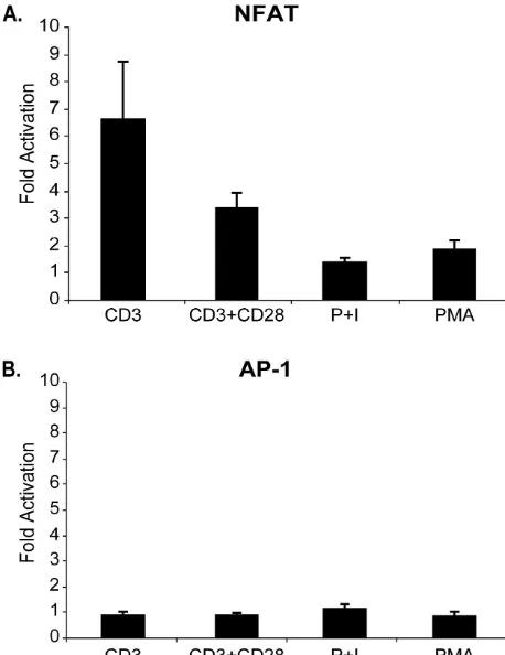
p12Iexpression enhanced NFAT transcriptional activities
six-fold in the presence of anti-CD3 alone and threesix-fold when cells were stimulated with anti-CD3 and anti-CD28 compared to the control cells expressing GFP alone (Fig. 4A). Of note, anti-CD3 and anti-CD28 dual stimulation in Jurkat T cells elicited increased NFAT reporter gene activities compared to anti-CD3 stimulation alone (data not shown). Jurkat T cells
that expressed p12I exhibited threefold-higher NFAT
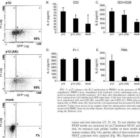
tran-FIG. 1. p12I expression in Jurkat T cells. (A) Schematic representation of the p12 transgene plasmids pHR⬘CMV/p12(⫹)/eGFP and
pHR⬘CMV/p12(⫺)/eGFP. (B) The HTLV-1 p12Igene was introduced into Jurkat T cells by lentiviral transduction as described in Materials and
Methods. Cells were transduced with sense p12I(p12I, expressing both p12Iand eGFP) or antisense p12I[p12(AS), expressing eGFP alone] by
lentiviral vectors or mock infected. At day 4 posttransduction, an aliquot of cells was stained with propidium iodide (PI) and anti-CD25 antibody by flow cytometry. GFP expression was concurrently analyzed. Approximately 50% of infected cells were found to be GFP positive in both p12I
and p12(AS) samples. Samples lysed from approximately 2 million cells were tested for p12Iexpression by immunoblot assay. The results were
representative of three independent infection experiments.
on November 8, 2019 by guest
http://jvi.asm.org/
[image:4.603.129.460.77.548.2]scription as indicated by enhanced luciferase reporter gene activity in the presence of both anti-CD3 and anti-CD28 stim-ulation compared to control cells expressing GFP alone. Cor-roborating our data of IL-2 production, this hyperactivation was not present in unstimulated cells (data not shown) in cells stimulated with PMA plus ionomycin or in cells stimulated with PMA alone. These data indicate that the NFAT activation
mediated by p12Iand T-cell receptor ligation likely result in
enhanced IL-2 production. Furthermore, the stimulatory effect
of p12I specifically involved NFAT-dependent transcription,
since the activity of AP-1 dependent transcription remained unchanged regardless of the stimulation conditions (Fig. 4B).
Expression of p12Iin PBMC enhances the IL-2 production
in the presence of PMA stimulation.As an extension of the
above studies in Jurkat T cells, we transduced the HTLV-1
natural target cells, PBMC, and analyzed the effect of p12I
expression on IL-2 production in PBMC. Preactivated PBMC
by PHA were transduced with pHR⬘CMV/p12(⫹)/eGFP or
pHR⬘CMV/p12(⫺)/eGFP vector, and GFP expression was
an-alyzed by flow cytometry (Fig. 5A). We observed a higher GFP expression (ca. 50 to 60%) at day 6 posttransduction than at the earlier time points (data not shown). Therefore, the IL-2 production in the supernatant was measured at day 6 post-transduction after the cells were stimulated with various agents. Receptor ligation by anti-CD3 in the presence or ab-sence of anti-CD28 did not significantly enhance IL-2
produc-tion in PBMC expressing p12Icompared to the control cells
expressing GFP alone (Fig. 5B and C). Expression of p12Ihad
[image:5.603.51.278.69.447.2]no effect on IL-2 production when cells were stimulated with
FIG. 2. p12Iincreases IL-2 production by T-cell antigen receptor
ligation in the presence or absence of CD28 costimulation. Transduced Jurkat T cells were activated by immobilized anti-CD3 (coated at 5 g/ml) (A), immobilized anti-CD3 plus anti-CD28 (coated at 5g/ml) (B), or PMA plus 2M ionomycin (Iono) (C) in 96-well plates. After 24 h at 37°C, IL-2 secreted in the supernatant was measured by ELISA techniques. p12I-transduced Jurkat T cells secreted significantly higher
[image:5.603.308.533.264.598.2]IL-2 than p12(AS)-transduced cells or mock-infected cells in the pres-ence of anti-CD3 with or without anti-CD28 stimulation.❋,P⬍0.05. Data represent mean IL-2 concentrations from triplicate wells. The results are representative of three independent infection experiments. Statistical significance was analyzed by using the Studentttest.
FIG. 3. Enhanced IL-2 production mediated by p12I is calcium
pathway dependent. Transduced Jurkat T cells were treated with BAPTA-AM (BAPTA) or cyclosporine before the T-cell receptor li-gation mimicked by anti-CD3 (A) or with anti-CD28 stimulation (B). p12I-transduced Jurkat T cells secreted significantly higher levels of
IL-2 than did p12(AS)-transduced cells or mock-infected cells in the presence of anti-CD3 alone (A) or with anti-CD28 stimulation (B). BAPTA-AM and cyclosporine treatment resulted in significantly re-duced IL-2 production in p12I- and p12(AS)-transduced cells, as well
as in mock-infected cells. However, IL-2 production in p12I-expressing
cells after the addition of either inhibitor was not significantly different from that seen in p12(AS)-transduced cells or in mock-infected cells. Values are the means of triplicate or six samples. Statistical signifi-cance was analyzed by using the Studentttest.❋,P⬍0.05.
on November 8, 2019 by guest
http://jvi.asm.org/
PMA plus ionomycin (Fig. 5D). Interestingly, PBMC
express-ing p12Ihad a sixfold-higher IL-2 concentrations in cell culture
supernatants compared to control cells after stimulation with PMA alone (Fig. 5E). These data likely represent the differ-ential response of primary PBMC compared to the trans-formed Jurkat T-cell line (39).
To address the issue of differential dose response with anti-CD3 and anti-CD28 in PBMC, we tested the IL-2 production from PBMC with various amounts of anti-CD3 (0.06, 0.2, 0.5,
and 1.0 g), with or without 1g of anti-CD28 (optimal
re-sponse dose to costimulate PBMC). We found differences of
⬍10% in IL-2 production between these various stimulation
protocols, indicating that the differential response between Jurkat T cells and PBMC was not due to saturating concen-trations of the stimuli for PBMC (data not shown).
We have previously demonstrated IL-2 responsiveness of HTLV-1 immortalized T-cell lines (8). To test whether the
quantities of IL-2 elicited from p12I-expressing PBMC could
stimulate normal PBMC to proliferate, we analyzed cell pro-liferation of PBMC in the presence of similar concentrations of
IL-2 measured from p12I-expressing PBMC. PBMC prepared
in a manner identical to that used in our vector trials were cultured in the presence of 0, 100, 200, 500, 1,000, and 2,000 pg of recombinant human IL-2/ml. At concentrations similar to
the levels of IL-2 produced from p12I-expressing PBMC, cell
proliferation was measurably increased (25% increase in opti-cal density in proliferation assay) after as little as 4 h of incu-bation (Fig. 6). At this rate of proliferation, PBMC would be predicted to double in number in approximately 16 h. Thus, the
quantity of IL-2 induced by p12I-expressing PBMC would be
predicted to have a functional influence on lymphocyte prolif-eration.
Identification of two positive (amino acids [aa] 33 to 47 and 87 to 99) and two negative regions (aa 1 to 14 and 70 to 86) in p12Ifor NFAT activation.To investigate the possible domains
in p12I required for NFAT activation, Jurkat T cells were
transiently cotransfected with full-length or p12I truncation
mutants in combination of pNFAT-luc plasmid, and the tran-scriptional activities of NFAT were analyzed. The expressions
of full-length and truncated p12Iwere tested by immunoblot
assay (Fig. 7A). The majority of p12I truncation mutants
ex-pressed at equivalent levels compared to full-length protein.
Mutant p12I15-99 expression induced higher NFAT luciferase
activity than full-length protein (Fig. 7B), suggesting the pres-ence of an inhibitory region in the amino-terminal 14 aa (first negative region). Further amino-terminal deletion displayed a
NFAT luciferase activity similar to full-length p12I in cells
transfected with mutant p12I 32-99. However, expression of
mutant p12I48-99 resulted in a reduced NFAT transcriptional
activity (Fig. 7B), indicating the region aa 33 to 47 contains a positive domain (first positive region). While the N-terminal
half of p12Icontained one negative region (aa 1 to 14) and one
positive region (aa 33 to 47) for NFAT activation, analysis of C-terminal mutants revealed another positive and another negative region. Carboxy-terminal deletion of 13 aa in mutant
p12I 1-86 resulted in a dramatic reduction of NFAT activity
(Fig. 7B). Thus, aa 87 to 99 likely contains the second positive region for NFAT activation. To our surprise, further
C-termi-nal deletion in mutant p12I1-47 almost restored the NFAT
activation mediated by full-length protein, implying the pres-ence of another inhibitory domain in region aa 48 to 86 (sec-ond negative region). Analysis of mutants containing both ami-no- and carboxy-terminal deletions further confirmed that the aa 1 to 15 is a negative region since a higher NFAT activity was
detected in cells expressing p12I 15-86 compared to cells
ex-pressing 1-86. Mutant p12I15-69 expression elicited a
twofold-higher NFAT luciferase activity than did the full-length protein (Fig. 7B), indicating the presence of the second negative region
in aa 70 to 86. Of note, all mutants except p12I15-47 localized
in the ER perinuclear region. Therefore, the differential NFAT activations in cells expressing individual mutants are likely due to the deleted sequence. The NFAT transcriptional
activities mediated by all p12Itruncation mutants, except
32-99, were significantly different (P⬍0.05) from those of
wild-type p12I. However, we cannot exclude the possibility that
alteration of the p12Iprotein structure is responsible for
dif-ferential NFAT activations. Expression of mutant p12I15-47,
which localizes in both the nucleus and the cytoplasm, failed to enhance NFAT luciferase activity (Fig. 7B), suggesting the ER
localization is necessary for p12Imediated NFAT activation.
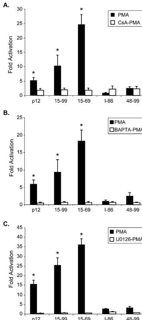
[image:6.603.50.279.70.367.2]To test whether NFAT transcriptional activation induced by individual mutants was calcium and Ras/MAPK pathway de-pendent, a number of inhibitors, including BAPTA-AM, cy-closporine, and an inhibitor of MAP kinase kinase (MEK-1),
FIG. 4. p12Ipromotes NFAT activation in response to CD3 with or
without CD28 stimulation. Transduced Jurkat T cells transfected with pNFAT-Luc (A) or AP-1-Luc (B) plasmid were stimulated with either PMA, PMA plus ionomycin (P⫹I), CD3, or CD3 plus anti-CD28. The graph depicts the fold induction of NFAT or AP-1 lucif-erase activity in cells expressing p12I (p12) versus those transduced
with antisense p12 (AS). Datum points are the means of triplicate samples from three independent experiments.
on November 8, 2019 by guest
http://jvi.asm.org/
U0126, were used to treat the transfected Jurkat T cells before the addition of PMA. All three inhibitors tested completely abolished the NFAT activation mediated by two highly active
mutants, p12I15-99 and p12I 15-69 (Fig. 8), indicating these
mutants activate NFAT in a similar calcium- and Ras/MAPK-dependent manner as full-length protein.
The third SH3-binding domain (aa 70 to 73) is responsible for the negative effect of region aa 70 to 86 on NFAT activation.
The two positive and two negative regions we identified for NFAT activation contain individual SH3-binding domains, the PXXP motif. The SH3-binding domain has been demonstrated to be involved in protein-protein interaction, and the PXXP motif in HIV Nef is necessary for Nef-mediated NFAT
acti-vation and viral infection (27, 29, 30). To test whether these
PXXP motifs are necessary for p12I-mediated NFAT
activa-tion, we mutated each proline residue in these motifs into alanine residues (Fig. 9A), and the effect of these mutations on NFAT activation were analyzed (Fig. 9B). Expression of the
third PXXP mutant, A70XXA73, in the second negative region
(aa 70 to 86), significantly increased NFAT transcriptional
activity (190%) compared to wild-type p12I. Thus, in parallel to
our data with serial deletion mutants, the third PXXP motif appears to mediate the negative effect of region aa 70 to 86 on NFAT activation. Expression of the first PXXP mutant
(A8XXA11) and second PXXP mutant (A35XXA38) minimally
enhanced and decreased NFAT activation, respectively. The
last PXXP mutant (A90XXA93), in the second positive region
(aa 86 to 99), enhanced the NFAT activation. The functional
significance of specific residues within the PXXP motifs of p12I
in NFAT activation remains to be further clarified.
ER localization is required for p12I-mediated NFAT
activa-tion.Although the mutant p12I15-47 contains a positive region (aa 33 to 47), expression of this mutant displayed a dramati-cally reduced NFAT transcriptional activity. In addition, this mutant fails to maintain ER localization; instead, it mainly accumulates in the nucleus (13). To test whether ER
local-FIG. 5. p12Ienhances the IL-2 production in PBMCs in the presence of PMA
stimulation. PBMCs were transduced with lentiviral vector containing sense p12I
(p12) or antisense p12(AS) sequence. At 6 days after transduction, aliquots of cells were analyzed for cell viability and GFP expression. Cells were left untreated (A) or were stimulated with anti-CD3 (B), anti-CD3 plus anti-CD28 (C), PMA plus iono-mycin (D), or PMA alone (E). Secreted IL-2 in supernatant was measured by ELISA methods. Values were mean of six samples from two independent infection experi-ments with PBMC from two healthy donors. Statistical significance was analyzed by using the Studentttest.
on November 8, 2019 by guest
http://jvi.asm.org/
[image:7.603.61.538.68.538.2]ization is necessary for p12I-mediated NFAT activation, we tagged the C terminus of this mutant with the sequence of a well-recognized ER targeting signal, KKLL (20, 43), and ana-lyzed the location of the new protein 15-47KKLL, as well as NFAT activation in Jurkat T cells expressing this chimera. As expected, the chimera 15-47KKLL accumulated in the perinu-clear region and colocalized with calreticulin, an ER luminal protein, in transfected HeLa-Tat cells (Fig. 10A). Expression of this chimera in Jurkat T cells partially restored the NFAT activation compared to the low NFAT activity in cells express-ing 15-47 (Fig. 10B). Thus, ER localization is required for
p12I-mediated NFAT activation. The chimera protein, which
regained the ER localization, only restored half of the NFAT activation induced by full-length p12I, suggesting that the first positive region (aa 33 to 47) is not sufficient for full activation of NFAT.
DISCUSSION
In this study, we used a lentiviral system to express p12I
protein in transduced Jurkat T cells and in primary human PBMC. Under the condition of T-cell receptor ligation and
anti-CD28 costimulation, expression of p12I enhanced IL-2
production in Jurkat T cells. As a downstream gene responsive to activation of multiple signal pathways in T cells, IL-2 is an indicator for T-cell activation. Activation of T lymphocytes results in cell division, which is a prerequisite for the proviral DNA of retrovirus, including HTLV-1, to integrate into the host cell genome and permit the subsequent viral replication
and productive infection. Thus, p12I expression appears to
augment T-cell activation to facilitate the viral replication and productive infection. This finding provides a mechanism for HTLV-1 to establish infection in resting T cells (1) and also is supportive of our previous finding that selective ablation of
mRNA containing p12I-coding sequence reduces viral
infectiv-ity in a rabbit model of infection (9). Consistent with our data presented here, T cells would become hypersensitive to T-cell
activation signals when HTLV-1 p12I is present and become
highly permissive for subsequent viral infection.
p12Ilocalizes in the ER and cis-Golgi compartments and
associates with the calcium-binding protein, calreticulin (13).
p12Iexpression in Jurkat T cells increases the level of
intra-cellular calcium (12) and selectively activates NFAT (2).
Therefore, the elevated IL-2 secretion in p12I-expressing T
cells likely results from the activation of calcium-dependent pathway. Indeed, the calcium chelator, BAPTA-AM and the calcineurin inhibitor, cyclosporine, inhibited the enhanced
IL-2 production in cells expressing p12I. In addition,
expres-sion of p12Iactivated the NFAT transcriptional activity in the
presence of surface receptor CD3 and CD28 stimulation but not in the presence of PMA stimulation. These results are in contrast to our previous finding in which we transiently
over-expressed p12I in Jurkat T cells and reported the selective
activation of NFAT in the presence of PMA. This discrepancy is likely related to the levels of p12Iexpression with each model
system. We observed much higher p12Iexpression in transient
transfected Jurkat T cells compared to transduction of p12I
FIG. 6. To test PBMC proliferation in response to IL-2, PBMC were stimulated with in the presence of similar concentrations of IL-2 measured from p12I-expressing PBMC. PBMC prepared in a manner identical to that used in our vector trials were cultured in the presence of 0, 100, 200,
500, 1,000, or 2,000 pg of natural human IL-2 (10,000 U⫽5g [Roche])/ml. Cells were tested for proliferation as previously described (8) by using a tetrazolium dye-based method (CellTiter 96 cell proliferation assay [Promega]). Linear analysis was performed to show the relationship between IL-2 concentration and PBMC proliferation. The range of IL-2 concentrations from PBMC infected with control or p12-expressing vectors is indicated on the graph.
on November 8, 2019 by guest
http://jvi.asm.org/
using the lentiviral vector. Therefore, the relatively lower levels
of p12Iexpression by an internal CMV promoter via the
lenti-viral system may not be enough to trigger the NFAT activation and IL-2 production in the presence of PMA alone. However, when relative strong stimulations, such as surface receptor
ligation, are provided, low amounts of p12Iexpression can
fa-cilitate the Jurkat T-cell activation. The production of HTLV-1 viral transcripts is very low, and the viral proteins are not easily detectable in naturally infected PBMC (16, 18, 41). Thus,
the low levels of p12Iexpression via the lentiviral system may
be more biologically representative of natural infection. In
contrast to Jurkat T cells, expression of p12Iin PBMC induced
an approximately sixfold higher level of IL-2 in the presence of
PMA. However, p12Iexpression in PBMC did not significantly
enhance IL-2 secretion during T-cell-receptor stimulation. These data may represent the differential responses in primary
FIG. 7. Two positive regions (aa 33 to 47 and 87 to 99) and two negative regions (aa 1 to 14 and 70 to 86) are identified in p12Ifor
NFAT activation. (A) Schematic representation and expression of wild-type p12Iand serial truncation mutants. The hatched box
indi-cates the PXXP motif, and the circle indiindi-cates the HA tag. 293T cells were transfected with pMEp12 or mutant plasmids. The expression of wild-type p12I and mutants was tested by immunoblot assay at 48 h
posttransfection. (B) Jurkat T cells were electroporated with pMEp12 or plasmids expressing truncation mutants in combination with pNFAT-Luc reporter plasmid, and the NFAT luciferase activities were deter-mined 18 h after PMA treatment. The NFAT transcriptional activities mediated by all p12Itruncation mutants, except for 32-99, were
signif-icantly different (P⬍0.05) from that of wild-type p12I. The data are
presented as relative NFAT luciferase activities compared to wild-type p12I. The values are the mean of four independent transfection
[image:9.603.308.536.136.648.2]exper-iments. Statistical significance was analyzed by using the Studentttest.
FIG. 8. Hyperactivated p12Itruncation mutants induce NFAT
ac-tivation in the same fashion as wild-type p12I. Jurkat T cells were
transfected with plasmids containing different mutants in combination with pNFAT-Luc plasmid, and cells were treated with cyclosporine (A), BAPTA-AM (B), or U0126 (C) for 30 min before PMA was add-ed. These inhibitors significantly reduced NFAT activation mediated by the wild type and the hyperactivated mutants 15-99 and 15-69. Datum points are the means of three independent transfection exper-iments. Statistical significance was analyzed by using the Studentttest.
on November 8, 2019 by guest
http://jvi.asm.org/
T lymphocytes compared to transformed T cells. Our group has observed the prolonged CREB phosphorylation and in-creased basal HTLV-1 transcription in PBMC after mitogen stimulation compared to Jurkat T cells (39). Therefore, pri-mary T lymphocytes may contain a relative intact cell signal machinery, which is more sensitive in response to activation signals after PMA stimulation, such as protein kinase C
acti-vation. Expression of p12I had no effect on IL-2 production
and NFAT activation when downstream strong stimulations, i.e., with PMA plus ionomycin, are provided, indicating that
the function of p12Iis calcium dependent and that ionomycin
stimulation overrides the effect of p12I on regulation of
cal-cium homeostasis.
The role of the IL-2/IL-2R signaling pathway in HTLV-1 early infection, virus-induced immortalization, and the trans-formation process is not clear. Earlier studies (47, 48, 51) demonstrated that Tax expression induces the activation of
IL-2R␣and IL-2 genes. These activation events mediated by
Tax presumably occur via NF-B activation and are
cyclospor-ine resistant. However, the temporal expression pattern of viral
taxmRNA is not consistent with the pattern of IL-2 mRNA in
HTLV-1-infected primary lymphocytes. IL-2 is transiently ex-pressed during the early phase of the infection when viral integration is polyclonal after HTLV-1 infection of human primary lymphocytes. It is undetectable at a later stage when
viral integration is oligoclonal (23). In contrast,taxmRNA is
scarcely expressed in the polyclonal phase but is abundantly expressed in the oligoclonal stage. Therefore, additional viral protein expressed in the early phase of viral infection may induce the IL-2 production in the initial phase. The quantities
of IL-2 elicited from p12I-expressing PBMC were able to elicit
proliferation PBMC and therefore would be predicted to have a functional influence on lymphocyte proliferation. Our
find-ings support the idea that p12Imay trigger early IL-2
expres-sion to facilitate HTLV-1 infection.
A similar viral protein, Nef, modulates calcium signaling through interaction with IP3 receptor (31) and thus activates NFAT (28), as well as enhances IL-2 production in T lympho-cytes (46, 50). Nef has been demonstrated to associate with HIV virion (26, 56) and is selectively expressed before the integration to modulate the resting T-cell activity (54).
There-fore, it will be important to determine whether p12Iis a
virion-associated protein or whether p12I mRNA and protein are
selectively expressed before integration.
HTLV-1-transformed T cells display constitutive tyrosine phosphorylation of IL-2R-coupled signaling proteins, includ-ing Jak1, Jak3, Stat3, and Stat5 (33, 34, 36, 55). Expression of
p12Iand signaling molecules involved in the IL-2R signaling
pathway in 293T cells induced the increased DNA-binding
activity of Stat5 (40). The mechanism of p12I-mediated Stat5
activation may be related to the interaction between IL-2R (
and␥) and p12I. Alternatively, expression of p12Imay activate Stat5 through the IL-2R pathway in an autocrine manner in-duced by elevated IL-2 production (40). Thus, our results
pro-vide a potential mechanism for p12I-mediated Stat5 activation.
p12Iis probably not involved in the transformation process
of HTLV-1-infected T cells. mRNA of ORF I can be detected in both IL-2-dependent and -independent HTLV-1-infected
T-cell lines (4–7, 17, 25), and p12Iis not necessary for
immor-talization of HTLV-1-infected cells in vitro (11, 45). Therefore,
the elevated IL-2 secretion mediated by expression of p12Imay
promote HTLV-1-infected T cells to proliferate through an autocrine or paracrine mechanism and allow HTLV-1 infec-tion to subsequently spread more effectively.
Using a variety of p12Itruncation mutants, we identified two
positive regions (aa 33 to 47 and 87 to 99) and two negative regions (aa 1 to 14 and 70 to 86) in the viral protein for NFAT activation. An SH3-binding domain (PXXP) is contained in each individual region. Further analysis with point mutants that replaced prolines with alanines in each PXXP motif
dem-onstrated that the third PXXP motif (P70XXP73) is responsible
for the inhibitory effect of region aa 70 to 86 on NFAT acti-vation. Interestingly, we have recently identified a conserved PxIxIT calcineurin-binding motif, encompassing the third
PXXP motif, in p12Ithat binds to calcineurin (22). Wild-type
p12Iand deleted mutants, which contained the motif, bound
calcineurin. In addition, an alanine substitution mutant (p12I
[image:10.603.47.279.71.391.2]AxAxAA) had greatly reduced binding affinity for calcineurin.
FIG. 9. The mutant A70XXA73significantly increases NFAT
activ-ity. (A) Schematic representation and expression of wild-type p12Iand
point mutants. The hatched box indicates the PXXP motif, and the black box indicates the mutated PXXP motif. (B) Jurkat T cells were transfected with p12Ipoint mutants that replaced proline with alanine
in PXXP motifs and pNFAT-Luc plasmid, followed by 18 h of PMA treatment and measurement of luciferase activities. Data are pre-sented as relative NFAT luciferase activities compared to wild-type p12I. The NFAT transcriptional activities mediated by all p12Ipoint
mutants were significantly different (P ⬍0.05) from wild-type p12I.
Values are means of at least three independent transfection experi-ments. Statistical significance was analyzed by using the Studentttest.
on November 8, 2019 by guest
http://jvi.asm.org/
Nef also contains the SH3-binding domain, and this motif is required for the binding between Nef and multiple cellular proteins, including Hck and PAK, and is necessary for Nef-mediated NFAT activation (27, 29, 30). Thus, further studies
designed to identify possible binding partners of p12Iand the
role of these motifs in HTLV-1 infection will be necessary to clarify the role of specific motifs in T-cell activation. Finally, we observed partially restored NFAT activation by tagging the mutant 15-47 with an ER targeting signal, KKLL. This finding
demonstrates the requirement of ER localization in p12I
me-diated NFAT activation, further indicating the role of this protein in modulating the ER calcium homeostasis.
In summary, expression of p12Iin primary T lymphocytes
and Jurkat T cells promotes IL-2 production during T-cell ac-tivation, suggesting the important role of this viral accessory protein in early HTLV-1 infection in vivo.
ACKNOWLEDGMENTS
This work was supported by National Institute of Health grants RR-14324, AI-01474, and CA-92009 (M.D.L.) and CA-70529 from the National Cancer Institute, awarded through the Ohio State University Comprehensive Cancer Center.
[image:11.603.130.456.73.524.2]We thank A. Oberyszyn and R. Meister for technical assistance in flow cytometric analysis. We also thank Patrick Green for critical FIG. 10. ER targeting partially restores the NFAT activation mediated by mutant p12I15-47. (A) Localization of mutant 15-47 and chimeric
protein 15-47KKLL. An ER targeting signal, KKLL, redirected the chimeric protein 15-47KKLL to ER compartments stained with the ER marker, calreticulin (CRT). The nuclei were stained with Hoechst stain. (B) Relative NFAT activities mediated by p12I, mutant 15-47, and chimeric protein
15-47KKLL. Data are presented as relative NFAT luciferase activities compared to wild-type p12I. Values are means of three independent
transfection experiments. Statistical significance was analyzed by using the Studentttest.❋,P⬍0.05.
on November 8, 2019 by guest
http://jvi.asm.org/
review of the manuscript, T. Vojt for preparation of figures, and G. Franchini and G. Crabtree for sharing valuable reagents.
REFERENCES
1. Albrecht, B., N. D. Collins, M. T. Burniston, J. W. Nisbet, L. Ratner, P. L. Green, and M. D. Lairmore.2000. Human T-lymphotropic virus type 1 open reading frame I p12Iis required for efficient viral infectivity in primary lymphocytes. J. Virol.74:9828–9835.
2. Albrecht, B., C. D. D’Souza, W. Ding, S. Tridandapani, K. M. Coggeshall, and M. D. Lairmore.2002. Activation of nuclear factor of activated T cells by human T-lymphotropic virus type 1 accessory protein p12I. J. Virol.76: 3493–3501.
3. Bangham, C. R.2000. HTLV-1 infections. J. Clin. Pathol.53:581–586. 4. Berneman, Z. N., R. B. Gartenhaus, M. S. Reitz, W. A. Blattner, A. Manns,
B. Hanchard, O. Ikehara, R. C. Gallo, and M. E. Klotman.1992. Expression of alternatively spliced human T-lymphotropic virus type 1 pX mRNA in infected cell lines and in primary uncultured cells from patients with adult T-cell leukemia/lymphoma and healthy carriers. Proc. Natl. Acad. Sci. USA 89:3005–3009.
5. Cereseto, A., Z. Berneman, I. Koralnik, J. Vaughn, G. Franchini, and M. E. Klotman.1997. Differential expression of alternatively spliced pX mRNAs in HTLV-I-infected cell lines. Leukemia11:866–870.
6. Ciminale, V., D. D’Agostino, L. Zotti, G. Franchini, B. K. Felber, and L. Chieco-Bianchi.1995. Expression and characterization of proteins produced by mRNAs spliced into the X region of the human T-cell leukemia/lympho-tropic virus type II. Virology209:445–456.
7. Ciminale, V., G. N. Pavlakis, D. Derse, C. P. Cunningham, and B. K. Felber. 1992. Complex splicing in the human T-cell leukemia virus (HTLV) family of retroviruses: novel mRNAs and proteins produced by HTLV type I. J. Virol. 66:1737–1745.
8. Collins, N. D., C. D’Souza, B. Albrecht, M. D. Robek, L. Ratner, W. Ding, P. L. Green, and M. D. Lairmore.1999. Proliferation response to interleu-kin-2 and Jak/Stat activation of T cells immortalized by human T-cell lym-photropic virus type 1 is independent of open reading frame I expression. J. Virol.73:9642–9649.
9. Collins, N. D., G. C. Newbound, B. Albrecht, J. L. Beard, L. Ratner, and M. D. Lairmore.1998. Selective ablation of human T-cell lymphotropic virus type 1 p12I reduces viral infectivity in vivo. Blood91:4701–4707. 10. Dekaban, G. A., A. A. Peters, J. C. Mulloy, J. M. Johnson, R. Trovato, E.
Rivadeneira, and G. Franchini.2000. The HTLV-I orfI protein is recognized by serum antibodies from naturally infected humans and experimentally infected rabbits. Virology274:86–93.
11. Derse, D., J. Mikovits, and F. Ruscetti.1997. X-I and X-II open reading frames of HTLV-I are not required for virus replication or for immortaliza-tion of primary T cells in vitro. Virology237:123–128.
12. Ding, W., B. Albrecht, R. E. Kelley, N. Muthusamy, S. Kim, R. A. Altschuld, and M. D. Lairmore.2002. Human T lymphotropic virus type 1 p12I expres-sion increases cytoplasmic calcium to enhance the activation of nuclear factor of activated T cells. J. Virol.76:10374–10382.
13. Ding, W., B. Albrecht, R. Luo, W. Zhang, J. R. Stanley, G. C. Newbound, and M. D. Lairmore.2001. Endoplasmic reticulum and cis-Golgi localization of human T-lymphotropic virus type 1 p12I: association with calreticulin and calnexin. J. Virol.75:7672–7682.
14. Franchini, G.1995. Molecular mechanisms of human T-cell leukemia/lym-photropic virus type I infection. Blood86:3619–3639.
15. Franchini, G., J. C. Mulloy, I. J. Koralnik, M. A. Lo, J. J. Sparkowski, T. Andresson, D. J. Goldstein, and R. Schlegel.1993. The human T-cell leu-kemia/lymphotropic virus type I p12Iprotein cooperates with the E5 onco-protein of bovine papillomavirus in cell transformation and binds the 16-kilodalton subunit of the vacuolar H⫹ATPase. J. Virol.67:7701–7704. 16. Franchini, G., F. Wong-Staal, and R. C. Gallo.1984. Human T-cell leukemia
virus (HTLV-I) transcripts in fresh and cultured cells of patients with adult T-cell leukemia. Proc. Natl. Acad. Sci. USA81:6207–6211.
17. Furukawa, K., K. Furukawa, and H. Shiku.1991. Alternatively spliced mRNA of the pX region of human T lymphotropic virus type I proviral genome. FEBS Lett.295:141–145.
18. Gessain, A., A. Louie, O. Gout, R. C. Gallo, and G. Franchini.1991. Human T-cell leukemia-lymphoma virus type 1 (HTLV-1) expression in fresh pe-ripheral blood mononuclear cells from patients with tropical spastiv parapa-resis/HTLV-1-associated myelopathy. J. Virol.65:1628–1633.
19. Gessain, A., and R. Mahieux.2000. A virus called HTLV-1: epidemiological aspects. Presse Med.29:2233–2239.
20. Gomord, V., E. Wee, and L. Faye.1999. Protein retention and localization in the endoplasmic reticulum and the Golgi apparatus. Biochimie81:607–618. 21. Hollsberg, P.1999. Mechanisms of T-cell activation by human T-cell
lym-photropic virus type I. Microbiol. Mol. Biol. Rev.63:308–333.
22. Kim, S. J., W. Ding, B. Albrecht, P. L. Green, and M. D. Lairmore.2003. A conserved calcineurin-binding motif in human T lymphotropic virus type 1 p12Ifunctions to modulate nuclear factor of activated T-cell activation. J. Biol. Chem.278:15550–15557.
23. Kimata, J. T., and L. Ratner.1991. Temporal regulation of viral and cellular
gene expression during HTLV-I mediated lymphocyte immortalization. J. Virol.65:4398–4407.
24. Koralnik, I. J., J. Fullen, and G. Franchini.1993. The p12, p13 and p30 proteins encoded by human T-cell leukemia/lymphotropic virus type-1 open reading frames I and II are localized in three different cellular compart-ments. J. Virol.67:2360–2366.
25. Koralnik, I. J., A. Gessain, M. E. Klotman, A. Lo Monico, Z. N. Berneman, and G. Franchini.1992. Protein isoforms encoded by the pX region of human T-cell leukemia/lymphotropic virus type 1. Proc. Natl. Acad. Sci. USA89:8813–8817.
26. Kotov, A., J. Zhou, P. Flicker, and C. Aiken.1999. Association of Nef with the human immunodeficiency virus type 1 core. J. Virol.73:8824–8830. 27. Lee, C. H., B. Leung, M. A. Lemmon, J. Zheng, D. Cowburn, J. Kuriyan, and
K. Saksela.1995. A single amino acid in the SH3 domain of Hck determines its high affinity and specificity in binding to HIV-1 Nef protein. EMBO J. 14:5006–5015.
28. Manninen, A., R. G. Herma, and K. Saksela.2000. Synergistic activation of NFAT by HIV-1 nef and the Ras/MAPK pathway. J. Biol. Chem.275:16513– 16517.
29. Manninen, A., M. Hiipakka, M. Vihinen, W. Lu, B. J. Mayer, and K. Saksela. 1998. SH3 domain binding function of HIV-1 Nef is required for association with a PAK-related kinase. Virology250:273–282.
30. Manninen, A., P. Huotari, M. Hiipakka, G. H. Renkema, and K. Saksela. 2001. Activation of NFAT-dependent gene expression by Nef: conservation among divergent Nef alleles, dependence on SH3 binding and membrane association, and cooperation with protein kinase C-theta. J. Virol.75:3034– 3037.
31. Manninen, A., and K. Saksela.2002. HIV-1 Nef interacts with inositol trisphosphate receptor to activate calcium signaling in T cells. J. Exp. Med. 195:1023–1032.
32. Merl, S., B. Kloster, J. Moore, C. Hubbell, R. Tomar, F. Davey, D. Kali-nowski, A. Planas, G. Ehrlich, D. Clark, R. Comis, and B. Poiesz.1984. Efficient transformation of previously activated and dividing T lymphocytes by human T cell leukemia-lymphoma virus. Blood64:967–974.
33. Migone, T. S., N. A. Cacalano, N. Taylor, T. L. Yi, T. A. Waldmann, and J. A. Johnston.1998. Recruitment of SH2-containing protein tyrosine phospha-tase SHP-1 to the interleukin 2 receptor; loss of SHP-1 expression in human T-lymphotropic virus type I-transformed T cells. Proc. Natl. Acad. Sci. USA 95:3845–3850.
34. Migone, T. S., J. X. Lin, A. Cereseto, J. C. Mulloy, J. J. Oshea, G. Franchini, and W. J. Leonard.1995. Constitutively activated Jak-STAT pathway in T cells transformed with HTLV-I. Science269:79–81.
35. Mulloy, J. C., R. W. Crowley, J. Fullen, W. J. Leonard, and G. Franchini. 1996. The human T-cell leukemia/lymphotropic virus type 1 p12Iprotein binds the interleukin-2 receptor beta and gamma(c) chains and affects their expression on the cell surface. J. Virol.70:3599–3605.
36. Mulloy, J. C., T. S. Migone, T. M. Ross, N. Ton, P. L. Green, W. J. Leonard, and G. Franchini.1998. Hum. and simian T-cell leukemia viruses type 2 (HTLV-2 and STLV-2) transform T cells independently of Jak/STAT acti-vation. J. Virol.72:4408–4412.
37. Nagai, M., K. Usuku, W. Matsumoto, D. Kodama, N. Takenouchi, T. Mori-toyo, S. Hashiguchi, M. Ichinose, C. R. Bangham, S. Izumo, and M. Osame. 1998. Analysis of HTLV-I proviral load in 202 HAM/TSP patients and 243 asymptomatic HTLV-I carriers: high proviral load strongly predisposes to HAM/TSP. J. Neurovirol.4:586–593.
38. Newbound, G. C., J. M. Andrews, J. Orourke, J. N. Brady, and M. D. Lairmore.1996. Human T-cell lymphotropic virus type 1 tax mediates en-hanced transcription in CD4⫹T lymphocytes. J. Virol.70:2101–2106. 39. Newbound, G. C., J. P. Orourke, N. D. Collins, J. Dewille, and M. D.
Lairmore.1999. Comparison of HTLV-I basal transcription and expression of CREB/ATF-1/CREM family members in peripheral blood mononuclear cells and Jurkat T cells. J. Acquir. Immune Defic. Syndr. Hum. R.20:1–10. 40. Nicot, C., J. C. Mulloy, M. G. Ferrari, J. M. Johnson, K. Fu, R. Fukumoto, R. Trovato, J. Fullen, W. J. Leonard, and G. Franchini.2001. HTLV-1 p12I protein enhances STAT5 activation and decreases the interleukin-2 require-ment for proliferation of primary human peripheral blood mononuclear cells. Blood98:823–829.
41. Pique, C., and M. C. Dokhelar.2000. In vivo production of rof and tof proteins of HTLV type 1: evidence from cytotoxic T lymphocytes. AIDS Res. Hum. Retrovir.16:1783–1786.
42. Pique, C., A. Uretavidal, A. Gessain, B. Chancerel, O. Gout, R. Tamouza, F. Agis, and M. C. Dokhelar.2000. Evidence for the chronic in vivo production of human T-cell leukemia virus type I Rof and Tof proteins from cytotoxic T lymphocytes directed against viral peptides. J. Exp. Med.191:567–572. 43. Plemper, R. K., A. L. Hammond, and R. Cattaneo.2001. Measles virus
envelope glycoproteins hetero-oligomerize in the endoplasmic reticulum. J. Biol. Chem.276:44239–44246.
44. Poiesz, B. J., F. W. Ruscetti, A. F. Gazdar, P. A. Bunn, J. D. Minna, and R. C. Gallo.1980. Detection and isolation of type C retrovirus particles from fresh and cultured lymphocytes of a patient with cutaneous T-cell lymphoma. Proc. Natl. Acad. Sci. USA77:7415–7419.
45. Robek, M. D., F. H. Wong, and L. Ratner.1998. Hum. T-Cell leukemia virus
on November 8, 2019 by guest
http://jvi.asm.org/
type 1 pX-I and pX-II open reading frames are dispensable for the immor-talization of primary lymphocytes. J. Virol.72:4458–4462.
46. Schrager, J. A., and J. W. Marsh.1999. HIV-1 Nef increases T-cell activation in a stimulus-dependent manner. Proc. Natl. Acad. Sci. USA96:8167–8172. 47. Siekevitz, M., M. Feinberg, N. J. Holbrook, F. Wong-Staal, and W. C. Greene.1987. Activation of interleukin 2 and interleukin 2 receptor (Tac) promoter expression by the trans-activator (tat) gene product of human T-cell leukemia virus type I. Proc. Natl. Acad. Sci. USA84:5389–5393. 48. Siekevitz, M., S. F. Josephs, M. Dukovich, N. Peffer, F. Wongstaal, and W. C.
Greene.1987. Activation of the HIV-1 LTR by T cell mitogens and the trans-activator protein of HTLV-1. Science238:1575–1578.
49. Uchiyama, T.1997. Human T-cell leukemia virus type I (HTLV-I) and human diseases. Annu. Rev. Immunol.37:15–37.
50. Wang, J. K., E. Kiyokawa, E. Verdin, and D. Trono.2000. The Nef protein of HIV-1 associates with rafts and primes T cells for activation. Proc. Natl. Acad. Sci. USA97:394–399.
51. Wano, Y., M. Feinberg, J. B. Hosking, H. Bogerd, and W. C. Greene.1988. Stable expression of thetaxgene of type I human T-cell leukemia virus in
human T cells activates specific cellular genes involved in growth. Proc. Natl. Acad. Sci. USA85:9733–9737.
52. Wilson, S. P., F. Liu, R. E. Wilson, and P. R. Housley.1995. Optimization of calcium phosphate transfection for bovine chromaffin cells: relationship to calcium phosphate precipitate formation. Anal. Biochem.226:212–220. 53. Wrzesinski, S., R. Seguin, Y. Liu, S. Domville, V. Planelles, P. Massa, E.
Barker, J. Antel, and G. Feuer.2000. HTLV type 1 Tax transduction in microglial cells and astrocytes by lentiviral vectors. AIDS Res. Hum. Retro-vir.16:1771–1776.
54. Wu, Y., and J. W. Marsh.2001. Selective transcription and modulation of resting T-cell activity by preintegrated HIV DNA. Science293:1503–1506. 55. Xu, X., S. H. Kang, O. Heidenreich, M. Okerholm, J. J. Oshea, and M. I.
Nerenberg.1995. Constitutive activation of different Jak tyrosine kinases in human T-cell leukemia virus type 1 (HTLV-1) Tax protein or virus-trans-formed cells. J. Clin. Investig.96:1548–1555.
56. Zhou, J., and C. Aiken.2001. Nef enhances human immunodeficiency virus type 1 infectivity resulting from intervirion fusion: evidence supporting a role for Nef at the virion envelope. J. Virol.75:5851–5859.