1
Randomized controlled trial of the use of drain
versus no drain in open incisional hernia mesh
repair.
A dissertation submitted in partial fulfilment of M.S.
3
Declaration Certificate
This is to certify that the dissertation titled “Randomized controlled trial of the use of drain versus no drain in open incisional hernia mesh repair.” which is submitted by me in partial fulfilment towards M.S. Branch I (General Surgery) Examination of the Tamil Nadu
Dr M.G.R. University, Chennai to be held in 2016 comprises only my original work and due acknowledgement has been made in text to all material used.
SIGNATURE:
Dr Rahul Lakshminarayanan
PG Registrar, Department of General Surgery
13
ACKNOWLEDGMENTS
It gives me pleasure to express my gratitude to my respected teacher and guide Dr
Sukria Nayak for his valuable guidance, support and encouragement in carrying out
this study.
I am also indebted to my co-investigators in Surgery Unit 4, Dr Suchita Chase and Dr
Rajesh Joseph Selvakumar, who have guided and helped me throughout the study.
I am also grateful to all teachers in the Department of Surgery, especially in Surgery
Unit 4, for the guidance, support and suggestions I received while preparing this
dissertation.
I am grateful to Dr KJ Selvaraj, who was instrumental in the initial randomisation and
help with regards to the design of the study and to Mr Bijesh Yadav ( both from the
Department of Biostatistics) who subsequently helped me with the data analysis.
14
Randomized controlled trial of the use of drain versus no drain in open incisional
hernia mesh repair.
Table of Contents
INTRODUCTION ... 7
AIMS AND OBJECTIVES ... 11
LITERATURE REVIEW ... 13
History of incisional hernia repair ... 14
Incisional hernia ... 16
Methods for repair ... 19
Complications following incisional hernia repair ... 26
Seroma occurrence in other operations ... 29
Types of drains ... 30
The use of drains in incisional hernia repair ... 31
The use of drain in other surgeries ... 32
JUSTIFICATION ... 36
MATERIALS AND METHODS ... 37
RESULTS ... 50
DISCUSSION ... 78
LIMITATIONS ... 82
CONCLUSIONS... 83
REFERENCES ... 85
ANNEXURES ... 89
INFORMATION SHEET ... 90
CONSENT FORM ... 92
DATA EXTRACTION FORM ... 94
15
TABLE OF TABLES
Table 1 : Who classification of body mass index ... 47
Table 2 : Analysis of comorbid illnesses ... 57
Table 3 : Diabetes Mellitus ... 58
Table 4 : Systemic Hypertension ... 59
Table 5 : Hypothyroidism ... 60
Table 6 : Comparison of Sex Distribution and Outcome ... 61
Table 7 : Smoking ... 62
Table 8: Comparison of alcohol consumption to outcome ... 63
Table 9: Comparison of size of defect to outcome ... 64
Table 10: Comparison of width of defect to outcome... 65
Table 11: Comparison of BMI to outcome ... 66
Table 12: Comparison of number of previous operations to outcome ... 67
Table 13 : Comparison of type of repair done versus outcome ... 68
Table 14: Comparison of duration of incisional hernia versus outcome... 69
Table 15: Comparison of drain insertion to seroma/ hematoma and infection occurrence ... 70
Table 16: Comparison of randomisation allocation to outcome ... 72
Table 17: Comparison of drain insertion to hospital stay ... 73
Table 18: Findings of ultrasonography done ... 74
Table 19: 30 day post-operative complications among ultrasound-assessed patients ... 75
16
TABLE OF FIGURES
Figure 1:Methods of placement of mesh in incisional hernia repair 5 ... 21
Figure 2 : Plane of mesh placement in Rives-Stoppa repair 10 ... 23
Figure 3 : After mesh placement in RIves - Stoppa repair 10 ... 24
Figure 4 : Distribution of patients in age groups ... 52
Figure 5 : Sex distribution ... 53
Figure 6: Type of occupation... 54
Figure 7: Regional distribution ... 55
Figure 8 : Incidence of comorbid illnesses ... 56
7
8
INTRODUCTION
Incisional hernias are protrusion of abdominal contents through weakness in the scar of the abdominal wall, following any abdominal operation. They may be primary or recurrent.
They commonly occur due to pre-existing risk factors which include age, obesity, chronic obstructive pulmonary disease (especially emphysema), diabetes mellitus, smoking, drug intake around the time of surgery (like steroids), infection at the surgical incision site.
These can be repaired surgically by different methods. For incisional hernias with less than 3cm defect primary suturing can be done. For defects of more than 3 cm, mesh hernioplasty is usually done: which can be by sublay / onlay / inlay/
9
The prosthetic materials available for incisional hernia repair may be of Biological or Synthetic type. Because of various factors, like the increased cost and non- availability, the biological mesh use is very less. The synthetic materials are used more frequently. Available synthetic meshes include polypropylene (Prolene, Marlex), expanded PTFE (Gore-tex) and polyester. Prolene mesh is the most commonly used material in our institute for open repair of incisional hernias.
Regardless of the technique employed in open repair of incisional hernias the use of drains is almost universal, especially for large hernias. Insertion of drain is usually to evacuate the blood and fluid collection, which might happen in the potential space created, and to allow tissue apposition and better healing. Hence traditional teaching tells us that drains reduce the accumulation of fluid and blood, which reduce the incidence of postoperative hematoma, seroma and wound infection, and there by reduce the recurrence of incisional hernia.
10
11
12
AIMS AND OBJECTIVES
Aim of the study:
The aim of the study is to assess the outcome of drain placement Vs no drain use, in patients undergoing open mesh repair of incisional hernias in the Department of General Surgery, Unit 4.
Primary objective:
To assess and compare the occurrence of seroma, hematoma and wound infection in the two groups.
Secondary objective:
13
14
LITERATURE REVIEW
History of incisional hernia repair
Since the dawn of history hernias or ‘ruptures’ have been of interest to
surgeons. More commonly groin and umbilical hernias. Only in the second half of the nineteenth century, after the advent of general anaesthesia and the practice of
abdominal surgery, the incidence of incisional hernia or ‘post-operative eventration’ ( as it was called then ) have been documented. In response to this frequent clinical problem different techniques were being developed and employed. Historically there have been numerous mentions about the importance of the integrity of the abdominal wall and the steps advocated in preventing its disruption during abdominal operations.
Descriptive anatomy of the anterior abdominal wall has dated back to as early as the Egyptian civilisation 6000 years ago, to the Ebers papyrus , where there was a description of abdominal swellings and tumours with a description of epigastric hernias.1
15
single mass layer and (ii) where the abdominal wall closed in many layers – apposing like tissue to like. He also included a detailed description of paramedian incisions, which were less prone to incisional hernias. It is to be noted that several decades later the same methods of gastrorrhaphy were used by Antoine Vesalius and Ambrose Pare for abdominal wall closure.2
During the 1700s, although there were descriptions and operations being done for hernias and particularly incisional hernias, the majority of operations had adverse outcomes. In France, Le Chausse in his dissertation ‘le Hernia Ventralis’ classified ventral hernias based on position. There were numerous descriptions of incisional hernias in this period by de Garengeot and August Richter.
The first incisional hernia repair was described and carried out by Pierre Nichollas Gerdy in 1836. This was followed forty years later, by the first detailed treatise on hernias by Greensville Dowell titled ‘A Treatise on Hernia with a New Process for Its Radical Cure’.
Since the onset of anaesthesia by Morton in 1846 and antisepsis by Joseph Lister in 1865, abdominal surgery became more survivable, and thus the incidence of incisional hernias also started increasing. In Modern times there have been more than 2000 peer reviewed articles about incisional hernia. The repair of incisional
16
prosthetic materials included braided silver wire and stainless steel meshes. Later tantalum gauze was also used. As these materials became scarce during World War II, manufacturers had to resort to developing plastics and polymers, which fortunately have come to be immensely useful in hernia repair.
The principles of abdominal wound closure by Jenkins have also contributed to the reduction of incisional hernia occurrence, whereby the rule of suture length to abdominal incision in a ratio of 4:1 with bites 2 cm from the fascial edge and 2 cm apart are applicable and still used for routine closure of abdominal incisions.
Irrespective of the type of abdominal closure used, the incisional hernias occurred in the past and do occur in the present era too.
Van't Riet et al. showed that any type of wound dehiscence led to an incisional hernia in 69% of patients at 10 years of follow-up3. They also looked at fascial
closure in a similar meta-analysis which showed that the incidence of incisional hernia did not differ much whether delayed absorbable or nonabsorbable sutures were used. They also concluded that non-absorbable sutures were associated with more post-operative pain.
Incisional hernia
17
There are many varieties of hernias: the common varieties include inguinal, femoral and ventral hernias: which include para-umbilical, epigastric and incisional hernias.
Incisional hernias are out pouching of abdominal contents through weakness in the abdominal wall following surgery. over the same site 4. The global incidence of incisional hernias has been described to be around 13-20% of all laparotomies5. Some studies mention the rate of 5-11 %, but the true incidence remains variable from centre to centre.
An ongoing meta-analysis by Gurusamy et al analysing the comparison of drains versus no drains in the repair of incisional hernias mentioned the incidence of incisional hernioplasties following laparotomies to be in the range of 5-11 % 6.
The Indian incidence is not known as there are no studies or long term follow up in this regard, on the incidence of incisional hernia or the outcome following their repair.
The usual incidence of incisional hernias occur around 6 months to one year after any operation, but majority of studies did not have long term follow up, thus the assumption that the incidence of incisional hernias might be grossly underestimated is not unfounded7 .
18
diabetes mellitus, obesity, chronic obstructive pulmonary disease, malnutrition, the use of steroids, and prior history of wound infection following the initial operation. There have been studies assessing the incidence of incisional hernias following patients who have had wound dehiscence following the initial laparotomy, which showed a higher incidence of incisional hernias following abdominal aortic aneurysm surgery (84%) and following wound dehiscence with evisceration (78%)3.
In obese patients, weight loss is very helpful prior to any subsequent repair and should strongly be considered prior to ventral hernia repair. Weight loss makes the surgery easier, because excess skin and fat can be excised, closure becomes much easier, and the eventual result is aesthetically more pleasing to the patient. As there is a decreased intra-abdominal pressure after significant weight loss, this is also thought to be a contributing factor. 8 Loss of domain after these operations is also thought to be a factor resulting in increased abdominal pressure post-operatively, thus increasing the risk of incisional hernia recurrence. 5
Left alone the incisional hernia’s natural history is such that it will grow in size and lead to complications. So with more delay in repairing the same, there is also a proportional increase in the complexity of the surgical repair, and the complications and morbidity associated with such a repair. Complications include incarceration and strangulation of viscera, atrophy of the subcutaneous tissues, thinning and ulceration of the overlying skin, and loss of domain of the viscera. In addition, the lateral
19 Methods for repair
There are numerous methods of repair of incisional hernias. The classically described methods include Primary Suturing – which is described for incisional
hernias which have a defect of less than 3 cm and which can be apposed with sutures.
However, recurrence of hernia after suture repair alone is high, while mesh repair in the United States gives recurrence rates usually in the 20% to 30% range. Luijendijk et al. Carried out a randomised controlled prospective trial which showed that, regardless of the size of the defect, mesh repair for incisional hernia repair was superior to suture repair. In fact, the 10-year cumulative rate of recurrence was 63% for suture repair and 32% for prosthetic repair 9.
Mesh/ prosthetic repair of incisional hernia has been recommended as the treatment of choice in incisional hernia repair following the dramatic reduction in the incidence of recurrence as reported by Burger and Luijendijk et al 9. Available meshes include polypropylene (Prolene, Marlex), expanded polytetrafluoroethylene -PTFE (Gore-tex) and polyester. The most commonly used meshes include
polypropylene and polytetrafluoroethylene. For meshes to be placed within the abdomen, PTFE is preferred as they cause a lesser degree of adhesions and fistulisation when placed in contact with the bowel. Biological mesh is another
20
Onlay repair (also known as the pre-fascial prosthetic technique or the Chevrel technique) is done by placing a prosthetic mesh (polypropylene or PTFE) over the defect with adequate overlap all around the defect and suturing it to the anterior rectus sheath or external oblique muscle beyond the boundaries of the defect. The mesh therefore lies in a plane deep to the subcutaneous fat and superficial to the anterior rectus sheath. This was seen to have wound complication rates of 4–26%, 2.5–13% recurrence rate and mortality up to 2.7%. Avoidance of onlay methods has been recommended because of minimal tissue incorporation of the prosthesis , excessive tension on the repair , and a possible increase in the risk of seroma and infection10.
The Inlay method of repair involves the use of mesh for repair of abdominal walls that are deficient or difficult to close. This involves the dissection of the sac, reduction of the contents followed by sac closure. The prosthetic mesh is sutured to the rectus sheath, end to end, to bridge the defect. This sort of repair is usually
21
Figure 1:Methods of placement of mesh in incisional hernia repair 5
The Sublay repair (also known as the subfascial prosthetic technique) involves placement of the mesh deep to the rectus sheath in one of the following three planes:
1. Between the anterior rectus sheath and the rectus muscle, 2. Between the rectus muscle and the posterior rectus sheath,
22
This is followed by closing the anterior rectus sheath over the mesh to ensure that there is no contact between the subcutaneous plane and the mesh. The sublay method of repair uses the Pascal’s principle, whereby it is proposed that intra-abdominal pressure acts against the mesh to hold it in place against the anterior rectus sheath (which is the strongest layer in the anterior abdominal wall). The sublay technique gives results ranging from 1–49% wound complications, 2–23% recurrence rate and mortality up to 4.5%.8
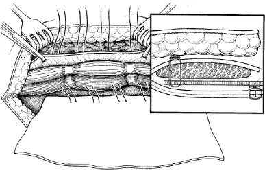
The Rives-Stoppa-Wantz retro-rectus repair is a subtype of the sublay method of mesh repair and involves the placement of the mesh deep to the rectus muscle and superficial to the posterior rectus sheath (where present) or deep to the rectus muscle and superficial to the fascia transversalis ( where the posterior rectus sheath is deficient)10. Recurrence rates in previous studies of Rives-Stoppa repairs range from zero to approximately 4%. Postoperative infections occurred in 0–18% of patients.
23
Figure 2 : Plane of mesh placement in Rives-Stoppa repair 10
24
Figure 3 : After mesh placement in Rives - Stoppa repair 10
The Underlay or intraperitoneal repair is done by placement of a dual sided mesh (with outer side formed by polypropylene and the inner peritoneal side formed by PTFE) intraperitoneally and fixing it to the anterior abdominal wall by using non absorbable synthetic sutures as shown in figure 3.
The Laparoscopic approach to ventral and incisional hernia repairs applies the underlay type of repair. Using the laparoscopic approach, a large prosthetic mesh is placed deep to posterior fascia or peritoneum), overlapping beyond the defect by several centimetres in all directions. With this technique, there is no need for the extensive soft tissue dissection seen in the open approach and its attendant
complications11. This type of repair has gained popularity because of the advances in laparoscopic instruments and easy availability of synthetic dual side mesh and
25
Additional methods are available as adjuncts when simple repair is inadequate or closure is not possible, because of tissue loss. The methods available include: Component separation method, and local flaps or free flaps.
The use of autologous tissue to repair abdominal wall hernias has also been described, but mainly used in the pre-mesh era. The tensor fascia lata, Sartorius and rectus femoris can be used as either free flaps or pedicled flaps to close large defects. However, the lack of sufficient tissue may require the insertion of prosthetic material or transposition of autologous material to bridge the fascial gap. These types of repairs were practised before the era of synthetic mesh use.
In 1990 Ramirez described a method of abdominal wall reconstruction without the use of a mesh. This method was initially described by a plastic surgeon and
26
complications occurred in a similar number. Thus it is concluded that this method for ventral hernia repair may be best used in the contaminated situation where the use of a mesh may not be safe. 8
Abdominoplasty or panniculectomy is a cosmetic procedure often used in obese patients with redundant fatty apron. This has been used along with incisional hernia repair in obese patients, in whom there is significant redundant abdominal fat. The abdominoplasty involved the use of a transverse suprapubic incision at the
superior aspect of the pubic tubercle till the anterior superior iliac spines or the use of an inverted t incision with the vertical limb in the midline. Following plication of the rectus diastases or the incisional hernia repair, the sufficient flaps of skin and
subcutaneous fat were excised, the umbilicus was repositioned and closure was carried out with subcutaneous vicryl and monocryl for skin closure. The main complications following these include seroma, hematoma, infection, wound-healing problems, and skin flap necrosis12.
Complications following incisional hernia repair
27
The incidence of seroma formation following open incisional hernioplasty have been found to vary from 4-21 % 51314. It is assumed that they occur due to serum and lymph leak, but also to some extent they are contributed to by the liquefied fat
following use of electrocautery as a method dissection 15. In several cases the seroma develops due to the presence of a dead space where fluid from the raw surfaces as well as unoccluded lymphatics and blood vessels can leak and form a collection at the surgical site. In most cases, they resolve over a period of few weeks to 3 months 10. Some studies have advocated on the placement of drains to reduce their incidence 16. In a few instances there have been infections to pre-existing seromas, complicating the wound healing resulting in readmission and prolonged hospital stay13. Seromas have been found to be almost double in cases where the onlay method of mesh repair has been used 14.
28
The incidence of infections following incisional hernia repair vary from 2% to 15 %
5,13,13). These infections varied from superficial to deep wound infections. The
manifestation may vary from erythema with tenderness, discharge; wound gaping to abdominal wall dehiscence. The follow up period was in-homogenous in the various studies evaluated. This remains the more serious among the common wound
complications following incisional hernia repair, as the extension of the infection to the peri-prosthetic space would entail eventual removal of the mesh and recurrence of the hernia.
Recurrence following incisional hernia repair still remains a potential problem on long term follow up of these patients. The recurrence rates vary depending on the period of follow up and are usually a late complication following incisional hernia repair. Studies have reported recurrence rates from 2.9 - 21 % 145,16.
The incidence of bowel injury, adhesions and fistulation vary depending on the method of mesh repair used, as also the mesh material used. Varying studies mention incidence of 2-4%15,16 . They are seen more often following inlay mesh repair, as there is contact of abdominal visceras with the mesh, which act as foreign bodies.
29
Seroma occurrence in other operations
The incidence of seroma following mastectomy and breast conservation surgery has been looked into in several studies. In summation, the cause of seroma formation has not been clearly elucidated, but is presumed to be due to exudates in response to the trauma of surgery and the acute inflammatory reaction which is a part of the process of wound healing. Some studies have described that the amount of seroma formation depends on the number and extent of lymph node involvement.17 While other studies have found that the only statistically significant predictor of seroma formation was the type of surgery done – that is, whether a mastectomy was done or a wide local excision with Axillary lymphadenectomy. They found that the incidence of seroma following a mastectomy was more than from other surgeries. No other factors, such as age, obesity, tumour size, neo-adjuvant therapy influenced the incidence of seroma formation 18,19. In mastectomies, it was found that damaged blood vessels and lymphatics continually oozed blood and lymphatic fluid
contributing to a seroma more in mastectomies and extensive dissections than it did for breast conserving surgeries. 20
30 Types of drains
A drain describes any material or equipment placed in a potential space within the body and usually brought out through a narrow opening on the skin surface to allow drainage of fluids accumulating in the space, allow tissue apposition and ensure faster healing rates. Allowing unwanted fluid or blood to remain in a wound may be a potential source of infection and may also impede healing, or result in wound
breakdown and dehiscence6.
There are a variety of drains available for use in different operations. Broadly wound drains are classified as closed or open drainage system.
Closed drains are constructed so as not to expose the contents to the
atmosphere. They include vacuum drains which apply negative pressure such as the Jackson-Pratt, Vario, Romovac drain, or non-vacuum systems such as the T-tube drain or the Robinson system. The use of a closed drainage system lowers the risk of
infection tracking into the potential space, avoids soiling and leaves the skin surface dry, easy to manage and reduces staff contact with body fluids.
31 The use of drains in incisional hernia repair
When repairing incisional hernias, the traditional practice involved to place drains to facilitate drainage of fluids which may collect in the potential space at the site of operation22. Following the incisional hernia repair, usually the preferred drain of choice is a closed drain, primarily because it is a closed system and the chance of infections entering the wound are lesser compared to the use of open drains. A closed drain is an artificial conduit that is left in the wound to allow drainage of fluids into a closed container. Presently, more than 50 % of the wounds following open incisional hernia repair have drains inserted14.
There have always been proponents for the use of drains following incisional hernioplasties and those who disagree that the routine use of drains has any additional benefit to the patients’ outcome. The proponents believe that all incisional hernias require drain placement to reduce the incidence of seromas or hematomas22. They also state that following incisional hernia repair, the occurrence of collections like hematomas predispose to recurrence.
32
chance of introducing infections into the wound24,14. Bauer et al were very
categorical in their recommendations that the routine use of subcutaneous drains at the time of surgery appears to have no effect on the complication rate. Seromas (19%) were the most common complications associated with incisional hernia repair, and virtually all seromas resorbed within weeks to 3 months following the operations.
A meta-analysis to look for previous studies comparing the use of drains in incisional hernia repair by Gurusamy et al did not find any studies comparing the outcome of drain versus no drain in incisional hernia repair. They found a study by Shafik et al which compared two types of drains – and electrified versus a corrugated drain placement following incisional hernia repair. A subgroup analysis by White et al did show that drains did not have any additional benefit or reduction in wound
complications14 .
The use of drain in other surgeries
Following face-lifts, there has been literature showing the incidence of seromas in the immediate post-operative period, which are prevented from accumulation by the placement of drains.
33
aspirations as opposed to 10.9% in the non-drain arm. They also showed a 2.8 % incidence of infection following drain placement as opposed to a 10.1% incidence in the no drain arm.25 Overall they had concluded that drain placement helped in reducing incidence of both seroma formations and infections following Axillary dissection and lumpectomy. In addition to the above, Scevola et al showed that following reconstruction following mastectomies with TRAM or DIEP flaps , the placement of 2 drains significantly helped in the reduction of seromas compared to only one drain placement.
A study by Tabaqchali et al failed to demonstrate that drainage in thyroid and parathyroid surgery was of any benefit to the patient. Other retrospective studies and randomised trials had also not shown any advantage in draining neck wounds
following thyroidectomy or parathyroidectomy. The studies showed that drains neither prevented postoperative haematoma nor facilitated their early diagnosis. The diagnosis of wound haematoma was made by observing the neck and noticing a progressive collection under the skin, as often, the neck drains became blocked with blood clots and did not function. 26 They also noted that the wound infection rate of 1% only occurred in patients who had drains placed in the neck.
The use of drains following head and neck procedures (submandibular gland excisions, parotidectomies – superficial as well as total parotidectomies,
34
There was a meta-analysis by Peng et al to assess the outcome following drain or no drain placement following pancreatic surgery. The studies considered had a high risk of bias and had low quality of evidence, but stated that there was inadequate evidence to establish the effect of drains on mortality at 30 days, mortality at 90 days, intra-abdominal infection, wound infection, morbidity, length of hospital stay, or additional open procedures for postoperative complications. There was one drain-related complication in the drainage group 27.
The meta-analysis by Charoenkwan et al, which looked at the rate of
lymphocoele after pelvic lymphadenectomy following surgeries for gynaecological malignancy, also showed no significant difference in retroperitoneal tube drain placement. In fact it showed that leaving the pelvic peritoneum open resulted in reduced risks of lymphocoele formation28.
Wang et al in a meta-analysis for gastrectomy found that there was no difference, between the two groups of - with drain and no-drain, in mortality; re-operations; post-operative complications ; wound infection; intra-abdominal abscess:; anastomotic leak; or initiation of soft diet. However, the addition of a drain prolonged the operation time and post-operative hospital stay and led to drain-related
complications29 .
35
36 JUSTIFICATION
The Null Hypothesis for the study was that the insertion of drains following open incisional hernia repair reduced the incidence of wound complications like seroma and hematoma formation, wound infections, but prolonged hospital stay.
According to a recent update of a meta-analysis looking into this very question, they were unable to select any randomized controlled trial with adequate blinding comparing 2 groups having undergone incisional hernia repair, with versus without the use of drains, but was able to find one study that compared two types of drains (corrugated drain versus electric drain). Majority of studies carried out about
incisional hernia repair; compare the outcomes of laparoscopic versus open method of incisional hernia repair. In addition, recent studies comparing the biochemical
37
38
MATERIALS AND METHODS
Study type: Non-inferiority, non-blinded randomised controlled trial.
Study design: Randomised controlled trial.
Setting: Surgery 4 unit, Department of General Surgery, CMC Hospital, Vellore.
Study population: Patients admitted to Surgery Unit 4 during the period of the study.
Study period: From 14th April 2014 to 26th August 2015.
Inclusion Criteria:
1. Age >18 years of age
2. Patients giving consent for participation in the trial.
3. Patients pre-operatively planned for sublay or onlay method of open incisional hernia repair
Exclusion criteria:
1. Patients not willing to participate in the study
2. Pre-operatively planned for laparoscopic or underlay method of hernioplasty
3. Patients planned for abdominoplasty, panniculectomy or components separation method of repair in association with mesh repair.
39
Withdrawal criteria:
Patients unwilling to continue participation in the study.
Sources of information:
1. Operation notes
2. Clinical evaluation pre and post-operatively 3. OPD Medical records
4. Telephonic follow-up
Outcome measures:
PRIMARY OUTCOME:
1. Incidence of seroma / hematoma formation
2. Incidence of Surgical site infections ( as described by CDC classification of Surgical site infections)
SECONDARY OUTCOME:
40
Statistical methods:
Data entry was done using the Epidata software version 3.1. Descriptive statistics were computed with use of the SPSS software (version 14). Sample size was calculated.
Data Analysis was done using SPSS software and p values were computed with Pearson’s Chi square.
Sample Size estimation:
Target sample size and rationale: 120 patients. Sample size: Seroma formation in
– 1. Previous study with drain - 23.3 % (n=86)~ 24 % = P – 2. Previous study without drain – 7.7 % (n=26) ~ 8% = Q – D= 16
– Formula: [(Zα + Z1-β)2 x P x Q ] / d2 – 10.4 x 24 x 8 / 16x16 = 55
– Sample Size – 55 in each study arm = 110 total – Considering drop-outs , Sample size = 120 total
With an α error of – 5%
41
Methodology:
Step 1: Recruitment
All patients fitting the inclusion criteria for the study were recruited by the primary investigator using a consent form in one of 4 suitable languages commonly spoken among patients.
Step 2: Randomisation
Method of randomization: Computer Generated randomization list
Method of allocation concealment: Allocation sheet to be kept by impartial administrator (department secretary). Envelopes were collected by the junior registrars and interns and placed in the charts of the consented patients prior to being shifted to the operation theatre. The envelopes were opened at the
completion of the mesh repair and prior to abdominal wall closure or skin closure as applicable. The randomisation was followed as per the allocation in the envelopes.
42
Step 3: Data collection:
Data collection was done in the ward on day 3 by the primary investigator or the concerned registrars posted in the unit on a rotatory basis. On day 7-10, corresponding roughly to the 1st visit to the outpatient clinic on follow-up, the data was entered into the electronic medical records by scanning the follow-up sheet. And the data was obtained by viewing the EMR records.
The patients were planned for an ultrasound between 7-10 days, and necessary arrangements done.
Telephone numbers were obtained at the time of consenting the patients or from the admission records and the patients were followed up at approximately 30 days from the date of the operation to assess if they had any wound complaints.
Data collected:
Name
Hospital number
Age
Sex
Residence
43 Phone no
Co morbidities: T2DM/ Sys HTN/ IHD/ CKD/ COPD/ Obstructive SAS/ BPH/ TB/ Malnutrition
H/o Smoking
If yes, No. of pack years
H/o Alcohol consumption
If yes, Amount per week
H/o Wound infection at previous surgical site
H/o Steroid intake
Weight
Height
BMI
Previous surgery done
Duration of incisional hernia
Size of defect
Type of repair done: Sublay/ Onlay / Other
Randomisation allocation
44
Postoperative day 3: Seroma / Hematoma: Clinical swelling? If yes, fluctuant?
Intervention done?
If drained – Nature of collection
Surgical site infection: Redness?
Tenderness
Purulent discharge
Wound Gaping
The same repeated on Post-operative Day 7
Ultrasound finding and volume of collection
Day 30: telephonic follow up to assess the presence or absence of any wound complications.
Date of operation
Date of discharge
45
Detailed diagrammatic algorithm of the study:
Study protocol:
46
history was taken and a physical examination done. The relevant risk factors of prior co-morbid illnesses, steroid intake and history of wound infections were taken.
They were randomised by picking an envelope which was numbered in serial order. The randomisation in the envelopes was done according to a computer generated randomisation list prepared prior to starting the study. The patients were randomised to drain placement or no drain placement.
Intraoperative procedure
– Open Incisional Hernia repair via incision over the defect.
– Plane to be created behind the rectus muscle (for sublay) and anterior to rectus sheath (for onlay).
– Polypropylene Mesh (Ethicon) to be used with a 5-6 cm extension beyond the defect.
– Linea alba to be closed by interrupted PDS.
Vario Drain to be placed in the subcutaneous plane or above the mesh.
– Skin to be closed by 3-0 monocryl
47
obvious seroma. There was telephonic follow up of any wound complications after being discharged home at post-operative day 30.
The outcomes of collection (seroma/ hematoma), surgical site infection and duration of hospital stay were recorded. The data collection was done in proformas.
Tools used:
[image:57.595.106.446.413.689.2]1. BMI calculation was done using the standard Body mass Index formula [BMI = Weight (in kg) / Height2 (in m)] and WHO approved classification of body mass index.
Table 1 : Who classification of body mass index32
Classification BMI(kg/m2)
Principal cut-off
points Additional cut-off points
Underweight <18.50 <18.50
Severe thinness <16.00 <16.00 Moderate thinness 16.00 - 16.99 16.00 - 16.99 Mild thinness 17.00 - 18.49 17.00 - 18.49
Normal range 18.50 - 24.99 18.50 - 22.99
23.00 - 24.99
Overweight ≥25.00 ≥25.00
Pre-obese 25.00 - 29.99 25.00 - 27.49 27.50 - 29.99
Obese ≥30.00 ≥30.00
Obese class I 30.00 - 34.99 30.00 - 32.49 32.50 - 34.99
48
2. CDC criteria for Surgical site infections
Centres for Disease Control and Prevention Criteria for Defining a Surgical Site Infection
Superficial Incisional
Infection less than 30 days after surgery
Involves skin and subcutaneous tissue only, plus one of the following: ▪ Purulent drainage
▪ Diagnosis of superficial surgical site infection by a surgeon ▪ Symptoms of erythema, pain, local oedema
Deep Incisional
Less than 30 days after surgery with no implant and soft tissue involvement
Infection less than 1 year after surgery with an implant; involves deep soft tissues (fascia and muscle), plus one of the following:
▪ Purulent drainage from the deep space but no extension into the organ space ▪ Abscess found in the deep space on direct or radiologic examination or on
reoperation
▪ Diagnosis of a deep space surgical site infection by the surgeon
▪ Symptoms of fever, pain, and tenderness leading to dehiscence of the wound or opening by a surgeon
Organ Space
Infection less than 30 days after surgery with no implant
Infection less than 1 year after surgery with an implant and infection; involves any part of the operation opened or manipulated, plus one of the following:
▪ Purulent drainage from a drain placed in the organ space
▪ Cultured organisms from material aspirated from the organ space ▪ Abscess found on direct or radiologic examination or during reoperation ▪ Diagnosis of organ space infection by a surgeon
49
Protocol variations: Any rules for
a. Interim analyses: Assessment for completeness of forms. Clarifications regarding criteria being used.
b. For withdrawal of participants: Patient discretion. c. For premature stopping of trial : Nil
Post Trial benefits and care: Post-operative care to be provided by clinicians of the concerned unit as per pre-existing protocol. Routine follow-up and management of complications to be done on follow-up OPD visit.
Funding – The ultrasound examinations done for the patients were paid for by the Fluid research grant given for the purpose of the study.
Cost of ultrasound – Abdomen and Pelvis (C rate):- Rs.600
No of patients: 120
Patients related expenditure: 600 x 120= 72,000
Stationary: 8,000
Total expenditure: 80,000.
Institutional Research Board approval and Ethical considerations:
50
51
CONSORT 2010 Flow Diagram
ENROLLMENT
ALLOCATION
FOLLOW-UP
ANALYSIS
Assessed for eligibility (n= 107)
Excluded (n=45 )
Not meeting inclusion criteria (n= 19)
Declined to participate (n= 10)
Other reasons (n= 16) – were missed
Analysed (n= 20)
Excluded from analysis (n=10) – randomisation not followed
Lost to follow-up (n= 1) – incorrect phone number
Discontinued intervention - not applicable (n=0) Allocated to intervention (No Drain placement) (n= 30)
Received allocated intervention (n=20)
Did not receive allocated intervention (n= 10)
Lost to follow-up (n=1) incorrect phone number
Discontinued intervention (n=0)
Allocated to intervention (Drain placement) (n=32)
Received allocated intervention (n= 32)
Did not receive allocated intervention (n=0)
Analysed (n=42)
52
DEMOGRAPHIC DETAILS
[image:62.595.115.481.181.400.2]AGE DISTRIBUTION
Figure 4 : Distribution of patients in age groups
53
[image:63.595.118.481.154.373.2]SEX DISTRIBUTION
Figure 5 : Sex distribution
The majority of patients in our study were females (82.3%).
54
[image:64.595.107.472.152.375.2]OCCUPATION
Figure 6: Type of occupation
The occupations of the patients were classified on the type of labour involved. Sedentary workers and housewives were classified as white collar and farmers and labourers were classified as blue collar workers.
55
[image:65.595.122.489.153.375.2]Regional Distribution
Figure 7: Regional distribution
The majority of the recruited patients were form the Tamil Nadu state, followed by Eastern and North-eastern states. 12.9% were from the neighbouring state, Andhra Pradesh.
The regional distribution of cases in our institution is because local patients are always more in number, but a number of patients – 27.5% come from East India to our
56
[image:66.595.120.511.150.414.2]COMORBID ILLNESSES
Figure 8 : Incidence of comorbid illnesses
Around 24.1% of patients in our study had Type 2 diabetes mellitus, 37.1% had systemic hypertension.
57
ANALYSIS
[image:67.595.103.530.315.633.2]In analysing our data, several parameters were looked into and their causative association with the outcome was assessed. The outcomes were classified into Seroma / Hematoma, Infections, and having had no adverse outcome by 1st OPD visit (7-10 days)
Table 2 : Analysis of comorbid illnesses
Variable Outcome P value
Infection No adverse outcome
Total number of patients Ischemic heart
disease
0 1 1 0.756
Chronic
kidney disease
1 1 2 0.077
COPD 0 1 1 0.756
Benign prostatic hypertrophy
0 1 1 0.756
Tuberculosis 0 0 0 N/A
58
[image:68.595.102.529.308.458.2]There were no patients who had developed a seroma with any of these comorbid illnesses. The incidence of infections was also minimal. Thus as can be seen (p values in table– Pearson’s Chi square), there is no correlation between the above comorbid illnesses and outcome.
Table 3 : Diabetes Mellitus
Seroma/Hematoma Infection No adverse outcome
Total
DM Present 3 (20%) 3 (20%) 9 (60%) 15 DM not
present
14 (29.8%) 2 (4.3%) 31 (66%) 47
Total 17 5 40 62
There were 15 patients in our study who had diabetes mellitus, of whom 40 % had adverse outcomes: 3 each had seroma / Infections.
59
Table 4 : Systemic Hypertension
Seroma/Hematoma Infection No adverse outcome
Total
Hypertension Present
7 (30.4%) 3 (13%) 13 (56.5%) 23 (100%)
Hypertension not present
10 (26.5%) 2 (5.1%) 27 (69.4%) 39 (100%)
Total 17 5 40 62
60
Table 5 : Hypothyroidism
Seroma/ Hematoma
Infection No adverse outcome
Total
Hypothyroidism absent
14 (26.9%) 4 (7.7%) 34 (56.4%) 52 (100%)
Hypothyroidism present
3 (30%) 1 (10%) 6 (60%) 10 (100%)
Total 17 5 40 62
61
Table 6 : Comparison of Sex Distribution and Outcome
Outcome
Sex Seroma/
Hematoma
Infection Uneventful Total
Male 6 (54.5%) 0 (0%) 5 (45.5%) 11 (100%) Female 11 (21.6%) 5 (9.8%) 35 (68.6%) 51 (100%) Total 17 (27.4%) 5 (8.1%) 40 (64.5%) 62 (100%)
There were a total of 11 males among whom there were no infections noted, but a 54.5% incidence of collections. There were a total of 51 females of whom 21.6% developed a collection and 9.8% developed an infection.
62
Table 7 : Smoking
Seroma/ Hematoma
Infection Collection Total
No smoking 16 (27.5%) 5 (8.4%) 38 (64.4%) 59 (100%) Smoking
present
1 (33.3%) 0 2 (66.67%) 3 (100%)
Total 17 5 40 62
There were 3 patients who were habituated to smoking among the study population of whom 1 had developed a seroma.
63
Table 8: Comparison of alcohol consumption to outcome
Seroma/ Hematoma
Infection Collection Total
No alcohol 17 (27.9%) 5 (8.2%) 39 (63.9%) 61 (100%)
Alcohol 0 0 1 1 (100%)
Total 17 5 40 62
64
Table 9: Comparison of size of defect to outcome
Seroma / Hematoma
Infection No adverse outcome
Total
Size <=20 cm2
14 (31.8%) 2 (4.5%) 28 (64.6%) 44 (100%)
Size > 20 cm2
3 (20%) 2 (13.3%) 10 (66.7%) 15 (100%)
Total 17 4 38 59
The size of the defect was calculated based on the length and width of the defect. Area of an ellipse =π x l/2 x b/2.
65
Table 10: Comparison of Width of defect to outcome
Seroma / Hematoma
Infection No adverse outcome
Total
Width <10cm 13 4 38 55
Width >=10cm
4 0 0 4
Total 17 4 38 59
There have been previous studies which showed the presence of more adverse outcomes following repair of incisional hernias with width > 10 cm.
66
Table 11: Comparison of BMI to outcome
As there were only 2 patients who were underweight, they were analysed with the group within normal BMI as opposed to the patients who were overweight and obese. The incidence of outcomes was not significantly reduced in patients with a normal BMI. (p value – 0.665 – Pearson’s Chi square)
BMI Seroma/Hematoma/
Infection
No adverse outcome
Total
Normal and underweight
8 (42.1%) 11 (57.9%) 19
Overweight 6 (28.6%) 15 (71.4%) 21
67
Table 12: Comparison of number of previous operations to outcome
Seroma / Hematoma
Infection No adverse outcome
Total
Single operation
11 (27.5%) 2 (5%) 27 (62.5%) 40 (100%)
Multiple operations
6 (27.3%) 2 (9.1%) 14 (63.6%) 22 (100%)
Total 17 4 41 62
Of 39 patients who had had a single operation 11 developed collections (27.5%),
2 developed infections (5%). Of 22 patients who had recurrent or multiple operations
6 developed collections (27.3%), 2 developed infections (9.1%). However, the p-value
68
Table 13 : Comparison of type of repair done versus outcome
There were a total of 6 cases in whom onlay repair was done of whom 50% developed collections. The other methods were a heterogenous group involving deviations from the preoperatively planned method of repair.
Studies have shown an increased incidence of seromas following onlay method 14 . But despite the above percentile correlation, the value as not found to be significant. (p value – 0.617 – Pearson’s Chi square)
Seroma / Hematoma
Infection No adverse outcome
Total
Sublay repair 12 (24.5%) 4 (8.1%) 33 (67.3%) 49 (100%)
Onlay repair 3 (50%) 0 3 (50%) 6 (100%)
Other (Underlay/ component’s sep)
2 (33.3%) 1 (16.7%) 3 (50%) 6 (100%)
69
Table 14: Comparison of duration of incisional hernia versus outcome
Seroma / Hematoma
Infection No adverse outcome
Total
<=1 year 11 (28.2%) 3 (7.7%) 25 (64.1%) 39 (100%) >1 year 6 (26.1%) 2 (8.7%) 15 (65.2%) 23 (100%)
Total 17 5 40 62
70
Table 15: Comparison of drain insertion to seroma/ hematoma and infection occurrence
Seroma/ Hematoma
Infection No adverse outcome
Total
Drain Not placed
7 (35%) 1 (5%) 12 (60%) 20 (100%)
Drain Placed 10 (23.8%) 4 (9.5%) 28 (66.7%) 42 (100%) Total 17 (27.4%) 5 (8.1%) 40 (64.5%) 62 (100%)
There were 20 cases in whom drain had not been placed and of them 7 (35%) had developed collections, 1(5%) had developed an infection, and 12 (60%) had had an uneventful recovery.
There was double the incidence of infection in the arm in which drain had been placed ( 9.5% vs. 5 %) supporting the assumption of some that drain placement had a higher incidence of infections14.
71
72
Table 16: Comparison of randomisation allocation to outcome
Seroma / hematoma
Infection No adverse outcome
Total
Randomised to no drain
8 (26.7%) 3 (10%) 19 (63.3%) 30 (100%)
Randomised to drain
9 (28.1%) 2 (6.3%) 21 (65.6%) 32 (100%)
17 5 40 62
There were 10 cases where randomisation had not been followed which were then analysed under the arm with drain placement.
What is significant to note is that, there is not significance seen in the outcome, had the randomisation been followed in the same 10 patients. (P value – 0.863 – Pearson’s Chi square)
73
Table 17: Comparison of Drain insertion to hospital stay
Hospital stay <=4 days
Hospital stay >4 days
Total
No Drain Group 15 (75%) 5 (25%) 20 (100%) With Drain
Group
11 (26.2%) 31 (73.8%) 42 (100%)
Total 26 36 62
There were 20 patients in whom a drain was not placed and 75 % among them had a hospital stay < or= 4 days. Of the 42 patients in whom a drain was
74
Figure 9 : Comparison of drain placement versus hospital stay
Table 18: Findings of Ultrasonography done
Collection Infection No adverse outcome
Total
No collection on ultrasound
1` 1 2 4
Collection on ultrasound
10 1 4 15
11 2 6 19
Of 62 patients in whom the study was done, 19 patients underwent an ultrasound scan.
[image:84.595.99.553.461.626.2]75
It was seen that majority of seromas (73.3%) were picked up clinically and showed a correlation with ultrasound detected collections. Thus, to assess the significance of collections detected on ultrasound, the patients followed up at day 30 were analysed to see if there was any long term morbidity associated with the seromas.
[image:85.595.134.407.355.526.2]Among the 15 Collections picked up by ultrasound, the long term morbidity on follow up till 30 days post-op showed the following:
Table 19: 30 day post-operative complications among ultrasound-assessed patients
No of patients Seroma / Hematoma 8 (53.33%)
Infection 1 (6.67%)
Infected collection 1 (6.67%) No adverse outcome 5 (33.33%)
Total 15 (100%)
Among 15 ultrasound-detected patients followed up till 30 days
76
Retrospectively analysing the data among the 10 patients in whom an adverse outcome was noted at day 30 post-operatively, it was found that
- 8 collections had initially been picked up clinically, while
- 1 patient who had been released from OPD follow up without an adverse outcome was found to have an adverse outcome at day 30
No of patients Initially picked up Collection 8
Initially picked up Infection 1
No adverse outcome 1
77
Table 20: Comparison of outcome to the need for intervention
Seroma/ Hematoma
Infection No adverse outcome
Total
Intervention done 8 (47.1%) 5 (100%) 0 13
Intervention not done
9 (52.9%) 0 40 (100%) 49
Total 17 (100%) 5 (100%) 40 (100%) 62
The interventions done included aspiration, drainage, laying open of wound and/ or reoperation.
In all the patients who had infections, there was a need to lay open the sutures and allow the wound to heal secondarily.
However, in 52.9% of seroma / hematoma formation no intervention was required, and 47.1% required aspiration or drainage.
78
79
The presence of wound related complications are common following incisional hernia repair. Overall, the incidence of wound related complications came to
approximately 1/3rd of the total hernia repairs (35%).
The incidence of seromas noted in our study was 24.1%. (15 / 62 patients), which is in keeping with the published literature.
The incidence of hematomas was 2 cases out of 62 (3.2%), in both of whom a drain had been placed.
As there were numerous collections for which no intervention was done and the nature of the contained fluid was not known, the seromas and hematomas were
grouped for the sake of the analysis as collections (27.3%). This was comparable to several prior studies which showed wound complication rates related to seroma and hematoma formation to be between 4-21% and 1.5-9.6% respectively.
The incidence of infections in our study was 8.1% (5 cases out of a total of 62), which compared to studies by White et al and Bauer et al as between 2-15%.
As per the per-protocol analysis for the data, there were 20 cases without drain placement and 42 cases with drain placement. As per the intention to treat analysis (ITT), there were 30 cases without drain placement and 32 cases with drain
80
There was no correlation between the obesity, co-morbid illnesses which included diabetes mellitus, systemic hypertension, hypothyroidism, asthma and the incidence of seroma, hematoma, wound infections following open incisional hernia repair. There was also no correlation between size of hernia defect [as measured by π x l/2 x b/2 (cm2)] , duration of the presence of incisional hernia, number of previous operations or type of repair done with the presence of wound related complications.
There was a higher incidence of wound complications in patients for whom the width of the incisional hernia was more than 10 cm. This is found to be concordant with White et al.14 The p valuefor the same was not significant though.
There was a significant reduction in the duration of hospital stay in those
patients on whom no drain was placed. 75 % of patients in whom no drain was placed had a hospital stay of <= 4 days. 73.8% of patients in whom drains were placed had a hospital stay of >4 days. This was found to be statistically significant (p value <0.05)
Among the patients for whom an ultrasound was done (19/62), there were collections in 15 patients detected by ultrasound. Of these:
- 10 were detected clinically also,
- 1 was detected to have an infection,
- 4 were not found to have significant collection clinically.
81
outcome. Of the total numbers of seroma / hematoma picked up 52.9% did not require any intervention and resolved by itself.
82 LIMITATIONS
Limitations noted in the study include:
1. The randomisation allocation was not followed for 12 cases, resulting in selection bias.
2. The sample size as calculated was not reached, due to a variety of causes: a. Cases missed.
b. Patients not consenting to participate in a trial.
3. The ultrasound examination in incisional hernia could not be uniformly applied as patients were not compliant due to
a. Logistical reasons – other appointments coiniciding with time slot of ultrasound.
b. Wound complications limiting the travel of the patient to the ultrasound room.
83
84 Conclusion 1:
A. The incidence of seroma or hematoma with or without the use of drain did not vary significantly.
B. The incidence of infection with or without the use of drain did not vary significantly
Conclusion 2:
85
REFERENCES
1. From ancient to contemporary times: a concise history of incisional hernia repair - Springer. at <http://link.springer.com/article/10.1007%2Fs10029-011-0870-5> 2. Papavramidou, N. & Christopoulou-Aletra, H. The Ancient Technique of
‘Gastrorrhaphy’. J. Gastrointest. Surg.13, 1345–1350 (2009).
3. van’t, R. M. T., De Vos Van Steenwijk, P. J., Bonjer, H. J., Steyerberg, E. W. & Jeekel, J. Incisional hernia after repair of wound dehiscence: incidence and risk factors. Am. Surg.70, 281–286 (2004).
4. Muysoms, F. E. et al. Classification of primary and incisional abdominal wall hernias. Hernia13, 407–414 (2009).
5. Kingsnorth, A., Sivarajasingham, N., Wong, S. & Butler, M. Open mesh repair of incisional hernias with significant loss of domain. Ann. R. Coll. Surg. Engl.86,
363–366 (2004).
6. Gurusamy, K. S., Allen, V. B. & Samraj, K. Wound drains after incisional hernia repair. Cochrane Database Syst. Rev.2, CD005570 (2012).
7. Ellis, H., Gajraj, H. & George, C. D. Incisional hernias: when do they occur? Br. J. Surg.70, 290–291 (1983).
8. Fischer Josef. Mastery of Surgery 5th edition.
9. Burger, J. W. A. et al. Long-term Follow-up of a Randomized Controlled Trial of Suture versus Mesh Repair of Incisional Hernia. Ann. Surg.240, 578–585 (2004). 10. Bauer, J. J., Harris, M. T., Gorfine, S. R. & Kreel, I. Rives-Stoppa procedure for
86
11. Franklin, M. E., Gonzalez, J. J., Glass, J. L. & Manjarrez, A. Laparoscopic ventral and incisional hernia repair: an 11-year experience. Hernia J. Hernias Abdom. Wall Surg.8, 23–27 (2004).
12. Grieco, M., Grignaffini, E., Simonacci, F. & Raposio, E. Analysis of
Complications in Postbariatric Abdominoplasty: Our Experience. Plast. Surg. Int.
2015, 209173 (2015).
13. Helgstrand, F., Rosenberg, J., Kehlet, H., Jorgensen, L. N. & Bisgaard, T.
Nationwide prospective study of outcomes after elective incisional hernia repair. J. Am. Coll. Surg.216, 217–228 (2013).
14. White, T. J., Santos, M. C. & Thompson, J. S. Factors affecting wound complications in repair of ventral hernias. Am. Surg.64, 276–280 (1998).
15. Porter, K. A., O’Connor, S., Rimm, E. & Lopez, M. Electrocautery as a factor in seroma formation following mastectomy. Am. J. Surg.176, 8–11 (1998).
16. Scevola, S., Youssef, A., Kroll, S. S. & Langstein, H. Drains and seromas in TRAM flap breast reconstruction. Ann. Plast. Surg.48, 511–514 (2002).
17. Petrek, J. A. et al. Axillary lymphadenectomy. A prospective, randomized trial of 13 factors influencing drainage, including early or delayed arm mobilization.
Arch. Surg. Chic. Ill 1960125, 378–382 (1990).
18. Hashemi, E. et al. Seroma formation after surgery for breast cancer. World J. Surg. Oncol.2, 44 (2004).
87
20. Budd, D. C., Cochran, R. C., Sturtz, D. L. & Fouty, W. J. Surgical morbidity after mastectomy operations. Am. J. Surg.135, 218–220 (1978).
21. Agrawal, A., Ayantunde, A. A. & Cheung, K. L. Concepts of seroma formation and prevention in breast cancer surgery. ANZ J. Surg.76, 1088–1095 (2006). 22. George, C. D. & Ellis, H. The results of incisional hernia repair: a twelve year
review. Ann. R. Coll. Surg. Engl. 68, 185–187 (1986).
23. Hesselink, V. J., Luijendijk, R. W., de Wilt, J. H., Heide, R. & Jeekel, J. An evaluation of risk factors in incisional hernia recurrence. Surg. Gynecol. Obstet.
176, 228–234 (1993).
24. Amir, I., Morar, P. & Belloso, A. Postoperative drainage in head and neck surgery.
Ann. R. Coll. Surg. Engl.92, 651–654 (2010).
25. Somers, R. G., Jablon, L. K., Kaplan, M. J., Sandler, G. L. & Rosenblatt, N. K. The use of closed suction drainage after lumpectomy and axillary node dissection for breast cancer. A prospective randomized trial. Ann. Surg.215, 146–149 (1992).
26. Tabaqchali, M. A., Hanson, J. M. & Proud, G. Drains for
thyroidectomy/parathyroidectomy: fact or fiction? Ann. R. Coll. Surg. Engl. 81,
302–305 (1999).
27. Peng, S. et al. Prophylactic abdominal drainage for pancreatic surgery. Cochrane Database Syst. Rev.8, CD010583 (2015).
88
formation in patients with gynaecological malignancies. Cochrane Database Syst. Rev.6, CD007387 (2014).
29. Wang, Z., Chen, J., Su, K. & Dong, Z. Abdominal drainage versus no drainage post-gastrectomy for gastric cancer. Cochrane Database Syst. Rev.5, CD008788 (2015).
30. Rondelli, F. et al. To drain or not to drain extraperitoneal colorectal anastomosis? A systematic review and meta-analysis. Colorectal Dis. Off. J. Assoc.
Coloproctology G. B. Irel.16, O35–42 (2014).
31. Daams, F., Wu, Z., Lahaye, M. J., Jeekel, J. & Lange, J. F. Prediction and diagnosis of colorectal anastomotic leakage: A systematic review of literature.
World J. Gastrointest. Surg.6, 14–26 (2014). 32. WHO :: Global Database on Body Mass Index. at
<http://apps.who.int/bmi/index.jsp?introPage=intro_3.html>
33. Mangram, A. J., Horan, T. C., Pearson, M. L., Silver, L. C. & Jarvis, W. R. Guideline for prevention of surgical site infection, 1999. Hospital Infection
89
ANNEXURES
I. INFORMATION SHEET II. CONSENT FORM
90
I. INFORMATION SHEET
INFORMATION SHEET
Randomised controlled trial comparing outcome following incisional hernia
repair with Vs without drains
You are being requested to participate in a study to assess whether the
presence or absence of drains during incisional hernia repair improves
outcome. The repair of incisional hernias is usually associated with placement
of drains. This study aims to assess whether drains have any measurable
benefit in the outcome following surgery. We hope to include about 110
people from this hospital in the study.
Aims of study –
To assess the outcome following incisional hernia repair in the two different
groups.
Reasons for this study –
Drains are traditionally placed in the plane of surgery following incisional
hernia repair, presumably to drain fluids accumulating in the surgical plane –
blood, serous discharge or pus. In addition, serous discharge, if localised, may
predispose for infection, which may require further operation and they lead to
mesh extraction. On the other hand, drains are known to have several
complications including wound discomfort, prolonged hospital stay, pain,
erosion into wound/ neurovascular structures, tract for introduction for
infection, blockage and failure of drain, drain fracture. Studies have shown
conflicting reports concerning the use of drains and there is no clear
91
Risk factors –
Complication in absence of drain insertion may include increased seroma or
hematoma accumulation and consequent infection. Complication in presence
of drain insertion may include wound discomfort, prolonged hospital stay,
pain, erosion into wound/ neurovascular structures, tract for introduction for
infection, blockage and failure of drain, drain fracture.
In case of development of any complications, the treatment done for the same
would be done free of cost. But there would be no additional remuneration.
Neither the investigator, nor the concerned unit will know in advance, if a
drain will be placed for you or not after the surgery.
Do I have the right to opt out of the concerned study, at a later date, if I
change my mind?
Yes. You may opt out of the study at any time and treatment would continue
as per the treatment guidelines of the concerned unit.
What do I gain out of this study? –
You may be the beneficiary of a favourable outcome. Your treatment will
continue as per the guidelines followed by the unit in charge, regardless of
whether you are participating in the study or not.
Will my identity be revealed by this study? –
The study may be taken up for publishing in which case your identity will be
preserved. However, your case notes and case information may be scrutinised
and used in the future, by the principal investigator or the unit treating you, for
which separate consent will not be taken at a later time.