A Dissertation on
PROGNOSTIC INDICATORS OF MORBIDITY AND MORTALITY IN NON DIABETIC SOFT TISSUE INFECTIONS
Dissertation submitted to
THE TAMIL NADU Dr.M.G.R.MEDICAL UNIVERSITY CHENNAI.
With partial fulfilment of the regulations
For the Award of the degree of
M.S. GENERAL SURGERY
Branch – I
MADRAS MEDICAL COLLEGE
THE TAMIL NADU Dr.M.G.R.MEDICAL UNIVERSITY CHENNAI
CHENNAI.
BONAFIDE CERTIFICATE
Certified that this dissertation is the bonafide work of Dr. ANITHA
MUTHUSAMI on “PROGNOSTIC INDICATORS OF MORBIDITY AND
MORTALITY IN NON DIABETIC SOFT TISSUE INFECTIONS”during her M.S.
(General Surgery) course from April 2011 to April 2014 at the Madras Medical College
and Rajiv Gandhi Government General Hospital, Chennai.
Prof. Dr. S.DEIVANAYAGAM M.S. Prof. Dr. R.G.SANTHASEELAN M.S.,
Professor and head of the department Prof. of General Surgery,
Dept. of General Surgery, Dept. of General Surgery,
Madras Medical College & RGGGH Madras Medical College & RGGGH
Chennai – 600 003. Chennai – 600 003.
Prof. Dr. V. KANAGASABAI, M.D.,
DEAN
Madras Medical College &Rajiv Gandhi Government General Hospital,
ACKNOWLEDGEMENT
I would like to express my sincere thanks to the Dean of the
Madras medical College Prof. Dr. V.KANAGASABAI, M.D., for permitting me to
conduct the study in Madras Medical College & Government General Hospital.
With deep sense of gratitude and respect, I take this opportunity to
thank my teacher and professor Dr.R. G. SANTHASEELAN M.S., for his constant
inspiration, able guidance and kind help which he rendered in preparing this dissertation
and in pursuit of my post graduate studies.
I also thank the Registrar of Department of Surgery DR. A.MUTHURAJA
M.S., who helped me to progress through this study with great interest.
I am very grateful to all the Assistant Professors of my unit
DR.G.GAJENDRARAJ M.S., DR. P. RAMADOSS M.S.,
DR. A. SAGAYA INBA SEKAR M.S., for the constant support and guidance through
my study.
Last but not the least my heartfelt gratitude to the patients for their
CONTENTS
Title
Page No.
INTRODUCTION
4
AIM OF STUDY
6
REVIEW OF LITERATURE
7
PATIENTS & METHODS
55
RESULTS
61
DISCUSSION
97
CONCLUSION
102
ANNEXURE
PROFORMA
105
Introduction
Soft tissue skin infections (SSTI’s) were first described in the Hippocratic
era. The principles of management, including early diagnosis with prompt and
repeated surgical debridement, aggressive resuscitation and physiological support,
broad spectrum antimicrobial drugs, and nutritional support, have been well
documented. Despite this well accepted management approach, the mortality rate
remains between 16 – 34% in most major published series. Due to the lack of
defined criteria to determine the type of treatment that has to be given for patients
at the time of admission, most patients undergo multiple surgical procedures which
increases the morbidity and mortality.
Aim of the study
The primary objective of this analysis is to create a simple clinical score to
aid in the prediction of morbidity defined by the number of days of hospital stay or
limb loss and mortality in patients with SSTIs at the time of first assessment. The
scoring system may further be used to predict limb loss at first assessment, thereby
reducing multiple surgeries for the same patient.
Methods
A retrospective review of 200 consecutive patients with necrotizing soft
tissue infections, treated at Rajiv Gandhi Government General Hospital during a
1-year period, was conducted. Using a model for logistic regression analysis,
characteristics of each patient and his/her clinical course were tested for impact on
Results
The scoring system decreased the number of surgeries undergone by each
patient significantly. The scoring system also reduced the number of days of
hospital stay per patients, though not significantly. The use of the scoring system
did not alter the mortality in any way.
Conclusion
Skin and soft tissue infections of the limbs have a high mortality and
morbidity especially if necrosis is present. The morbidity is in the form of
prolonged hospital stay and limb loss. Further detailed studies are required to
produce repeated significant results, which is essential for the scoring system to be
INTRODUCTION
Soft tissue skin infections (SSTI’s) were first described in the
Hippocratic era. In time various surgeons described the disease process in
details. The most well documented among these is the work of Joseph Jones
a confederate army surgeon, who reported 2,642 cases of “hospital
gangrene” with a mortality rate of 46%. Since then, multiple reports and
classification systems have been published in an attempt to define this
disease better and achieve lower mortality rates with better outcomes. The
principles of management, including early diagnosis with prompt and
repeated surgical debridement, aggressive resuscitation and physiological
support, broad spectrum antimicrobial drugs, and nutritional support, have
been well documented. Despite this well accepted management approach,
the mortality rate remains between 16 – 34% in most major published series.
Over the last decade, there has been an interest in understanding SSTIs
better. Some investigators have focused on methods that aid in early
diagnosis so that surgical debridement can be accomplished promptly,
whereas other researchers have focused on identifying patients at higher risk
of death. Although several predictors of death have been identified,
Due to the lack of defined criteria to determine the type of treatment
that has to be given for patients at the time of admission, most patients
undergo multiple surgical procedures which increases the morbidity and
mortality. The primary objective of this analysis is to create a simple clinical
score to aid in the prediction of morbidity defined by the number of days of
hospital stay or limb loss and mortality in patients with SSTIs at the time of
first assessment. The scoring system may further be used to predict limb loss
AIMS OF THE STUDY
The aims of the study is,
1) To establish a scoring system to predict the outcome of a patient with
non diabetic soft tissue limb infection at the time of admission.
2) To determine the factors which increase the morbidity of a patient with
non diabetic soft tissue limb infection as determined by no. Of days of
REVIEW OF LITERATURE HISTORY
Skin and soft tissue infections have been described since 5th century BC. The first clear reference to necrotizing fasciitis (NF) dates back to the
5th century BC, with Hippocrates’ description of a fatal infection,
“Many were attacked by the erysipelas all over the body when the
exciting cause was a trivial accident. The erysipelas would quickly spread
widely in all directions. Flesh, sinews and bones fell away in large
quantities…Fever was sometimes present and sometimes absent…There
were many deaths. The course of the disease was the same to whatever part
of the body it spread."[1]
From the 18th and 19th century the British naval surgeons referred to
the necrotizing fasciitis (NF) as "hospital gangrene." Indeed the first modern
report that describes a detailed case of "hospital gangrene" was reported by
Joseph Jones, a Confederate Army Surgeon during the American Civil War.
He was the first person to describe this disorder in a large group
of patients. He reported 2,642 cases and found
patients.[3] The condition that bears his name is now described in both male and female patients.
A major advance took place in 1924 when Meleney and Breuer
isolated streptococcal infection as the prime cause of lethal NF[4]. Before the
advent of the antibiotics, NF was treated successfully with "bear-claw
scratch debridement" and tubes irrigating the tissues with Dakin's solution of
chlorinated sofa[5]. In 1924, a Beijing missionary surgeon reported similar conditions among the opium addicts.
Over the years, many terms have been developed and used such as
flesh-eating bacteria syndrome, suppurative fasciitis, and streptococcal
gangrene."Meleney's gangrene" is commonly used for abdominal fasciitis,
but strictly speaking should be streptococcal dermal gangrene on any part of
body. The term "necrotizing fasciitis" was coined by Wilson in 1952, to
delineate the histological appearance of the disease, that is, an invasive
necrotizing infection involving deep fascia and soft tissue. [6]
From 1987 to 1990, scattered outbreaks of NF were reported in both
the USA and Scandinavia, while significant media attention focused on a
there has been an increase in the incidence of severe streptococcal infection
throughout the 20th century, resulting in more cases of NF being identified and treated.[9] While most Westernised countries are said to have an incident rate of around one in every 100,000 people, the latest Indian statistics
suggests an incidence of 4 in every 100,000 people.[10] Recently, the term necrotizing soft tissue infection
has been adopted.[11]
DEFINITION
Skin and soft tissue infections (SSTI) are classified into complicated
and uncomplicated infections.[11] Uncomplicated SSTI’s include cellulitis, erysipelas, simple abscesses,impetigo, ecythyma, follicilitis, furunculosis
and carbuncles. They are superficial infections and have a low mortality and
morbidity (limb loss). They can be treated by antibiotic therapy and drainage
procedures.
Complicated SSTI’s includes necrotizing soft tissue
infections, complicated abscesses, infected burn wound, infected ulcers,
infections with significant underlying disease states that complicates
response to treatment (e.g. DM). These are usually deeper infections with
Necrotizing soft tissue infections by definition include the presence of
necrotic or devitalized tissue as part of the pathophysiology[13].Necrotic tissue provides a growth medium for bacteria and precludes delivery
of host defence mechanisms and antimicrobial agents[13]. Necrotizing soft
tissue infections includes, necrotizing cellulitis (involvement of dermal and
subcutaneous layers), necrotizing fasciitis (involvement of deep fascia) and
pyomyositis or myonecrosis or a combination of any of the above.[14]
PATHOPHYSIOLOGY
Necrotizing fasciitis is characterized by widespread necrosis of the
subcutaneous tissue and the fascia. Although the pathogenesis of necrotizing
fasciitis is still open to speculation, the rapid and destructive clinical course
of necrotizing fasciitis is thought to be due to multibacterial symbiosis and
infection is usually associated with an underlying cause, such as
diabetes,[16] atherosclerotic vascular disease, or venous insufficiency with oedema. GABS usually affects the extremities; approximately two thirds of
the GABS infections are located in the lower extremities.[17]
During the last 2 decades, researchers have found that necrotizing
fasciitis is usually polymicrobial rather than monomicrobial.[18, 19,
20]
Anaerobic bacteria are present in most necrotizing soft-tissue infections,
usually in combination with aerobic gram-negative organisms. Anaerobic
organisms proliferate in an environment of local tissue hypoxia in those
patients with trauma, recent surgery, or medical compromise. Facultative
aerobic organisms grow because polymorphonuclear neutrophils (PMNs)
exhibit decreased function under hypoxic wound conditions. This growth
further lowers the oxidation/reduction potential, enabling more anaerobic
proliferation and, thus, accelerating the disease process. Carbon dioxide and
water are the end products of aerobic metabolism. Hydrogen, nitrogen,
hydrogen sulphide, and methane are produced from the combination of
aerobic and anaerobic bacteria in a soft tissue infection. These gases, except
necrotizing fasciitis, group A haemolytic streptococci and Staphylococcus
aureus, alone or in synergism, are frequently the initiating infecting bacteria.
However, other aerobic and anaerobic pathogens may be present, including
the following:
Bacteroides Clostridium
Peptostreptococcus Enterobacteriaceae
Coliforms (eg, Escherichia coli)
Proteus
Pseudomonas
Klebsiella
Bacteroides fragilis is usually noted as part of a mixed flora in
combination with E coli. B fragilis does not directly cause these infections,
but it does play a part in reducing interferon production and the phagocytic
capacity of macrophages and PMNs.
A variant synergistic necrotizing cellulitis is considered to be a form of
necrotizing fasciitis, but some authorities feel that it is actually a
necrotizing fasciitis, but it progresses rapidly to involve wide areas of deeper
tissue and muscle at an earlier stage than might be expected. Severe systemic
toxicity occurs.
Anaerobic streptococci, occasionally seen in intravenous drug users,
cause many forms of nonclostridial myonecrosis. Some cases of necrotizing
fasciitis can be caused by Vibrio vulnificus. This organism is seen more
often in patients with chronic liver dysfunction, and it often follows the
consumption of raw seafood. V vulnificus may cause subcutaneous
bleeding.[23,24]
Organisms spread from the subcutaneous tissue along the superficial
and deep fascial planes, presumably facilitated by bacterial enzymes and
toxins. This deep infection causes vascular occlusion, ischemia, and tissue
necrosis. Superficial nerves are damaged, producing the characteristic
localized anaesthesia. Septicaemia ensues with systemic toxicity.
A subset of virulence factors, for instance, SpeB and Ska/ Plasmin
directly damage the host tissues, degrade the extracellular matrix proteins,
and induce vascular dissemination via their enzymatic pathway[26]. Other virulence factors such as SpyCEP and Mac1/IdeS indirectly damage the host
of proapoptotic molecules. Host matrix metalloproteninases (MMPs) and
coagulopathy are also implicated.
Important bacterial factors include surface protein expression and toxin
production. M-1 and M-3 surface proteins, which increase the adherence of
the streptococci to the tissues, also protect the bacteria against phagocytosis
Organisms undergo a very complex molecular transition during the
progression from a localized to an invasive infection. SpeB, a
broad-spectrum cysteine protease virulence factor, is regulated by multiple
intersecting and collateral pathways that respond to different environmental
stimuli. SpeB is repressed, activated and regulated by multiple regulatory
pathways. The combined effects result an in vivo temporal-spatial
expression pattern of SpeB.
Streptococcal pyrogenic exotoxins[27] (SPEs) A, B, and C are directly toxic and tend to be produced by strains causing necrotizing fasciitis. These
pyrogenic exotoxins, together with streptococcal superantigen (SSA), lead to
the release of cytokines and produce clinical signs such as hypotension. The
enterotoxin gene cluster seg, sei, sem, sen, and seo, but lacking all common
toxin genes, including Panton-Valentine leukocidin.[28]
The poor prognosis associated with necrotizing fasciitis has been
linked to infection with certain streptococcal strains. Community-acquired
methicillin-resistant S aureus (MRSA) has also been associated with
necrotizing fasciitis.[29]
Single-nucleotide changes are the most common cause of natural
genetic variation among members of the same species. They may alter
bacterial virulence; a single-nucleotide mutation in the group
A Streptococcus genome was identified that is epidemiologically associated
with decreased human necrotizing fasciitis.[30]
It was found that wild-type mtsR function is required for group
A Streptococcus to cause necrotizing fasciitis in mice and nonhuman
primates. It was speculated that a naturally occurring single-nucleotide
mutation dramatically alters virulence by dysregulating a multiple gene
virulence axis.
Severe myositis accompanying septic necrotizing fasciitis may be
aureus strain.[31] Immunostaining may document strong binding of the Panton-Valentine leukocidin toxin to necrotic muscle tissues.
Although necrotizing fasciitis most frequently develops after trauma
that compromises skin integrity, it may rarely develop in a healthy person
after minor trauma such as an isolated shoulder sprain that occurred without
a break in skin barrier.[32]
ETIOLOGY
Surgical procedures may cause local tissue injury and bacterial
invasion, resulting in necrotizing fasciitis. These procedures include surgery
for intraperitoneal infections and drainage of ischiorectal and perianal
abscesses. Intramuscular injections and intravenous infusions may lead to
necrotizing fasciitis.
Minor insect bites may set the stage for necrotizing infections.
Streptococci introduced into the wounds may be prominent initially, but the
bacteriologic pattern changes with hypoxia-induced proliferation of
anaerobes.
Local ischemia and hypoxia can occur in patients with systemic
systemic diseases favouring the development of these infections. Illnesses
such as diabetes or cancer have been described in over 90% of cases of
progressive bacterial gangrene.
Of patients with necrotizing fasciitis, 20-40% are diabetic. As many as
80% of Fournier gangrene cases occur in people with diabetes. In some
series, as many as 35% of patients were alcoholics. However, approximately
one half of the cases of streptococcal necrotizing fasciitis occur in young and
previously healthy people.
Studies have shown a possible relationship between the use of
nonsteroidal anti-inflammatory agents (NSAIDs), such as ibuprofen, and the
development of necrotizing fasciitis during varicella infections. Additional
studies are needed to establish whether ibuprofen use has a causal role in the
development of necrotizing fasciitis and its complications during varicella
infections. This has not previously been described.
Group A beta-haemolytic streptococci have historically been noted as a
cause of necrotizing fasciitis, but Haemophilus aphrophilus and S aureus are
also associated with the condition, and some patients have mixed infections
involving multiple species of bacteria, including mycobacteria, as well as
A synergistic infection with a facultative anaerobic bacterium may be
significant. In 1 patient, Phycomycetes appeared to be responsible for
necrotizing fasciitis.
Streptococcus pneumoniae is a rare cause of necrotizing fasciitis.[33] In one patient, S pneumoniae serotype 5 was also isolated. This serotype 5
antigen is included in the polysaccharide 23-valent pneumococcal vaccine,
highlighting the value of pneumococcal immunization.
In type I necrotizing fasciitis[21], anaerobic and facultative bacteria work synergistically to cause what may initially be mistaken for a simple
wound cellulitis. A variant of type I necrotizing fasciitis is saltwater
necrotizing fasciitis in which an apparently minor skin wound is
contaminated with saltwater containing a Vibrio species.
In type II necrotizing fasciitis,[22] varicella infection and the use of nonsteroidal anti-inflammatory drugs may be predisposing factors.
Type III necrotizing fasciitis is usually caused by Clostridium
perfringens[25]. When type III necrotizing fasciitis occurs spontaneously, C septicum is more likely to be the etiologic agent; these cases usually occur in
Unusual causes include injection anthrax.[35] Rapidly progressive necrotizing fasciitis following a stonefish sting has been described in 2
patients.[36]
PREDISPOSING OR RISK FACTORS FOR NECROTIZING FASCIITIS[37,38]
PROGNOSIS
The reported mortality in patients with necrotizing fasciitis has ranged
from 20% to as high as 80%.[25, 27, 39] Pathogens, patient characteristics, infection site, and speed of treatment are among the variables that affect
Poor prognosis in necrotizing fasciitis has been linked to infection with
certain streptococcal strains. However, McHenry et al found that
monomicrobial infection with S pyogenes was not associated with an
increased mortality.[27]
A retrospective study by Hsiao et al found
that Aeromonas infection, Vibrio infection, cancer, hypotension, and band
form WBC count greater than 10% were independent positive predictors of
mortality in patients with necrotizing fasciitis, while streptococcal and
staphylococcal infections were not identified as predictors of mortality.
Hemorrhagic bullae appeared to be an independent negative predictor of
mortality. However, accuracy of these factors needs to be verified.[40]
In another study, pre-existing chronic liver dysfunction, chronic renal
failure, thrombocytopenia, hypoalbuminemia, and postoperative dependence
on mechanical ventilation represented poor prognostic factors in
monomicrobial necrotizing fasciitis. In addition, patients with gram-negative
monobacterial necrotizing fasciitis had more fulminant sepsis.[41]
The mean age of survivors is 35 years. The mean age of non survivors
A retrospective review by Cheng et al showed that upper extremity
necrotizing fasciitis has a high mortality rate. In their review, about 35% of
patients died. A state of altered consciousness and respiratory distress at
initial presentation were found to be statistically significant factors for
eventual mortality. Early diagnosis and referral for aggressive surgical
treatment prior to the development of systemic toxic signs are essential for
survival.[42]
In a retrospective review of craniocervical necrotizing fasciitis, Mao et
al reported a survival rate of 60% for patients with thoracic extension (6 of
10) compared with 100% for those without thoracic extension. Lower
overall survival for the patients in the thoracic extension group was
attributed to older patient age, greater co morbidity, need for more extensive
surgical debridement, and increased postoperative complications.
Better survival of the patients without thoracic extension was attributed
to aggressive wound care and surgical debridement, broad-spectrum
intravenous antibiotics, and care in the surgical intensive care unit.[43]
In a study by Rouse et al, the overall mortality rate was 73% (20 of 27
necrotizing fasciitis was essential: Of 12 patients whose treatment was
delayed for more than 12 hours, 11 patients died.[25]
Similarly, McHenry et al reported that the average time from
admission to operation was 90 hours in nonsurvivors of necrotizing
soft-tissue infections; in survivors, this average time was 25 hours.[27] Early debridement of the infection was obviously associated with a significant
decrease in mortality.
Necrotizing fasciitis survivors may have a shorter life span than
population controls, owing to infectious causes such as pneumonia,
cholecystitis, urinary tract infections, and sepsis.[44]
COMMON SITES OF INFECTION
While any area of the body can succumb to NF, the most common sites
are the extremities, the abdominal wall, the perianal and groin area and
post-operative wounds.
CLINICAL PRESENTATION
Diagnosis of necrotizing fasciitis can be difficult and requires a high
degree of suspicion. In many cases of necrotizing fasciitis, antecedent
trivial, such as an insect bite, minor abrasion, boil, or injection site.
Idiopathic cases are not uncommon, however.
Olafsson et al indicate that the hallmark symptom of necrotizing
fasciitis is intense pain and tenderness over the involved skin and underlying
muscle.[45] The intensity of the pain often causes suspicion of a torn or ruptured muscle. This severe pain is frequently present before the patient
develops fever, malaise, and myalgias.
In some cases, the symptoms may begin at a site distant from the initial
traumatic insult. Pain may be out of proportion to physical findings. Over the
next several hours to days, the local pain progresses to anaesthesia.
Other indicative findings include oedema extending beyond the area of
erythema, skin vesicles, and crepitus. McHenry et al and others have noted
that the subcutaneous tissue demonstrates a wooden, hardened feel in cases
of necrotizing fasciitis.[26] The fascial planes and muscle groups cannot be detected by palpation.
A history of co morbid factors, including diabetes mellitus, should be
Physical findings may not be commensurate with the degree of patient
discomfort. Early in the disease course, the patient may look deceptively
well; unfortunately, this may interfere with early detection, which is key to a
favourable outcome. Soon, however, the patient will usually begin to appear
moderately to severely toxic.
Typically, the infection begins with an area of erythema that quickly
spreads over a course of hours to days. The redness quickly spreads, and its
margins move out into normal skin without being raised or sharply
demarcated. As the infection progresses, the skin near the site of insult
develops a dusky or purplish discoloration. Multiple identical patches
expand to produce a large area of gangrenous skin, as the erythema
continues to spread.
Iwata et al reported that 2 of 3 patients who lacked inflammatory signs
such as redness and heat experienced fulminant progression of necrotizing
fasciitis and death.[46]
The initial necrosis appears as a massive undermining of the skin and
subcutaneous layer. If the skin is open, gloved fingers can pass easily
between the 2 layers and may reveal yellowish-green necrotic fascia. If the
The normal skin and subcutaneous tissue become loosened from the
rapidly spreading deeper necrotic fascia that is a great distance from the
initiating wound. Fascial necrosis is typically more advanced than the
appearance suggests.
Anaesthesia in the involved region may be detected, and it usually is
caused by thrombosis of the subcutaneous blood vessels, leading to necrosis
of nerve fibres. Without treatment, secondary involvement of deeper muscle
layers may occur, resulting in myositis or myonecrosis. Normally, however,
the muscular layer remains healthy red with normal bleeding muscle under
the yellowish-green fascia.
Usually, the most important signs are tissue necrosis, putrid discharge,
bullae, severe pain, gas production, rapid burrowing through fascial planes,
and lack of classical tissue inflammatory signs.
Usually, some degree of intravascular volume loss is detectable on
clinical examination. Other general signs, such as fever and severe systemic
reactions, may be present. Local crepitation can occur in more than one half
of patients. This is an infrequent finding, specific but not sensitive,
At this point the patient is grossly unwell, experiencing shock, reduced
perfusion, fluid and electrolyte disturbances and an altered mental state.
Death from disseminated intravascular coagulation and multi-organ system
failure can occur in at least 30 per cent of cases.[1,8,47,48]
PHYSICAL FINDINGS OF CLINICAL FEATURES[49]
FINDINGS PERCENTAGE
PAIN 100
ERYTHEMA 95
OEDEMA 82
CELLULITIS 75
FEVER 70
DISCOLOURATION 49
CREPITATION 25
VESICLES 16
Fournier gangrene in males begins with local tenderness, itching,
oedema, and erythema of the scrotal skin. This progresses to necrosis of the
scrotal fascia. The scrotum enlarges to several times its normal diameter. If
the process continues beyond the penile-scrotal region to the abdomen or the
upper legs, the normal picture of necrotizing fasciitis can be seen.
In males, the scrotal subcutaneous layer is so thin that most patients
present after the skin is already exhibiting signs of necrosis. In 2-7 days, the
this infection may resemble acute orchitis, epididymitis, torsion, or even a
strangulated hernia.
In women, Fournier gangrene acts more like necrotizing fasciitis
because of the thicker subcutaneous layers involving the labia majora and
the perineum.
Complications may include the following:
Renal failure
Septic shock with cardiovascular collapse Scarring with cosmetic deformity
Limb loss
Sepsis
Toxic shock syndrome
Metastatic cutaneous plaques may occur in necrotizing fasciitis.
Septicaemia is typical and leads to severe systemic toxicity and rapid death
CLINICAL STAGES OF NECROTIZING FASCIITIS[55]
STAGE FEATURES
1- EARLY Tenderness beyond skin involvement
Erythema
Swelling
Calor
2- INTERMEDIATE Blisters or bullae formation
3- LATE Crepitus
Skin anaesthesia
Skin necrosis
INVESTIGATIONS
Laboratory tests, along with appropriate imaging studies, may facilitate
the diagnosis of necrotizing fasciitis.[50] Laboratory evaluation should
include the following:
Complete blood count with differential
Serum chemistry studies
Arterial blood gas measurement Urinalysis
Skin and superficial tissue cultures may be inaccurate because samples
may not contain the infected tissue. Deeper tissue samples, obtained at the
time of surgical debridement, are needed to obtain proper cultures for
microorganisms. New techniques include rapid streptococcal diagnostic kits
and a polymerase chain reaction (PCR) assay for tissue specimens that tests
for the genes for streptococcal pyrogenic exotoxin (SPE; eg, SPE-B)
produced by group A streptococci. B-mode and possibly colour Doppler
ultrasonography, contrast-enhanced computed tomography (CT) scanning, or
magnetic resonance imaging (MRI) can promote early diagnosis of
necrotizing infections.[51] In addition, these studies permit visualization of the location of the rapidly spreading infection. More importantly, MRI or CT
scan delineation of the extent of necrotizing fasciitis may be useful in
directing rapid surgical debridement. However, when the patient is seriously
ill, necrotizing fasciitis is a surgical emergency with high mortality.
Therefore, laboratory tests and imaging studies should not delay surgical
intervention.[52]
Most fluid collections in the tissue, especially in the musculoskeletal
system, can be localized and aspirated under ultrasonographic guidance.
ultrasonographic characteristics; however, laboratory analysis of the
aspirated fluid can help in identifying the pathogen.[53]
In a study of 13 patients with thoracic and abdominal wall infections,
Sharif et al reported that CT and MRI were superior to sonography,
scintigraphy, and plain radiography in providing useful information about
the nature and extent of infections.[54] Furthermore, they point out that while
CT compares favourably with MRI in accurate diagnosis of soft tissue
infection, multiplanar MRI images can be obtained without ionizing
radiation and the use of intravenous contrast agents.
Although the laboratory results may vary in a given clinical setting, the
following may be associated with necrotizing fasciitis:
Elevated white blood cell (WBC count), possibly to more than 14,000/µL Elevated blood urea nitrogen (BUN) level, possibly to greater than 15
mg/mL
Reduced serum sodium level, possibly to less than 135 mmol/L
SCORING SYSTEMS
A numerical score sheet, called the laboratory risk indicator for
necrotizing fasciitis (LRINEC), was devised from lab parameters as a
has a positive predictive value of 92% and a negative predictive value of
96%. [56]
IMAGING STUDIES Radiographs
Plain radiographs, often obtained to detect soft-tissue gas that is
sometimes present in polymicrobial or clostridial necrotizing fasciitis, are of
no value in the diagnosis of necrotizing infections.[57] Indeed, nondiagnostic plain radiographs may even hinder the diagnosis of necrotizing
delay in operative intervention and consequent increased morbidity and
mortality.[58]
The presence of subcutaneous gas in a radiograph does not necessarily
indicate a clostridial infection, as Escherichia coli,
Peptostreptococcus species, and Bacteroides species may produce gas under
appropriate conditions. Misleading subcutaneous gas can also result from the
undermining of tissue planes during surgical debridement. Perforations of
the oesophagus, the respiratory tract, or the GI tract related to endoscopy or
chest tube insertion can result in the radiographic appearance of gas.
Ultrasonography
Bedside ultrasonography may be useful in patients with necrotizing
fasciitis, as well as other soft-tissue infections including cellulitis, cutaneous
abscess, and peritonsillar abscess. It may be superior to clinical judgment
alone in determining the presence or the absence of occult abscess
formation.[59]
Sonography may reveal subcutaneous emphysema spreading along the
tissue with interlacing fluid collections, allowing for early surgical
debridement and parenteral antibiotics.[60]
Parenti et al retrospectively reviewed the ultrasonographic appearances
of 32 pathologically proven cases of necrotizing fasciitis.[51] Ultrasonography revealed changes in the subcutaneous fat (28 of 32 patients), investing fascia
(18 of 32 patients), and muscle (15 of 32 patients), which correlated well
with histological findings. However, in some cases, ultrasonography missed
histologically apparent inflammation in the subcutaneous tissues (3 of 32
patients) or muscle (8 of 32 patients).[61]
CT and MRI
CT scanning can pinpoint the anatomic site of involvement by
demonstrating necrosis with asymmetric fascial thickening and the presence
of gas in the tissues. However, note that early on, CT findings may be
minimal.
While no published, well-controlled, clinical trial has compared the
efficacy of various diagnostic imaging modalities in the diagnosis of
necrotizing infections, MRI is the preferred technique to detect soft tissue
detecting soft-tissue fluid, its spatial resolution, and its multiplanar
capabilities.[62, 63]
The usefulness of MRI in the diagnosis of necrotizing fasciitis has been
supported in a study by Rahmouni et al, who were able to differentiate
nonnecrotizing cellulitis that would respond to medical treatment from
severe necrotizing infections that required rapid life-saving surgery.[64] In
necrotizing fasciitis, MRI can provide dramatic evidence of an inflammatory
process infiltrating the fascial planes.[51]
Craig notes that the combined use of MRI and aspiration under
ultrasonographic guidance is very useful in complicated infections (eg,
septic arthritis and osteomyelitis) and that its role in the diagnosis of
necrotizing fasciitis should be considered.[65] Early muscle necrosis may be apparent.
Absence of gadolinium contrast enhancement in T1 images reliably
detects fascial necrosis in those requiring operative debridement. Combined
with clinical assessment, MRI can determine the presence of necrosis and
the need for surgical debridement. T2-weighted MRI may show well-defined
regions of high signal intensity in the deep tissues. However, the sensitivity
OTHER TESTS
Finger Test and Biopsy
The finger test should be used in the diagnosis of patients who present
with necrotizing fasciitis.[67, 68] The area of suspected involvement is first
infiltrated with local. A 2-cm incision is made in the skin down to the deep
fascia. Lack of bleeding is a sign of necrotizing fasciitis. On some occasions,
a dishwater-colored fluid is noticed seeping from the wound.
A gentle, probing manoeuvre with the index finger covered by a sterile
powder-free surgical double glove puncture indication system is then
performed at the level of the deep fascia. If the tissues dissect with minimal
resistance, the finger test is positive.
Tissue biopsies are then sent for frozen section analysis. The
characteristic histologic findings are obliterative vasculitis of the
subcutaneous vessels, acute inflammation, and subcutaneous tissue necrosis.
If either the finger test or rapid frozen section analysis is positive, or if the
patient has progressive clinical findings consistent with necrotizing fascia,
Excisional deep skin biopsy
Excisional deep skin biopsy may be helpful in diagnosing and
identifying the causative organisms.[69] Specimens can be taken from the spreading periphery of the necrotizing infection or the deeper tissues,
reached only in surgical debridement, to obtain proper cultures for
microorganisms.
This procedure is not done from the actual necrosis or granulating
centre, as many bacteria that neither cause nor add to the infection would be
detected.
ASPIRATION AND GRAM STAIN
Uman et al recommended percutaneous needle aspiration followed by
prompt Gram staining and culture for a rapid bacteriologic diagnosis in
soft-tissue infections.[70] A needle aspirate should be taken on the advancing edge
of the infection, where group A beta-haemolytic Streptococcus (GABS) is
plentiful.[71]
The Gram stain usually shows a polymicrobial flora with aerobic
gram-negative rods and positive cocci when polymicrobial infection is
methicillin-resistant Staphylococcus aureus[MRSA], Clostridium) may be causing the
infection, while cultures, including blood cultures, may spuriously reveal a
polymicrobial infection. The presence of plentiful cocci on the Gram stain is
characteristic of necrotizing infection, whereas cocci are rarely identified
in erysipelas.[71]
Polymicrobial infections are often associated with previous surgical
procedures, pressure ulcers, penetrating trauma, perianal abscesses, and
intravenous drug use. In the study by Andreasen et al, 71% of their patients
had polymicrobial infections.[19]
Histologic Findings
Sections from necrotizing fasciitis tissue show superficial fascial
necrosis with blood vessels occluded by thrombi. A dense infiltration of
neutrophils may be observed in deeper parts of the subcutaneous tissue and
fascia. Subcutaneous fat necrosis and vasculitis are also evident. Eccrine
glands and ducts may be necrotic. Alcian blue or periodic acid-Schiff
staining with diastase may show clusters of bacteria and fungi (see the image
TREATMENT AND MANAGEMENT
Approach Considerations
Once the diagnosis of necrotizing fasciitis is confirmed, treatment
should be initiated without delay. Because of the complexity of this disease,
a team approach is best. Hemodynamic parameters should be closely
monitored, and aggressive resuscitation initiated immediately if needed to
maintain hemodynamic stability.
Because necrotizing fasciitis is a surgical emergency, the patient
should be admitted immediately to a surgical intensive care unit in a setting
such as a regional burn centre or trauma centre, where the surgical staff is
skilled in performing extensive debridement and reconstructive surgery.
Such regional burn centres are ideal for the care of these patients because
A regimen of surgical debridement is continued until tissue necrosis
ceases and the growth of fresh viable tissue is observed. If a limb or organ is
involved, amputation may be necessary because of irreversible necrosis and
gangrene or because of overwhelming toxicity, which occasionally occurs.
Prompt surgery ensures a higher likelihood of survival.
Antibiotic therapy is a key consideration. Possible regimens include a
combination of penicillin G and an amino glycoside (if renal function
permits), as well as clindamycin (to cover streptococci, staphylococci,
gram-negative bacilli, and anaerobes).
While the literature appears to support the use of hyperbaric oxygen as
an adjunctive treatment measure in patients with necrotizing fasciitis.
However, transfer to a hospital equipped with a hyperbaric oxygen chamber
should not delay emergency surgical intervention.
Surgical Debridement
Surgery is the primary treatment for necrotizing fasciitis. .Surgeons
must be consulted early in the care of these patients, as early and aggressive
addition, early surgical treatment may minimize tissue loss, eliminating the
need for amputationof the infected extremity.[75,76]
It is recommended to do a wide, extensive debridement of all tissues
that can be easily elevated off the fascia with gentle pressure. Wide
debridement of all necrotic and poorly perfused tissues is associated with
more rapid clinical improvement.
Controversy exists regarding how much tissue should be initially
excised because the skin may often appear normal. Andreasen et al
examined the normal-appearing tissues microscopically and reported that the
tissues had extensive early vascular thrombosis as well as vasculitis.[19] Their findings indicate that these tissues, though they have a normal appearance,
have a high potential for full-thickness loss.
After the initial debridement, the wound must be carefully examined.
Hemodynamic instability is usually present after surgery, and it may cause
progressive skin necrosis. After debridement, the patient may return as often
as necessary for further surgical debridement. The anaesthesiologist is an
important member of the operative team because continued resuscitative
The surgical regimen can be summarized as follows:
Surgical incisions should be deep and extend beyond the areas of necrosis
until viable tissue is reached
The entire necrotic area should be excised
The wound should be well irrigated
Haemostasis should be maintained, and the wound should be kept open
Surgical debridement and evaluations should be repeated almost on a daily
basis
The wound should be inspected in the operating room
Dressings
Following each debridement of the necrotic tissue, daily antibiotic
dressings are recommended.[77] Silver sulfadiazine (Silvadene) remains the
most popular antimicrobial cream. This agent has broad-spectrum
antibacterial activity and is associated with relatively few complications in
these wounds.
The current formulation of silver sulfadiazine contains a lipid-soluble
carrier, polypropylene glycol, which has certain disadvantages, including
pseudoeschar formation. When this antibacterial agent is formulated with
because of its water solubility, making dressing changes considerably more
comfortable.
If the patient is allergic to sulpha, alternative agents include
Polysporin, Bacitracin, and Bactroban. While these agents are relatively
inexpensive, they may induce allergies.
Mafenide is an alternate agent that penetrates eschar more effectively
than silver sulfadiazine. Consequently, it is frequently used on infected
wounds that do not respond to silver sulfadiazine. Use mafenide with caution
because it can induce metabolic acidosis.
Barrier dressings provide the beneficial antimicrobial properties of the
silver ion by coating the dressing material with a thin, soluble silver film.
This dressing appears to maintain antibacterial levels of silver ions in the
wound for up to 5 days. Because these can remain on the wound for up to 5
days, the patient is spared the pain and expense associated with the dressing
changes. Additional studies are now under way to determine the ultimate
Soft-tissue reconstruction
Once all of the affected tissues have been debrided, soft tissue
reconstruction can be considered. According to literature, this may take at
least 2 debridements. When the debridement involves relatively small (<
25%) body surface areas, skin grafts and flaps can provide coverage. When
donor-site availability is limited, alternatives to standard skin graft
construction must be considered, including Integra artificial skin (Integra
Life Sciences, Plainsboro, NJ) and AlloDerm (LifeCell Corporation,
Blanchburg, NJ).[78, 79]
Antimicrobial Therapy
Empiric antibiotics should be started immediately. Initial antimicrobial
therapy should be broad-based, to cover aerobic positive and
gram-negative organisms and anaerobes. A foul smell in the lesion strongly
suggests the presence of anaerobic organisms. The maximum doses of the
antibiotics should be used, with consideration of the patient's weight and
liver and renal status.
Antibiotic therapy is a key consideration. Possible regimens include a
permits), as well as clindamycin (to cover streptococci, staphylococci,
gram-negative bacilli, and anaerobes).
A more specifically targeted antibiotic regimen may be begun after the
results of initial gram-stained smear, culture, and sensitivities are available.
Although some necrotizing infections may still be susceptible to penicillin,
clindamycin is the treatment of choice for necrotizing infections, for the
following reasons[57] :
Unlike penicillin, the efficacy of clindamycin is not affected by the
inoculum size or stage of bacterial growth[80,81]
Clindamycin is a potent suppressor of bacterial toxin synthesis[82, 83]
Sub inhibitory concentrations of clindamycin facilitate the phagocytosis of GABS[51]
Clindamycin reduces the synthesis of penicillin-binding protein, which, in
addition to being a target for penicillin, is also an enzyme involved in cell
wall synthesis and degradation[81]
Clindamycin has a longer post antibiotic effect than β-lactams such as penicillin[83]
Clindamycin suppresses lipopolysaccharide-induced mononuclear synthesis
Consequently, the success of clindamycin also may be related to its ability to
modulate the immune response.[85]
Broad-spectrum beta-lactam drugs such as imipenem cover aerobes,
including Pseudomonas species. Ampicillin sulbactam also has
broad-spectrum coverage, but it does not cover Pseudomonas species; however,
necrotizing fasciitis caused by Pseudomonas aeruginosa is unusual.[86]
If staphylococci or gram-negative rods are involved, vancomycin and
other antibiotics to treat gram-negative organisms other than amino
glycosides may be required. The use of vancomycin to treat
methicillin-resistant Staphylococcus aureus (MRSA) may depend on the clinical
situation. For example, use may depend on whether a nasocranial infection is
present, or it may need to be avoided in patients who are likely to be carriers
of MRSA (eg, those with diabetes, those who use illicit drugs, those
undergoing haemodialysis).
Fluid, Nutritional Support, IVIG
Because of persistent hypotension and diffuse capillary leak, massive
amounts of intravenous fluids may be necessary after the patient is admitted
patients with necrotizing fasciitis. This supplementation should be initiated
as soon as hemodynamic stability is achieved. Enteral feeding should be
established as soon as possible to offset the catabolism associated with large
open wounds.
Successful use of intravenous immunoglobulin (IVIG) has been
reported in the treatment of streptococcal toxic shock syndrome (STSS).[87,
88]
In a multicenter, randomized, double-blind, placebo-controlled trial of the
efficacy and safety of high-dose polyspecific IVIG as adjunctive therapy in
21 patients with soft-tissue STSS, mortality at 28 days was 3.6-fold higher in
the placebo group.[89]
Norrby-Teglund et al successfully used high-dose polyspecific IVIG,
along with antimicrobials and a conservative surgical approach, in 7 patients
with severe group A streptococcal soft tissue infections.[90] However, Sarani et al indicate that this therapy has not been approved by the FDA for the
treatment of necrotizing fasciitis.[91]
Hyperbaric Oxygen Therapy
Once other modalities, including surgical debridement and antibiotic
considered, if available.[92, 40, 93] The literature suggests that HBOT can reduce mortality when used as part of an aggressive treatment regimen for
necrotizing fasciitis.[94, 45, 95, 49, 96]
Well-controlled, randomized, clinical trials demonstrating a
statistically significant benefit of HBOT are lacking, however, and
consequently its use as an adjunctive therapy for necrotizing fasciitis
remains controversial.[97, 98, 99] Transfer to a hospital equipped with HBOT should not delay emergency surgical intervention.
RECENT CONTROVERSIES
In the modern era, SSTIs were the focus of one of the first published
clinical studies of an antibacterial agent[100], as well as one of the first active-controlled studies demonstrating the superiority of an antibacterial agent
versus background medical therapy[101]. Given such a venerable and well-documented history, perhaps it is surprising that SSTIs have become such a
dynamic—even contentious—contemporary topic.
Just in the last decade, the remarkable spread of methicillin-resistant
Staphylococcus aureus (MRSA) as a cause of community-acquired
infections has resulted in substantial changes in the epidemiology and
antibacterial prescriptions for such infections has markedly increased[107]. Meanwhile, considerable controversy has arisen regarding the need to treat
skin abscesses (including those caused by MRSA) with adjunctive
antibacterial therapy, in addition to incision and drainage[108,109]. Such
controversy has been exacerbated by the fact that most investigations
exploring this issue have been highly underpowered and yet have still often
shown trends toward a benefit of antibacterial therapy[109]. Furthermore, patients with complicated abscesses (eg, those accompanied by systemic
signs of illness) have been excluded from such studies.
Another recent controversy has developed regarding the precise
magnitude of the therapeutic benefit of antibacterial therapy for other forms
of SSTIs, such as cellulitis and wound/ulcer infections[110,111] . This new controversy has resulted in a complete rethink of regulatory standards
governing the conduct of clinical trials of new antibacterial agents for the
treatment of complicated SSTIs. In the face of such dramatically changing
clinical, scientific, and regulatory landscapes, new research in SSTIs is
clearly needed to guide clinical practice, resolve scientific controversies, and
It is in this context that the importance of the study by Jenkins et al[112] that appears in this issue of Clinical Infectious Diseases should be
appreciated. They systematically described the presentation, treatment, and
outcomes of 322 cases of SSTIs at a comprehensive urban health care
system in the United States during the year 2007. The high frequency of
SSTIs seen during the year of study underscores the magnitude of the global
societal problem. Furthermore, the authors described a general overuse of
radiographic procedures (x-rays, computerized tomography, and magnetic
resonance imaging scans) and laboratory testing (erythrocyte sedimentation
rate and C-reactive protein) in patients with SSTIs. These tests resulted in
very low diagnostic yields and thus likely substantially contributed to
unnecessary health care expenditures related to SSTIs.
Another factor affecting health care resources was selection of
antimicrobial therapy. Appropriately, empiric treatment against MRSA was
administered to most patients. Of great concern, however, is that a high
percentage of patients received treatment with broad-spectrum antibacterial
agents that had activity against gram-negative bacilli and anaerobes. Such
patients also often received combinations of 3 or more antibacterial agents.
caused by streptococci and staphylococci[112,113]. Treatment of infections caused by such a narrow spectrum of etiologic microbes with combinations
of multiple agents, including those with broad activity against gram-negative
bacilli and anaerobes, is antithetical to critically needed antibacterial
stewardship. Hence, there is much work to be done to improve antibacterial
prescribing behaviours for SSTIs.
In previous years, cellulitis was considered by US Food and Drug
Administration guidance to be indicative of an uncomplicated skin
infection[114]. However, analysis of historical data has demonstrated that the
mortality rate of cellulitis (or erysipelas as it was called before the 1950s)
was ∼11% in the preantibiotic era, underscoring that cellulitis is a
complicated infection that is made relatively benign only in the context of
effective antibacterial therapy[109]. Furthermore, while historical data do demonstrate a substantial effect of antibacterial therapy for wound infections
and carbuncles/major abscesses, much of the available data are from
historically controlled studies or a systematic review of single-armed cohort
studies[109]. Therefore, the US Food and Drug Administration will likely allow only patients with cellulitis to be considered evaluable for primary
and patients with complicated abscesses or wound/ulcer infections, in the
absence of concomitant cellulitis of 5 cm in diameter, are likely to be
excluded from such studies[110]. However, Jenkins and colleagues classified only 20% of SSTIs as cellulitis. Some additional cases of cellulitis were
probably classified as SSTIs with additional complicating factors because of
the presence of other cofactors, such as health care contact, bacteraemia, and
significant co morbidities. Nevertheless, the overall proportion of SSTIs
identified as cellulitis was low, and the majority of patients seen had other
skin infections. Therefore, insistence that only patients with cellulitis be
enrolled in future clinical trials of SSTIs will make completion of enrolment
of such studies very difficult and will leave clinicians in the unacceptable
position of not knowing the efficacy of new antibacterial agents for
complicated abscesses and wound and ulcer infections—regulatory thinking
on this matter should be readdressed.
The severity of the infections seen was also of crucial importance. For
example, nearly 10% (10/103) of patients with abscesses as their SSTI
manifestation were bacteraemia. This finding puts into sharp relief the
debate regarding whether patients with cutaneous abscesses require
Adjunctive antibacterial therapy must not be withheld from patients who are
potentially bacteraemia. Furthermore, the high rate of concomitant
bacteraemia in patients with abscesses in the study by Jenkins and
colleagues and the 6% mortality rate for complex abscesses in the
preantibiotic era (which was largely due to sequelae from concomitant
bacteraemia)[109] underscore that these infections indeed can be “complicated.” Finally, the lack of mortality seen in the antibiotic era,
including in the study by Jenkins and colleagues, underscores that
antibacterial therapy is very effective in the treatment of complicated
abscesses and that patients with these infections should be included in
noninferiority studies of antibacterial therapy for SSTIs[109].
Limitations to the study by Jenkins and colleagues include the
retrospective design, the lack of data capture on wound infections (resulting
from the search criteria used), the commingling of severe SSTIs of several
types within the broad category of SSTI with additional complicating
factors, and the exclusion of paediatric data collection. Important
information might have been gleaned by separately capturing data on
cellulitis, wounds, ulcers, and abscesses and by analyzing these categories
is greatly needed to advance clinical care, improve antibacterial stewardship,
help reduce overuse of imaging and laboratory medical resources, and
establish critical parameters in support of conduct of antibacterial clinical
trials for these infections. Foci of study necessary to facilitate future
antibacterial clinical trials include the following: quantification of the
efficacy of active versus inactive antibacterial therapy for SSTI subtypes,
establishment of a severity of illness scoring system for SSTIs, and
identification of appropriate clinical end points for efficacy analysis.
Skin infections have been around ever since the invention of skin, have
been written about by Homo sapiens for >2500 years, and have been studied
in the context of antibacterial therapy since the discovery of antibacterial
therapy. But these infections are ever evolving, and our understanding must
evolve with them to facilitate optimal clinical care and rational investigation
PATIENTS AND METHODS
My study was conducted in the Department of general surgery,
Rajiv Gandhi Government General hospital, Chennai for a period of 18
months from April 2012 to October 2013.
My study was to establish a scoring system to predict the
outcome of a patient with non diabetic soft tissue infection of the lower
limbs at admission using a multivariate analysis. My study also aims to
determine the factors which increase the morbidity of a patient with a non
diabetic soft tissue infection as determined by the no. Of days of hospital
stay or limb loss or death of the patient.
Two hundred cases of non diabetic soft tissue infections of the lower
limb were studied retrospectively and analysed statistically to determine the
factors that altered the outcome. This analysis was then used to establish a
scoring system which was then applied on fifty cases of non diabetic patients
with soft tissue infections of lower limb at the time of their hospital
SELECTION OF SUBJECT
All patients with soft tissue infections of the lower limbs including
cellulitis, abscesses, necrotising fasciitis who were admitted to Rajiv Gandhi
Govt. General Hospital during the study period were included.
EXCLUSION CRITERIA
Patients who were diabetics, either known cases or newly diagnosed
were excluded from the study. Patients whose X-rays showed osteomyelitic
changes were also excluded from the study. Patients who had prior surgeries
for the same problem elsewhere were also excluded from the study.
DESIGN OF STUDY
Retrospective analysis on consecutive patients admitted and treated for
non diabetic soft tissue infections of lower limbs, followed by a prospective
analysis on consecutive patients admitted for non diabetic soft tissue
infections.
STUDY POPULATION
Retrospective analysis included 200 patients who were admitted in
Department of general surgery, Rajiv Gandhi Govt. General hospital during
included 50 patients admitted with non diabetic soft tissue infection of lower
limbs between May 2013 and October 2013.
METHODS
The following materials were evaluated in each patient included in the
retrospective study
1. Clinical data
2. Laboratory data
The following variables were evaluated and compared between survivors
and non survivors and also between those who underwent a limb salvaging
procedure or an amputation.
1. Age in years
2. Gender of the patient
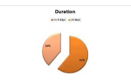
3. Duration of symptoms prior to admission in days
4. Co morbid conditions
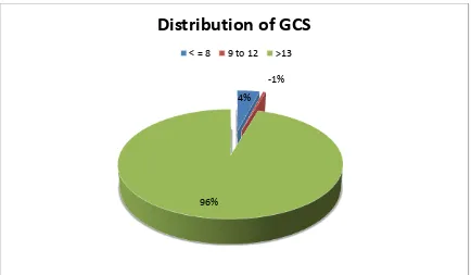
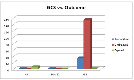
5. Glasgow coma scale at admission
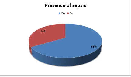
6. Presence of sepsis as determined by the presence of two or more of
the following - fever/ hypothermia, raise/fall of total leukocyte
count, tachycardia and tachypnoea
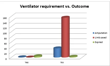
8. Requirement of ionotropic support at admission
9. Urea and creatinine at admission
10. Erythrocyte sedimentation rate at admission
11. Total bilirubin at admission
12. Surface area of body involved
13. Haemoglobin in gm% at admission
14. Depth of involvement
Because of the large number of potentially interdependent parameters
examined in this retrospective analysis, it was believed that a more suitable
test for significance would reside in a multivariate analysis, using a model of
logistic regression analysis. From the large pool of univariately significant
variables (p < 0. 05), a smaller and more manageable group of 10 clinically
relevant variables were selected for inclusion in the first step of the stepwise
regression model. The selected parameters were age in years, duration of
symptoms prior to admission in days, co morbid conditions, glasgow coma
scale at admission, presence of sepsis as determined by the presence of two
or more of the following - fever/ hypothermia, raise/fall of total leukocyte
admission, requirement of ionotropic support at admission, surface area of
body involved, haemoglobin in gm% at admission, depth of involvement.
The variables found on logistic regression analysis to significantly increase
the risk of death or limb loss were used to form a scoring system. This score
was then re applied to the retrospective study to analyze the actual outcome
with the expected outcome. After taking the difference between the expected
and actual outcomes into account, cut offs for the scoring system were
established. This scoring system was then applied to a group of 50 patients
and these patients were treated according to the score protocol. The results of
this prospective study were then statistically analyzed.
DATA ANALYSIS:
To assess possible risk factors for morbidity and mortality, univariate
analyses were completed initially to aid in determining the variables that
should be included in a stepwise logistic regression model. Comparisons of
proportions were made using Pearson's chi square statistic to identify
univariate differences among defined variables with respect to mortality.
Fisher's exact test for 2 X 2 tables was used in the small-sample case. For
measured variables, the F statistic was used to compare means between
the large pool of variables with uni variate p values less than 0.05 for
inclusion in the initial step of the logistic regression analysis. A p value of
0.05 also was chosen as the criterion by which to judge the entry and
removal of variables at each step of the regression procedure. Results of the
logistic regression analysis were expressed using beta coefficient values,
odds ratios (defined as exp[coefficient]), and 90% confidence limits for the
odds ratios. Statistical analysis was performed using SPSS software version
RESULTS
A total of 200 cases which satisfied the inclusion criteria, admitted during a
period of April 2012 to April 2013 were analysed retrospectively.
SAMPLE SIZE = 200 (n)
The following are the individual parameters studied
Age:
Age group – 14 years to 91 years
Mean Age of the Sample – 52.5 years
FIGURE (1)
10%
<