Copyright © 2001, American Society for Microbiology. All Rights Reserved.
Effects of Mutations within the Herpes Simplex Virus Type 1
DNA Encapsidation Signal on Packaging Efficiency
PAUL D. HODGEANDNIGEL D. STOW*
MRC Virology Unit, Institute of Virology, Glasgow G11 5JR, United Kingdom
Received 9 May 2001/Accepted 3 July 2001
Thecis-acting signals required for cleavage and encapsidation of the herpes simplex virus type 1 genome lie within the terminally redundant region orasequence. Theasequence is flanked by short direct repeats (DR1) containing the site of cleavage, and quasi-unique regions, Uc and Ub, occupy positions adjacent to the genomic L and S termini, respectively, such that a novel fragment, Uc-DR1-Ub, is generated upon ligation of the genomic ends. The Uc-DR1-Ub fragment can function as a minimal packaging signal, and motifs have been identified within Uc and Ub that are conserved near the ends of other herpesvirus genomes (pac2 andpac1, respectively). We have introduced deletion and substitution mutations within thepacregions of the Uc-DR1-Ub fragment and assessed their effects on DNA packaging in an amplicon-based transient transfection assay. Within pac2, mutations affecting the T tract had the greatest inhibitory effect, but deletion of sequences on either side of this element also reduced packaging, suggesting that its position relative to other sequences within the Uc-DR1-Ub fragment is likely to be important. No single region essential for DNA packaging was detected withinpac1. However, mutants lacking the G tracts on either side of thepac1 T-rich motif exhibited a reduced efficiency of serial propagation, and alteration of the sequences between DR1 and thepac1 T element also resulted in defective generation of Ub-containing terminal fragments. The data are consistent with a model in which initiation and termination of packaging are specified by sequences within Uc and Ub, respectively.
Herpesviruses have linear double-stranded DNA genomes of 125 to 245 kbp that are circularized upon infection and replicate in an “endless” form, generating branched concate-meric structures. During the assembly of progeny particles, the concatemers are cleaved at specific sites corresponding to the genomic termini and, in a tightly coupled process, the viral DNA is packaged into preformed capsids (reviewed in refer-ences 13, 20, and 23).
In the case of herpes simplex virus type 1 (HSV-1), the
cis-acting sequences required for cleavage and packaging re-side within the terminally redundant region orasequence (21, 29, 32, 34). Theasequence is 250 to 500 bp long and present as a single copy at the S terminus and one or more tandem copies at the L terminus. In addition, one or more copies are also present in inverted orientation at the junction between the L and S segments (23).
Flanking theasequences are direct repeats (DR1) of 17 to 20 bp, with single copies of DR1 separating tandem a se-quences. The site of genomic cleavage occurs within the DR1 element such that a single copy is regenerated upon ligation of the two genomic ends (see Fig. 1). The central portion of the
asequence comprises multiple tandem reiterations of one or two other short sequence elements (11 to 24 bp) referred to as DR2 and DR4. The quasi-unique sequences, each of ca. 80 bp, which lie between DR1 and either side of the DR2 and DR4 repeats are termed the Ub and Uc elements. In virion DNA the Uc element is adjacent to the L terminus and Ub is adja-cent to the S terminus (23). Within the Ub and Uc regions are two domains, defined aspac1 andpac2, respectively, that
con-tain several highly conserved motifs present near the ends of other herpesvirus genomes, including those lacking a terminal redundancy (5, 8, 9, 11). The presence of these conserved sequences near the termini of herpesvirus genomes, the fact that they are brought into close proximity upon circularization, and the fact that in most instances cleavage of concatemeric DNA occurs between the pac1 and pac2 signals led to the suggestion that they have important roles in the cleavage pack-aging process.
The analysis of herpesvirus cleavage packaging signals has employed two principal approaches. In the first, plasmids con-taining a viral origin of DNA replication and putative packag-ing signal (so-called amplicons) are transfected into tissue cul-ture cells and helper functions are provided either by cotransfection with intact viral DNA or superinfection with virus particles. The amplicon DNA replicates autonomously and, if functional packaging signals are present, it is encapsi-dated in the form of long concatemeric molecules consisting of head-to-tail repeats of the input plasmid. Encapsidation of the amplicon DNA confers resistance to exogenously added DNase and permits serial propagation as defective genomes in the presence of the helper (10, 22, 29, 32, 34). Alternatively, additional copies of putative cleavage-packaging signals can be inserted at ectopic sites within the viral genome and assessed for functionality by determining whether concatemeric DNA becomes cleaved at novel sites corresponding to the inserted sequences (7, 18, 21, 27, 34). However, particularly in the case of HSV-1, delimiting the sequences required for packaging has frequently been complicated by the repair of mutated se-quences through recombination with wild-type copies of thea
sequence. There is a strong selection for such repair when amplicons are serially propagated prior to analysis, and se-quence homology between ectopically inserted a sequences * Corresponding author. Mailing address: Institute of Virology,
MRC Virology Unit, Church Street, Glasgow G11 5 JR, United King-dom. Phone: 44(0)141-330-4017. Fax: 44(0)141-337-2236. E-mail: n [email protected].
8977
on November 9, 2019 by guest
http://jvi.asm.org/
and the resident genomic copies, possibly involving the DR2-DR4 region, plays an important role (7, 11, 27, 28).
Single copies of the HSV-1asequence were initially shown to be sufficient for cleavage and packaging (10, 32, 34). In this instance, thepac1 andpac2 homologies reside at opposite ends of theasequence separated by up to 300 bp of the DR2 and DR4 repeats and, in order to explain how packaged molecules come to possess anasequence at each terminus, it is necessary to invoke either specific amplification of asequences or the “wastage” of DNA lacking a terminalasequence (10, 11, 34). Circularization of the HSV-1 genome, however, fuses the ter-minalasequences and, as shown in Fig. 1b, generates a novel junction (Uc-DR1-Ub) in which thepac2 andpac1 homologies are close together. Indeed, an⬃200-bp fragment correspond-ing to the Uc-DR1-Ub junction has been shown to represent the minimal functional packaging signal for HSV-1 (22). More-over, if cleavage of concatemers occurs at the novel junctions between such fusedasequences, genomes withasequences at each end can readily be generated without the necessity for eitherasequence amplification or wastage of DNA. The novel Uc-DR1-Ub junction may therefore represent an important in
vivo substrate for the HSV-1 cleavage and packaging machin-ery.
Conservedpac1 andpac2 motifs were defined by Deiss et al. (11), and their locations within the HSV-1 Uc-DR1-Ub ele-ment are illustrated in Fig. 1c. Within Uc a consensus sequence CGCCGCG (pac2 consensus) lies ca. 70 bp from the S termi-nus, followed in turn by a relatively poorly conserved region (pac2 unconserved), a highly conserved T-rich region (pac2 T element), and a terminal region of high G⫹C content (pac2 GC element). The 40 to 50 bp of Ub adjacent to the DR1 direct repeat represent a region of the S terminus of high G⫹C content (pac1 proximal GC element). This is followed by a T-rich element (pac1 T element) and another region of high G⫹C content (pac1 distal GC element). Characteristically, the
pac1 T element is flanked by the sequences CnGnfrom the
proximal GC element and Gnfrom the distal GC element, and
this unit has been proposed to represent the functionalpac1 signal (5, 8, 11, 18). Experiments with deletion mutants remov-ing sequences from either end of the HSV-1asequence have shown that regions critical for cleavage and packaging reside within both Ub and Uc (11, 22, 27, 34). In addition, strong FIG. 1. Structure and cloning of the HSV-1 Uc-DR1-Ub element. (a) Structure of the HSV-1 genome showing the positions and relative orientations of copies of theasequence. For simplicity, only single copies are shown at the L terminus and joint. (b) Circularization of linear genomes by direct ligation of the termini brings together two copies of theasequence (dashed arrows, orientation as in panel a) separated by a single DR1 repeat. The site of ligation, and of cleavage of concatemers, is shown by the arrow. (c) Motifs within the 194-bp Uc-DR1-Ub element spanning tandemasequences. The poly(G) and poly(C) stretches (within the proximal and distal GC elements) that flank thepac1 T element are indicated by diagonal hatching. (d) Structure of the amplicon, pSA1. oriSand the Uc-DR1-Ub fragment are shown as thickened lines with the site of cleavage indicated by the arrow. E, H, B, S, and P indicate the positions of theEcoRI,HindIII,BamHI,SalI, andPstI restriction endonuclease sites, respectively. (e) The upper line illustrates the structure of a concatemer generated by pSA1 replication (only two complete copies of the monomeric plasmid are depicted). As with full-length HSV-1 genomes, cleavage within DR1 generates linear packaged pSA1 molecules with Uc and Ub at opposite ends (lower line). Digestion of packaged DNA withPstI (P) orSalI (S) yields fragments corresponding to pSA1 monomers plus diagnostic terminal fragments (sizes indicated in kilobase pairs). The larger terminal fragment produced byPstI orSalI cleavage represents the Uc or Ub terminus, respectively.
on November 9, 2019 by guest
http://jvi.asm.org/
evidence has been presented that the DR1 element is not essential for cleavage and packaging, with the S and L termini being generated as a result of cleavage at defined distances from the two T elements. This suggests that the T elements or sequences flanking them are of pivotal importance in the pro-cess (34). Although several of the analyses were complicated by the occurrence of DNA rearrangements, a 15-bp sequence withinpac1 that contributed to the S-terminal cleavage signal was identified (27), and very recently this was shown to lie within a region bound specifically by the HSV-1 DNA pack-aging protein, UL28 (3).
To date, only in the case of the murine cytomegalovirus (MCMV) packaging signal have the effects of specific muta-tions within the conservedpac1 andpac2 motifs been analyzed. In that study (18) the wild-type MCMV cleavage signal (gen-erated by fusion of the genomic termini), and mutated forms thereof, were recombined into the viral genome at an ectopic site. Stocks of cell-released virus were derived, and the occur-rence of cleavage at the novel signal was determined by ana-lyzing the terminal fragments of virion DNA. Several muta-tions within both the pac1 and pac2 regions resulted in significant reductions in progeny that had been cleaved at the ectopic signal, while other mutations had little effect. The G tract distal to thepac1 T element, but not the T element itself, was very important for DNA packaging. Withinpac2 both the T element and adjacent sequences within the relatively uncon-served region were essential for cleavage and packaging, while thepac2 consensus sequence played a contributory role.
In order to analyze the role of individual motifs within the HSV-1 cleavage-packaging signals, we have employed an am-plicon-based assay and conditions designed to reduce the op-portunity for recombination with helper virus. A minimal cleavage packaging signal (Uc-DR1-Ub), lacking the DR2-DR4 repeats, was incorporated into the amplicon, and encap-sidation was examined during a single cycle of infection with helper virus. Using this assay, we have tested the effects of several deletion and substitution mutations within thepac1 and
pac2 regions on DNA packaging.
MATERIALS AND METHODS
Cells and viruses.Baby hamster kidney 21 clone 13 (BHK) cells were grown in Glasgow minimal essential medium (MEM) supplemented with 10% tryptose phosphate broth, 10% newborn calf serum, 100 U of penicillin/ml, and 100g of streptomycin/ml (ETC10). Monolayers of BHK cells were set up in ETC10 and, after transfection or infection, were maintained in Glasgow MEM supplemented with 5% newborn calf serum, 100 U of penicillin/ml, and 100g of streptomy-cin/ml (EC5). Stocks of HSV-1 (strain 17 syn⫹[16]) and HSV-2 (strain HG52
[12]) were prepared and titrated in BHK cells.
Plasmids.Plasmid pS1 (30) contains a 540-bp fragment including a functional HSV-1 oriScloned into theBamHI site of the vector pAT153. Plasmids pY1 and pZ1 contain 1,762- and 2,161-bpHinfI fragments from the junction of the L and S segments of HSV-1 strain 17 syn⫹, including one and two copies, respectively,
of theasequence (9), cloned between theEcoRI andHindIII sites of pS1 (32). The 194-bp MnlI fragment spanning the junction between the tandem a se-quences in pZ1 was isolated, and blunt ends were generated using T4 DNA polymerase in the presence of all four deoxyribonucleoside triphosphates. The resulting fragment (Uc-DR1-Ub element) was inserted, using synthetic linkers, between theEcoRI andHindIII sites of pS1 to generate plasmid pSA1 (Fig. 1d). Site-directed mutagenesis of the Uc-DR1-Ub fragment was performed using the method of Kunkel (14). TheEcoRI-to-HindIII fragment of pSA1 was first cloned between the corresponding sites of the vector pTZ18U, and the resulting plas-mid, pTZ2, was transferred toEscherichia colistrain CJ236 (Dut⫺Ung⫺; New
England Biolabs). Uracil-rich single-stranded DNA was prepared after infection with helper phage R408 and annealed to the appropriate mutagenic
oligonucle-otides. The oligonucleotides were designed to introduce substitution and dele-tion mutadele-tions at various posidele-tions within the Uc-DR1-Ub fragment (see Fig. 3) and contained ca. 14 additional bases complementary to the single-stranded DNA on either side of the region being mutated. In the case of the substitution mutations, novel restriction endonuclease sites were incorporated to facilitate screening. After second-strand synthesis, the products were transformed intoE. coliXL1-Blue (Stratagene). Colonies containing the desired plasmids were iden-tified by restriction enzyme analysis, and in each case the DNA sequence of the complete Uc-DR1-Ub insert was confirmed. The mutatedEcoRI plusHindIII fragments were finally transferred back into pS1, generating the pPH series of plasmids. Plasmid pTZ2 was also digested withSacII orBanII, recircularized, and used to transformE. coli. Colonies containing the expected deletions were identified and the fragments similarly inserted into pS1, generating pPH3 and pPH4, respectively.
Transient DNA packaging assay.Monolayers of BHK cells in 35-mm petri dishes (2⫻106cells per plate) were transfected by using the calcium phosphate technique followed by treatment with dimethyl sulfoxide (DMSO) at 4 h as previously described (31). Each monolayer received 0.5 ml of precipitate con-taining 0.5g of plasmid and 12g of calf thymus carrier DNA. At 2 h after DMSO treatment the cells were infected with 5 PFU of HSV-1 or HSV-2/cell. Incubation was continued for 16 h at 37°C, the medium was removed, the cells were resuspended in TBS (137 mM NaCl, 5 mM KCl, 0.7 mM Na2HPO4, 5.5 mM glucose, 25 mM Tris-HCl [pH 7.4]) and divided into two equal samples which were used to prepare total cellular and DNase-resistant (encapsidated) DNA. The cells from both samples were pelleted and resuspended in 184l of RSB (10 mM Tris-HCl [pH 7.5], 10 mM KCl, 1.5 mM MgCl2) containing 0.5% Nonidet P-40 (NP-40). An equal volume of 20 mM Tris-HCl (pH 7.5)–2 mM EDTA– 1.2% sodium dodecyl sulfate–1 mg of protease (Sigma grade XIV)/ml was either added immediately (total cell DNA) or after incubation in the presence of 200g of DNase I/ml, with occasional mixing, for 20 min at 37°C (encapsidated DNA). After addition of protease, all samples were incubated for 1 h at 37°C, extracted sequentially with phenol and chloroform, and precipitated with ethanol, and the nucleic acids were redissolved in 10 mM Tris-HCl (pH 7.5)–1 mM EDTA containing 5g of RNase A and 50 U of RNase T1/ml. DNase-resistant DNA was also similarly prepared from isolated nuclei. In this case the cells were first incubated 10 min on ice in RSB containing 0.5% NP-40 and then the nuclei were pelleted and incubated in 184l of RSB containing 0.5% NP-40 and 200g of DNase I/ml prior to DNA isolation. Samples of DNA corresponding to the yield from 4⫻105cells were cleaved withDpnI (which cleaves only unreplicated input plasmid molecules) and a second enzyme (usuallyEcoRI) that converts the concatemeric products of plasmid replication into monomers. The digested DNA was fractionated by agarose gel electrophoresis and transferred to a Hybond-N membrane (Amersham), and replicated (DpnI-resistant) plasmid DNA was de-tected by hybridization to a32P-labeled probe prepared from the vector pAT153. Phosphorimages of Southern blots were acquired using the Personal Molecular Imager and analyzed with Quantity One software (Bio-Rad). In control experi-ments with DNA from cells that were mock infected after transfection, no
DpnI-resistant species were detected, demonstrating that the bands present con-tain only replicated plasmid molecules.
Serial propagation of amplicons.Supernatant medium was removed from monolayers of transfected cells at 16 h postinfection, and debris was pelleted by centrifugation for 5 min at 1,300⫻g. Fresh monolayers of BHK cells in 35-mm petri dishes were infected with 0.5 ml of the supernatant. At 1 h after virus addition, the inoculum was removed, the residual virus was inactivated with an acid-glycine wash (1), and incubation was continued at 37°C for 18 h in 2 ml of EC5. Total cellular DNA was prepared and analyzed as described above.
RESULTS
Evaluation of the Uc-DR1-Ub element as a packaging sig-nal.Since Nasseri and Mocarski (22) had previously demon-strated that the Uc-DR1-Ub element could function as an HSV-1 packaging signal, a similar fragment spanning the junc-tion of tandem a sequences of HSV-1 strain 17 syn⫹ was
isolated and cloned between theEcoRI andHindIII sites of the HSV-1 oriS-containing plasmid, pS1. The relative efficiency of
this fragment to direct encapsidation of HSV-1 DNA was eval-uated in a transient assay employing four plasmids, namely, the parental plasmid pS1 and derivatives containing a single a
sequence (pY1), two tandem asequences (pZ1), or the
on November 9, 2019 by guest
http://jvi.asm.org/
DR1-Ub element (pSA1). BHK cells were transfected with each of the plasmids and superinfected with HSV-1 prior to the preparation of total cellular DNA or DNase-resistant DNA. Samples of DNA were digested withEcoRI plusDpnI and analyzed as described in Materials and Methods. Figure 2 shows that, although all four plasmids replicated with similar
efficiency (lefthand four lanes), only pY1, PZ1, and pSA1 were readily detectable in the DNase-resistant DNA samples (right-hand four lanes). These data confirm the previous observations that single or tandem copies of theasequence direct packaging with similar efficiency (32) and that the Uc-DR1-Ub element represents a functional packaging signal (22). Moreover, for the first time, they show that in a single cycle of infection the minimal Uc-DR1-Ub fragment functions as efficiently as full-lengthasequences containing the DR2 reiterations. Quantifi-cation of a large number of experiments indicated that 5 to 10% of replicated pSA1 DNA was generally recovered in the DNase-resistant DNA sample. Although longer exposures al-lowed the detection of small amounts of packaged pS1 DNA, this represented⬍1% the amount of packaged pSA1.
Analysis of Uc-DR1-Ub mutants.In order to investigate the role of individual motifs within the Uc and Ub regions, ampli-cons similar to pSA1 were ampli-constructed containing copies of the Uc-DR1-Ub fragment into which substitution and deletion mutations had been introduced. The regions chosen for mu-tagenesis were essentially those originally defined by Deiss et al. (11), and the DNA sequences of the mutated fragments are shown in Fig. 3. In addition to the plasmids in which individual motifs were altered, two larger deletion mutants removing all of the Uc region (pPH3) and most of Ub (pPH4) were con-structed.
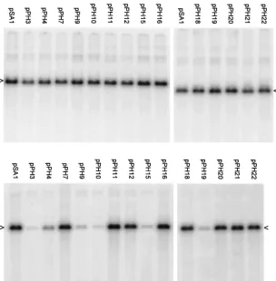
[image:4.612.61.286.79.232.2]The packaging abilities of the resulting 14 plasmids were compared to those of pSA1 in a transient assay (Fig. 4). The results indicate that each of the mutated plasmids replicated to the same extent as pSA1 (top panels), but there were signifi-cant differences in the amount of DNA encapsidated (lower FIG. 2. Replication and packaging of plasmids pS1, pY1, pZ1, and
pSA1 in a transient assay. BHK cells were transfected with pS1 (4.2 kbp), pY1 (6.0 kbp), pZ1 (6.2 kbp), or pSA1 (4.4 kbp) as indicated and superinfected with HSV-1, and total (left-hand 4 lanes) and DNase-resistant (right-hand 4 lanes) DNAs were prepared. Aliquots were digested withEcoRI plusDpnI, and the fragments were separated by electrophoresis through an agarose gel. The gel was blotted, the mem-brane was hybridized to32P-labeled pAT153 DNA and, after being washed, the membrane was exposed to a phosphorimager screen. The arrowheads indicate replicated (DpnI-resistant) pSA1 monomers.
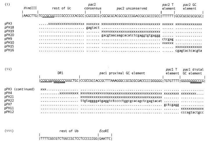
FIG. 3. Sequence of the HSV-1 Uc-DR1-Ub element and of the mutants used in this study. The sequence of the cloned insert in plasmid pSA1 is depicted on three lines (i, ii, and iii) with the characteristic motifs and regions corresponding to those shown in Fig. 1c. The substitutions and deletions in the mutants are indicated below the sequence. The modified sequences of the relevant regions are shown in lowercase for the substitution mutants, and the nucleotides missing in the deletion mutants are indicated by “x.” Plasmid pPH3 contains an 80-bp deletion between the twoSacII sites (single underlined) and pPH4 contains a 69-bp deletion between theBanII sites (double underlined).
on November 9, 2019 by guest
http://jvi.asm.org/
[image:4.612.123.479.440.683.2]panels). Quantitative data were obtained from three separate experiments in which the full set of mutant plasmids was ex-amined (Table 1). The amount of each plasmid packaged was expressed as a percentage of the pSA1 value and the mean and
[image:5.612.150.454.75.385.2]standard deviation were calculated. The two plasmids most impaired for packaging were pPH3 (Uc deletion) and pPH10 (pac2 T element deletion), which exhibited relative packaging efficiencies of 2.4 and 2.3%, respectively. Plasmids pPH15 FIG. 4. Packaging ability of plasmids containing mutated Uc-DR1-Ub fragments. A transient packaging assay using HSV-1 as helper was performed with the pPH series of plasmids as described in Materials and Methods and in the legend to Fig. 2. The upper and lower panels are for total and DNase-resistant DNA, respectively, and the positions of the replicated amplicon monomers (4.4 kbp) are indicated by arrowheads.
TABLE 1. Properties of the plasmids used in this study
Plasmid Mutation
Mean packaging efficiency⫾
SD with: Serial
propagationc Generation ofUb terminusd
HSV-1a HSV-2b
pSA1 Wild type 100 100 ⫹⫹ ⫹
pPH3 pac2 (Uc) 80-bp deletion 2.4⫾1.6 1.2 ND ND
pPH18 pac2 consensus substitution 168⫾106 102 ⫹⫹ ⫹
pPH19 pac2 unconserved deletion 12.9⫾5.0 26 ND ND
pPH20 pac2 unconserved substitution 132⫾86 58 ⫹⫹ ⫹
pPH9 pac2 T element substitution 6.9⫾3.6 4.6 ND ND
pPH10 pac2 T element deletion 2.3⫾0.6 1.9 ND ND
pPH15 pac2 GC element deletion 5.0⫾1.8 3.2 ND ND
pPH16 pac2 GC element substitution 152⫾57 105 ⫹⫹ ⫹
pPH4 pac1 (Ub) 69-bp deletion 9.6⫾5.8 4.3 ND ND
pPH21 pac1 proximal GC element deletion 75⫾39 22 ⫹ ⫺
pPH22 pac1 proximal GC element substitution 82⫾34 39 ⫹ ⫺
pPH7 pac1 T element substitution 177⫾55 176 ⫹⫹ ⫹
pPH11 pac1 distal GC element deletion 76⫾40 37 ⫹ ⫹
pPH12 pac1 distal GC element substitution 118⫾65 53 ⫹ ⫹
aPackaging efficiencies were relative to pSA1 and are presented as the mean of three independent determinations⫾the standard deviation. bPackaging efficiencies were relative to pSA1 as determined from Fig. 5.
c⫹⫹, serially propagated with similar efficiency to pSA1;⫹, propagated with reduced efficiency; ND, not done.
d⫹, Ub-containing terminal fragment present in packaged DNA;⫺, Ub-containing terminal fragment not detected; ND, not done.
on November 9, 2019 by guest
http://jvi.asm.org/
[image:5.612.51.557.521.700.2](pac2 GC element deletion), pPH9 (pac2 T element substitu-tion), pPH4 (Ub delesubstitu-tion), and pPH19 (pac2 unconserved re-gion deletion) were also significantly impaired (relative pack-aging efficiencies of 5.0, 6.9, 9.6, and 12.9%, respectively). The remaining eight plasmids all exhibited packaging efficiencies that differed ⬍2-fold from pSA1. The values obtained for pPH21, pPH22 (pac1 proximal GC element deletion and sub-stitution mutants), pPH11, pPH12 (pac1 distal GC element deletion and substitution mutants), pPH20, pPH16, pPH18 (pac2 substitution mutants affecting the unconserved region, the GC element and the consensus sequence), and pPH7 (pac1 T element substitution) were 75, 82, 76, 118, 132, 152, 168, and 177%, respectively.
These results confirm that the Uc region contains sequences important for DNA packaging and identify thepac2 T element as being of particular importance. Deletions of the elements to either side ofpac2 T also significantly impaired encapsidation, but substitution mutations within these regions had relatively little effect, suggesting that the position of thepac2 T element relative to other motifs in the Uc-DR1-Ub fragment may be important for its activity. The removal of most of the Ub region, and part of DR1, impaired packaging ability but, sur-prisingly, none of the deletion or substitution mutations intro-duced into the component pac1 elements had a significant effect.
Ability of HSV-2 to function as a helper virus. As noted above, repair of mutated a sequences may sometimes occur through recombination with wild-type copies (11, 27). Smiley et al. (28) previously demonstrated that HSV-2 was able to recognize the HSV-1asequence for DNA cleavage and pack-aging but that recombinational inversion events did not occur
between the twoasequences, presumably as a result of their overall low DNA sequence homology (9).
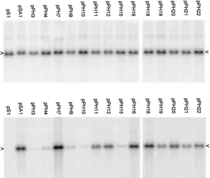
In order to reduce the possibility that recombinational re-pair might be having a significant influence on the phenotypes of our mutants, we assessed their packaging ability when HSV-1 was replaced by HSV-2 as the superinfecting helper virus. The results (Fig. 5 and Table 1) are very similar to those obtained with HSV-1 helper (Fig. 4 and Table 1). However, the plasmids affecting thepac1 proximal and distal GC elements (pPH21, pPH22, pPH11, and pPH12) all exhibited a small reduction in packaging efficiency relative to pSA1 when HSV-2 was helper, while pPH19 (pac2 unconserved region deletion) showed a small increase. These effects, which were also noted in an independent experiment, are readily illustrated by com-parison of the pPH19, pPH21, and pPH22 lanes in Fig. 4 and 5. Thus, although there may be small differences between pack-aging signal recognition in HSV-1 and HSV-2, these data sug-gest that it is unlikely that the high-efficiency packaging seen with several mutants is a consequence of recombinational re-pair.
Further characterization of mutants relatively unimpaired for DNA packaging.Since the data presented in Fig. 4 showed that eight of the mutants were packaged with a similar effi-ciency to pSA1, we sought to determine whether the lesions in these plasmids might play a role in amplicon propagation at a stage other than the initial encapsidation process. Supernatant medium from BHK cells that had been transfected with pS1, pSA1, or one of the eight mutants was passaged on fresh cell monolayers, and total DNA was prepared and examined for the presence of replicated plasmid molecules (Fig. 6 and Table 1). In agreement with previous results that showed a require-FIG. 5. Packaging assay with HSV-2 as superinfecting virus. The assay was performed as in Fig. 4 except that the helper virus was HSV-2. The upper and lower panels are for total and DNase-resistant DNA, respectively, and the positions of the replicated amplicon monomers (4.4 kbp) are indicated by arrowheads.
on November 9, 2019 by guest
http://jvi.asm.org/
[image:6.612.151.455.78.336.2]ment for the Uc-DR1-Ub element for serial propagation (22), replicated pSA1 but not pS1 was detected in the total DNA. Plasmids pPH7, pPH16, pPH18, and pPH20 were propagated with the same efficiency as pSA1 but the other four plasmids (pPH11, pPH12, pPH21, and pPH22, carrying alterations to thepac1 distal and proximal GC elements) accumulated to at least fourfold lower levels. When the blot was reprobed to detect fragments of the HSV-1 helper DNA similar levels were detected in each of the lanes. Thus, although alteration of the
pac1 distal and proximal GC elements does not appear to affect the amount of DNA encapsidated, the packaged DNA is impaired in its ability to be serially propagated.
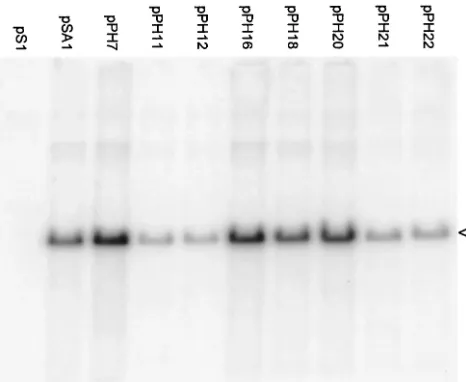
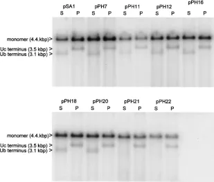
One possible explanation for this above result is that pack-aged concatemers of pPH11, pPH12, pPH21, or pPH22 may have abnormal termini that could affect their ability to reini-tiate infection. To examine the termini of packaged amplicons, the DNase-resistant DNA isolated from the nuclei of trans-fected cells superintrans-fected with HSV-1 was cleaved with two enzymes, SalI orPstI, that generate distinctive terminal frag-ments. In addition to amplicon monomers, cleavage of DNase-resistant DNA withSalI would be expected to generate frag-ments of 3.1 and 1.3 kbp representing the termini bearing the Ub and Uc sequences, respectively (Fig. 1e). The sizes of the correspondingPstI fragments terminating with Ub and Uc are 0.9 and 3.5 kbp, respectively.
The results are shown in Fig. 7 and summarized in Table 1. When DNA from cells that had been transfected with pSA1 was cleaved withSalI orPstI, the larger terminal fragment was readily detected as a band of faster mobility and lower abun-dance than unit length monomers (upper band). The Uc-con-taining PstI terminal fragment was detectable in DNA from cells transfected with each of the packaging-competent mutant plasmids. In contrast, although detectable for six of the mu-tants, the Ub-containing SalI terminal fragment was not ap-parent in the DNAs of cells transfected with pPH21 or pPH22
(pac1 proximal GC element mutants). Thus, although these two plasmids give rise to DNase-resistant DNA with normal Uc-containing termini, fragments from the other terminus are probably heterogeneous in size and, as a consequence, are not detected as a discrete band. Such heterogeneity might occur through a loss of cleavage specificity or, alternatively, cleavage might occur at the correct position but the terminal sequences may remain susceptible to added DNase because they are not correctly inserted into the capsid.
In an attempt to estimate the lengths of the packaged mono-mers, we measured the radioactivity in the bands correspond-ing to amplicon monomers and terminal fragments (where detectable). Although the presence of a background smear in the lanes made it difficult to determine reliably the amount of terminal fragment, the signal in the amplicon monomers band was generally 10- to 20-fold higher than in the terminal frag-ment band. If we assume that the signal intensity is propor-tional to the amount of vector sequence in the DNA fragment, this suggests that the packaged concatemers contain, on aver-age, ca. 12 to 24 monomeric units. None of the mutant plas-mids differed significantly from pSA1 in this regard.
DISCUSSION
The transient assay we employed in this study differs in two important aspects from other approaches that have been used to analyze the signals required for herpesvirus DNA packag-ing. Most significantly, in contrast to previous experiments with amplicons in which mixed virus stocks containing helper and defective genomes were serially propagated prior to analysis (10, 11, 22, 34), we examined DNA packaging during a single cycle of infection. Additionally, the preparations of packaged DNA we analyzed included DNA from the infected cell nu-cleus.
[image:7.612.56.289.77.268.2]The approach confers several probable advantages: (i) the opportunity for recombination with the helper virus is mini-mized; (ii) there is no selection for molecules that may have repaired lethal or debilitating mutations through such a pro-cess; (iii) quantitative comparison of the effects of different mutations is facilitated; and (iv) the identification of novel packaging phenotypes is possible. For example, it is conceiv-able that a class of mutant exists in which significant amounts of DNA are packaged in the nucleus but the resulting nucleo-capsids fail to progress to enveloped virions. In an assay de-pendent on serial propagation, this mutant would exhibit a negative phenotype similar to that of a mutant that was defec-tive in the initiation of packaging. A clear precedent for this type of defect is provided by the studies of defective HSV-1 genomes by Vlazny et al. (35). They reported that capsids in the nuclei contained defective genomes comprising integral numbers of copies of a basic monomeric unit up to the length of a standard HSV-1 genome. However, only the largest de-fective genomes were detected in the cytoplasm, suggesting the existence of selectivity in the envelopment and translocation of capsids into the cytoplasm. Studies employing ectopic copies of packaging signals to identifycis-acting herpesvirus DNA pack-aging signals may similarly have failed to differentiate between mutations affecting the initiation and termination of DNA packaging since again DNA from only cytoplasmic or cell-released virus particles was analyzed (18, 27).
FIG. 6. Serial propagation of amplicons. Samples of cell-released virus from BHK cell monolayers transfected with the indicated plas-mids were used to infect fresh cells, and total cellular DNA was prepared and analyzed as described in the legend to Fig. 2. The position of the amplicon monomers (4.4 kbp) is indicated by an ar-rowhead.
on November 9, 2019 by guest
http://jvi.asm.org/
We initially confirmed the observation of Nasseri and Mo-carski (22) that a 200-bp Uc-DR1-Ub fragment of HSV-1 could act as a packaging signal, and we also showed that it functions with an efficiency similar to that of larger fragments containing one or two copies of the complete a sequence, including the highly reiterated DR2 regions (Fig. 2). Because the small size of the Uc-DR1-Ub fragment and the absence of high-copy-number repeats might be expected to minimize re-combination with the helper virus, this fragment was chosen for site-directed mutagenesis experiments. Although we can-not conclusively exclude the possibility that recombination may have occurred between the helper virus and the replicated amplicon DNA to repair the sequences at the termini of pack-aged molecules, several observations argue that it is unlikely to have had a significant influence on packaging efficiency. First, deletions or substitutions of similar sizes frequently exhibited quite-different phenotypes (e.g., compare pPH7 and pPH9 in Fig. 4). Second, each of the mutants exhibited a similar phe-notype when HSV-2, which shares insufficientasequence ho-mology with HSV-1 to drive recombinational genome isomer-ization (28), was used as a helper (Fig. 5). Finally, four plasmids with lesions in pac1 (pPH11, pPH12, pPH21, and pPH22), which exhibited wild-type levels of DNA packaging, were impaired in their ability to be serially propagated, and two of these (pPH21 and pPH22) were defective in the gen-eration of a normal Ub-containing terminal fragment (Fig. 6 and 7). If recombinational repair of the lesions had been nec-essary to achieve high-efficiency packaging, these plasmids would have been expected to be unimpaired in the serial
prop-agation assay and packaged pPH21 and pPH22 DNAs would have been expected to generate normal Ub-containing termini. In agreement with previous results, both the Ub and Uc regions were shown to play important roles in DNA packaging (11, 22, 27, 34). Both the Ub (pPH4) and Uc (pPH3) deletion mutants nevertheless exhibited residual packaging activity (Fig. 4) at a level greater than that observed with the parental plasmid, pS1, suggesting that there may be some functional redundancy between the regions.
Within the Uc region, the pac2 T element was the single most critical sequence; deletion of this motif had as great an effect as deletion of all of Uc, and the substitution mutant was also significantly impaired. Deletion of the pac2 unconserved region andpac2 GC element resulted in much greater reduc-tions in packaging than substitureduc-tions that did not affect the lengths of these regions, suggesting that the position of the
pac2 T element relative to other motifs within the Uc-DR1-Ub fragment is likely to be important. These results are broadly similar to those obtained with an ectopic cleavage-packaging signal in MCMV, in which substitution of thepac2 T element or insertions of 6 or 47 bp on either side of it abolished detectable cleavage activity (18). A more significant reduction in activity was noted with an MCMVpac2 consensus substitu-tion mutant than with the corresponding HSV-1 mutant. Since this mutant (pPH18) is also unimpaired in its ability to be serially propagated (Fig. 6), it is possible that any important function of the HSV-1pac2 consensus may be compensated for by the presence of closely related GC-rich motifs in the adja-cent regions of Uc. When deletions were progressively intro-FIG. 7. Terminal fragments of packaged amplicon DNA. DNase-resistant DNA, prepared from the nuclei of cells transfected with the indicated plasmids and superinfected with HSV-1, was cleaved withDpnI plus eitherSalI (S) orPstI (P) and analyzed as described in the legend to Fig. 2. The positions of the larger of the terminal fragments generated bySalI orPstI (3.1 and 3.5 kbp, respectively) and of linear monomeric plasmid are indicated.
on November 9, 2019 by guest
http://jvi.asm.org/
[image:8.612.145.454.83.346.2]duced into the Uc side of anasequence, some reduction in ability to function as a cleavage site was noted, but it was not possible to identify the contribution of individual pac2 ele-ments because of the likely occurrence of recombinational repair (27).
Analysis of the mutants affecting the Ub region yielded sev-eral surprising results. Although deletion of the three pac1 elements (and part of DR1) greatly reduced DNA packaging (plasmid pPH4), the substitution and deletion mutants affect-ing the individualpac1 motifs all exhibited near-wild-type ac-tivity (Fig. 4). This paradoxical result is not fully understood. The impairment of pPH4 packaging is unlikely to be a conse-quence of the removal of the DR1 seconse-quences since it has previously been demonstrated thatpac1-directed cleavage can occur in the absence of this element (34). However, it remains possible that the DR1 element plays an important role in the context of a minimal Uc-DR1-Ub packaging signal. Alterna-tively, there may be functional redundancy among the pac1 motifs, such that the removal of an individual motif has rela-tively little effect. For example, both the proximal and distal GC elements contain tracts of at least six G residues, and runs of at least three T residues occur in both the proximal GC and T elements.
The results obtained with the mutants affecting the individ-ualpac1 motifs also contrast markedly with data from experi-ments in which the effects ofpac1 mutations on the generation of terminal fragments of HSV-1 or MCMV DNA were exam-ined (18, 27). In the latter case, the poly(G) tracts on either side of thepac1 T element were shown be important for the generation of virions containing genomes cleaved at an ectopi-cally inserted packaging signal. The apparent discrepancy be-tween the sets of data may be explained if these motifs are involved in relatively late stages of the encapsidation process such as the maturation of nuclear DNA-containing capsids into virions. Consistent with this, all four amplicons carrying muta-tions within the twopac1 GC elements exhibited reduced rep-lication when virus stocks were passaged on fresh cells (Fig. 6), and both mutants with lesions in the proximal GC element were additionally defective in the generation of normal Ub-containing terminal fragments (Fig. 7). However, an alterna-tive possibility that the pac1 GC element mutants may be impaired in the efficiency with which virus particles can initiate new cycles of infection, rather than in virion production per se, cannot be excluded.
Further experiments are clearly required to elucidate the stage(s) at which thepac1 mutants are impaired. This analysis, however, may be more complex with amplicons than ectopic packaging signals. For example, ifpac1 is involved in the late stages of DNA packaging, it is probable that the generation of concatemers containing multiple tandem copies of an ampli-con may allow packaging to be completed with relatively higher efficiency when the signals retain partial functionality. In ad-dition, packaged amplicons with an abnormal terminus may be able to circularize relatively efficiently by homologous recom-bination to reinitiate infection even though their termini may be incapable of direct ligation. An interesting approach would be to examine whether the Uc-DR1-Ub mutants allow the rescue of progeny virus when inserted into an HSV-1 bacterial artificial chromosome lacking functionalpacsignals (24).
It has previously been shown that during herpesvirus DNA
packaging only the genomic terminus containing thepac2 sig-nal is found associated with the high-molecular-weight con-catemeric DNA (15, 17, 19, 25, 26, 36), and this observation has formed the basis for a proposal that the pac2 sequence controls the initiation and directionality of DNA packaging (19). Our observations that mutations withinpac2 have rela-tively greater effects during the first cycle of infection and that motifs in pac1 may be involved in the late stages of DNA encapsidation are compatible with this model. In the case of HSV-1 genomes, the pac2 signal at the L terminus would promote the initial cleavage of a concatemeric molecule and the initiation of DNA packaging into the preformed capsid. A second cleavage event, driven bypac1, would be necessary for completion of the process and the generation of an authentic S terminus (i.e., the directionality of packaging would be from L to S). It should be noted, however, that until other possible mechanisms can be excluded, this remains a tentative model. Moreover, it remains to be determined whether cleavage re-actions are identical at junctions containing only a single a
sequence which, unless duplication first occurs, does not con-tain a Uc-DR1-Ub junction. An attractive feature of the above model, however, is that the independent functioning of the
pac1 andpac2 regions may explain how a singleasequence, in which these two signals are separated by several hundred base pairs can efficiently direct cleavage and packaging (7, 10, 11, 27, 32, 34).
The HSV-1 UL28 packaging protein has recently been shown to interact specifically with thepac1 signal and the G tracts at either side of thepac1 T element have been shown to be key elements in the binding (3). Our data are consistent with this interaction being involved at a late stage in packaging, such as the termination cleavage event. Studies with UL28 mutants, however, suggest that no cleavage of concatemers occurs in the absence of UL28, implicating this protein also in the initiation of packaging (2, 6, 33). This suggests that UL28 may also interact with thepac2 signal, as previously reported for the homologous protein of human cytomegalovirus (4), or that binding to thepac1 signal may also be involved in initia-tion of packaging. In this regard, it is interesting to recall that
pac1 mutants that retained at least one of the flanking G tracts were unimpaired for packaging, whereas the mutant lacking the entire Ub region was severely impaired.
ACKNOWLEDGMENTS
We are grateful to Andrew Davison and Frazer Rixon for critical reading of the manuscript, and we thank Leslie Taylor for carrying out DNA sequencing.
P.D.H. was supported by a Medical Research Council Ph.D. Stu-dentship.
REFERENCES
1.Abbotts, A. P., V. G. Preston, M. Hughes, A. H. Patel, and N. D. Stow.2000. Interaction of the herpes simplex virus type 1 packaging protein UL15 with full-length and deleted forms of the UL28 protein. J. Gen. Virol.81:2999– 3009.
2.Addison, C., F. J. Rixon, and V. G. Preston.1990. Herpes simplex virus type 1 UL28 gene product is important for the formation of mature capsids. J. Gen. Virol.71:2377–2384.
3.Adelman, K., B. Salmon, and J. D. Baines.2001. Herpes simplex virus DNA packaging sequences adopt novel structures that are specifically recognized by a component of the cleavage and packaging machinery. Proc. Natl. Acad. Sci. USA98:3086–3091.
4.Bogner, E., K. Radsak, and M. F. Stinski.1998. The gene product of human cytomegalovirus open reading frame UL56 binds the pac motif and has
on November 9, 2019 by guest
http://jvi.asm.org/
specific nuclease activity. J. Virol.72:2259–2264.
5.Broll, H., H.-J. Buhk, W. Zimmermann, and M. Goltz.1999. Structure and function of the prDNA and genomic termini of the␥2-herpesvirus bovine herpesvirus type 4. J. Gen. Virol.80:979–986.
6.Cavalcoli, J. D., A. Baghian, F. L. Homa, and K. G. Kousoulas.1993. Resolution of genotypic and phenotypic properties of herpes simplex virus type 1 temperature-sensitive mutant KOStsZ47: evidence for allelic comple-mentation in the UL28 gene. Virology197:23–34.
7.Chou, J., and B. Roizman.1985. Isomerization of herpes simplex virus 1 genome: identification of thecis-acting and recombination sites within the domain of theasequence. Cell41:803–811.
8.Davison, A., and F. Rixon.1985. Cloning of the DNA of Alphaherpesvirinae, p. 103–124.InY. Becker (ed.), Recombinant DNA research and virus. Martinus Nijhoff Publishing, Boston, Mass.
9.Davison, A. J., and N. M. Wilkie.1981. Nucleotide sequences of the joint between the L and S segments of herpes simplex virus types 1 and 2. J. Gen. Virol.55:315–331.
10. Deiss, L. P., and N. Frenkel.1986. Herpes simplex virus amplicon: cleavage of concatemeric DNA is linked to packaging and involves amplification of the terminally reiteratedasequence. J. Virol.57:933–941.
11. Deiss, L. P., J. Chou, and N. Frenkel.1986. Functional domains within the
a sequence involved in the cleavage-packaging of herpes simplex virus DNA. J. Virol.59:605–618.
12. Dolan, A., F. E. Jamieson, C. Cunningham, B. C. Barnett, and D. J. Mc-Geoch.1998. The genome sequence of herpes simplex virus type 2. J. Virol. 72:2010–2021.
13. Homa, F. L., and J. C. Brown.1997. Capsid assembly and DNA packaging in herpes simplex virus. Rev. Med. Virol.7:107–122.
14. Kunkel, T. A.1985. Rapid and efficient site-specific mutagenesis without phenotypic selection. Proc. Natl. Acad. Sci. USA82:488–492.
15. Martinez, R., R. T. Sarisky, P. C. Weber, and S. K. Weller.1996. Herpes simplex virus type 1 alkaline nuclease is required for efficient processing of viral DNA replication intermediates. J. Virol.70:2075–2085.
16. McGeoch, D. J., M. A. Dalrymple, A. J. Davison, A. Dolan, M. C. Frame, D. McNab, L. J. Perry, J. E. Scott, and P. Taylor.1988. The complete DNA sequence of the long unique region in the genome of herpes simplex virus type 1. J. Gen. Virol.69:1531–1574.
17. McVoy, M. A., and S. P. Adler.1994. Human cytomegalovirus DNA repli-cates after early circularization by concatemer formation, and inversion occurs within the concatemer. J. Virol.68:1040–1051.
18. McVoy, M. A., D. E. Nixon, S. P. Adler, and E. S. Mocarski.1998. Sequences within the herpesvirus-conservedpac1 and pac2 motifs are required for cleavage and packaging of the murine cytomegalovirus genome. J. Virol. 72:48–56.
19. McVoy, M. A., D. E. Nixon, J. K. Hur, and S. P. Adler.2000. The ends of herpesvirus DNA replicative concatemers containpac2cis cleavage/packag-ing elements and their formation is controlled by terminalcissequences. J. Virol.74:1587–1592.
20. Mocarski, E. S.1996. Cytomegaloviruses and their replication, p. 2447–2492.
InB. N. Fields, D. M. Knipe, and P. M. Howley (ed.), Fields virology, 3rd ed. Lippincott-Raven Publishers, Philadelphia, Pa.
21. Mocarski, E. S., and B. Roizman.1982. Structure and role of the herpes
simplex virus DNA termini in inversion, circularization and generation of virion DNA. Cell31:89–97.
22. Nasseri, M., and E. S. Mocarski.1988. The cleavage recognition signal is contained within sequences surrounding ana-ajunction in herpes simplex virus DNA. Virology167:25–30.
23. Roizman, B., and A. E. Sears.1996. Herpes simplex viruses and their repli-cation, p. 2231–2295.InB. N. Fields, D. M. Knipe, and P. M. Howley (ed.), Fields virology, 3rd ed. Lippincott-Raven Publishers, Philadelphia, Pa. 24. Saeki, Y., T. Ichikawa, A. Saeki, E. A. Chiocca, K. Tobler, M. Ackermann,
X. O. Breakfield, and C. Fraefel.1998. Herpes simplex virus type 1 DNA amplified as bacterial artificial chromosome inEscherichia coli:rescue of replication-competent virus progeny and packaging of amplicon vectors. Hum. Gene Ther.9:2787–2794.
25. Severini, A., A. R. Morgan, D. R. Tovell, and D. L. Tyrrell.1994. Study of the structure of replicative intermediates of HSV-1 DNA by pulsed-field gel electrophoresis. Virology200:428–435.
26. Slobedman, B., and A. Simmons. 1997. Concatemeric intermediates of equine herpesvirus type 1 DNA replication contain frequent inversions of adjacent long segments of the viral genome. Virology229:415–420. 27. Smiley, J. R., J. Duncan, and M. Howes.1990. Sequence requirements for
DNA rearrangements induced by the terminal repeat of herpes simplex virus type 1 KOS DNA. J. Virol.64:5036–5050.
28. Smiley, J. R., C. Lavery, and M. Howes.1992. The herpes simplex virus type 1 HSV-1asequence serves as a cleavage/packaging signal but does not drive recombinational genome isomerization when it is inserted into the HSV-2 genome. J. Virol.66:7505–7510.
29. Spaete, R. R., and N. Frenkel.1982. The herpes simplex virus amplicon: a new eukaryotic defective-virus cloning-amplifying vector. Cell82:295–304. 30. Stow, N. D., and E. C. McMonagle.1983. Characterisation of the TRS/IRS
origin of DNA replication of herpes simplex virus type 1. Virology130:427– 438.
31.Stow, N. D., and N. M. Wilkie.1976. An improved technique for obtaining enhanced infectivity with herpes simplex virus type 1 DNA. J. Gen. Virol. 33:447–458.
32. Stow, N. D., E. C. McMonagle, and A. J. Davison.1983. Fragments from both termini of the herpes simplex virus type 1 genome contain signals required for the encapsidation of viral DNA. Nucleic Acids Res.11:8205–8220. 33. Tengelsen, L. A., N. E. Pederson, P. R. Shaver, M. W. Wathen, and F. L.
Homa.1993. Herpes simplex virus type 1 DNA cleavage and encapsidation require the product of the UL28 gene: isolation and characterization of two UL28 deletion mutants. J. Virol.67:3470–3480.
34. Varmuza, S. L., and J. R. Smiley.1985. Signals for site-specific cleavage of HSV-1 DNA: maturation involves two separate cleavage events at sites distal to the recognition sequences. Cell41:793–802.
35. Vlazny, D. A., A. Kwong, and N. Frenkel.1982. Site-specific cleavage/pack-aging of herpes simplex virus DNA and the selective maturation of nucleo-capsids containing full-length viral DNA. Proc. Natl. Acad. Sci. USA79: 1423–1427.
36. Zhang, X., S. Efstathiou, and A. Simmons.1994. Identification of novel herpes simplex virus replicative intermediates by field inversion gel electro-phoresis: implications for viral DNA amplification strategies. Virology202: 530–539.