SAFETY AND EFFICACY OF CORNEAL
COLLAGEN CROSSLINKING WITH RIBOFLAVIN
AND ULTRAVIOLET-A IN THE TREATMENT OF
PROGRESSIVE KERATOCONUS
DISSERTATION SUBMITTED FOR
MS (Branch III) Ophthalmology
THE TAMILNADU DR. M.G.R. MEDICAL UNIVERSITY
CHENNAI
CERTIFICATE
This is to certify that the thesis entitled “SAFETY AND EFFICACY
OF CORNEAL COLLAGEN CROSSLINKING WITH
RIBOFLAVIN AND ULTRAVIOLET-A IN THE TREATMENT OF
PROGRESSIVE KERATOCONUS” is the original work of Dr.
Gitansha Sachdev and was conducted under our direct supervision and
guidance at Aravind Eye Hospitals and Postgraduate Institute of
Ophthalmology, Madurai
Dr.Mano Ranjan Das Dr. N.Venkatesh Prajna
Guide, Medical Officer &
Senior Consultant, Head of Department
Cornea Services Cornea Services
Aravind Eye Hospital, Aravind Eye Hospital,
Madurai. Madurai.
Dr.M.S.Srinivasan Director Emeritus, Aravind Eye Hospital,
ACKNOWLEDGEMENT
I take this opportunity to pay my respect and homage to
Dr.G.Venkatasamy, our founder and visionary, whose dynamism had led
Aravind against all odds to its epitope.
It is a proud privilege and pleasure to express my thanks and
indebtedness towards my revered mentor and guide Dr. Mano Ranjan Das,
Consultant, Cornea services, Aravind Eye Hospital Madurai, for being a
constant source of motivation and encouragement, which ultimately structured
my thesis .
I am grateful to Dr. N.V.Prajna, Director of academics, Aravind Eye
Care System, who offered his excellent guidance and support throughout my
residency programme.
I am very grateful to Dr.R.D.Ravindran, Chairman of Aravind Eye
Care System for having created and environment enriched with all the facilities
for learning and gaining knowledge. I am privileged to have on my side
Dr. P. Namperumalsamy, Chairman emeritus director of research,
Dr.G.Natchiar Director Emeritus (Human Resource Department),
Dr.M.Srinivasan, Director Emeritus and other scholars of ophthalmology at
My sincere thanks to Dr.Hemalatha, cornea fellow and all paramedical
staff for their excellent co- operation during the study. Last but not the least; I
CONTENTS
S.No Title Page Number
Introduction 1
Review of Literature 9
Lacunae in Knowledge 27
Aim of Study 28
Materials and Methods 29
Results 39
Discussion 73
Conclusion 81
Annexure
a) Bibliography
b) Proforma
SAFETY AND EFFICACY OF CORNEAL COLLAGEN CROSSLINKING WITH RIBOFLAVIN AND ULTRAVIOLET-A IN THE TREATMENT OF
PROGRESSIVE KERATOCONUS
KEYWORDS
Keratoconus, collagen crosslinking, riboflavin, ultraviolet A, corneal ectasia
INTRODUCTION
Keratoconus is a non inflammatory progressive corneal thinning of unknown etiology
in which the cornea assumes a conical shape, resulting in mild to marked impairment
of visual acuity due to irregular astigmatism, progressive myopia and central corneal
scarring. A reduced number of collagen cross-links and a pepsin digestion higher than
normal induce an overall structural weakness of the corneal tissue, resulting in a
stiffness that is only 60% of normal cornea. Changes in corneal collagen structure,
organization and intercellular matrix as well as apoptosis and necrosis of
keratinocytes, prevalently or exclusively involving the central anterior stroma and
Bowman’s lamina are documented in literature. Corneal collagen cross-linking (C3R)
is a new approach to increase the mechanical and biochemical strength of corneal
tissue.
BASIC PRINCIPLE-The aim of this treatment is to create additional chemical bonds
inside the corneal stroma by means of highly localised photopolymerisation while
minimising exposure to surrounding ocular structures.
Using UVA at 370nm and the photosensitizer riboflavin, the photosensitizer is
mainly in the form of singlet oxygen and to a much less degree, they may be in the
form of superoxide anion radicals. The ROS then reacts with various molecules,
including chemical covalent bonds, bridging amino groups of collagen fibrils(type 2
photochemical reaction).The wavelength of 370nm was chosen because of an
absorption peak of riboflavin at this wavelength.
AIMS AND OBJECTIVES
-To evaluate the safety of C3R as measured by intra operative and postoperative
complications
-To measure the efficacy of C3R in bringing progressive keratoconus to a halt
MATERIALS AND METHODS TYPE OF STUDY
Prospective interventional clinical trial
CASE COLLECTION PERIOD - 1 year FOLLOW UP PERIOD - 1 year
SAMPLE SIZE - 50 patients
SELECTION CRITERIA Inclusion criteria
Progressive keratoconus
Age between 12 to 30 years
Corneal pachymetry> 400 microns at thinnest point
Normal corneal endothelium
Willing for follow up
Exclusion criteria
Corneal thickness < 400 microns at thinnest point
H/O herpetic keratitis
Central or paracentral opacities
Prior corneal surgery
Severe dry eye and ocular surface disorders
Concurrent corneal infections
Concomitant autoimmune diseases
Pregnant/nursing women
Hormone therapy
EVALUATION PARAMETERS
• UDVA
• CDVA
• CLVA
• Refraction
• Slit lamp biomicroscopy
• Fundus evaluation
• Non contact tonometry
• Tear Film break up time
• OrbscanIIz (Bausch & Lomb)
• Specular microscopy (Topcon SP 3000 P)
SAFETY PARAMETERS Post operative complications: -
• Non healing/Persistent Epithelial Defect
(a) <2mm (b) 2-5 mm (c ) >5 mm
• Permanent stromal Haze
(a) mild (b) moderate (c) severe
• Corneal Scarring
(a) nebular (b) macular (c) leucomatous
• Infective keratitis
• Corneal melting
• Corneal Infiltrate-
1. Number (a) single (b) multiple
2. Size (a) <2mm (b) 2-5 mm (c )>5 mm
3. Type (a) bacterial (b) fungal (c ) sterile
• Endothelial cell loss
• Cataract formation
• Retinal pathology
• Loss of 2 or more lines in BCVA
EFFICACY PARAMETERS
• Best corrected distance visual acuity-
1. Spectacle corrected
2. Contact lens corrected
• Refraction-
1. Spherical
3. Mean spherical equivalent
• ORBSCAN IIz
1. Sim K
2. K max
3. Thinnest pachymetry
4. Anterior float BFS difference
5. Posterior float BFS difference
6. 3mm zone irregularities
7. 5mm zone irregularities
METHODOLOGY
-All patients with documented progression of keratoconus(more than 0.5 diopter
increase in the past 1 year) were included.
-Preoperative measurements included uncorrected visual acuity,best corrected and
contact lens visual acuity,refraction, intra ocular tension (Non contact tonometry),
keratometry, specular microscopy and ORBSCAN IIzreadings.
-The eye with advanced keratoconus was treated with C3R while the other eye served
as the control.
-The patients were called for follow-up at 1 week,1 month,3 months,6 months and 12
months postoperatively. All necessary investigations were done at each visit.
-The progression of keratoconus (if any) was documented and was compared with the
fellow control eye.
RESULTS
DEMOGRAPHICS
The mean age of presentation was 17.72 years (SD= 2.98) with a range of 12 to 26
years. A total of 50 patients were enrolled in the study, with 31 males (62%) and 19
females (38%)
VISUAL ACUITY
In our study,we found a significant improvement in uncorrected visual acuity
(UDVA) at 12 months follow up visit by 0.11 log MAR units which was significant
with respect to the controls where no significant change was observed.
In the corrected distance visual acuity, no significant improvement was observed at
6 months and 12 months in both the case and the control group.
REFRACTION
In our study, a mean decrease in refractive cylinder by 0.44D was observed at 6
months and 0.50D at 1 year, which however was not significant with respect to the
control group. The spherical equivalent showed no significant change in the cases but
a significant increase in the control group at 6 months and 12 months. There was no
significant change in refractive sphere in either group
ORBSCAN IIz
There was a significant reduction in simulated keratometry, maximum keratometry,
anterior float, posterior float, 3mm and 5 mm zone irregularities in the cases with
respect to preoperative values and values at 6 months and 12 months. The thinnest
pachymetry underwent significant reduction from one-month postoperative followup.
There was no significant change the intraocular pressure at follow up visits in either
group. We also observed transient stromal haze in a majority of our patients, which
persisted beyond 3 months in 30 patients (60%). Mild scarring was seen in 13(26%)
of our patients. No cases of keratitis, infiltrate or corneal melting were seen.We
observed no lens changes or induced retinal pathologies in our study.
CONCLUSION
Collagen crosslinking is a safe and effective procedure to halt the progression of
INTRODUCTION
Keratoconus is a bilateral non-inflammatory progressive corneal thinning
and ectasia in which the cornea assumes a conical shape. It is associated
with irregular astigmatism, central corneal scarring and progressive
myopia resulting in impaired visual acuity. A reduction in the number of
collagen crosslinks and an increase in pepsin digestion causes a structural
weakness within the cornea, with a resultant stiffness that is about 60 %
of normal tissue.1The disease process involves variations in structure of corneal collagen and it’s organization, along with apoptosis of
keratocytes in the anterior corneal stroma and Bowman’s lamina.
Epidemiology- The reported incidence of keratoconus varies, with most
estimates being approximately 1 per 2,000 in the general population.2 The onset of keratoconus classically begins at puberty and progresses till the
third to fourth decade, following which it usually arrests. It commonly
occurs in isolation. The associations of keratoconus include Down’s
syndrome, Turner’s syndrome, Marfan’s syndrome, Lebers congenital
amaurosis, Atopic keratoconjunctivitis, connective tissue disorders and
disorders of collagen metabolism.3 Keratoconus occurs in all age groups with no sex predominance, though few reports have reported a higher
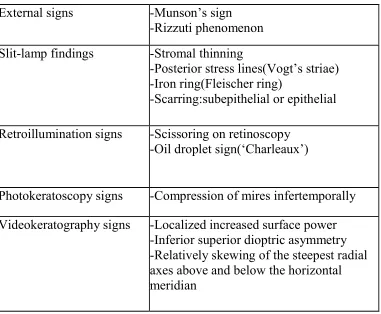
Signs of keratoconus
External signs of Keratoconus
Munson’s sign: V-shaped conformation of the lower lid produced
by the ectatic cornea in down gaze in advanced keratoconus.3
Rizzuti phenomenon: A beam of light focused near the nasal
limbus is produced by lateral illumination of the cornea in patients
with advanced keratoconus.3 Slit-lamp findings
Stromal thinning
Posterior stress lines (Vogt’s striae) 5
Iron ring (Fleischer ring)
Prominent corneal nerves
Scarring - epithelial, subepithelial or anterior stromal 6
Subepithelial fibrillary lines 6
Epithelial nebulae
Increased intensity of endothelial reflex 6 Retroillumination signs
Scissoring on retinoscopy 3
Early cases of keratoconus may be difficult to diagnose based on slit
lamp examination. Topography holds the key to diagnosis of these cases
and has today become essential in the diagnosis and assessment of
[image:15.612.113.494.215.529.2]keratoconus.
Table -1
External signs -Munson’s sign
-Rizzuti phenomenon
Slit-lamp findings -Stromal thinning
-Posterior stress lines(Vogt’s striae) -Iron ring(Fleischer ring)
-Scarring:subepithelial or epithelial
Retroillumination signs -Scissoring on retinoscopy -Oil droplet sign(‘Charleaux’)
Photokeratoscopy signs -Compression of mires infertemporally
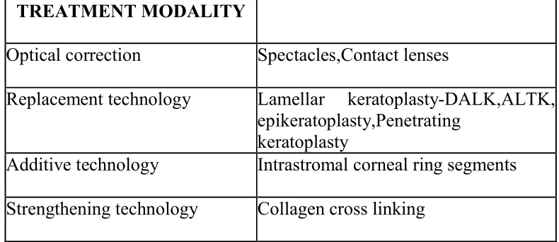
Management
The management options in keratoconus include optical correction,
replacement technology, additive technology and strengthening
technology.
1. Optical correction-The optical correction can be done with the help of spectacles and contact lenses.
Spectacles
They may be helpful in early stages by correcting the refractive
error but do not alter the shape of the cornea.
Contact lenses
Contact lenses are optical aids used in 90% of the patients.7 Early in the disease process soft lenses of toric design may suffice. However in
more advanced disease, rigid gas permeable lenses, including multicurve
spherical based lenses, aspheric lenses and bispheric lenses may be
required. A hybrid lens that has a rigid central portion for obtaining best
optics and a soft hydophilic peripheral skirt is also popular.8The complications associates with contact lenses include corneal abrasion,
apical scarring, hypoxia 9, neovascularization 10 and lens discomfort.
2. Replacement technology- Replacement therapy involves corneal
transplantation surgery in which the diseased layers of the cornea are
lamellar keratoplasty depending upon the depth of involvement and
presence or absence of scarring.
Lamellar keratoplasty- It is an extra ocular procedure in which only
diseased anterior part of corneal tissue is removed, leaving the recipients
normal anatomical structures intact.11
Epikeratoplasty- It is a type of onlay lamellar keratoplasty in which the
partial thickness donor cornea is placed on de-epithelized recipient
cornea. It may be preferred over penetrating keratoplasty in selected cases
like Down’s syndrome because of it’s non invasive nature and decreased
potential for graft rejection.12
Deep anterior lamellar keratoplasty (DALK)- It is a type of
lamellar keratoplasty in which lamellar dissection is performed up to the
descemet’s membrane and then the donor corneal button is sutured in its
place. It decreases the incidence of endothelial graft rejection.13
Automated lamellar therapeutic keratoplasty (ALTK)- In this
procedure a microkeratome is used to excise the pathological part of a
host cornea up to a particular depth and a healthy donor cornea, which is
also cut using an automated microkeratome and an artificial chamber is
Penetrating keratoplasty-It is the procedure of choice for
management of cases of keratoconus not adequately rehabilitated by
contact lenses. The success rate varies from 93%-96%.14 The indications for penetrating keratoplasty include contact lens failures or intolerance,
central scarring and poor visual acuity despite contact lenses. However a
full thickness graft is associated with complications like graft rejection,
post operative astigmatism and recurrence of keratoconus.15
3. Additive technology- The additive technology for the treatment of
keratoconus consists of the use of intrastromal corneal ring segments
(ICRS) which are manufactured under two names- Intacs prescription
inserts and Ferrara intrastromal corneal ring segments.16
Intrastromal corneal ring segments- Two thin arcs made up of
PMMA are slid between the layers of the corneal stroma through
incisions made in the corneal periphery. The segments flatten the peak of
the cone, thus reducing the amount of myopia and make patients more
contact lens tolerant.17,18 The potential complications include accidental penetration through the anterior chamber, infection, migration or
extrusion of segments.
4.Strengthening technology
Corneal collagen cross-linking is a new approach to increase the
BASIC PRINCIPLE- Crosslinking is a widespread method in the
polymer industry to harden material e.g. chemical crosslinking with
glutaraldehyde is used in the preparation of prosthetic heart valves and
physical cross linking by Ultraviolet-A (UVA) is used in dentistry to
harden filling material. Cross linking of human collagen is a
physiological process and stiffening of the connective tissue is well
known in diabetes and ageing.19
This is related to the age related glycosylation of collagen
molecules. The aim of this treatment is to create additional chemical
bonds inside the corneal stroma by means of highly localised
photopolymerisation while minimising exposure to surrounding ocular
structures.
Using UVA at 370nm and the photosensitizer riboflavin, the
photosensitizer is excited into it’s triplet state generating the reactive
oxygen species (ROS). These are mainly in the form of singlet oxygen
and to a much less degree, they may be in the form of superoxide anion
radicals. The ROS then reacts with various molecules, including chemical
covalent bonds, bridging amino groups of collagen fibrils (type 2
photochemical reaction). The wavelength of 370nm is chosen because of
Table -2
TREATMENT MODALITY
Optical correction Spectacles,Contact lenses
Replacement technology Lamellar keratoplasty-DALK,ALTK,
epikeratoplasty,Penetrating keratoplasty
Additive technology Intrastromal corneal ring segments
REVIEW OF LITERATURE
ANIMAL STUDIES
Wollensak et al20 in 2003 first introduced the procedure of collagen
cross-linking (CXL). They evaluated the effect of treatment using
riboflavin and ultraviolet A on human and porcine corneas. A total of 25
corneal strips were subjected to treatment, 5 of which were obtained from
porcine eyes and the remaining from human cadavers. The corneas were
treated with UV-A (370 nm, irradiance-3mW/cm2 ) for half an hour following treatment with riboflavin. The treated corneas were subjected
to a static stress test using a biomaterial tester. A significant increase in
the rigidity was noted, indicated by a 71.9 % and 328.9 % increase in
stress of porcine and human corneas respectively. The greater stiffening
effect of the human corneas was due to the relatively thinner nature.
Wollensak et al 21 in 2003 evaluated possible in vitro cytotoxic
effects on corneal keratocytes following treatment with riboflavin and
ultraviolet-A irradiation. They treated endothelial cell cultures of porcine
corneas with varying levels of UV-A irradiances ranging from 0.4 to 1.0
mW/cm2 , after treatment with riboflavin. They used Yopro fluorescence and tryphan blue staining to evaluate cell death in endothelial cultures, 24
irradiation ranging from 0.4 to 9 mW/cm2) was also tested. An abrupt cytotoxic irradiance level was found at 0.5 mW/cm2 after UVA irradiation combined with photosensitizer riboflavin, which was 10 fold
lower than the cytotoxic irradiance of 5mW/cm2 after UV-A irradiation alone. Riboflavin alone was not cytotoxic. They noted a cytotoxic effect
upto a corneal depth of 300um following combined treatment.
Spoerl et al22 in 2004 evaluated the resistance of collagen
cross-linking treated corneas to enzymatic degradation. The study group
included 80 porcine corneas, 60 of which were treated with riboflavin and
ultraviolet-A while the remaining served as controls. They exposed the
trephined treated corneal buttons to collagenase, trypsin and pepsin
enzymes. The buttons were then examined by light microscopy. The
treated corneas underwent dissolution by pepsin enzyme by day 14 while
the untreated cases underwent digestion by day 6. Corneal resistance to
collagen digesting enzymes markedly increased following collagen
cross-linking.
Wollensak et al23 in 2004 studied corneal keratocytes for the
possible cytotoxic effect of riboflavin and UV-A treatment. They
subjected corneas of thirty-four New Zealand white rabbits to cross
linking treatment, following which they euthanized the rabbits four to
twenty four hours postoperatively. Four eyes were treated with corneal
evaluation of the treated corneas was done. They detected keratocyte
apoptosis using TUNEL technique and transmission electron microscopy.
The apoptotic keratocytes were found within the anterior 50 microns of
the control eyes. The depth of apoptotic cells in the treated eyes varied
depending upon the strength of the irradiance applied. A strength of
0.5-0.7 mw/cm2of UVA irradiance was found to be toxic. Dose dependent keratocyte damage up to a depth of 300um in human corneas can be
expected following treatment with UVA dose of 5.4J/cm2.
Wollensak et al24 in 2004 studied changes in the diameter of
collagen fiber following cross-linking of rabbit corneas. The right eyes of
10 New Zealand white rabbits underwent cross linking and the fellow
eyes served as the control. They divided the control group into three
groups- eyes left untreated (1-4), eyes epithelialized only (5-7) and
de-epithelialized eyes treated with riboflavin alone (8-10). There was a
significant increase in the diameter of collagen fiber by 12.2% and 4.6%
in the anterior and posterior stroma respectively, compared to the left
eyes. There was also a significant increase in the diameter of anterior
corneal fibers compared to fibers in the posterior stroma of the same eye.
There is a stronger effect of cross-linking in the anterior half of the
stroma due to rapid decrease in irradiance across the cornea following
Kohlhaas et al 25 in 2006 evaluated the depth of corneal tissue up to
which stiffening effect of crosslinking was biomechanically detectable.
They evaluated 40 enucleated porcine eyes, half of which underwent
treatment with riboflavin and Ultraviolet-A while the remaining half
served as control. Following treatment 2 flaps of 200 microns each were
cut using a microkeratome. Corneal strips of 7mm length and 5mm width
were prepared and were subjected to stress-strain behaviour with a
material tester. There was a stronger stiffening effect in the anterior
treated flaps as compared to the posterior treated flaps and the control
group (p=0.001). There was a significant increase in stress of treated
anterior corneal flaps compared to those of the control group. There was
however no significant difference between the posterior treated flaps and
the control group. The greater stiffening effect in the anterior stroma was
attributed to the absorption of 65-70% of UV-A by the anterior 200
microns and the remaining 20% by the next 200 microns. Thus
crosslinking has no effect on deeper structures and endothelium.
Wollensak et al 26 in 2009 studied the efficacy of cross-linking
treatment without epithelial debridement in rabbit eyes. The cross-linked
eyes were divided into three groups- standardized crosslinking following
epithelial debridement (Group 1), using benzalkonium
chloride-containing proxymetacaine eye drops without epithelial removal (Group
removal (Group 3). All three groups were treated with riboflavin solution
and were irradiated with an ultraviolet-A double diode for 30
minutes(irradiance 3 mW/cm2).The rabbits were euthanised 1 day following crosslinking. The corneas were subjected to biochemical and
histological analyses. Group 1 (102.45%) and Group 2 (21.30%) showed
a significant increase in Young’s modulus. No significant changes were
observed in Group 3. Histological evaluation revealed complete loss of
keratocytes and endothelium in Group 1 and an inhomogeneous loss of
keratocytes in Group 2. Group 3 showed no changes.Biochemical effect
of crosslinking without epithelial debridement was reduced probably due
to restricted and heterogenous stromal distribution of riboflavin. The
cytotoxic effect was however restricted to 200 mm.
CLINICAL STUDIES
Wollensak et al 27 in 2003 were the first to evaluate the clinical
effect of corneal cross-linking using riboflavin and UV-A for halting the
progression of keratoconus. The study included twenty-three eyes of
twenty-two patients who presented with moderate or advanced
progressive keratoconus. The procedure involved application of
riboflavin drops and UVA irradiation (370 nm,3mW/cm2) following corneal epithelial debridement. Post-operative evaluation included visual
acuity, slit lamp evaluation, corneal topography, endothelial cell count
years.All eyes in the study failed to progress following treatment. 16 eyes
(70%) showed a regression of keratoconus with a 2.01D reduction in
maximum keratometric value and a 1.14 D reduction in refractive error.
There was no effect on intraocular pressure, corneal transparency and
endothelial density. A slight improvement in visual acuity was noted in
15 eyes (65%).
Wollensak et al28 in 2006 evaluated the effect of cross-linking on
the progression of keratoconus in 60 eyes over a period of 3 to 5 years.
They concluded a halt in the progression of the disease in all eyes.
Moreover, there was a minimal reversal of the keratoconus in 31 eyes. An
improvement of 1.4 lines was noted in the best-corrected visual acuity.
Caporossi et al29 in 2006 carried out a prospective non-randomized
study to evaluate the efficacy of crosslinking in halting the progression of
keratoconus. The study included 10 eyes of 10 patients with progressive
disease, while the fellow eyes of 8 patients served as controls. Clinical
evaluation included measurement of uncorrected and best-corrected
visual acuity. Corneal topographic evaluations, linear scan optical
tomography, endothelial cell density, ultrasound pachymetry, intraocular
pressure measurement and HRT 2 system confocal was performed at
1,2,3 and 6 months. The study showed a significant improvement in
uncorrected and best spectacle corrected visual acuity.Topographic
the central 3 mm. There were no significant difference in the intraocular
pressure and endothelial cell density within the cases. 37.5% of the eyes
showed a progression of disease within the control group.
Seiler et al 30 in 2006 performed cross-linking in 16 patients of
keratoconus with a maximum keratometry of 60D and central corneal
thickness of at least 400 microns. The corneal epithelium was
mechanically removed with a diameter of 6 mm and riboflavin drops
0.1% instilled repeatedly for 20 minutes. UVA radiations were given at
irradiance of 3mW/cm2 at a working distance 1 cm. Biomicroscopic and topographic evaluation of eyes was carried out preoperatively and at
subsequent follow-ups. A thin demarcation line was seen at around 300
microns of corneal depth on slit lamp evaluation in 14 eyes.
Mazzotta et al 31 in 2007 evaluated changes in the corneal stroma
of eyes with advanced keratoconus following treatment with collagen
cross-linking.10 patients with progressive keratoconus were treated by
collagen cross-linking and assessed by means of Heidelberg Retinal
Tomography II Rodstock Corneal Module (HRT II-RCM) in vivo
confocal microscopy. The eye that had progressed further in the disease
process was treated while the fellow eye served as the control. Eyes were
evaluated at 1, 3 and 6 months postoperative with HRT II-RCM confocal
microscopy. Stromal edema with refraction of keratocytes in the anterior
with associated keratocyte repopulation was observed 3 months
postoperatively. Complete keratocyte repopulation with increased stromal
density was noted at 6 months. There was no endothelial cell damage
noted postoperatively.
Wittig-Silva et al32 in 2008 randomized 66 eyes of 49 patients
with documented progressive keratoconus into treatment and control
groups. Collagen cross-linking was performed in all the eyes in
accordance with previously published protocols.On every follow-up a
complete ocular evaluation was conducted including confocal microscopy
and endothelial cell count. Statistical analysis of treated eyes revealed a
significant flattening of the simulated keratometry value (K max) at 3, 6
and 12 months postoperatively. The cases also showed an improvement
in best-corrected visual acuity. On the other hand, a significant steeping
of K max and associated reduction of best spectacle-corrected visual
acuity was noted in the control eyes at 3, 6 and 12 months
postoperatively. No statistically significant changes in spherical
equivalent and endothelial cell density were observed. Postoperative
confocal microscopy revealed some highly reflective stria in mid to
posterior stroma between 1-3 months after treatment, which became less
marked in subsequent visits. One patient with a highly atopic
predisposition developed an inflammatory reaction in anterior chamber
paracentral infiltrate, after prematurely resuming wear of his rigid contact
lens on day 3 with no persistent scarring.
Dhaliwal et al33 in 2008 used confocal, electron and light
microscopy to study changes in corneas treated with collagen
cross-linking. The procedure involved removal of central epithelium followed
by treatment with riboflavin 0.1 % and ultraviolet – A light. Preoperative
evaluation revealed normal appearing corneas with reduced stromal detail
on confocal microscopy. Postoperative evaluation revealed a superficial
layer of hyper reflective structures upto a depth of 300 microns.
Keratocyte apoptotic changes within the superficial layers of the cornea
were seen on electron microscopy.
Kymionis et al34 in 2008 evaluated changes in corneal tissue
following crosslinking in eyes with post laser in situ
keratomileusis(LASIK) keratectasia and keratoconus. The study group
included five patients with progressive keratoconus and five with post
LASIK ectasia. The treated eyes were evaluated by corneal in vivo
confocal microscopy. The control group included three healthy corneas
and three post LASIK eyes with no evidence of ectasia. Corneal
evaluation within the first three postoperative months revealed apoptosis
of keratocyte nuclei and alterations in collagen structure.Over subsequent
changes seen were similar in both keratoconic and post LASIK ectasia
patients.
Mazzotta et al 35 in 2008 used Heidelberg Retinal Tomography
(HRT) II confocal microscopy to evaluate morphological changes in
cross-linked corneas. The study included 44 eyes with progressive
keratoconus that were treated based on the Siena protocol: Pilocarpine
1% drops 30 minutes before, topical anaesthesia with lidocaine 4% drops
15 minutes before irradiation, mechanical scraping of epithelium (9mm
diameter area),pre irradiation soaking for 10 minutes in riboflavin
solution 0.1% (Ricolin, Sooft, Italy) applied every 2.5 minutes for 30
minutes,30 minutes exposure to solid state UVA illuminator (Caporossi;
Baiocchi; Mazzotta; X linker, CSO, Italy),8-mm diameter irradiated area,
energy delivered 3 mW/cm2. Confocal scans were taken preoperatively and at subsequent postoperative follow-ups at 1, 3, 6 months and 1, 2 and
3 years. Complete epithelial regrowth was noted within four days of
removal of bandage contact lens removal. Sub-epithelial plexus was
restored to original anatomical structure within first postoperative year. A
late demarcation line was noted at a depth of 340 microns. 5 out of 44
eyes presented with transitory corneal opacity similar to corneal haze.
Resolution of opacity was seen within a month following administration
of topical steroid drops. An increased evidence of preoperative Vogt
Raiskup-Wolf et al36 in 2008 conducted a long-term study on the
dampening effect of collagen crosslinking on progressive keratoconus.
The study included 488 eyes of 272 patients with a mean age of 30.04 +/-
10.46 years. Investigations done preoperatively and at all subsequent
follow-ups included uncorrected and best-corrected visual acuity, corneal
pachymetry, corneal topography and intraocular pressure evaluation. The
period of follow-up ranged from 6 months to a maximum of 6 years.
There was a significant decrease in the steepest keratometry reading at
first, second and fourth postoperative year. There was a significant
improvement in BCVA by at least one Snellen’s line in 53% of 142 eyes
in the first year,57% of 66 eyes in the second year, and 58% of 33 eyes in
the first year or remained stable(no lines lost) in 20%,24% and 29%
respectively. Keratoconus continued to progress in two patients at 18 and
24 months follow-up following acute exacerbation of neurodermatitis.
The procedure was repeated in both eyes.
Koller et al 37 in 2008 used Scheimpflug imaging to compare
geometrical shape factors of post corneal crosslinking corneas with
untreated fellow eyes. The study group included 21 patients with
progressive disease, all of whom underwent Scheimpflug imaging
(Pentacam) of the corneal surface. The eye of the patient more advanced
in the disease process was treated, while the fellow eye served as the
cases. On the other hand 8 out of 21 eyes showed progression in the
control group. There was a significant decrease in the minimal curvature
radius between the preoperative and one year postoperative readings in
the cases, while a significant increase was noted in the control group. A
significant reduction in the thinnest pachymetry was noted following
treatment. No intraoperative or postoperative complications were seen.
Santonja et al 38 in 2008 reported a case of a 29-year-old woman
presenting with multiple corneal infiltrates in the upper quadrant of her
right eye. She underwent uneventful corneal crosslinking in the same eye
previously. Microbiological evaluation confirmed staphylococcus
epidermis keratitis, following which treatment with fortified antibiotics
was initiated. There was a significant increase in the best spectacle
corrected visual acuity and decrease in spherical equivalent between
preoperative and 5 months postoperative values. Mild residual haze
following treatment was seen in the corneal stroma.
Rama et al 39 in 2008 reported a case of corneal melt in 32-year-old
male, five days following treatment with corneal crosslinking for
keratoconus. Microbiological evaluation was positive for acanthamoeba
keratitis. The patient gave history of washing his face repeatedly with tap
Sharma et al 40 in 2009 reported a case of a 19-year-old woman
presenting on fourth postoperative day with complaints of redness, pain
and defective vision in her right eye. Clinical examination revealed
corneal infiltration measuring 7mm x 8 mm. Microbiological evaluation
of corneal and contact lens scraping confirmed Pseudomonas aeruginosa.
Posterior segment analysis on ultrasound revealed no abnormalities. The
infiltrate responded to treatment with antibiotics leaving behind a
leucomatous corneal opacity.
Koppen et al 41 in 2009 reported severe keratitis in four eyes from a
total of 117 eyes treated with corneal crosslinking. Patients experienced
delayed signs and symptoms of inflammation. Clinical features included
circumciliary congestion, diffuse keratic precipitates on the corneal
endothelium, anterior chamber reaction and multiple white infiltrates
along the edge of the treated cornea. The symptoms improved following
administration of topical or subconjunctival steroids. Two eyes showed a
persistent decrease in visual acuity secondary to corneal scarring.
Koller et al 42 in 2009 studied the rate of complications of
crosslinking procedure. The study included 117 eyes of 99 patients
presenting with primary corneal ectasia. Clinical evaluation at
preoperative and postoperative visits at 6 and 12 months included
uncorrected and best corrected visual acuity, slitlamp examination,
Statistical analysis included analysis of variance and the Mann-Whitney
U test to detect risk factors for complications. 2.9 % of the eyes two or
more Snellen lines of visual acuity. 7.6 % of the eyes continued to
progress in the disease process. The studies identified old age (of more
than 35 years) and a preoperative corrected distance visual acuity better
than 20/ 25 as significant risk factors for complications.7.6 % of the
patients presented with sterile corneal infiltrates while 2.8 % showed
stromal scarring. A resolution of stromal infiltrates was seen within one
month following administration of topical steroids. There was no
significant loss of final corrected visual acuity in any of the
complications. Corneal scarring faded almost completely within first
postoperative year.
Vinciguerra et al43 in 2009 evaluated refractive, topographic,
tomographic and abberometric outcome 12 months after corneal
cross-linking in 28 eyes with progressive advanced keratoconus. There was an
improvement in mean UCVA and BSCVA between preoperative and 12
months postoperative values. A significant decrease was seen in the mean
spherical equivalent. Mean baseline simulated keratometry (SIM K)
flattest, steepest and SIM K average decreased from 46.10 D,50.37 D and
48.08 D to 40.22 D,44.21 D and 42.01 D respectively at 12 months, a
difference that was significant for all three indices(P<0.005).Mean
41.40D at 12 months(P<0.005) and apical keratometry(AK) from 58.94
to 55.18(P<0.05).The treated eyes showed no deterioration of the Klyce
indices at 6 months postoperatively whereas the untreated contralateral
eyes did show deterioration. For a 3mm pupil there was a significant
reduction (P<0.05) in whole eye, corneal, higher order, and astigmatic
wavefront aberrations. They observed a significant difference in the total
coma and spherical aberration following the procedure. There was no
significant decrease in the endothelial counts.
Agrawal et al44 in 2009 studied the results of corneal collagen
cross-linking with riboflavin using ultraviolet-A light in sixty-eight eyes
of 41 patients with progressive keratoconus. The mean age was 16.9 +/-
3.5 years. Thirty-seven eyes with a follow up of atleast 12 months were
analysed. BCVA improved at least one line in 54% (20/37) of eyes and
remained stable in 28%(10/37) of eyes (P=0.006).Astigmatism decreased
by a mean of 1.20D in 47%(17/37) of eyes and remained stable(within +/-
0.50D) in 42% (15/37) of eyes. The K value of the apex decreased by a
mean of 2.73D in 66%(24/37) of eyes and remained stable(within
+/-0.50D) in 22% (8/37) of eyes. The maximum K value decreased by a
mean of 2.47 D in 54% (20/37) eyes and remained stable (within +/-
0.50D) in 38%(14/37) eyes. Corneal wavefront analysis revealed that
spherical and higher-order aberrations did not show significant variations
reduction at six months after treatment and persisted throughout the
follow up period(P=0.003).
Grewal et al45 in 2009 studied the effects of CXL on the corneal
elevation, curvature and thickness in 102 eyes with progressive
keratoconus. Clinical evaluation included uncorrected and best-corrected
visual acuity, Scheimpflug imaging and optical coherence tomography at
preoperative and postoperative visits at 1,3,6 and 12 months. No
significant difference in BCVA, spherical equivalent or cylindrical
refraction was noted between preoperative and postoperative values.
Similarly no significant changes were noted in corneal curvature and
pachymetry between preoperative and postoperative values.
Goldich et al46 in 2009 evaluated biomechanical changes in the
cornea after treatment with CXL. The study included 10 eyes of
progressive keratoconus, with a mean age of 26.5 years. Investigations
done on every visit included Ocular Response Analyzer (ORA) to
measure corneal hysteresis(CH),corneal resistance factor (CRF) and
intraocular pressure analysis using Goldmann applanation tonometry.
There was no statistically significant increase in the CH and CRF
between preoperative and postoperative values. There was a significant
increase in the IOP at 1 and 3 months postoperative compared to
Caporossi et al47 in 2009 studied the effects of CXL on 44 eyes
over a follow-up period of 48 months. Clinical investigations at each visit
included uncorrected and best corrected spectacle visual acuity,
endothelial cell count(I Konan, Non Con Robo; Konan Medical
Inc.,Hyogo, Japan),optical (Visante OCT; Zeiss, Jena, Germany) and
ultrasound (DGH; Pachette, Exton, Pennsylvania, USA) pachymetry,
corneal topography and surface abberometry (CSO EyeTop, Florence,
Italy),tomography (Orbscan IIz; Bausch & Lomb Inc., Rochester, New
York, USA), posterior segment optical coherence tomography(Stratus
OCT; Zeiss, Jena, Germany), and in vivo confocal microscopy(HRT II;
Heidelberg Engineering, Rostock, Germany). There was a stabilization of
keratoconus noted in 44 out of 48 eyes, while the fellow eyes showed a
progression of the disease process. A significant decrease in mean k value
and coma aberration was noted. There was a statistically significant
improvement in mean UCVA and BCVA between preoperative and
postoperative values. The study noted no side effects of the treatment,
either intraoperative or postoperatively.
Hersh et al48 in 2011 evaluated 1-year treatment outcomes
following corneal crosslinking in eyes with keratoconus and corneal
ectasia. The treatment group received standard CXL treatment and the
sham group received treatment with riboflavin alone. Parameters
(CDVA), refraction and corneal topography. There was a significant
improvement in the UDVA and CDVA within the treated eyes. Fifteen
patients (21.1%) gained and 1 patient lost (1.4 %) 2 or more Snellen lines
of CDVA. There was a significant decrease in K max in both keratoconic
and corneal ectasia eyes. Both CDVA and K max value worsened
between baseline and 1 month, followed by improvement between
1,3,and 6 months postoperatively and stabilization thereafter.
George D kymionis et al49 in 2011 studied 14 eyes of 21 patients with
corneal thickness less than 400 microns (following epithelial
debridement). The patients underwent CXL procedure based on
standardized treatment protocols. Preoperative and postoperative
evaluation included uncorrected and best-corrected distance visual acuity
and corneal topographic examination at 1,3,6 and 12 months
postoperatively. Corneal endothelium was evaluated using Confocal
scanning laser ophthalmoscope. No intraoperative and postoperative
complications were noted. A significant decrease of endothelial cell
LACUNAE IN KNOWLEDGE
There are limited prospective clinical trials available which
evaluated efficacy and safety of collagen cross linking in keratoconus in
AIM OF STUDY
To conduct a clinical trial to evaluate the safety and efficacy of
corneal collagen cross linking using riboflavin and UVA in progressive
MATERIALS AND METHODS
STUDY DESIGN – Prospective interventional clinical trial
PLACE OF STUDY – Aravind Eye Hospital And Postgraduate Institute,
Madurai – a tertiary eye care hospital
CASE COLLECTION PERIOD – 1 Year
FOLLOW UP PERIOD – 1 Year
SAMPLE SIZE – 50 patients
Case – Eye subjected to CXL procedure
Control – Fellow eye
INCLUSION CRITERIA
Progressive Keratoconus (1 dioptre or more within 1 year)
Age 12 to 30 years
Corneal pachymetry > 400 microns at thinnest point
Normal corneal endothelium
Maximum corneal curvature < 60 D
EXCLUSION CRITERIA
Corneal thickness < 400 microns at thinnest point
H/O herpetic keratitis
Central or paracentral opacities
Prior corneal surgery
Severe dry eye and ocular surface disorders
Concurrent corneal infections
Concomitant autoimmune diseases
Pregnant/nursing women
Hormone therapy
EVALUATION PARAMETERS
UDVA
CDVA
CLVA
Refraction
Slit lamp biomicroscopy
Fundus evaluation
Non contact tonometry
Tear Film break up time
Specular microscopy (Topcon SP 3000 P)
Clinical Photograph (Haag Streit)
SAFETY PARAMETERS
Post operative complications: -
• Non healing/Persistent Epithelial Defect
(a) <2mm (b) 2-5 mm (c ) >5 mm
• Permanent stromal Haze
(a) mild (b) moderate (c) severe
• Corneal Scarring
(a) nebular (b) macular (c) leucomatous
• Infective keratitis
• Corneal melting
• Corneal Infiltrate-
1. Number (a) single (b) multiple
2. Size (a) <2mm (b) 2-5 mm (c ) >5 mm
3. Type (a) bacterial (b) fungal (c ) sterile
• Endothelial cell loss
• Cataract formation
• Retinal pathology
EFFICACY PARAMETERS
Best corrected distance visual acuity-
1. Spectacle corrected
2. Contact lens corrected
Refraction-
1. Spherical
2. Cylindrical with axis
3. Mean spherical equivalent
ORBSCAN IIz
1. Sim K
2. K max
3. Thinnest pachymetry
4. Anterior float BFS difference
5. Posterior float BFS difference
6. 3mm zone irregularities
7. 5mm zone irregularities
SURGICAL TECHNIQUE
Procedure was performed under all aseptic precautions. Patient’s
eye was cleaned and draped. Proparacaine 0.5 % was instilled thrice at 5
minute intervals, 15 minutes before the procedure. A 15mm blade was
using a corneal trephine. One drop of Riboflavin 0.1 % in 20 % dextran
(Isotonic, 3ml vial by Medio-Cross Italy) was instilled every 2 minutes
for first 30 minutes and one drop every 2 minutes under UVA radiation
for the next 30 minutes. UVA radiations 365 nm with desired irradiance
of 3 mW/cm2 was used at a distance of 5 cm (UV-X Zurich Switzerland). On completion of the procedure a bandage contact lens was applied,
which was removed on the third postoperative day.
POSTOPERATIVE THERAPY
The postoperative therapy included-
Eye drops Vigamox 0.5% QID for 1 month
FOLLOW UP
The follow up schedule was as follows
1 week
1 month
3 months
6 months
12 months
ORBSCAN IIz (Bausch & Lomb)
It is based on the principle of placido disc and slit scanning system.
Patient details are entered in the proprietary software information window
and “acquisition” is selected. The patient is comfortably seated on the
device with chin at chinrest and forehead placed against the forehead rest.
He is directed to fix on the center of the target and to maintain a steady
gaze. Following appropriate alignment of the instrument the acquisition
sequence is triggered. The placido disc in the Orbscan is illuminated,
causing mires to reflect from the anterior surface of the cornea. The
machine stores the reflected mires. The machine projects 40 slits in total,
20 each from the right and left side onto the anterior corneal surface.
Each slit measures 12.50 mm by 0.30 mm and is projected onto the
cornea at an axis of 45 degrees from the instrument. The light from the
slit on passing the cornea is scattered in various directions. A part of this
light is back scattered towards the camera of the device and a two
dimensional image is recorded. The assessment of the acquired images is
the first step in processing. The acquired image is rejected when the
patient moves his eyes excessively, and a new image is required. On the
other hand, if the acquired image is satisfactory, a proprietary technique
compensates for minute eye movements. The anterior edge of each slit is
topographic image is created. Subsequent software processing detects the
edges of the reflected ring mires of the Placido disc, which allows
curvature reconstruction of the anterior surface of the cornea. Further
processing using the sampled data enables the Orbscan system to digitally
recreate the internal surface of the eye i.e. posterior cornea. This
procedure requires more sophisticated triangulation, integrating refractive
variables and the use of two previously generated anterior topographic
representations, elevation and curvature. The entire procedure typically
takes 30 seconds or less, and a total of more than 30 anterior segment
topographic maps can subsequently be created.
SPECULAR MICROSCOPY (Topcon SP 3000P)
Specular microscopy is based on the principle of specular
reflection where the angle of incidence is equal to the angle of reflection.
As the beam of light strikes the posterior corneal surface, almost all of it
is transmitted into the aqueous humour. Because there is a change in the
index of refraction at the endothelium aqueous humour interface, about
0.022 percent of total incident light is reflected, this reflected light is
captured by the clinical specular microscope and forms the endothelial
UV-XTM ILLUMINATION SYSTEM VERSION 1000 (IROC, SWITZERLAND)
It is a portable optoelectronic device in which light emitting diodes
of the device create UV-A light at a wavelength of 365 nm. An internal
microprocessor unit that controls the electric current to drive the unit
controls the device.
Parts of the device
1. Mechanical stand- It is used to mount the UV-X light source on a
stable table
2. Power supply – Low voltage is delivered to the light source with a DC
cord
3. UV-X light source – the light source has a beam aperture of 25mm
diameter. The treatment plane is about 50mm distance from the beam
aperture. The aperture wheel determines the diameter of the treatment
plane. Three sizes available are small (7.5mm), medium (9.5 mm) and
large (11.5mm)
4. UV light meter – it is used to check the correct UV light irradiation.
The meter is battery operated
5. Sensor probe adapter- The sensor probe attached to the UV light meter
is mounted in the beam aperture. The device is switched on and the
The nominal value for correct irradiance is 3.0 +/- 0.3 mW/cm2
To use the device, it is first mounted on a table and the irradiation
checked using the UV light meter. Then medium aperture (9.5mm) is
selected and the device is switched on. The beam is adjusted on the
patients cornea at a distance of 50 mm. UV radiations at the appropriate
dosage is given for half an hour and the device gets switched off
automatically after 30 minutes.
STATISTICAL ANALYSIS
Data was recorded on a predesigned proforma and managed on an
excel spreadsheet. All the entries were checked for any possible keyboard
error. All the quantitative variables were assessed for normal
distributions. Variables following normal distribution were summarized
by mean and standard deviation and other variables which were
non-normative as median (minimum-maximum) values at each point for both
case and control eyes. For all parameters following analysis were
performed,
1. Before comparing the post interventional values, the baseline for
variables following normal distribution were compared by student t test
and baseline for non-normative variables were compared using Freidman
test.
2. Repeated measure analysis of variance were used to compare the mean
3. To compare two groups at different follow up times, percentage change
at each time point from baseline were computed for every patient. Since
the percentage change was non-normally distributed, median was used as
summary measure and Wilcoxon rank sum test was used to compare the
median percentage change from baseline between the two groups at
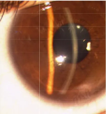
CLINICAL PHOTOGRAPH
[image:51.612.163.373.112.337.2]Figure 1 a -The preoperative clinical photograph showing clear stroma
Figure 1 b -Three month postoperative photograph showing CLINICAL PHOTOGRAPH
The preoperative clinical photograph showing clear stroma
Three month postoperative photograph showing scarring The preoperative clinical photograph showing clear stroma
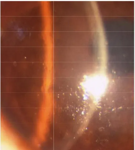
Figure 2 a -1 week postoperative photograph showing stromal haze
Figure 2 b - At three months there was no haze with clear stroma 1 week postoperative photograph showing stromal haze
due to oedema
At three months there was no haze with clear stroma 1 week postoperative photograph showing stromal haze
RIBOFLAVIN (MEDIO-CROSS ITALY) – RIBOFLAVIN IS AVAILABLE AS 0.1 % PRE PREPARED ISOTONIC SOLUTION
IN 20 % DEXTRAN AS A 3 ML VIAL
UV LIGHT FOCUSSED ON PATIENTS CORNEA
UV LIGHT SOURCE WITH SENSOR PROBE AND UV LIGHT UV LIGHT FOCUSSED ON PATIENTS CORNEA
UV LIGHT SOURCE WITH SENSOR PROBE AND UV LIGHT
METER
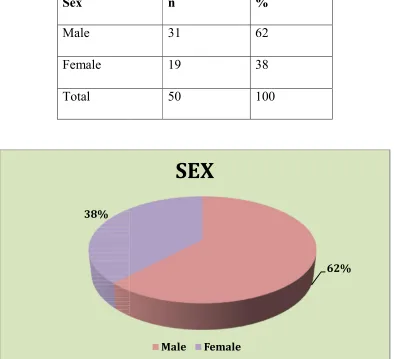
The mean age of presentation was 17.72 years (SD= 2.98) with a range of
[image:56.612.111.505.264.623.2]12 to 26 years.
Table -3
Sex
Male
Female
Total
A total of 50 patients were enrolled in the study, with 31 males (62%)
19 females (38%)
38%
RESULTS
AGE
The mean age of presentation was 17.72 years (SD= 2.98) with a range of
SEX DISTRIBUTION
n %
31 62
19 38
50 100
A total of 50 patients were enrolled in the study, with 31 males (62%)
62%
SEX
Male Female
The mean age of presentation was 17.72 years (SD= 2.98) with a range of
A total of 50 patients were enrolled in the study, with 31 males (62%) and
UNCORRECTED DISTANCE VISUAL ACUITY
Table – 4 UDVA in LogMAR
UDVA
Case Control
P-value (between groups)*
P-value Case (within group)*
P-value Control (within group)* n Mean(SD) Min-max n Mean(SD) Min-max
Pre-op 49 1.04(0.28) 0.48-1.78 50 0.63(0.46) 0-1.78 <0.001 - - 1 week 35 1.12(0.35) 0.3-1.78 32 0.77(0.44) 0-1.78 0.0001 0.224 0.026 1 month 50 1.00(0.29) 0.3-1.78 48 0.66(0.44) 0-1.48 <0.001 0.160 0.100 3 month 50 0.97(0.32) 0.3-1.78 49 0.67(0.47) 0-1.78 <0.001 0.007 0.030 6 month 46 0.95(0.31) 0.3-1.78 47 0.66(0.47) 0-1.78 <0.001 0.004 0.092 12
months
48 0.93(0.30) 0-1.3 48 0.64(0.45) 0-1.78 <0.001 0.009 0.163
*Wilcoxon signed-rank test
1.04 1.12 1
0.97 0.95 0.93
0.63 0.77
0.66 0.67 0.66
0.64
0.2 0.4 0.6 0.8 1 1.2
Pre-op 1Week 1 Month 3 Months 6 Months 12 Months
UDVA
The mean (SD) uncorrected visual acuity in eyes that underwent CXL
was 1.04 (0.28) log MAR preoperatively, 0.97 (0.32) at 3 months, 0.95
(0.031) at 6 months and 0.93 (0.30) at 1 year. There was a significant
improvement in UCVA among the cases with respect to preoperative
vision and vision and 3 months (p value = 0.007), 6 months (p value =
0.004) and 12 months (p value = 0.09).
The mean (SD) uncorrected visual acuity in control eyes was 0.63 (0.46)
log MAR preoperatively, 0.67 (0.47) at 3 months, 0.66 (0.47) at 6 months
and 0.64 (0.45) at 12 months. There was no statistically significant
change with respect to preoperative vision and final vision at 6 months
BEST SPECTACLE CORRECTED VISUAL ACUITY
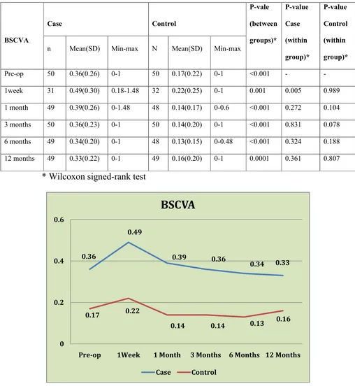
Table – 5 BSCVA in logMAR
BSCVA
Case Control
P-vale (between groups)*
P-value Case (within group)*
P-value Control (within group)* n Mean(SD) Min-max N Mean(SD) Min-max
Pre-op 50 0.36(0.26) 0-1 50 0.17(0.22) 0-1 <0.001 - - 1week 31 0.49(0.30) 0.18-1.48 32 0.22(0.25) 0-1 0.001 0.005 0.989 1 month 49 0.39(0.26) 0-1.48 48 0.14(0.17) 0-0.6 <0.001 0.272 0.104 3 months 50 0.36(0.23) 0-1 50 0.14(0.20) 0-1 <0.001 0.831 0.078 6 months 49 0.34(0.20) 0-1 48 0.13(0.15) 0-0.48 <0.001 0.324 0.188 12 months 49 0.33(0.22) 0-1 49 0.16(0.20) 0-1 0.0001 0.361 0.807
* Wilcoxon signed-rank test
0.36
0.49
0.39 0.36
0.34 0.33
0.17 0.22
0.14 0.14 0.13 0.16
0 0.2 0.4 0.6
Pre-op 1Week 1 Month 3 Months 6 Months 12 Months
BSCVA
The mean (SD) best spectacle corrected visual acuity in eyes that
underwent CXL was 0.36 (0.26)log MAR preoperatively, 0.36 (0.23) at 3
months, 0.34 (0.020) at 6 months and 0.33 (0.22) at 1 year. There was no
statistically significant improvement in the BSCVA with respect to
preoperative vision and vision at subsequent follow-ups.
The mean (SD) best spectacle corrected visual acuity in control
eyes was 0.17(0.22) logMAR preoperatively, 0.14 (0.20) at 3 months,
0.13 (0.15) at 6 months and 0.16 (0.20) at 1 year. There was no
CONTACT LENS VISUAL ACUITY
Table -6 CLVA in logMAR
CLVA
Case Control
P-vale (between groups)
P-value Case (within group)
P-value Control (within group) n Mean(SD)
Min-max
N Mean(SD) Min-max
Pre-op 49 0.06(0.10) 0-0.3 49 0.02(0.06) 0-0.3 0.014 - - 3 months 48 0.06(0.11) 0-0.48 46 0.01(0.05) 0-0.3 0.003 0.769 0.317 6 months 49 0.05(0.09) 0-0.3 47 0.01(0.05) 0-0.3 0.030 0.438 0.317 12 months 48 0.03(0.07) 0-0.3 49 0.02(0.06) 0-0.3 0.115 0.122 >0.99
Wilcoxon signed-rank test
0.06 0.06
0.05
0.03
0.02
0.01 0.01
0.02
0 0.02 0.04 0.06 0.08
Pre-op 3 Months 6 Months 12 Months
CLVA
The mean (SD) contact lens corrected visual acuity in eyes that
underwent CXL was 0.06 (0.10)logMAR preoperatively, 0.06 (0.11) at 3
months, 0.05(0.09) at 6 months and 0.03 (0.07) at 1 year. There was no
significant improvement in the CLVA in the cases.
The mean (SD) contact lens visual acuity in control eyes was 0.02
(0.06) log MAR preoperatively, 0.01 (0.05) at 3 months, 0.01 (0.05) at 6
months and 0.02 (0.06) at 1 year. There was no significant change in
REFRACTIVE SPHERE
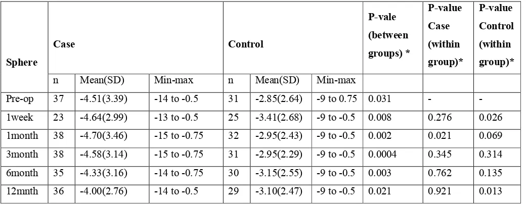
Table-7 Refractive sphere in Dioptre
Sphere
Case Control
P-vale (between groups) *
P-value Case (within group)*
P-value Control (within group)* n Mean(SD) Min-max n Mean(SD) Min-max
Pre-op 37 -4.51(3.39) -14 to -0.5 31 -2.85(2.64) -9 to 0.75 0.031 - - 1week 23 -4.64(2.99) -13 to -0.5 25 -3.41(2.68) -9 to -0.5 0.008 0.276 0.026 1month 38 -4.70(3.46) -15 to -0.75 32 -2.95(2.43) -9 to -0.5 0.002 0.021 0.069 3month 38 -4.58(3.14) -15 to -0.75 31 -2.95(2.29) -9 to -0.5 0.0004 0.345 0.314 6month 35 -4.33(3.16) -14 to -0.75 30 -3.15(2.55) -9 to -0.5 0.003 0.762 0.135 12mnth 36 -4.00(2.76) -14 to -0.5 29 -3.10(2.47) -9 to -0.5 0.021 0.921 0.013
The mean (SD) spherical value in eyes that underwent CXL was
-4.51D (3.39) preoperatively, -4.58D (3.14) at 3 months, -4.33D (3.16) at
6 months and -4.00D (2.76) at 12 months. There was no significant
decrease in the spherical value of the cases.
-4.51 -4.64 -4.7 -4.58 -4.33
-4
-2.85 -3.41 -2.95 -2.95 -3.15 -3.1
-5 -4 -3 -2 -1 0
Pre-op 1Week 1 Month 3 Months 6 Months 12 Months
Refractive Sphere
The mean (SD) spherical value in the control eyes was -2.85D
(2.64) preoperatively, -2.95D (2.29) at 3 months, -3.15D (2.55) at 6
months and -3.10 (2.47) at 12 months. There was a significant increase in
the spherical value of the control group at 1 year as compared to
REFRACTIVE CYLINDER
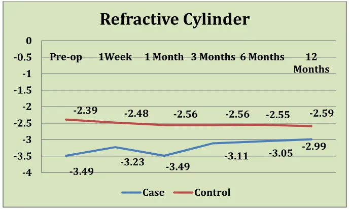
Table- 8 Refractive cylinder in Dioptre
Cylinder
Case Control
P-value (between groups) * P-value Case (within group)* P-value Control (within group)* n Mean(SD) Min-max n Mean(SD) Min-max
Pre-op 46 -3.49(1.39) -6 to -1 36 -2.39(1.58) -6 to -0.5 0.0004 - - 1 week 24 -3.23(1.42) -6 to -1 25 -2.48(1.62) -6 to -0.5 0.0066 0.322 0.415 1 months 46 -3.49(1.40) -6 to -0.75 36 -2.56(1.64) -6 to -0.5 0.0033 0.785 0.682 3 months 48 -3.11(1.31) -6 to -0.75 35 -2.56(1.62) -6 to -0.5 0.071 0.066 0.449 6 months 44 -3.05(1.28) -6 to -0.75 33 -2.55(1.55) -6 to -0.5 0.032 0.114 0.212 12 months 46 -2.99(1.26) -6 to -0.75 33 -2.59(1.40) -6 to -0.75 0.080 0.133 0.212
The mean (SD) cylindrical value in eyes that underwent CXL was
-3.49D (1.39) preoperatively, -3.11D (1.31) at 3 months, --3.05D (1.28) at
6 months and -2.99D (1.26) at 12 months. There was no significant
decrease in the cylindrical value of the cases.
-3.49 -3.23 -3.49
-3.11 -3.05 -2.99 -2.39 -2.48 -2.56 -2.56 -2.55 -2.59
-4 -3.5 -3 -2.5 -2 -1.5 -1 -0.5 0
Pre-op 1Week 1 Month 3 Months 6 Months 12 Months
Refractive Cylinder
The mean (SD) cylindrical value in control was -2.39D (1.58)
preoperatively, -2.56D (1.62) at 3 months, -2.55D (1.55) at 6 months and
-2.59D (1.40) at 12 months. There was no significant increase in the
SPHERICAL EQUIVALENT
Table- 9 Spherical equivalent in Dioptre
SPH EQ
Case Control
P-value (between groups) *
P-value Case (within group)*
P-value Control (within group)* n Mean(SD) Min-max n Mean(SD) Min-max
Pre-op 47 -5.60(3.28) -15.25 to -1.00 39 -3.57(2.92) -11.5 to -0.5 0.0002 - - 1 week 26 -5.88(3.16) -14.75 to -1.25 29 -4.08(3.33) -11.5 to -0.5 0.0005 0.807 0.271 1 month 46 -5.76(3.47) -16.25 to -0.75 39 -3.77(2.88) -11.5 to -0.5 <0.001 0.244 0.101 3 months 49 -5.56(3.46) -16.25 to -0.75 38 -3.78(2.75) -11.5 to -0.5 0.0007 0.497 0.099 6 months 43 -5.09(3.05) -15.25 to -0.75 37 -3.94(2.96) -11 to -0.5 0.0004 0.078 0.032 12 months 45 -4.78(2.94) -15.25 to -0.75 38 -3.74(2.73) -10.88 to -0.68 0.017 0.074 0.008
*Wilcoxon signed-rank test
-5.6 -5.88 -5.76 -5.56
-5.09 -4.78 -3.57
-4.08 -3.77 -3.78 -3.94 -3.74
-7 -6 -5 -4 -3 -2 -1 0
Spherical Equivalent
The mean (SD) spherical equivalent in eyes that underwent CXL
was -5.60 (3.28) preoperatively, -5.56 (3.46) at 3 months, -5.09(3.05) at 6
months and -4.78 (2.94) at 1 year. There was a no significant reduction in
the spherical equivalent in the cases.
The mean (SD) spherical equivalent in control eyes was -3.57
(2.92) preoperatively, -3.78 (2.75) at 3 months, -3.94 (2.96) at 6 months
and -3.74 (2.73) at 1 year. The increase in spherical equivalent was
statistically significant at 6 months (p value = 0.032) and 12 months (p