Dissertation submitted to
THE TAMILNADU Dr. M.G.R. MEDICAL UNIVERSITY In partial fulfillment for the Degree of
MASTER OF DENTAL SURGERY
BRANCH III
This is to certify that this dissertation titled “TUMORS OF MAXILLARY SALIVARY GLANDS” is a bonafide record of work done by Dr.V.R.RAJINIKANTH under my guidance during his postgraduate study period between 2012-2015.
This dissertation is submitted to THE TAMILNADU Dr. M.G.R. MEDICAL UNIVERSITY, in partial fulfillment for the degree of MASTER OF DENTAL SURGERY in Branch III – ORAL AND MAXILLOFACIAL SURGERY.
It has not been submitted (partially or fully) for the award of any other degree or diploma.
Professor, HOD and Guide Principal
Dr.L.DEEPANANDAN, M.D.S., Dr.V.PRABHAKAR, M.D.S., Department of Oral & Maxillofacial surgery, Sri Ramakrishna Dental College & Hospital,
Sri Ramakrishna Dental College & Hospital, Coimbatore. Coimbatore.
Candidate
Dr.V.R.RAJINIKANTH
Department of Oral & Maxillofacial surgery, Sri Ramakrishna Dental College & Hospital,
Coimbatore.
Date:
ACKNOWLEDGEMENT
Foremost, I would like to express my sincere gratitude to my guide
Dr. L.Deepanandan, M.D.S., Professor and Head, Department of Oral and Maxillofacial Surgery, Sri Ramakrishna Dental College, for his unwavering guidance,
for his patience, motivation, enthusiasm, and immense knowledge. His guidance helped
me in getting a better shape during the time of my course, to understand and complete the
dissertation. I could not have imagined having a better guide for my dissertation.
I also express my sincere heartfelt gratitude Dr. M.S.Senthil kumar, M.D.S., Associate Professor Department of Oral and Maxillofacial Surgery, Sri Ramakrishna Dental College, for his constant support and encouragement throughout the duration of my
course, scholarly support throughout this journey.
I also express my sincere heartfelt gratitude to Dr.R.Kannan, MDS , Dr.M.A.I.Munshi, MDS and Dr.R.S.Karthik,MDS, Readers Department of Oral and Maxillofacial Surgery, Sri Ramakrishna Dental College, for their innovative ideas, suggestions,
valuable criticism and constant encouragement throughout the duration of my course.
I also express my sincere heartfelt gratitude to Dr.V.Sundararajan, MDS and Dr.R.Vijay, MDS Senior Lecturers, Department of Oral and Maxillofacial Surgery, Sri Ramakrishna Dental College, for their valuable help, support and guidance.
I also express my sincere heartfelt gratitude to Dr. Guhan, M.D, D.M., Director,
my course,
It would be unfair of me if I fail to acknowledge the timely help and constant
encouragement from my colleague Dr.M.Suganthi, whose support helped me to overcome difficulties.
I also express my sincere thanks to my juniors Dr.V.Kiruthika, Dr.M.Geetha, Dr. Bhargavi, Dr.Gayatri for their help and support.
Above all I wish the Almighty for blessing me with such a wonderful parents and brother. Their support, love, sacrifices and encouragement have made me to achieve my dream.
I thank the almighty for guiding me throughout my life.
CONTENTS
TITLE
PAGE NO
1. Introduction
1
2. Aim and Objective
6
3. Review of Literature
7
4. Materials and Methods
41
5. Results
57
6. Discussion
60
7. Summary and Conclusion
71
1
Tumors of maxillary salivary glands are a heterogeneous group of malignancies1 with unknown etiology. The unique nature of maxillary salivary gland tumors is that it should be considered malignant until proven otherwise. Tumors arising from the smaller glands are more likely to be malignant.
Salivary gland tumors represent 3% of all the head and neck malignancies 2,3 The tumors of minor salivary glands range from 9-23% of all salivary gland tumor4,5,6,7 Minor salivary gland tumors of hard palate accounts for 5% of all oral cavity tumors. Eight percent of salivary gland tumors occur in hard palate and 80% of them are malignant. Minor salivary gland tumors of paranasal sinuses are very rare accounting for less than 0.48% of all nasopharyngeal malignancies8.
2
minor salivary glands. The frequency of distribution in decreasing order of frequency is hard palate (55.8%), soft palate (13.4%), buccal mucosa (7.8%) and lip (7%).13
The classification of salivary gland tumors was first done in 1972 by WHO, the classification was revised after 19 years in the year 1991. In the second edition of the histological classification of salivary gland tumors new entities were included, the concept of monomorphic adenoma was suppressed and 11 independent histological entities were introduced. In the year 2005 new classification of salivary gland tumors was published taking in to account the great morphological diversity of salivary gland
tumors21.
The oral cavity contains 450-750 minor salivary glands 22 .The minor salivary glands are located beneath the epithelium in nearly all parts of oral cavity, consist of multiple small groups of secretory units opening through short ducts in to the oral cavity. Minor salivary glands do not have a distinct capsule; they mix with the connective tissue of the submucosa or muscle fibers of tongue or cheek.
3
The lingual glands are divided in to several groups. The anterior lingual glands also called as Glands of Blandin and Nuhn are located near the apex of tongue. The anterior region of glands is predominantly mucous, while posterior portion are mixed. The ducts open on the ventral surface of tongue near lingual frenum. The posterolateral mucous glands are located lateral and posterior to vallate papillae in association with lingual tonsil. They are purely mucous and their ducts open in to the dorsal surface of tongue. The posterolateral serous glands also called Von Ebner Glands are purely serous glands. They are located between muscle fibers of tongue, below vallate papillae. The ducts of the glands open in to the trough of vallate papillae and at the rudimentary foliate papillae on the side of tongue23.
Mucoepidermoid carcinoma is the most prevalent tumor in India24, Libya25, UK26, Venezula27 and USA28. Adenoid cystic carcinoma is the most prevalent tumor in China9, Germany20 and Brazil9. In Tamil Nadu, India, the most common benign tumor is
pleomorphic adenoma, the most common malignant tumors in decreasing frequency is mucoepidermoid carcinoma, adenoid cystic carcinoma, polymorphous low grade adenocarcinoma, adenocarcinoma not specified, basal cell adenocarcinoma1.
4
The time duration for diagnosis of malignant tumor is more than one year, for benign tumor it is less than six months. The malignant tumors progressed faster than benign tumors9. The clinical presentation is that of a nodule, pain and ulceration, with pain being most common presentation30.
The following features like difficult surgical access, difficulty in obtaining three dimensional marginal clearence and negative surgical margins, high primary site
5 The purpose of the study is
1. To evaluate the surgical outcome in patients with tumors of maxillary salivary gland.
2. To evaluate the combined surgical and radiotherapeutic outcomes. 3. To evaluate the recurrence in patients after treatment.
6 1. James et al;(1999)31
made a clinicopathological study of 164 cases of polymorphous low grade adenocarcinoma. The literature states polymorphous low grade
adenocarcinoma occurs only in minor salivary gland, most frequent in females than males, with clinical presentation as mass lesion and presenting for average duration of two years. The palate is the most common site of occurrence, with average tumor size of 2.2cm. The literature states that adjuvant radiotherapy did not alter the survival, as patients who underwent radiotherapy were more likely to have evidence of disease at last follow up. But the overall survival of polymorphous low grade adenocarcinoma was considered by the literature as excellent. Lesion in soft palate tended to have a shorter mean duration of symptoms. Complete surgical excision is the appropriate therapy, with excellent long term prognosis. Local recurrence was seen after an average period of 7.2 years but treated adequately with surgical excision. The literature states that statistically the tumors located in hard palate were significantly more likely to be associated with tumor recurrence, persistence or death. Patients with tumors described to occur on hard palate, or “palate, Not Specified” were 1.6 times more likely to have evidence of disease
7
embedded sections. Macroscopically polymorphous low grade adenocarcinoma is firm to solid, ovoid mass, typically in close proximity to overlying surface epithelium,
unencapsulated. Polymorphous low grade adenocarcinoma infiltrates in to perisalivary gland adipose connective tissue, but true skeletal muscle invasion is uncommon. Skeletal muscle involvement presents clinically as a compression of muscle fibers. Infiltration in to adjacent salivary gland is quite common. Normal ducts and acini are seen at the periphery of polymorphous low grade adenocarcinoma. The surface epithelium is intact occasionally ulcerated. When intact the epithelium usually is not invaded by tumor cells. Polymorphous low grade adenocarcinoma display a mixture of growth patterns within a single tumor including 1.glandular profiles, 2.tubules, 3.trabeculae, 4.cribriform nests and 5.linear single cell “Indian file infiltration”, with less frequent focal papillary pattern.
Slate gray colouration is characterization of this neoplasm due to myxoid, mucoid matrix background. Tumor cells arranged concentrically around a central nidus, creating
targetoid appearance (“onion skinning”).The nidus often was found to be a small nerve
bundle (neurotropism) and was quite characteristic for polymorphous low grade adenocarcinoma. Perineural invasion was more accentuated in targetoid pattern
2. Catherine et al ;(2000)32
8
surface. The literature summarizes that pediatric Minor salivary gland tumors are rare but more likely to be malignant in contrast to adult. Clinically polymorphous low grade adenocarcinoma has innocuous appearance mimicking a reactive or benign salivary gland lesion. If not diagnosed earlier it leads to significant delay in appropriate referral and treatment. The specimen in this study was formalin fixed, embedded in wax. Palatal specimens were routinely decalcified before sectioning.
3. Hannen et al;(2000)33
reports a case of palate with metastasis to lung. The literature states that there is growing evidence that polymorphous low grade adenocarcinoma are a low grade malignancy, with potential to metastasize to distance organs. The literature suggests periodic follow up of patients with polymorphous low grade adenocarcinoma, including chest x-rays, to obtain information regarding distant metastasis as the tumor can give rise to widely spread metastasis as the present case.
4. Lester et al;(2004)34
states that complete surgical excision is the treatment of choice for polymorphous low grade adenocarcinoma. The typical clinical presentation of polymorphous low grade adenocarcinoma is non-specific painless mass in oral cavity, in palate specifically. Patients may complain of loose fitting denture. The larger extent of surgery is due to frequent association with perineural invasion. Post operative
9
radiotherapy appears to be palliative than curative. Polymorphous low grade adenocarcinoma has excellent overall survival rate. Metastasis to lungs is most uncommon. Polymorphous low grade adenocarcinoma of hard palate is more likely to be associated with tumor recurrence/persistence. Women are more likely to develop recurrence than men. Size of primary tumor does not appear to influence disease progression or patient outcome.
5. Hyam et al;(2004)35
reviewed the details of patients diagnosed with a minor salivary gland tumors of oral cavity and oropharynx. All patients with stage IV disease had clinical T4N0 disease with underlying bone involvement. Patients with positive surgical
margins were recommended adjuvant radiotherapy of range from 60-70 Gy. About 50-60 % of tumors from minor salivary glands are carcinomas. Adjuvant radiotherapy is recommended in positive or close margins, high grade carcinomas or local invasion in to bone. Radiotherapy aims to reduce the risk of local recurrence.
10
when assessing intraoral pathology. In smokers the most common diagnosis of squamous cell carcinoma should be considered. But any lesion arising from the hard palate should be considered as possible minor salivary gland tumors. The best outcomes of Minor salivary gland tumors are achieved in early stage disease, early diagnosis and early treatment. Patients with hard palate lesion after surgery underwent reconstruction with radial forearm flap, their functional outcome was excellent. The use of obturator provides opportunity for further surgery or close observation, in the setting of close or incomplete margin.
6. B.Bianchi et al;(2007)36
retrospectively analyzed 67 patients with intraoral adenoid cystic carcinoma treated surgically and studied regarding the treatment outcomes, identified the factors that influence the survival and locoregional or distant failure. Locoregional recurrences were observed more in patient with cribriform subtype (32.3%). A trend towards better survival was observed in patients showing predominantly tubular pattern. Recurrence rate were low at the primary site and neck in stage T1-T2
patients. Locoregional recurrence developed in 16.7% patients with clinically positive lymph node, compared with 23% of patients with negative lymph nodes. Higher locoregional control was observed in patients with negative surgical margins. Higher rate of distant spread was observed in solid subtype (35.5%). Higher distant control was observed in patients with early T-stage primaries (95.7%) and in patients with N0 neck
11
Surgical metastectomy was performed in 8 cases (40%) with isolated primary lesions, as they are potentially curable by surgical resection.
7. Maria et al ;(2007)37
describes polymorphous low grade adenocarcinoma
as a rare malignant neoplasm, with a clinical behavior similar to that of benign neoplasm, with low symptomatology. Due to slow growth of tumor and initial presentation as a small mass in palate, physical examination before any dental treatment must be accurate; all perceptible swellings must be evaluated radiographically. Polymorphous low grade adenocarcinoma occurs exclusively in minor salivary glands with 60% of cases occurring in hard or soft palate. The lesion is normally described as painless slow growing mass, covered by non-ulcerated mucosa. Polymorphous low grade adenocarcinoma expresses large amount of vimentin, which differentiates it from canalicular adenoma.
8. J.P.Agarval et al;(2008)38
retrospectively reviewed 80 patients with intraoral adenoid cystic carcinoma (oral cavity and oropharynx) and studied the definitive loco-regional therapy in an attempt to identify clinicopathological variables correlating with outcomes. Various patterns of perineural invasion including complete encirclement, crescent like encirclement, sandwich ‘onion-skin’ laminations, partial invasion by tumor and neural
12
to the patients with primary site rich in capillary lymphatics. The 5 year locoregional control and disease free survival was higher for oral cavity primaries than for
oropharyngeal primaries. Excellent local control was observed in patients with perineural invasion, but without named nerve involvement.
9. Copelli et al;(2008)22
retrospectively analyzed 43 patients with minor salivary gland tumors. The literature infers that palate is the most common site of occurrence. The most common histological subtype is adenoid cystic carcinoma followed by mucoepidermoid carcinoma. Surgery was the prime mode of treatment along with neck dissection and radiotherapy mucoepidermoid carcinoma has best survival rate compared to adenoid cystic carcinoma. Higher survival rate was observed in patients with low grade
mucoepidermoid carcinoma and in patients with tubular adenoid cystic carcinoma. The presence of positive surgical margins was associated with poor survival rate. The most common failure patterns was distant metastasis for adenoid cystic carcinoma and local recurrence for mucoepidermoid carcinoma
10. Bushra et al;(2008)39
13
common than mucoepidermoid Carcinoma, the difference between the numbers of the two tumors does not seem to be of any significance. Minor salivary gland tumors are more common in 5th decade.
11. Hideo kurokawa et al;(2008)40
discusses about a case of spontaneous extensive necrosis of pleomorphic adenoma in soft palate in a 34 year old patient. Only 14 cases of necrotic pleomorphic adenoma have been reported in literature, it may be due to
spontaneous occurrence, after FNAC or incision biopsy. The pathogenesis may be due to trauma, drug induced vasoconstriction, thrombo-occlusive vascular changes, compression of greater palatine artery, poor systemic condition due to diabetes. The patient discussed in this literature had good systemic condition. The necrosis was limited to center of lesion, consistent with an infarctive cause. The feature highly suggestive of benign process include well circumscribed or encapsulated periphery, lack of calcification in tumor tissue, absence of abnormal mitosis. The entire mass was removed under general anesthesia, after 13 months follow up; the patient was healthy with no evidence of local recurrence.
14
immunohistochemistry. It showed a papillary endophytic epithelial mass peripherally separated from mucosal epithelium by thin band of connective tissue. The most frequent location of inverted papilloma is lips, buccal mucosa; followed by palate clinically inverted papilloma appears firm, discrete submucosal mass beneath normal mucosa, sometimes with small surface pore contiguous with lumen of underlying tumor. The differential diagnosis of inverted papilloma is mucocele, lipoma, fibroma and salivary gland tumor. Diagnosis can be established only after histological examination. Inverted papilloma appears to be the result of a process of proliferation and squamous metaplasia of a minor salivary gland excretory duct. Inverted papilloma infected with human papilloma virus in the surface epithelium consists with koilocytosis, binucleated keratinocytes and papillamatosis.
13. Juliana et al;(2008)41
15
misinterpreted as adenoid cystic carcinoma. Metachromatic granules are typical of adenoid cystic carcinoma, more common in cribriform adenoid cystic carcinoma, not reported in polymorphous low grade adenocarcinoma and highly uncommon in
pleomorphic adenoma. Compared to adenoid cystic carcinoma, basal cell adenoma has narrow intercellular space, abundant eosinophilic cytoplasm, smaller nuclei, evident chromatin, indistinct nucleoli and minor atypia. Pleomorphic adenoma shows plasmacytoid ovoid and spindle shaped cells with dense abundant cytoplasm, with fibrillar irregular metachromatic substances. Embedding of neoplastic cells with extracellular matrix is characteristic of pleomorphic adenoma, in contrast to smooth interface between tumor cells and intercellular matrix that forms cylinders and spheres in adenoid cystic carcinoma. The literature states that the most important cytological
differential diagnosis of adenoid cystic carcinoma is polymorphous low grade
16
technically easy, cost effective and less invasive with high specificity and suggests their use as additional diagnostic tool.
14. Laura Ciccolallo et al;(2009)42
retrospectively analyzed survival from salivary gland adenoid cystic carcinoma in European population and stated that adenoid cystic
carcinoma accounts for about 10% of all salivary gland neoplasm and 1% of all head and neck tumors. The literature could able to show the impact on survival of the site of tumor origin in other sites, but negative for adenoid cystic carcinoma arising in minor salivary gland. Localized adenoid cystic carcinoma originating in oral cavity had a better outcome as compared to that diagnosed in major salivary gland.
15. William Barrett et al;(2009)43
in his review of literature states adenoid cystic carcinoma as a basaloid tumor with epi and myoepithelial cells. Perineural invasion occurs via contiguous spread along perineural spaces or within the nerve itself. Even when the surgical margins are clear skip deposits of adenoid cystic carcinoma
along nerves could compromise the outcome. Age, bone invasion, vascular invasion, muscle or extraglandular invasion and lymph node metastasis are been implicated as adverse finding in adenoid cystic carcinoma. Large primary adenoid cystic carcinoma, adenoid cystic carcinomas of advanced clinical stages, recurrent adenoid cystic
17
affected by adenoid cystic carcinoma has been shown to be consistently prone to spared perineural invasion. Dilemma exists between association of perineural invasion and histological subtype. Adenoid cystic carcinoma is more prone to skip deposits even when surgical margins are clear. Age, bone invasion, vascular invasion, muscle or
extraglandular invasion, lymph node metastasis are other factors affecting prognosis of adenoid cystic carcinoma. The literature assumes that perineural invasion is the tumor growth along path of least resistance and concludes that the histological evidence of perineural invasion affects patients’ prognosis.
16. Thomas et al;(2009)44
18
and UICC stage as major prognostic factors for outcomes after diagnosis of minor salivary gland tumors. Salvage surgery followed by radiotherapy should be considered for patients with relapse of minor salivary gland tumors as a serious modality. The literature also states that there is high rate of recurrence in patients suffering from Minor salivary gland tumors; the proportion of patients with Polymorphous low grade
adenocarcinoma developing regional metastasis is also very high.
17. Adna et al;(2010)9
19 18. Sunil et al;(2010)45
states that polymorphous low grade adenocarcinoma is proposed as arising from intercalated duct system. Polymorphous low grade adenocarcinoma is a slow growing tumor that can recur over a long period of time and may even metastasis to regional lymph nodes, but distant metastasis does not occur and death attributable to polymorphous low grade adenocarcinoma is extremely rare. Neurotropism is found in majority of tumor along with perivasular invasion.
19. Daver et al;(2010)46
states that palatal mucoepidermoid carcinoma is most frequently misdiagnosed and treated as palatal odontogenic infection. The literature also discusses about the first case of mucoepidermoid carcinoma misdiagnosed as odontogenic
infection. Pleomorphic adenoma, polymorphous low grade adenocarcinoma, adenoid cystic carcinoma, acinic cell carcinoma is other differential diagnosis for
mucoepidermoid carcinoma occurring in palate. The literature concludes that the most factor for presumptive diagnosis of tumor lesions on the hard palate are time of presence, relation to middle palatal line, presence of pain and nature of bone destruction.
20. Jagdeep et al;(2010)47
discusses a case of pleomorphic adenoma of minor salivary gland arising from palate encroaching nasopharynx which was misdiagnosed as
20
adenoma can simulate as carcinoma. No patient should be taken for chemo-radiation without any histopathological evidence.
21.Tian et al;(2010)48
analyzed relative frequency, location, patient sex and age of a variety of histological tumors of the oral and maxillofacial region in Eastern Chinese population over a period of 23 years. The literature found minor salivary gland tumors were most common in palate. Pleomorphic adenoma being most common benign minor salivary gland tumors in palate, adenoid cystic carcinoma, mucoepidermoid carcinoma, carcinoma EX pleomorphic adenoma were most common malignant tumors. Benign tumors were more common in males, malignant in females. The peak decade of incidence for patients with benign salivary gland tumors and for minor salivary gland tumors was 4th decade of life. The data from the study indicates that polymorphous low grade
adenocarcinoma is a relatively rare tumor entity in China, the incidence of minor salivary gland tumors is very less in sublingual salivary gland. Ductal papillomas are more
common in minor salivary gland. Palate is the most common organ of occurrence. The incidence of minor salivary gland tumors is more common in Eastern Chinese population.
22. DeAngelis et al;(2011)49
21
delayed cervical lymph node metastasis 2 years after initial treatment. Cervical nodal metastasis occurred in T3, T4 tumors, with 2 cases from base of tongue and posterior
palate, 1 case from floor of mouth. Perineural invasion was found in 11 cases (79%), deceased patients had perineural invasion in their primary resected specimen. One inoperable case developed radiation induced sarcoma following 50 Gy radical
radiotherapy over 1 week in 1-2 Gy fractions with lesion regression. Glossectomy and hemimandibulectomy was done to treat sarcoma. The size of the tumor at present is by far the most important factor for survival.
23. Vani et al;(2011)50
studied the relative frequency and distribution of minor salivary gland tumors cases between 1971 and 2008 in Tamil Nadu Government Dental College, Chennai. The study represented mainly Australoid/Dravidian/Tamil population found in southern India state of Tamil Nadu. In the said population pleomorphic adenoma is the most frequently encountered benign tumor. Mucoepidermoid carcinoma, adenoid cystic carcinoma, polymorphous low grade adenocarcinoma, not specified and basal cell adenocarcinoma are the most frequently encountered malignant tumor. Assessment revealed a gradual increase in number of malignant minor salivary gland tumors and predilection towards male gender. The 3rd to 7th decades accounted for most minor salivary gland tumors, 4-5th decade being peak for benign tumor, 5-6th for malignant tumor. Palate was the most frequent site for pleomorphic adenoma and mucoepidermoid carcinoma followed by adenoid cystic carcinoma and polymorphous low grade
22
gland tumors might differ from region to region, where geographic and ethnic factors have an effect.
24. Imad et al;(2011)51
shares the experience in treating patients diagnosed with
polymorphous low grade adenocarcinoma. Primary diagnosis of polymorphous low grade adenocarcinoma is controversial because of its morphological diversity and histological overlap. Adenoid cystic carcinoma, pleomorphic adenoma and myoepithelioma are the most frequent misdiagnosis of polymorphous low grade adenocarcinoma. These subtle histological differences can go unnoticed and leads to markedly different treatment regimens and prognosis. From their limited experience the author advocates that for small lesions (1-2cm), a single large enough incision biopsy will suffice, but in large tumors (>2.5cm) several incision biopsy specimens will be needed to achieve an accurate diagnosis. The authors also state that reconstruction of palatal defect may be immediate or delayed. The author advises obturator reconstruction in maxillary defects as there is low post operative morbidity, almost immediate phonetic and masticatory function, also allows direct visualization of primary site for recurrence detection.
25. Sepulveda et al;(2011)52
states that diagnostic workup for mucoepidermoid
23
carcinoma with clinical stage (T1 or T2N0) is surgical excision, for high grade tumors
surgery and radiotherapy is recommended.
26. Qun-Lie et al;(2011)53
retrospectively analyzed surgery, surgery and post-operative radiotherapy in adenoid cystic carcinoma of palate in 58 cases and concluded that the patient who received greater than 60 Gy had overall 5 (83.3%),10 (45.8%) years
recurrence free survival rate than the patient underwent surgery alone (75%, 38%) and in patients who received less than 60 Gy (40%,60%) group respectively. The recurrence was the main feature deciding the survival. The lymph nodes metastatic patients had less survival rate. The common areas of recurrence were at palate, maxillary sinus and lymph nodes, skull base and nasal cavity.
27. Chunying shen et al;(2012)54
retrospectively analyzed 101 patients treated surgically to evaluate the efficacy of post-operative radiotherapy in the management of adenoid cystic carcinoma of head and neck. 63 patients received post-operative radiotherapy. Improved locoregional control was observed in patients with T1-T2 stage (79.2%) and in
patients who underwent post-operative radiotherapy (81%), with improved disease free survival rate. Tumor size determines the treatment outcomes with no significance in relation to surgical margin status or perineural invasion. The high risk of distant
24 28.Min et al;(2012)55
reports that the incidence of adenoid cystic carcinoma in minor salivary gland is twice compared to major salivary glands, frequently involving hard and soft palate, followed by submandibular and parotid gland. Adenoid cystic carcinoma of soft/hard palate has less incidence of cervical lymph node metastasis. Classic
‘tunnel-style’ metastasis (i.e. along lymphatic’s or vascular channels) was observed in majority of
cases. Adenoid cystic carcinoma of palate exhibited level II nodal metastasis. The author recommends that for primary cN0 a selective neck dissection is not necessary in majority
of cases. For primary or recurrent cN+ patients, wide resection of tumors, radical neck
dissection, followed by post-operative irradiation is strongly recommended. Cervical lymph node metastasis was a strong predictor for a poor 5-year survival rate.
29. Yu-Chin Li et al;(2012)B56
25
morbidity and prohibits radical surgery. Anticipated surgical morbidity might lead to delayed treatment which has possible impact on stage migration.
30. Achille Tarsitano et al;(2012)57
26 31. Priyanshi et al;(2012)58
states that Mucoepidermoid Carcinoma have a female predilection. Mucoepidermoid Carcinoma is uncommon in first decade of life, most common in hard and soft palate or both. Mucoepidermoid carcinoma appears fluctuant submucosal lump with light blue hue to purplish colour, firm to palpation with pink or flesh coloured surface, may or may not ulcerated. In case of lump or mass in palate Mucoepidermoid Carcinoma must be considered in differential diagnosis and mucocele must be the second option. The histological subtype of Mucoepidermoid carcinoma is low and to lesser degree, intermediate in the first and second decade of life.
32. Priyanshi et al;(2012)59
27
patient and parents when benign minor salivary gland tumors are diagnosed in pediatric and adolescent patients.
33. Kerry et al;(2012)60
reports a case of low grade mucoepidermoid carcinoma of palate metastasizing to liver. Literature states that the intraoral mucoepidermoid carcinoma presents as a painless, fixed, slow growing swelling over the hard palate.
Mucoepidermoid Carcinoma appears reddish blue, may ulcerate, occassionally invade bone. Symptoms include tenderness, otalgia, trismus and dysphagia. The clinical presentation for the patient was night sweat, lethargy and breathlessness with indurate ulcerated lesion affecting right hard palate. The patient’s ipsilateral neck node and
hepatic metastasis were confirmed by PET-CT. The patient was given palliative treatment and died several months later.
34. Mahnaz et al; (2012)61
describes mucoepidermoid carcinoma as an indolent
28 35. Robert et al;( 2012)62
made a retrospective investigation on a bone sparing approach to resection of low grade mucoepidermoid carcinoma of hard palate in18 Patients. Literature states that large bulky tumors or tumors with radiographic evidence of bone invasion should be treated by partial maxillectomy or palatal fenestrations. Tumors with no clinical or radiological evidence of bone invasion may be treated by resection of soft tissue alone. The long term prognosis of mucoepidermoid carcinoma is largely dependent on the stage of tumor, clear surgical margins and histological grade. The literature states that periosteum is a good barrier and recurrence is not seen with close or invaded
histological margins. But this result cannot be applied to other minor salivary gland tumors even if they are low grade. The literature recommends soft tissue resection with 1cm mucosal margins for T1 low grade mucoepidermoid carcinoma without clinical or
radiographic signs of bony invasion. The literature quotes about Brown and Lewis –Jones
statement that “decision to resect the mandible as part of management of oral cancer
should be taken on the evidence of clinical examination, periosteal stripping and at least two imaging techniques that complement each other in specificity and sensitivity”. The
29 36. Mathew et al;(2012)63
reports a case of malignant sinonasal papilloma with neck metastasis. The literature states that Sinonasal Papilloma arises from mucosal surface of sinonasal tract. Sinonasal Papilloma is rare in children and young adults, unilateral occurrence, tendency for local recurrence and malignant transformation. Squamous cell carcinoma is most malignant neoplasm associated with sinonasal papilloma; other rare tumors are adeno carcinoma, small cell carcinoma. The literature reports a case of Sinonasal Papilloma with palatal swelling, the patient underwent left subtotal
Maxillectomy. After 2 weeks the patient developed neck nodes in level II region. Neck dissection was done to clear level I to V nodes. Histopathology reveals positive level I, IIB and III nodes. The patient underwent post-op radiotherapy. HPV is associated with
sinonasal papilloma, both low risk subtypes (HPV 11, HPV 6) and high risk subtypes (HPV 16, HPV 18) had been identified in sinonasal papilloma. Fungiform papilloma has no malignant potential, cylindrical papilloma has a higher frequency of malignant association. The recurrence of sinonasal papilloma is highly variable depending mainly on type of surgical approach and completeness of resection. The recurrence rate is low after lateral rhinotomy and maxillectomy, compared with transnasal excision with Caldwell-luc operation or non endoscopic transnasal excision. Unilateral nasal
obstruction is the most common clinical presentation. Cannady et al classification was followed for surgical planning. Radiotherapy is effective in sinonasal papilloma
30
rate and possibility of multicentric origin of sinonasal papilloma needs management and aggressive treatment.
37. Stijn Van Weert et al;(2013)64
states that the most important features to be taken in to account while treating head and neck adenoid cystic carcinoma is high T-stage, N –stage, grade III histology (solid type), positive surgical margins, close surgical
margins and old age, as all these factors have negative prognostic value in adenoid cystic carcinoma. Distant metastasis develop in the first 5 years of post treatment, with local recurrence developing even later, warranting long term follow up.
38. Gao et al;(2013)65
31
treatment. Radical surgery to resect primary tumor might be more effective as local recurrent adenoid cystic carcinoma are more prone to develop distant metastasis, post-operative radiotherapy is recommended when surgical margins are positive. Larger the tumor higher is the distant metastasis.
39. Primoz Strojan et al;(2012)66 in their review states inverted papilloma as a benign but destructive tumor, originating from ciliated respiratory mucosa of sinonasal tract, characteristically from lateral nasal wall in the region of middle turbinate or ethmoid recesses. Inverted papilloma constitutes a group of papillomas referred to as Schneiderian papillomas, along with morphologically distinct and less frequent fungiform and
oncocytic variant. Inverted papilloma is more common in males, between 5th and 7th decades. Causes unilateral nasal obstructions with locally aggressive pattern of growth, tends to recur, with occasional malignant alteration. Inverted papilloma is said to be associated with carcinoma, carcinoma insitu, synchronous and metachronous carcinoma. The malignancies are usually squamous cell histology. Inverted papilloma
has been linked with HPV, bone invasion, absence of inflammatory polyp, increased neoplastic epithelium/stroma ratio, mitotic activity, hyperkeratosis, appearance of squamous epithelial hyperplasia, decrease in number of eosinophils, P53 gene mutation, EGF receptor elevation along with TGF-α and desmoglein-3, reduced expression of
32
more susceptible to viral infection than adults. Role of HPV in altering cell cycle protein is implicated in malignant transformation of inverted papilloma to inverted papilloma-squamous cell carcinoma. Surgery is the treatment of choice with lateral rhinotomy or midface degloving procedure and medial maxillectomy as a gold standard, permitting enbloc removal of lateral nasal wall and adjacent structure involved by the tumor. CT and MRI is the precise diagnostic tool. Site of tumor attachment can be identified on CT by localized neo-osteogenesis, occurring typically at the site of tumor origin. Regardless of endoscopic or non-endoscopic an important key is removal of bone at the site of
attachment. Radiotherapy is advocated for inverted papilloma with associated malignancy, inoperable tumor, multiple recurrence or presence of residual disease.
Radiotherapy is the only alternative for inoperable inverted papilloma and for patients not fit for surgery. Dose ranged from 45-70.4 Gy was used. There is possibility of regional dissemination of inverted papilloma/ squamous cell carcinoma, so radiotherapy for regional lymphatics is indicated for patients with extensive involvement of nasopharynx or those with proven neck metastasis.
40. Cai-Neng Caoet al;(2013)67
33
recurrence.11 of 36 patients had distant metastasis to lungs as the tumor had propensity for hematogenous spread. No significant difference for overall survival, locoregional failure free survival and distant metastasis failure free survival between radiotherapy only group and radiotherapy plus surgery or surgery plus radiotherapy group was observed. The literature states that dose from 60-70 Gy should be achieved in macroscopic tumors; dose larger than 80 Gy was advised for primary lesion.
41. Chun-Ye Zhazeng et al;(2013)68
34 42. Harischandra et al;(2013)69
state that polymorphous low grade adenocarcinoma is a rare Minor salivary gland tumors with clinical behavior similar to that of benign
neoplasm with low symptomatology and long duration. Because of its benign nature polymorphous low grade adenocarcinoma is considered most of time as one of the differential diagnosis of benign salivary gland tumor.
43. Mansur et al;(2013)70
states that most salivary gland tumors should be dissected due to the possibility of becoming malignant. Wide excision with negative margins is optimal treatment of pleomorphic adenoma due to lack of encapsulation. Adequate surgical excision corresponds with lower risk of recurrence.
44. Faith et al;(2013)71
discusses a case of mucoepidermoid carcinoma of hard palate in 12 year old girl. The swelling was firm, painless and non tender. The literature states that swelling in palatal area may resemble a palatal abscess secondary to dental infection, deep mucocele and hemangiomas. Through proper clinical and radiological examination the swelling must be differentiated and should arrive to a proper diagnosis.
45. Michel et al;(2013)72
35
damage. The location of tumor were nasal cavity, ethmoid and maxillary sinus. The patient underwent surgery alone, surgery followed by radiotherapy,
concurrent radiochemotherapy and chemotherapy as treatment. Treatment failure was observed in T4a patients, recurrence in T2, T4a, T4b cases. Recurrence was observed in
maxillary sinus and nasal cavity. Patients with recurrence were treated with surgery and chemotherapy. The literature states that surgery followed or not by radiotherapy results in better survival. No better disease free survival was observed in post operative
radiotherapy. No significant difference observed between different treatment modalities in terms of overall survival .No statistical significant results was obtained regarding the most frequent and most aggressive histological subtype. The author infers that the main problem in sinonasal Adenoid cystic carcinoma is long term disease control. The treatment for Adenoid cystic carcinoma must be surgical resection with clear margins followed by adjuvant radiotherapy as lack of postoperative radiotherapy is a predictive factor for recurrence.
46. Zameer Pasha et al;(2013)73
post-36
operative radiotherapy is effective to achieve local and regional control of disease. Mucoepidermoid Carcinoma is believed to arise from pleuripotent reserve cells of excretory ducts, capable of differentiating in to squamous, columnar and mucous cells. Clinically mucoepidermoid carcinoma appears as firm swelling, mimics mucocele or vascular lesions. The covering mucosa can be papillary, with superficial erosion of cortical bone. Mucoepidermoid carcinoma is usually painless; symptoms include pain, paresthesia, dysphagia and bleeding. The blue to red colour of lesion suggestive of vascular or salivary gland origin can also be attributed partly to cystic spaces of tumor associated vascular ectasia. Mucoepidermoid carcinoma is well known to display widely diverse biological behavior and variable clinical manifestation correlating tumor stage and grade. Distant Metastasis in Mucoepidermoid carcinoma shows unfavorable
prognosis, but biological behavior of metastatic deposits has slow progression. The lung is the most common site of metastasis. Mucoepidermoid carcinoma is considered radioresistant but post-operative radiotherapy for patients with positive surgical margins reported to decrease local failure. Local recurrence is low, occur within 1 year of
treatment and tend to occur rapidly in high grade than low grade neoplasm.
37
literature states that palatal tumors are almost found on posterolateral aspects of palate, presenting as smooth surface, dome shaped mass. Pleomorphic adenoma is typically well circumscribed encapsulated tumor, the capsule may be incomplete or infiltrated by tumor cells. The tumors of hard palate usually are excised down to periosteum, including overlying mucosa. Malignant degeneration is a potential complication, resulting in Carcinoma EX Pleomorphic Adenoma.
48. Roung-Xin Deng et al;(2014)75
retrospectively studied 16 patients to improve the diagnosis, management and treatment of primary intraosseous adenoid cystic carcinoma of jaw. Swelling, pain epistaxis and paresthesia are the symptoms noted .Recurrence was observed in a single patient, patients who underwent surgery but not radiotherapy or chemotherapy. The survival rate for patients with solid type tumor was 66.7%, with tubular and cribriform type sharing 100%. Strict diagnostic criteria for primary intraosseous adenoid cystic carcinoma followed by the literature was 1) radiographic evidence of osteolysis, 2) the presence of intact cortical plates, 3) absence of any primary tumor within the major or minor salivary glands, 4) histological confirmation of the typical architectural and morphological features of adenoid cystic carcinoma.
38
metastasis. CT, MRI and FNAC are important for determining the differential diagnosis and confirming the correct diagnosis.
49. Maya Ramesh et al;(2014)1
retrospectively studied intraoral minor salivary gland tumors from dental and maxillofacial surgery centre, in Salem, Tamil Nadu. The most common tumors were mucoepidermoid carcinoma followed by pleomorphic adenoma and adenoid cystic carcinoma. The location of tumors in decreasing frequency is hard palate, buccal mucosa and lip, with more female predilection. Mucoepidermoid
carcinoma, adenoid cystic carcinoma and polymorphous low grade adenocarcinoma were the most frequently encountered minor salivary gland tumors and also the most common malignant histological subtype. Palate is the most common site of origin of
mucoepidermoid carcinoma and adenoid cystic carcinoma. The average age of
occurrence for mucoepidermoid carcinoma is 40, for adenoid cystic carcinoma is 38.3% and for polymorphous low grade adenocarcinoma is 49 years.
50. Eesha et al;(2014)76
states that polymorphous low grade adenocarcinoma can be summarized as tumor of cytological uniformity, morphological diversity and low metastatic potential. The literature also adds that polymorphous low grade
adenocarcinoma is exclusive of minor salivary gland, neurotrophic with a tendency towards peri and intra neural spread, with rare metastatic spread. The literature
39
grade adenocarcinoma is being reported with regional metastasis and dreadful
complications. Polymorphous low grade adenocarcinoma must be differentiated from adenoid cystic carcinoma, pleomorphic adenoma and canalicular adenoma. The literature stresses that physical examination before any dental treatment must be accurate and all perceptible swellings must be evaluated by means of appropriate radiographic or other complementary exams.
51. Seema et al;(2014)77
reports a case low grade polymorphous low grade adenocarcinoma of hard palate. The literature stresses on adequate size of biopsy specimen and immunohistochemistry to diagnose polymorphous low grade
adenocarcinoma. Polymorphous low grade adenocarcinoma must be differentiated from pleomorphic adenoma and adenoid cystic carcinoma. Being a low grade tumor the author preferred wide local excision with a follow up period of 6 months.
52. Ivica Luksic et al;(2014)78 studied about perineural invasion and its relationship to size of primary tumor, local extension, histological state of surgical margins, presence of distant metastasis and outcomes in 26 patients. The indications for adjuvant radiotherapy was perineural invasion, invaded or close resected margins, advanced disease, deep infiltration to bone, cartilage or muscle and regional metastasis. Half of the patients had T1-T2 lesions and half T3-T4 lesions. Perineural invasion was associated with half of
40
41
STUDY DESIGN
The cases for this study have been selected from Sri Ramakrishna Institute Of Oncology and research, Coimbatore, Tamil Nadu. Data’s regarding surgical outcome of patients with tumors of maxillary salivary glands, combined surgical and radiotherapeutic outcomes, the prognosis of patient after surgical and radiotherapeutic treatments, the recurrence of tumors in patients who underwent treatment has been assessed.
MATERIALS
42 INCLUSION CRITERIA
1. Patients with histologically proven tumors of maxillary salivary glands. 2. Patients who underwent surgical therapy with or without radiotherapy. 3. Retrospective cases with complete details.
4. Patients who were medically fit. EXCLUSION CRITERIA
1. Retrospective cases with inadequate information.
2. Cases with other tumors along with tumors of maxillary salivary glands. 3. Patients with pre-existing systemic disease, not medically fit for surgery. 4. Maxillectomy cases other than tumors of maxillary salivary glands. PRE-OPERATIVE INVESTIGATIONS
1. All patients with clinical presentations of minor salivary gland tumors underwent incision biopsy and histopathological examination.
2. Extend of tumor was assessed with CT scan.
3. Presence of regional and distant metastasis was assessed with CT scan and chest X-ray.
4. Systemic conditions of patients were evaluated.
43 SURGICAL PROCEDURE
1. Surgical management consists of total or subtotal maxillectomy through Weber Ferguson incision or wide local excision with 3-dimensional marginal clearance.
2. Maxillectomy defect were closed by split thickness skin graft harvested from the patient and stabilized with impression compound.
3. Surgical specimen was sent for histopathological examination.
4. Interim obturator was given in the third post operative day for maxillectomy cases
5. Definite obturator was placed after complete healing or after completion of radiotherapy.
6. Patients with malignant tumors, with and without positive margins post surgically, underwent radiotherapy.
7. Patients were assessed periodically for recurrence SAMPLE SIZE
44
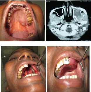
FIGURE-1.ADENOID CYSTIC CARCINOMA
[image:55.612.179.490.214.530.2]
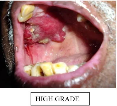
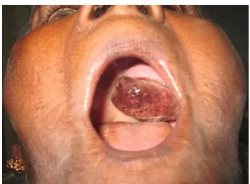
FIGUR-2.-MUCOEPIDERMOID CARCINOMA
[image:56.612.240.438.219.398.2]FIGURE-3. POLYMORPHOUS LOW GRADE ADENOCARCINOMA
FIGURE-4 PLEOMORPHIC ADENOMA
[image:57.612.216.398.212.346.2]
FIGURE-5 CARCINOMA EX PLEOMORPHIC ADENOMA
[image:58.612.226.397.173.314.2]
FIGURE-6 INVERTED PAPILLOMA
FIGURE-7.INTRAOPERATIVE PICTURES
[image:59.612.164.481.200.566.2]
45
SURGICAL OUTCOME
1. Tumors originating from palate grow and spread faster than tumors of maxillary sinus and nasal cavity as no T4 lesions were seen in maxillary sinus or nasal cavity in our study.
2. Tumors of maxillary sinus and nasal cavity has good surgical outcome than tumors of palate in terms of positive margins, perineural invasion and recurrence. 3-Dimensional clearance is difficult to achieve in palatal tumors, so tendency for nerve involvement, positive margins and recurrence is more.
3. Tendency for perineural invasion is more in palatal tumors, in our study 2 of 3 patients with perineural invasion is from palate.
4. The chance for recurrence is more in high grade mucoepidermoid carcinoma.
Recurrence was observed in patient with high grade mucoepidermoid carcinoma of palate within a short period of 3 months post operatively and the patient did not survive. No recurrence was observed in patients with low grade mucoepidermoid carcinoma.
5. Polymorphous low grade adenocarcinoma shows good surgical outcome if 3-Dimensional clearance is obtained with negative margins, T4a patient with tumor
46
6. T-stage does not influence the surgical outcome. The tumor involvement of nerve, bone, soft tissue and the grade of tumor influence the surgical outcome. A patient with T4a stage adenoid cystic carcinoma of palate did not survive. The patient had infraorbital
nerve involvement, breach in posterolateral wall of maxillary sinus, positive margins post-operatively, and suffered brain metastasis. Another patient with T4a stage adenoid
cystic carcinoma of palate did not suffered any recurrence and survives. The patient had tumor confined to maxillary sinus, with greater palatine nerve involvement and positive margins post-operatively. A patient with T2 high grade mucoepidermoid carcinoma of
palate suffered recurrence and did not survive, but patients with T2 low Grade
mucoepidermoid carcinoma of palate did not suffered any recurrence and continue a disease free survival.
SURGICAL AND RADIOTHERAPEUTIC OUTCOME
47 RECURRENCE
1. The only recurrence seen in our study was in a patient with high grade
mucoepidermoid carcinoma of palate. The patient with adenoid cystic carcinoma with and without positive margins, with and without radiotherapy did not developed recurrence.
2. No recurrence was seen in patients with polymorphous low grade adenocarcinoma, pleomorphic adenoma, carcinoma EX pleomorphic adenoma and inverted papilloma. All the patients had adequate 3-Dimensional marginal clearance and negative surgical margins.
PROGNOSIS
1. The prognosis of patients with adenoid cystic carcinoma, who underwent surgery, surgery and radiotherapy, is good irrespective of surgical margin status, but not for the patients with distant metastasis.
2. The prognosis for patients with low grade mucoepidermoid carcinoma is good, compared to high grade mucoepidermoid carcinoma.
48
Minor salivary glands constitute important components of the oral cavity. The glands are embedded in the submucosa. Minor salivary glands consist of a groups
secretory end-pieces which are made up of mucous acinar cells and serous or seromucous demilune cells.
The ductal systems is made up of intercalated ducts, intra-lobular ducts usually lacking basal striations, and excretory ducts opening in to oral cavity directly through the mucosa. Minor salivary glands secrete highly glycosylated mucins, also contain blood group determinants, and act as tissue lubrication and prevent bacterial aggregation. They also secrete several antimicrobial proteins like peroxidase, lysozymes and
immunoglobulin like secretory IgA. The lingual serous glands also called von Ebner's glands secrete digestive enzymes like lipase and proteins with possible taste perception functions 79.
The minor salivary gland salivary flow varies between different oral sites. Buccal saliva secretion is higher than labial salivary secretion, which is higher than the palatal gland secretion rate. Minor gland saliva secretion is important for oral saliva composition and especially for the secretory immunoglobulin A- SIgA and mucins. The secretion from these glands wets the mouth and helps in general wellbeing80.
49
behavior and morphological diversity. The most common clinical presentation of benign minor salivary gland tumors are, a well delimited, smooth, uniform nodular tumor with a normal overlying surface colour. The lesion is asymptomatic, can be displaced, usually single, and do not adhere to either superficial or deep layers.
The clinical features which differentiates between benign and malignant minor salivary gland tumors is the evolutive course, benign tumors tend to be insidious and slow-growing, with an average duration of 3-6 years, while malignant tumors are fast-growing typically less than a year, can ulcerate, become overinfected and cause external or interstitial bleeding which gives rise to superficial telengiectasias. The clinical pictures suggestive of malignancy are pain, adherence to deeper or superficial layers, epidermal involvement, ulceration and the presence of neck lymph adenopathies81.
Minor salivary gland tumor is said to be a heterogeneous group of malignancy because individual cells of minor salivary gland gives rise to wide range of tumors. This heterogeneous property is due to neoplastically altered epithelial cells with
multidirectional proliferation. These neoplastically altered cells may be myoepithelial cells, intercalated duct cells or reserve cells. Minor salivary gland are not encapsulated which accounts for its local aggressive nature, recurrence, perineural spread and distant metastasis.
50
Earlier studies regarding minor salivary gland tumors were retrospective; to the best of our knowledge, our study is the first prospective and retrospective study of minor salivary gland tumors. Minor salivary gland tumors have varied geographical
distribution; some tumors may be common in particular geographical distribution, while some tumors may be rare. The most common malignant minor salivary gland tumor treated in our Institution is adenoid cystic carcinoma, followed by mucoepidermoid carcinoma. The most common benign minor salivary gland tumor is pleomorphic adenoma. A study by vani et al shows mucoepidermoid carcinoma as the most common malignant minor salivary gland tumor50.
According to our study males were more commonly affected, representing 53.33% and females representing 46.66%. The study by pandey et al states that males are more commonly affected, but the study by Everson et al states that more females are
affected.4,24
51
The most common clinical presentation is palatal swelling in 66.66% of patients (10/15), 26.66% (4/15) of patients had tumor in maxillary sinus, and 06.66% (1/15) of patient had tumor in nasal cavity. Study by Thomas et al,44 shows that the palate is the most common site of occurrence for minor salivary gland tumors. Left side is the most common area of tumor presentation. Left side accounts for 60% (9/15), right side accounts for 40% (5/15) and 06.66% (1/15) of tumor in the midline at the junction of hard and soft palate abutting the uvula.
The most common tumor is adenoid cystic carcinoma, accounts for 40% (6/15), mucoepidermoid carcinoma accounts 20% (3/15), pleomorphic adenoma and inverted papilloma accounts for 13.33% (2/15), carcinoma EX pleomorphic adenoma and polymorphous low grade adenocarcinoma accounts for 06.66% (1/15), coinciding with the reports of Toida et al,10 but differing with the reports of Vani et al50 were
mucoepidermoid carcinoma is the most common tumor. The age range for adenoid cystic carcinoma is from 43 to 82, with the mean age of 62.16. Males were affected more with 66.66% (4/6), followed by 33.33% (2/6) of females. Our study coincides with the study of Bushra et al39 were 60% of patients were males, 40% were females, representing
52
the mean age of 53.5, affecting one male and one female. Carcinoma EX pleomorphic adenoma and polymorphous low grade adenocarcinoma had one male and female patient each.
Regarding tumor size 53.33% of patient presented with T1-T2 tumor, 46.66% of
patients presented with T3-T4 tumor with T1 accounts for 20.00%, T2 accounts for
33.33%, T3 accounts for 26.66% and T4 accounts for 20%. In palate the most common T
stage is T2. T1 accounts for 10%, T2 accounts for 40%, T3 accounts for 20% and T4
accounts for 30%. In maxillary sinus the most common T stage is T3. T1 and T2 account
for 25%, T3 50%. Nasal cavity accounts for one case of T1 tumor. In patients with
adenoid cystic carcinoma, one patient each of palate and nasal cavity had T1 tumor, one
patient with tumor of maxillary sinus had T3 tumor and two patients of palate had T4a
tumors. In patients with palatal mucoepidermoid carcinoma, two patients had T2 and one
patient had T3 tumor. One patient with polymorphous low grade adenocarcinoma had T4a
tumor. Two patients with maxillary sinus inverted papilloma and pleomorphic adenoma had one T1 and T2 tumors each. A patient with Carcinoma EX Pleomorphic adenoma had
T3 tumor.
53
marginal clearance, reflecting the study of Robert et al62 that large bulky tumors or tumors with radiographic evidence of bone invasion should be treated by partial maxillectomy or palatal fenestrations.
Of the patient treated surgically 26.66%. (4/11) patient had positive margins, all affected by adenoid cystic carcinoma. Out of six patients with adenoid cystic carcinoma 66.66% (4/6) had positive margins, 33.33% (2/6) patient had negative margins.
Radiotherapy was instituted in patients with and without positive margins. Total fraction of 60 gray was given for 6 weeks, 5 days a week, 2 gray per day. Radiotherapy was received by 83.33% of patients with adenoid cystic carcinoma out of which 80% of patients had positive margins. Brain metastasis was present in one out of six patient with adenoid cystic carcinoma, the patient did not survive in spite of radiotherapy, confirming the statement of Chunying et al68 that high risk of distant metastasis impairs the benefit of post-operative radiotherapy and also the statement of Gao et al65 thatadenoid cystic carcinoma patients with distant metastasis had significant lower survival rate. Lung is the most common organ for distant metastasis, but none of our patient had lung metastasis.
Perineural invasion was observed in 50% of patients with adenoid cystic
carcinoma, the patients had T4a, and T3 lésions. The statement by William Barret et al43
54
significant association between perineural invasion and size of primary tumor was found. Old age is considered to be a negative prognostic factor for adenoid cystic carcinoma, but an 84 year old nasal cavity adenoid cystic carcinoma patient with negative surgical margins, without post-operative radiotherapy is leading a disease free survival.
Adenoid cystic carcinoma is amenable to surgery and radiotherapy. Patients with positive surgical margins respond well to radiotherapy. Palate is the most common site of occurrence. The treatment outcome depends upon site of origin, involvement of named nerve and adjacent structures. Only one case (1/6) of brain metastasis has been reported in our study, it may be due to the fact that the patient suffered the disease for nearly four years. Though controversies exist regarding neck dissection in patients with adenoid cystic carcinoma, we did not do neck dissection as none of our patients had reactive lymph nodes and also adenoid cystic carcinoma very rarely metastasize to regional lymph nodes. Controversies exist regarding radiotherapeutic outcome of adenoid cystic
carcinoma, but in our study we found that adenoid cystic carcinoma responds to radiotherapy and no recurrence has been found till date.
In our study we found that the prognosis of patients with nasopharyngeal adenoid cystic carcinoma is favorable than palatal adenoid cystic carcinoma. Our study reflects the study of Cai-Neng Cao et al67.
55
shafers82 and Catherine et al32regarding the clinical presentation of low and high grade mucoepidermoid carcinoma.
Out of 15 patients 06.66% (1/15) had recurrence and was affected with high grade mucoepidermoid carcinoma. The study by vander Poortee et al83 shows that patients suffering from higher stage minor salivary gland tumors have higher probability of tumor recurrence. Out of 3 patients one high grade tumor patient received pre-operative
radiotherapy to down size the tumor and he did not survive due to recurrence and one low grade tumor patient received post operative radiotherapy, leading a disease free survival. Zameer pasha et 73 states that mucoepidermoid carcinoma is considered radioresistant but post-operative radiotherapy for patients with positive surgical margins is reported to decrease recurrence.
High grade mucoepidermoid carcinoma is more aggressive and challenging to treat regardless of T stage, more chance for local recurrence was found. Surgical outcome of low grade mucoepidermoid carcinoma is good, long term follow up showed no
recurrence. Study on mucoepidermoid carcinoma by Zameer pasha et al73 shows that local recurrence is low, occur within 1 year of treatment and tend to occur rapidly in high grade than low grade neoplasm.
56
both were treated by maxillectomy. The study by Primoz et al66 shows that inverted papilloma is more common in lateral nasal wall, with the clinical presentation of unilateral nasal blockade and maxillectomy, the treatment of choice.
WHO 2005 classification of tumors placed polymorphous low grade
adenocarcinoma as the second most common malignant salivary gland tumor. In our study polymorphous low grade adenocarcinoma is the least common tumor; only one case has been reported and treated. A 70 year old elderly female who presented with a palatal swelling was treated with maxillectomy. She had disease free survival for nearly 8 months, till her end of survival by self fall injury. A study by James et al31 shows that polymorphous low grade adenocarcinoma presents as a palatal swelling in elderly
females, another study by Jansisyanont et al84 and castle et al85 shows that polymorphous low grade adenocarcinoma has excellent cure rate after complete surgical excision, with infrequent recurrences and rare regional metastasis.
We treated two cases of pleomorphic adenoma. The first case was a 37 year old female who had T2 tumor, with firm rubbery palatal swelling. The second case was a 12
year old female patient who had T1 tumor with irregular corrugated appearance. Both
patients underwent wide local excision of the tumor, with no evidence of recurrence. A study by Vellios et al shows that pleomorphic adenoma may occur at any age, mainly affecting patients in the fourth, fifth and sixth decades. Forty percent of them were males and 60% were females10. Callender et al ranks pleomorphic adenoma as the most
57
tumors86. Byakodi et al states that wide local excision with removal of periosteum and involved bone is the treatment of choice87. A 70 year old male patient with long standing pleomorphic adenoma of palate for nearly 10 years developed carcinoma EX
pleomorphic adenoma. The patient underwent maxillectomy, with 3 dimensional marginal clearances and leads a disease free survival.Sołkiewicz et al states that the potential risk of the pleomorphic adenoma becoming malignant is about 6%.88 Shafer states that pleomorphic adenoma when presented in elderly for nearly 10 year may turn to carcinoma EX pleomorphic adenoma82.
It is a general acceptance that clear surgical margins are a road map for good prognosis. Patients with polymorphous low grade adenocarcinoma, pleomorphic
adenoma, carcinoma EX pleomorphic adenoma and inverted papilloma treated surgically with clear margins survive without any recurrence till date.
58
59
Tumors originating from smaller glands are more aggressive. Maxillary minor salivary glands tumors are aggressive because of lack of encapsulation. Our study
concludes that any palatal swelling must be considered a minor salivary gland tumor until proven otherwise. Palatal minor salivary gland tumor are aggressive, compared to