Copyright © 2004, American Society for Microbiology. All Rights Reserved.
Cell-to-Cell, but Not Long-Distance, Spread of RNA Silencing That
Is Induced in Individual Epidermal Cells
Eugene V. Ryabov,
1* Rene van Wezel,
1John Walsh,
2and Yiguo Hong
1*
Horticulture Research International, East Malling, West Malling, Kent ME19 6BJ,1and Horticulture
Research International, Wellesbourne, Warwick CV35 9EF,2United Kingdom
Received 26 August 2003/Accepted 17 November 2003
ATurnip crinkle virus(TCV)-based system was devised to discriminate cell-to-cell and systemic long-distance
spread of RNA silencing in plants. Modified TCV-GFP⌬CP, constructed by replacing the coat protein (CP)
gene with the green fluorescent protein (GFP) gene, replicated in single epidermal cells but failed to move from
cell to cell inNicotiana benthamiana. Mechanical inoculation of TCV-GFP⌬CP induced effective RNA silencing
in single epidermal cells which spread from cell to cell to form silenced foci on inoculated leaves, but no
long-distance systemic spread of RNA silencing occurred. Agroinfiltration of TCV-GFP⌬CP was, however, able
to induce both local and systemic RNA silencing. TCV coinfection arrested TCV-GFP⌬CP-mediated local
induction of RNA silencing. Possible mechanisms involved in cell-to-cell and long-distance spread of RNA silencing are discussed.
RNA silencing, including gene quelling, RNA interference, and posttranscriptional gene silencing are sequence-specific RNA degradation mechanisms that operate in fungi, animals, and plants (6, 7, 30). RNA silencing is triggered by double-stranded RNA and requires a conserved set of gene products (1, 13, 14). The double-stranded RNA is processed into small interfering RNAs (siRNAs) of 21 to 25 nucleotides (nt), and these siRNAs become associated with an RNA-induced silenc-ing complex that degrades specific target RNA sequences (3, 11, 12). RNA silencing plays a natural role in protecting fungi, plants, and animals against viral infection. To withstand the RNA-silencing defense, viruses across kingdoms have evolved diverse mechanisms either to avoid or actively suppress RNA silencing (22, 35, 40).
One intriguing feature of RNA silencing is that it is not cell autonomous. In plants, RNA silencing can be induced locally and then spread to distal parts (16, 36–38). While RNA-silenc-ing induction and RNA degradation have been elucidated in detail, much less is known about how RNA silencing moves from cell to cell and spreads systemically in plants. No mobile silencing signal has been characterized, although the sequence specificity of RNA silencing implies that nucleic acids, possibly siRNAs, may be a component of such an RNA-silencing signal (26). We have usedTurnip crinkle virus(TCV) to explore the requirements for cell-to-cell and long-distance spread of RNA silencing in plants.
TCV, a member of the Carmovirus genus, has a positive single-strand genomic RNA (4,053 nt), packaged in icosahe-dral capsids, which contains five major open reading frames (5). The p28 and p88 proteins are translated from genomic RNA, by readthrough of the p28 terminator, and are involved in viral RNA replication. Two overlapping proteins, p8 and p9, are expressed from subgenomic RNA1 and are required for cell-to-cell movement and systemic spread of the virus (10, 23).
The 3⬘-proximal open reading frame encodes the 38,000-mo-lecular-weight coat protein (CP), which also plays an essential role in cell-to-cell movement of TCV inNicotiana benthamiana (8) and acts as an effective suppressor of RNA silencing (27, 31). A full-length infectious cDNA of TCV was generated by reverse transcription (RT)-PCR with a genomic RNA tem-plate extracted from purified viral particles of a United King-dom isolate derived from wild brassicas (GenBank accession no. AY312063) (Fig. 1A). Two primers specific to TCV geno-mic RNA contained the 5⬘-terminal 19 nt following a T7 RNA polymerase promoter sequence and a sequence complemen-tary to the 3⬘-terminal 24 nt following aPacI site. The resulting RT-PCR fragment was cloned into pCR-BluntII-TOPO (In-vitrogen) to produce pT7.TCV. Plasmid pT7.TCV was used to construct pT7.TCV-GFP⌬CP, in which the CP gene was re-placed with the coding sequence for green fluorescent protein (GFP). Briefly, nt 2753 to 3388, encoding the R domain and the majority of the S domain of TCV CP (18), were deleted by overlap extension PCR (15) with a pair of primers (5⬘-GAAA cGGAAAATGagatctggaccggtgggtttaaacCACCTACGGCC AAGGAGC-3⬘and 5⬘-GGCCGTAGGTGgtttaaacccaccggtcca gatctCATTTTCCgTTTCCAGTGTTG-3⬘). Modified nucleo-tides are shown in lowercase, and introduced restriction en-donuclease sites forBglII andPmeI are underlined. The CP initiation codon (boldface) was also mutated. UniqueBglII and PmeI sites were inserted downstream of the stop codon for the p9 movement protein in the modified TCV genome. The GFP coding sequence was PCR amplified with a pair of primers (5⬘-ggaaagatctATGGCTAGCAAAGGAGAAGAAC-3⬘ and 5⬘-ggaagtttaaacTTATTTGTAGAGCTCATCCATG-3⬘), di-gested with BglII andPmeI, and cloned into theBglII and PmeI sites of the modified TCV cDNA to produce pT7.TCV-GFP⌬CP (Fig. 1A).
To test whether the cloned TCV and recombinant TCV-GFP⌬CP were infectious, RNA transcripts were produced in vitro from pT7.TCV or pT7.TCV-GFP⌬CP after linearization with PacI with the mMESSAGE mMACHINE T7 kit (Am-bion). Viral RNA transcripts were mechanically inoculated
* Correspondence author. Mailing address: Horticulture Research International, Wellesbourne, Warwick CV35 9EF, United Kingdom. Phone: 44 178 947 0382. Fax: 44 178 947 0552. E-mail: eugene.ryabov @hri.ac.uk (E. V. Ryabov) and yiguo.hong@hri.ac.uk (Y. Hong).
3149
on November 8, 2019 by guest
http://jvi.asm.org/
separately or as a mixture onto youngN.benthamianaplants. Plants were maintained in an insect-free growth room at 25°C with a continuous 12-h photoperiod. Infection with transcripts of pT7.TCV alone or with pT7.TCV-GFP⌬CP produced local and systemic symptoms indistinguishable from those induced by sap transmission of virions onto N. benthamiana plants. With RT-PCR assays done as previously described (34), a TCV genome-specific product (nt 3388 to 4053) was readily detect-able in inoculated and systemically infected young leaves at 12 days postinoculation (dpi) (Fig. 1B). Agfpgene-specific frag-ment was detected in leaves inoculated with mixed viral RNA transcripts (Fig. 1C). N. benthamiana plants inoculated with the pT7.TCV-GFP⌬CP transcript alone produced no obvious lesions on inoculated leaves and developed no systemic symp-toms at 24 dpi, although TCV-GFP⌬CP RNA replicated effi-ciently in the initially infected cells. Indeed, both viral and gfp-specific sequences were detected by RT-PCR in inoculated leaves at 3, 6, 12, and 24 dpi (Fig. 1C).
Fluorescence microscopy ofN.benthamianaplants
mechan-ically inoculated with TCV-GFP⌬CP RNA at 3, 6, and 12 dpi revealed that GFP fluorescence was clearly visible in individual epidermal cells in inoculated leaves (Fig. 1D). Green fluores-cence was confined to single epidermal cells even at 24 dpi. No GFP fluorescence was observed in young, newly emerging noninoculated leaves, consistent with the RT-PCR results for TCV andgfpRNA. Taken together, our data demonstrate that cloned TCV and recombinant TCV-GFP⌬CP accumulated ef-ficiently in initially infected single cells and that CP deletion resulted in the inability of TCV-GFP⌬CP RNA to move from cell to cell inN.benthamiana. Moreover, CP expressed from TCV was unable to complement long-distance transport of TCV-GFP⌬CP in plants. These results suggested that TCV-GFP⌬CP may be used as a functional tool to explore the single-cell induction and cell-to-cell and long-distance move-ment of RNA silencing in plants.
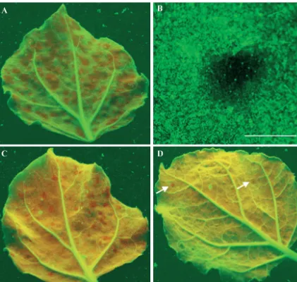
[image:2.603.44.281.65.370.2]To test the potential utility of TCV-GFP⌬CP,N. benthami-ana plants (line 16c) carrying a highly expressed GFP trans-gene (4) were inoculated with pT7.TCV-GFP⌬CP transcripts. Local RNA silencing was induced rapidly in individual epider-mal cells on inoculated leaves, and tinygfpRNA-silencing foci, each with dozens of cells, showing red chlorophyll fluorescence under long-wavelength UV light illumination (9), were de-tected at 48 h postinoculation. Expansion of these localizedgfp RNA-silencing foci continued, reaching up to 6 mm in diam-eter at 24 dpi (Fig. 2A). Leaves of line 16c plants inoculated with wild-type TCV transcripts showed no development ofgfp RNA silencing. Fluorescence microscopy of TCV-GFP⌬CP RNA-inoculated leaves showed that cell-to-cell spread of gfp RNA silencing was established efficiently in upper and lower epidermal cells and in palisade and spongy mesophyll cells (Fig. 2B). It is likely that the effectiveness of TCV-GFP⌬CP RNA at inducting silencing may be due to the lack of CP, an RNA-silencing suppressor (27, 31). However, the RNA-silenc-ing suppressor activity of TCV CP had been shown only in transient agroinfiltration assays and not in natural TCV infec-tions. To clarify this inconsistent result, leaves of line 16c plants were mechanically inoculated with TCV-GFP⌬CP RNA and then 3 days later inoculated with TCV RNA. The number ofgfpRNA-silencing foci was reduced approximately fourfold (Fig. 2C and 3A) compared with line 16c plant leaves mock inoculated 3 days after inoculation with TCV-GFP⌬CP RNA. At 24 dpi with TCV-GFP⌬CP, the sizes of the gfp RNA-silencing foci in TCV-reinoculated leaves ranged from 0.1 to 2 mm, smaller than the 1- to 6-mm foci in mock-reinoculated plants. More dramatic suppression of localgfpRNA-silencing induction was observed when the 16c plants were coinoculated with TCV and TCV-GFP⌬CP RNAs. The number of gfp RNA-silencing foci decreased about 70-fold compared with TCV-GFP⌬CP RNA inoculation alone, and foci were reduced in size to between 0.1 and 1 mm (Fig. 2D and 3A). Our results demonstrate that TCV infection arrestedgfpRNA-silencing in plants, probably owing to expression of TCV CP, which acted as an RNA-silencing suppressor. It is likely that CP produced during TCV infection interfered with the induction of RNA silencing rather than turned off RNA silencing already estab-lished. This view is supported by an earlier finding that TCV CP suppressed RNA silencing at the initiation stage by pre-venting siRNA production (27). Moreover, our data also imply
FIG. 1. Expression of GFP from TCV-GFP⌬CP in singleN. ben-thamianaepidermal cells. Construction of TCV and TCV-GFP⌬CP is shown in panel A. TCV (B) and GFP (C) RNAs were detected by RT-PCR with RNA samples extracted from mock-inoculated (Mo) or TCV-GFP⌬CP RNA-inoculatedN.benthamianaleaves harvested at 3, 6, 12, and 24 dpi. In the case of TCV infection (TCV) or coinoculation with TCV-GFP⌬CP and TCV RNAs (Mix), total RNAs were extracted from inoculated (IL) or systemically infected young (YL) leaves at 12 dpi. The sizes (nucleotides) and positions of a double-stranded DNA ladder (M) are indicated. Epifluorescence microscopic examination of GFP expression in a single epidermal cell in a TCV-GFP⌬ CP-inocu-latedN.benthamianaleaf at 6 dpi with a Zeiss Axiophot microscope through a green filter is shown in panel D. Bar⫽100m.
on November 8, 2019 by guest
http://jvi.asm.org/
that TCV CP could block cell-to-cell movement of RNA si-lencing.
To further demonstrate the local induction of RNA silencing by TCV-GFP⌬CP RNA, and suppression of silencing by coin-fection with TCV, levels of viral andgfpRNA accumulation in line 16c plants inoculated with TCV-GFP⌬CP RNA alone or with TCV RNA were analyzed by RT-PCR (Fig. 3B and C). TCV-GFP⌬CP RNA was detected at 3 dpi. The level of TCV-GFP⌬CP RNA declined significantly at 6 dpi and was below detectable levels at 12 dpi. However, TCV RNA was easily detectable in inoculated and systemically infected young leaves of line 16c plants treated with both RNAs or with TCV tran-scripts alone (Fig. 3B). Consistent with limited local induction and suppression of RNA silencing,gfpRNA was detected in all of the leaf samples tested (Fig. 3C). These results suggest that
thegfpsequence in TCV-GFP⌬CP RNA acted as an inducer of RNA silencing that targeted both transgene-derivedgfpRNA and recombinant TCV-GFP⌬CP RNA. Infection by TCV-GFP⌬CP RNA in line 16c plants, as in nontransformed N.
benthamiana, was confined to primarily inoculated, individual
epidermal cells. Thus, TCV-GFP⌬CP RNA effectively initi-ated and induced RNA silencing in single epidermal cells, which then spread from cell to cell to formgfpsilenced foci containing hundreds to thousands of cells within the inoculated leaves.
Although TCV-GFP⌬CP RNA-mediated induction of gfp RNA silencing in inoculated leaves was effective, with up to 50% of the lamina exhibiting silencedgfptransgene expression, no systemic long-distance spread of RNA silencing was ob-served up to 6 weeks after inoculation (Fig. 4A). This
phenom-FIG. 2. Local induction, cell-to-cell spread, and suppression of RNA silencing.N.benthamiana(line 16c) plants were mechanically inoculated with TCV-GFP⌬CP RNA (A and B), inoculated with TCV-GFP⌬CP RNA and then reinoculated 3 days later with TCV RNA (C), or coinoculated with TCV-GFP⌬CP and TCV RNAs (D). Inoculated leaves (A, C, and D) were photographed at 24 dpi with a Nikon Coolpix990 digital camera under long-wavelength UV illumination through a yellow filter.gfpRNA-silencing foci show red chlorophyll fluorescence, andgfpRNA-expressing tissues show green fluorescence. Only twogfpRNA-silencing foci (arrow) are seen in the leaf coinoculated with TCV-GFP⌬CP and TCV RNAs (D). OnegfpRNA-silencing focus, examined by epifluorescence microscopy of a TCV-GFP⌬CP RNA-inoculated leaf through a green filter (B), appears dark with some green fluorescent cells. Bar⫽6 mm.
on November 8, 2019 by guest
http://jvi.asm.org/
[image:3.603.82.503.69.469.2]enon was observed consistently in 12 individual plants in three separate experiments. Thus, although gfp RNA silencing in-duced by TCV-GFP⌬CP RNA in individual epidermal cells was able to move from cell to cell in the inoculated leaf, gfp silencing did not spread systemically to distal parts of the plant through the phloem. These results contrast with the effective induction of both local and systemicgfpRNA silencing in line 16c plants by infiltration with Agrobacterium carrying a 35S promoter-GFP binary vector (Fig. 4B) (4). It is notable the total area of local silencing in agroinfiltrated line 16c leaves did not exceed the area covered by all of the RNA-silencing foci induced by mechanical inoculation with TCV-GFP⌬CP. One of the differences between the initial induction of RNA silenc-ing by mechanical inoculation with TCV-GFP⌬CP RNA and agroinfiltration of the 35S-GFP cassette is related to cell types. Mechanical inoculation of line 16c plants with TCV-GFP⌬CP RNA inducedgfpsilencing only in individual epidermal cells, while agroinfiltration may lead to RNA silencing in a range of
cells including epidermal and mesophyll cells and cells of vas-cular tissues in which the 35S promoter is frequently active (2). To establish if the initial induction of RNA silencing by TCV-GFP⌬CP RNA in a range of cell types apart from epi-dermal cells would have an effect on the long-distance spread of RNA silencing, we constructed p35S.TCV-GFP⌬CP, with the TCV-GFP⌬CP cDNA inserted between the Cauliflower
mosaic virus35S promoter and terminator sequences of a
bi-nary vector inA.tumefaciensLBA4404 (17). In three indepen-dent experiments, 12 line 16c plants were infiltrated with an
Agrobacterium culture carrying p35S-TCV-GFP⌬CP. Six line
16c plants were also infiltrated independently with
Agro-bacteriumcarrying 35S-GFP. In the leaves of line 16c plants
agroinfiltrated with 35S-TCV-GFP⌬CP or 35S-GFP, local si-lencing of transgenic gfp expression was first visible 7 days postinfiltration; dark red rings without GFP fluorescence en-circled the infiltrated areas and were clearly visible under UV light. Approximately 2 weeks after agroinfiltration with either construct, systemicgfpRNA-silencing foci appeared in upper, noninfiltrated leaves along the minor veins and in growing “carbon sink” areas. All experimental plants developed this form of systemicgfpRNA silencing (Fig. 4B and C). Our data demonstrate that TCV-GFP⌬CP RNA was able to induce both local and systemic RNA silencing when introduced by agroin-filtration to different types of cells. In contrast, gfpRNA si-lencing induced exclusively in individual epidermal cells by mechanical inoculation with TCV-GFP⌬CP transcripts only
[image:4.603.44.280.72.357.2]FIG. 3. Effects of induction and suppression of local RNA silencing on accumulation of TCV andgfpRNAs. Average numbers of silencing foci per inoculated leaf are shown (A). Numbers were counted at 24 dpi in line 16c plants inoculated with TCV-GFP⌬CP RNA, inoculated with TCV-GFP⌬CP and then reinoculated 3 days later with TCV (TCV-GFP⌬CP 3dpi⫹TCV), or coinoculated with both transcripts on day 0 (TCV-GFP⌬CP⫹TCV). TCV (B) and GFP (C) RNAs were detected by RT-PCR with total RNA samples extracted from mock-inoculated (Mo) or TCV-GFP⌬CP RNA-inoculated line 16c plant leaves harvested at 3, 6, 12, and 24 dpi. In the case of TCV infection (TCV) or coinoculation with TCV-GFP⌬CP ⫹ TCV (Mix), total RNAs were extracted from inoculated (IL) and systemically infected young (YL) leaves at 12 dpi. The sizes (nucleotides) and positions of a double-stranded DNA ladder (M) are indicated.
FIG. 4. Spread of RNA silencing induced by TCV-GFP⌬CP via mechanical inoculation and agroinfiltration. No RNA silencing oc-curred in noninoculated older and newly emerging young leaves of line 16c plants mechanically inoculated with TCV-GFP⌬CP RNA (A). Infiltration of line 16c plants withAgrobacteriumcultures carrying a 35S-GFP expression cassette (B) or a TCV-GFP⌬CP cassette (C) induced systemic RNA silencing in noninfiltrated young leaves. Leaves were photographed at 42 days post-RNA inoculation or -agroinfiltra-tion.gfpRNA-silenced tissues show red fluorescence.
on November 8, 2019 by guest
http://jvi.asm.org/
moved from cell to cell within the inoculated leaf and failed to spread long distance.
The mechanism(s) of spread of RNA silencing in plants is poorly understood, and a mobile silencing signal remains to be elucidated (16, 25, 26). The sequence specificity of RNA si-lencing suggests that the mobile sisi-lencing signal contains an RNA component. siRNAs are an attractive candidate as they are short enough to move easily through plasmodesmata. Other candidates for the long-distance silencing signal include aberrant RNAs that could be transported by a host RNA-trafficking system(s). Plasmodesmal (local) and phloem (sys-temic long-distance) trafficking of endogenous mRNAs has been reported in plants (19, 20, 24, 28). Regardless of the nature of the RNA-silencing signal, we have provided direct evidence that the spread of RNA silencing is a two-step pro-cess involving both cell-to-cell and long-distance movement of the silencing signal. The spatial or multidirectional spread of RNA silencing from TCV-GFP⌬CP RNA-infected epidermal cells to adjacent cells indicates that the silencing signal(s) moves between cells by a passive diffusion mechanism. How-ever, we cannot rule out the possibility that cell-to-cell move-ment of the RNA-silencing signal may require host proteins or hijack viral cell-to-cell movement proteins. For example, the two small TCV proteins, p8 and p9, may act in this regard as both proteins are essential for viral RNA cell-to-cell movement in plants (10, 23). It is obvious that TCV CP is not involved in facilitating cell-to-cell movement of the RNA-silencing signal. More likely, CP expressed during TCV infection may impede RNA-silencing signal cell-to-cell movement in addition to its role in inhibiting the production of siRNA. A more recent finding demonstrated that limited and extensive cell-to-cell movements of RNA silencing occur in plants (16). These two types of RNA-silencing cell-to-cell movement could be oper-ated by different mechanisms involving 21- and/or 25-nt siRNA molecules. Interestingly, limited cell-to-cell movement spreads RNA silencing over only 10 to 15 cells that is thought to involve only the primary siRNAs moving from cells where RNA si-lencing is initially established and therefore is independent of homologous transcripts (16). In contrast, extensive cell-to-cell movement of RNA silencing requires homologous transcripts and occurs via relay amplification (16). Although we cannot distinguish these two types of cell-to-cell movement of RNA silencing in the TCV-based system, our data indeed indicate that silencing movement can be effectively promoted from one single epidermal cell to immediately neighboring cells in a three-dimensional manner and then spread to adjacent cells. This may involve both limited and extensive cell-to-cell move-ment of RNA silencing in TCV-GFP⌬CP-inoculated leaves of line 16c plants.
Furthermore, since both local and systemic induction of RNA silencing occurs after agroinfiltration of TCV-GFP⌬CP RNA into line 16c plants, it suggests that RNA silencing in-duced in different cell types, including phloem cells, may be a prerequisite for long-distance spread of the RNA-silencing signal. Remarkably, the patterns of spread of RNA silencing suggest that the signal moves both from cell to cell and through the phloem, mimicking viral RNA movement in plants (37). Plant viruses encode different proteins that are required for cell-to-cell and long-distance movement of genomic RNAs, respectively (21). Indeed, our data indicate that cell-to-cell
RNA-silencing signal movement can be independent of long-distance spread. A recent finding reveals that systemic, but not local, spread of RNA silencing induced in plants by agroinfil-tration can be blocked in the presence of low levels of cad-mium (33). Intriguingly, the same concentrations of cadcad-mium inhibited systemic movement of tobamoviruses, suggesting that the systemic spread of RNA silencing and that of viral RNA may have steps in their transport pathways in common (33). Moreover, RNA-silencing suppressors such as P1 and AC2 have contrasting effects on cell-to-cell movement and long-distance transport of RNA silencing in the vasculature (16).
Understanding the mechanisms of systemic viral movement restriction may provide a valuable insight into systemic move-ment of RNA silencing. It has been shown that the spread of viruses in hosts which allow cell-to-cell but not systemic move-ment is often restricted to the interface between bundle sheath cells and phloem parenchyma cells and the companion cell-sieve element complex (29, 32, 39). These observations suggest that the plasmodesmata which connect mesophyll cells, as well as mesophyll and bundle sheath cells, are structurally and func-tionally different from the plasmodesmata connecting bundle sheath cells and phloem. It is possible that the local silencing signal could move through one type of channel but would be unable to go through the other owing to an as yet unknown mechanism. In the case of TCV-GFP⌬CP RNA-mediated sin-gle-cell induction of RNA silencing, the silencing signal moves between epidermal and mesophyll cells and subsequently in-duces RNA-silencing foci within inoculated leaves but proba-bly was not able to enter the phloem and spread long distance to noninoculated leaves. Thus, it would be likely that lack of long-distance spread of RNA silencing initially induced in single epidermal cells by mechanical inoculation of TCV-GFP⌬CP occurred at the stage of entry into the vasculature. This view is consistent with the idea that RNA silencing which is established in phloem by phloem-limited virus spreads readily into various types of cells of lamina of newly emerging young leaves (16). Nevertheless, the system devised in this study may be exploited to dissect how RNA silencing spreads in plants.
We thank D. C. Baulcombe for providing transgenicN.benthamiana line 16c seeds, J. I. Cooper for the TCV isolate, and T. M. A. Wilson for encouragement throughout this work and for critical reading of the manuscript.
This project was supported by HRI-core BBSRC funding to E.V.R. and Y.H.
REFERENCES
1. Bass, B. L.2000. Double-stranded RNA as a template for gene silencing. Cell101:235–238.
2. Benfey, P. N., and N. H. Chua.1990. The cauliflower mosaic virus 35S promoter: combinatorial regulation of transcription in plants. Science250: 959–966.
3. Bernstein, E., A. A. Caudy, S. M. Hammond, and G. J. Hannon.2001. Role for a bidentate ribonuclease in the initiation step of RNA interference. Nature409:363–366.
4. Brigneti, G., O. Voinnet, W.-X. Li, L.-H. Ji, S.-W. Ding, and D. C. Baul-combe.1998. Viral pathogenicity determinants are suppressors of transgene silencing inNicotiana benthamiana. EMBO J.17:6739–6746.
5. Carrington, J. C., L. A. Heaton, D. Zudema, B. I. Hillman, and T. J. Morris. 1989. The genome structure of turnip crinkle virus. Virology281:219–226. 6. Carrington, J. C., K. D. Kasschau, and L. K. Johansen.2001. Activation and
suppression of RNA silencing by plant viruses. Virology281:1–5. 7. Cogoni, C., and G. Macino.2000. Post-transcriptional gene silencing across
kingdoms. Curr. Opin. Genet. Dev.10:638–643.
8. Cohen, Y., A. Gisel, and P. C. Zambrysky.2000. Cell-to-cell and systemic
on November 8, 2019 by guest
http://jvi.asm.org/
movement of recombinant green fluorescent protein-taggedTurnip crinkle virus. Virology273:258–266.
9. Dong, X., R. van Wezel, J. Stanley, and Y. Hong.2003. Functional charac-terization of the nuclear localization signal for a suppressor of posttranscrip-tional gene silencing. J. Virol.77:7026–7033.
10. Hacker, D. L., I. T. Petty, N. Wei, and T. J. Morris.1992. Turnip crinkle virus genes required for RNA replication and virus movement. Virology186:1–8. 11. Hamilton, A. J., and D. C. Baulcombe.1999. A novel species of small antisense RNA in post-transcriptional gene silencing. Science286:950–952. 12. Hamilton, A. J., O. Voinnet, L. Chappell, and D. C. Baulcombe.2002. Two classes of short interfering RNA in RNA silencing. EMBO J.21:4671–4679. 13. Hammond, S. M., A. A. Caudy, and G. J. Hannon.2001. Post-transcriptional
gene silencing by double-stranded RNA. Nat. Rev. Genet.2:110–119. 14. Hannon, G. J.2002. RNA interference. Nature418:244–251.
15. Higuchi, R., B. Krummel, and R. K. Saaki.1988. A general method ofin vitro
preparation and specific mutagenesis of DNA fragments: study of protein and DNA interactions. Nucleic Acids Res.16:7351–7367.
16. Himber, C., P, Dunoyer, G. Moissiard, C. Ritzenthaler, and O. Voinnet. 2003. Transitivity-dependent and -independent cell-to-cell movement of RNA silencing. EMBO J.22:4523–4533.
17. Hoekema, A., P. R. Hirsch, P. J. J. Hooykaas, and R. A. Schilperoort.1983. A binary plant vector based on separation ofvirand T-region of the Agrobac-terium tumefaciensTi-plasmid. Nature303:179–180.
18. Hogle, J. M., A. Maeda, and S. C. Harrison.1986. Structure and assembly of turnip crinkle virus. I. X-ray crystallographic structure analysis at 3.2 A˚ resolution. J. Mol. Biol.191:625–638.
19. Kim, M., W. Canio, S. Kessler, and N. Sinha.2001. Developmental changes due to long-distance movement of homeobox fusion transcript in tomato. Science293:287–289.
20. Kuhn, C., V. R. Franceschi, A. Schulz, R. Lemione, and W. B. Frommer. 1997. Macromolecular trafficking indicated by localisation and turnover of sucrose transporters in enucleate sieve elements. Science275:1298–1300. 21. Lazarowitz, S. G., and R. N. Beachy.1999. Viral movement proteins as
probes for intracellular and intercellular trafficking in plants. Plant Cell11: 535–548.
22. Li, H., W. X. Li, and S. W. Ding.2002. Induction and suppression of RNA silencing by an animal virus. Science296:1319–1321.
23. Li, W.-Z., F. Qu, and T. J. Morris.1998. Cell-to-cell movement of turnip crinkle virus is controlled by two small open reading frames that function in
trans. Virology244:405–416.
24. Lucas, W. J., S. Bouche-Pillon, D. P. Jackeson. L. Nguyen, L. Baker, B. Ding, and S. Hake.1995. Selective trafficking of KNOTTED1 homeodomain pro-tein and its mRNA through plasmodesmata. Science270:1980–1983. 25. Mallory, A. C., L. Ely, T. H. Smith, R. Marathe, R. Anandalakshmi, M.
Fagard, H. Vaucheret, G. Pruss, L. Bowman, and V. B. Vance.2001. HC-Pro
suppression of transgene silencing eliminates the small RNAs but not trans-gene methylation or the mobile signal. Plant Cell13:571–583.
26. Mlotshwa, S., O. Voinnet, M. F. Mette, M. Matzke, H., Vaucheret, S. W. Ding, and V. B. Vance.2002. RNA silencing and the mobile silencing signal. Plant Cell14:S289–S301.
27. Qu, F., T. Ren, and T. J. Morris.2003. The coat protein of turnip crinkle virus suppresses posttranscriptional gene silencing at an early initiation step. J. Virol.77:511–522.
28. Ruiz-Medrano, R., B. Xoconostle-Cazares, and W. J. Lucas.1999. Phloem long-distance transport of CmNACP mRNA: implications for supracellular regulation in plants. Development126:4405–4419.
29. Ryabov, E. V., I. M. Roberts, P. Palukaitis, and M. Taliansky.1999. Host-specific cell-to-cell and long-distance movements of cucumber mosaic virus are facilitated by the movement protein of groundnut rosette virus. Virology 260:98–108.
30. Sharp, P. A.2001. RNA interference 2001. Genes Dev.15:485–490. 31. Thomas, C. L., V. Leh, C. Lederer, and A. J. Maule.2003. Turnip crinkle
virus coat protein mediates suppression of RNA silencing in Nicotiana benthamiana. Virology306:33–41.
32. Thompson, J. R., and F. Garcia-Arenal.1998. The bundle sheath-phloem interface ofCucumis sativusis a boundary to systemic infection byTomato aspermy virus. Mol. Plant-Microbe Interact.11:109–114.
33. Ueki, S., and V. Citovsky.2000. Inhibition of systemic onset of post-tran-scriptional gene silencing by non-toxic concentrations of cadmium. Plant J. 28:283–291.
34. Van Wezel, R., X. Dong, H. Liu, P. Tien, J. Stanley, and Y. Hong.2002. Mutation of three cysteine residues inTomato yellow leaf curl virus-China C2 protein causes dysfunction in pathogenesis and post-transcriptional gene silencing suppression. Mol. Plant-Microbe Interact.15:203–208.
35. Voinnet, O.2001. RNA silencing as a plant immune system against viruses. Trends Genet.17:449–459.
36. Voinnet, O., and D. C. Baulcombe.1997. Systemic signalling in gene silenc-ing. Nature389:553.
37. Voinnet, O., C. Lederer, and D. C. Baulcombe.2000. A viral movement protein prevents spread of the gene silencing signal inNicotiana benthami-ana. Cell103:157–167.
38. Voinnet, O., P. Vain, S. Angell, and D. C. Baulcombe.1998. Systemic spread of sequence-specific transgene RNA degradation in plants is initiated by localised induction of ectopic promoterless DNA. Cell95:177–187. 39. Wang, H.-L., Y. Wang, D. Giesman-Cookmeyer, S. A. Lommel, and W. J.
Lucas.1998. Mutations in viral movement protein alter systemic infection and identify an intercellular barrier to entry into the phloem long-distance transport system. Virology245:75–89.
40. Waterhouse, P. M., M. B. Wang, and T. Lough.2001. Gene silencing as an adaptive defence against viruses. Nature411:834–842.