JOURNAL OFVIROLOGY,
0022-538X/01/$04.00⫹0 DOI: 10.1128/JVI.75.15.7149–7160.2001Aug. 2001, p. 7149–7160 Vol. 75, No. 15 Copyright © 2001, American Society for Microbiology. All Rights Reserved.
Pocket Protein p130/Rb2 Is Required for Efficient Herpes Simplex
Virus Type 1 Gene Expression and Viral Replication
GINGER L. EHMANN,1HEATHER A. BURNETT,2ANDSTEVEN L. BACHENHEIMER1,2,3*
Curriculum in Genetics and Molecular Biology,1Department of Microbiology and Immunology,2and
Lineberger Comprehensive Cancer Center,3University of North Carolina,
Chapel Hill, North Carolina 27599-7290
Received 1 February 2001/Accepted 7 May 2001
We have reported previously that herpes simplex virus type 1 (HSV-1) infection disrupts normal progression
of the mammalian cell cycle, causing cells to enter a G1-like state. Infected cells were characterized by a decline
in cyclin-dependent kinase 2 (CDK2) activities, loss of hyperphosphorylated retinoblastoma protein (pRb), accumulation of E2F-pocket protein complexes, and failure to initiate cellular DNA replication. In the present study, we investigated the role of the pocket proteins pRb, p107, and p130 in HSV-1-dependent cell cycle inhibition and cyclin kinase regulation by infecting murine 3T3 cells derived from wild-type (WT) mouse
embryos or embryos with deletions of pRb (pRbⴚ/ⴚ), p107 (p107ⴚ/ⴚ), p130 (p130ⴚ/ⴚ), or both p130 and p107
(p130ⴚ/ⴚ/p107ⴚ/ⴚ). With respect to CDK2 inhibition, viral protein accumulation, viral DNA replication, and
progeny virus yield, WT, pRbⴚ/ⴚ, and p107ⴚ/ⴚcells were essentially identical. In contrast, after infection of
p130ⴚ/ⴚcells, we observed no inhibition of CDK2 activity, a 5- to 6-h delay in accumulation of viral proteins,
an impaired ability to form viral DNA replication compartments, and reduced viral DNA synthesis. As a result,
progeny virus yield was reduced 2 logs compared to that in WT cells. Notably, p130ⴚ/ⴚ/p107ⴚ/ⴚ
double-knockout cells had a virus replication phenotype intermediate between those of the p107ⴚ/ⴚand p130ⴚ/ⴚcells.
We conclude from these studies that p130 is a key factor in regulating aspects of cell cycle progression, as well as the timely expression of viral genes and replication of viral DNA.
Precise temporal regulation of E2F transcription factor ac-tivities is critical to cell growth control, as E2Fs promote ex-pression of genes necessary for DNA replication and cell cycle progression. A family of pocket proteins, including retinoblas-toma protein (pRb), p107, and p130, function as the primary regulators of E2F activities, binding to and repressing the ac-tivity of E2Fs at specific points in the cell cycle. This repression regulates the cascade of E2F-dependent gene transcription, thus controlling progression through G1 and S phases. The
growth-inhibitory activities of the pocket proteins are likewise regulated by a family of cyclin-dependent kinases (CDKs) that phosphorylate and inactivate pRb, p107, and p130 in a cell cycle-dependent manner. Targeted disruption in both pRb al-leles caused embryonic lethality in mice of mixed background (9, 29, 37, 52). Mice deficient in either p107 or p130 showed no apparent abnormalities (11, 38), while mice deficient in both p107 and p130 displayed neonatal lethality (11). However, generation of p130-null mice in a pure BALB/cJ background resulted in embryonic lethality (36). Recent studies have shown that targeted disruption of the genes encoding all three pocket proteins resulted in the complete loss of G1checkpoint
control, leading to cell immortalization (14, 63).
Quiescent cells are characterized by the presence of p130-E2F4 complexes (47, 50, 70). As cells reenter the cell cycle, p130 is phosphorylated by G1-specific cyclin kinases and
sub-sequently targeted for ubiquitin-dependent proteolysis, though some p130 persists in cycling cells (6, 8, 47, 70). p107-E2F4 and
pRb-E2F4 complexes appear in G1cells (50), but pRb
com-plexes are disrupted in late G1as a result of sequential
phos-phorylation by cyclin D-CDK4,6 and cyclin E-CDK2 com-plexes (22, 26, 34, 42). S-phase cells are characterized by pRb-E2F1 complexes, as well as by p107-E2F4 complexes containing the cyclin A-CDK2 complex (50). Analysis of pro-teins bound at promoters of S-phase genes has shown that p107-E2F4 and p130-E2F4 complexes are abundant in prolif-erating cells (75).
Pocket proteins mediate transcriptional repression via E2F by a variety of mechanisms that are not yet fully understood. In addition to binding E2F and blocking the ability of free E2F to activate transcription, the pocket proteins can complex with E2F to form active transcriptional repressor complexes that occupy E2F binding sites on DNA (reviewed in references 17, 25, and 53). Pocket protein-E2F complexes function as active repressors of transcription by recruiting chromatin remodeling enzymes such as histone deacetylase (2, 43, 44) or the SWI/ SNF complex (reviewed in reference 25), as well as other transcriptional repressors, including HBP1 (73) and the core-pressors CtIP and CtBP (49).
In addition to their role as regulators of E2F, p107 and p130 (but not pRb) have been identified as inhibitors of CDKs (7, 16, 35, 77). The pocket region of p107 and p130, which is larger than that of pRb, contains a motif that mediates CDK binding. The binding of p107 or p130 to cyclin A-CDK2 or cyclin E-CDK2 negates the activities of these kinases, and p130-cyclin A-CDK2 complexes with or without E2F have been identified in vivo (76). Indeed, the level of inhibition of cyclin E-CDK2 and cyclin A-CDK2 by p107 has been demonstrated to be comparable to that of the CDK inhibitor p21 (7). Thus, p107 and p130 have dual roles in the control of cell cycle entry and * Corresponding author. Mailing address: Department of
Microbi-ology and ImmunMicrobi-ology, University of North Carolina, 837 Jones, CB#7290, Chapel Hill, NC 27599-7290. Phone: (919) 966-2445. Fax: (919) 962-8103. E-mail: [email protected].
7149
on November 9, 2019 by guest
http://jvi.asm.org/
progression as regulators of both the E2F family of transcrip-tion factors and the CDKs.
We and others have previously reported that herpes simplex virus type 1 (HSV-1) infection results in the accumulation of cells in a G1-like state, characterized by loss of
hyperphospho-rylated pRb, decline in G1CDK activities, and failure to
ini-tiate cellular DNA replication (18, 27, 54, 71). Coincident with the onset of viral DNA replication there is an increase in the E2F-p107-cyclin A-CDK2 complexes, hyperphosphorylation and nuclear translocation of E2F4, and posttranslational mod-ification and cytoplasmic translocation of E2F1 and a decrease in E2F DNA binding activities and E2F-dependenttrans -acti-vation (1, 27, 54). Quiescent human fibroblasts infected with HSV-1 are unable to exit G0in response to mitogen, as
char-acterized by persistence of E2F-p130 complexes and failure to upregulate G1CDK activities (18). Common to HSV-1
infec-tion of both quiescent and asynchronously cycling cells is the disregulation of CDK2 activities.
Based on the knowledge that p107 and p130 function as inhibitors of both E2F and CDK2, we investigated their role in HSV-1-induced cell cycle aberrations. Specifically, we wanted to establish whether one or both of these pocket proteins are necessary for HSV-1-dependent inhibition of CDK2 activities. To address these questions, we investigated the effects of HSV-1 on cell lines derived from mice deficient for pRb, p107, p130, or both p107 and p130 (10). The growth properties of these 3T3 fibroblast cell lines have been described previously (10). Briefly, the pRb⫺/⫺and p130⫺/⫺/p107⫺/⫺cell lines
dis-play a shortened G1 and lengthened S phase compared to
wild-type (WT) cells, and each cell line shows differential reg-ulation of E2F-dependent gene promoters and CDK activities. We report here that after infection of WT, pRb⫺/⫺, and
p107⫺/⫺cells by HSV-1, CDK2 activity was reduced 60 to 80%
compared to that in mock-infected cells, while p130⫺/⫺ cells
showed no decline in CDK2 activity. Additionally, pRb⫺/⫺and
p107⫺/⫺cells supported HSV-1 replication at levels
compara-ble to that of WT cells, while p130⫺/⫺cells were impaired in
their ability to support viral replication. HSV-1 infection of p130⫺/⫺cells was characterized by a 5-h delay in
immediate-early (IE) and delayed-immediate-early (DE) viral protein production, a delay in viral DNA replication, a lag and reduction in late (L) viral protein accumulation, and a 2-log reduction in virus yield. Cells deficient for both p130 and p107 (p130⫺/⫺/p107⫺/⫺) had
an intermediate phenotype. These findings establish a critical role for p130 in HSV-1 replication, which may point to its involvement in HSV-1-dependent cell cycle regulation.
MATERIALS AND METHODS
Cells and viruses.Vero cells, originally obtained from D. Knipe, Harvard University, were maintained in Dulbecco’s modified Eagle medium with high glucose (DMEM-H), supplemented with 5% bovine calf serum (CS); 129/BL6 3T3 cell lines derived from WT mouse embryos or mouse embryos with deletions
of pRb (pRb⫺/⫺), p107 (p107⫺/⫺), p130 (p130⫺/⫺), or both p130 and p107
(p130⫺/⫺/p107⫺/⫺) (10) were obtained from M. Classon and E. Harlow, MGH
Cancer Center, and were maintained in DMEM-H supplemented with 10% CS. HSV-1 strain KOS1.1 was used for all experiments unless otherwise indicated.
HSVd107 and HSVd99 viruses were a kind gift from N. DeLuca, University of
Pittsburgh (64). A green fluorescent protein (GFP)-expressing Sindbis virus derived from TR339 consensus AR339 strain (32) was a kind gift of W. Klimstra, University of North Carolina—Chapel Hill.
Detection of proteins.Cellular and viral proteins were detected by Western blot analysis essentially as described previously (18). The following antibodies
were used: anti-CDK2 (M2, sc-163), anti-CDK2 (D12, sc-6248), anti-p107 (C18, sc-318), anti-p130 (C20, sc-317), and anti-mouse immunoglobulin G (IgG)– horseradish peroxidase (sc-2005), all from Santa Cruz Biotechnology; anti-pRb (14001A) from Pharmingen; anti-rabbit Ig–horseradish peroxidase (NA 934), from Amersham; anti-ICP27 (H1113), from the Goodwin Institute; anti-ICP0 (H1083) and anti-ICP4 (H943), a gift from Lenore Pereira, University of Cali-fornia at San Francisco; anti-ICP8 (3–83), a gift from David Knipe, Harvard University; anti-gC (R47), a gift from Roselyn Eisenberg and Gary Cohen, University of Pennsylvania; and anti-Us11 (28*), a gift from Bernard Roizman, University of Chicago.
Immunofluorescence.3T3 cells (ca. 5⫻104) were seeded in wells of 24-well
tissue culture dishes each containing a 12-mm glass cover circle. After 1 day, cells were infected at a multiplicity of infection (MOI) of 20 with HSV for 1 h at 37°C. Following removal of the inoculum, cells were overlaid with DMEM-H with 2% CS. At various times postinfection (p.i.), cells were fixed at room temperature for 8 min in paraformaldehyde (Fisher) diluted to 1% in phosphate-buffered saline (PBS). Circles were then rinsed in PBS for 5 min and permeabilized for 8 min in Triton X-100 (Sigma) diluted to 0.02% in PBS. Following rehydration in PBS, cells were incubated in blocking solution containing 2.5% normal goat serum and 2.5% normal horse serum in PBS for 15 min at room temperature. Cells were then incubated for 60 min at 37°C with a 1:800 dilution of rabbit anti-ICP8 antiserum (3–83) in blocking solution. After three 5-min rinses in PBS and one 5-min incubation in blocking solution, cells were next incubated with a 1:50 dilution of a Texas red-coupled goat anti-rabbit IgG for 60 min at 37°C.
Follow-ing three 5-min rinses in PBS, the glass circles were rinsed in distilled H2O and
mounted on microscope slides in polyvinyl alcohol and glycerol containing 0.25%
diazabicyclo[2.2.2]octane, 2%n-propyl gallate, and 0.1% DAPI (4⬘,6⬘
-diamidino-2-phenylindole) and stored at 4°C. Microscopy was performed on a Zeiss micro-scope fitted with a multipass filter for Texas red. Images were captured digitally using the Scion Image program, saved in TIF format, and transferred to Adobe Photoshop.
Immunocomplex kinase assay.CDK2 protein complexes were
immunopre-cipitated from protein equivalent amounts (200 to 300g) of whole-cell lysate
with anti-CDK2 rabbit polyclonal antibody (Santa Cruz; M2; sc-163) for 12 h at 4°C. Protein complexes were collected on protein A-agarose beads (Boehringer Mannheim) for 1 h at 4°C and washed twice with 0.2% Tween 20 lysis buffer (50 mM HEPES [pH 7.3], 150 mM NaCl, 2.5 mM EGTA, 1 mM EDTA, 0.2% Tween
20) and twice with kinase buffer (50 mM HEPES [pH 7.3], 10 mM MgCl2). An
immunocomplex kinase assay was performed using histone H1 as the substrate exactly as described previously (18).
GFP expression.Subconfluent monolayers of WT and p130⫺/⫺cells in 60-mm
dishes were infected with HSVd107 at 10 or 20 PFU/cell, or Sindbis-GFP at 10
PFU/cell. At 5, 24, and 36 h p.i., cells were examined by UV microscopy (Nikon TE300) to visually confirm GFP expression and then harvested by trypsinization.
Cells were pelleted and then resuspended in 250l of 1⫻PBS and fixed by the
addition of 250l of 1⫻PBS–2% paraformaldehyde. The proportion of
GFP-positive cells was determined by flow cytometry analysis using a FACScan in-strument (Becton-Dickinson). Fifteen thousand cells were sorted for each sam-ple type, and GFP-positive cells were determined to be those with FL1 fluorescence above the level detected in mock-infected control cells of the ap-propriate genotype.
Southern blot analyses of viral DNA.Subconfluent cultures of WT, p107⫺/⫺,
p130⫺/⫺, and p130⫺/⫺/p107⫺/⫺cells in 100-mm dishes were infected with 20
PFU of HSV-1/cell. At various times p.i., cells were harvested by trypsinization
and resuspended in 5 ml of 1⫻PBS. Cells were pelleted through 1 ml of CS and
then resuspended in 200l of 1⫻PBS with 0.2 mg of RNase A and 0.4 mg of
protease K. Cellular and viral DNA were extracted using the QIAamp DNA minikit (Qiagen) according to the manufacturer’s protocol for adherent cells.
DNA was eluted in 200l (final volume) of buffer AE (10 mM Tris-HCl–0.5 mM
EDTA; pH 9.0), giving a final concentration of⬃0.1g/l. For each sample, 10
g of DNA was diluted to 400l in 0.4 M NaOH–10 mM EDTA and boiled for
10 min. DNA was vacuum slot blotted onto a positively charged nylon membrane
(Boehringer Mannheim), and each well was then rinsed with 500l of 0.4 M
NaOH. Following disassembly of the slot blot apparatus, the membrane was
rinsed in 2⫻SSC, dried, and UV cross-linked. The membrane was prehybridized
for 1 h at 42°C in DIG Easy Hyb buffer (Roche) and then hybridized for 16 h at 42°C with a digoxigenin-labeled DNA probe, specific for the HSV-1 terminal and
internal inverted repeats (BamHI-SP2) (58), at a concentration of 25 ng/ml.
Following hybridization, the membrane was washed and the digoxigenin probe was detected exactly according to the DIG High Prime DNA labeling and detection starter kit II protocol (Roche). Following detection, the membrane was exposed in a Lumi-Imager (Boehringer Mannheim) for 10 min, and band inten-sity was determined as the number of Boehringer luminescent units (BLU) per
on November 9, 2019 by guest
http://jvi.asm.org/
slot. Membranes were also exposed to BioMax-MR film (Kodak) for 5 min to 1 h to obtain an image on film.
Cell synchronization.WT and p130⫺/⫺cells were synchronized by contact inhibition. Cells were grown to confluence by maintaining them in DMEM-H with 10% CS for 4 days after plating in 100-mm dishes and then released into cell
cycle by replating at a lower density (8⫻105cells/dish) in fresh DMEM-H with
10% CS on 100-mm dishes. Synchronization was verified by flow cytometry analysis for DNA content of propidium iodide-stained cells exactly as described previously (18).
Virus yield assay.Replicate 60-mm monolayers of WT, pRb⫺/⫺, p107⫺/⫺,
p130⫺/⫺, and p130⫺/⫺/p107⫺/⫺cells were infected with 5 PFU of HSV-1/cell,
and cells and medium were harvested at various times p.i. Following three cycles of freezing and thawing, monolayers of Vero cells in 12-well dishes were inoc-ulated in triplicate with serial 10-fold dilutions of the lysates. After 1 h, mono-layers were covered with DMEM-H containing 2% CS and 0.3% methylcellulose. Following 3 days of incubation at 37°C, medium was aspirated from the wells and plaques were stained with 0.8% crystal violet in 50% ethanol. Plaques were counted, and virus yield was determined as the number of PFU generated per cell.
Generation of figures.Figures 1 to 4 and 6 to 7 were created using Microsoft PowerPoint. Images of original autoradiograms were generated using a desktop scanner, saved as bit-map files, and then imported into Microsoft PowerPoint. Figure 5 was created using Adobe Photoshop.
RESULTS
CDK2 inhibition by HSV-1 is independent of pRb and p107
but requires p130. To explore the potential role of pocket
proteins in the HSV-1-dependent down regulation of CDK2 activities, 3T3 fibroblasts derived from WT, pRb⫺/⫺, p107⫺/⫺,
p130⫺/⫺, and p130⫺/⫺/p107⫺/⫺ mouse embryos were mock
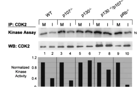
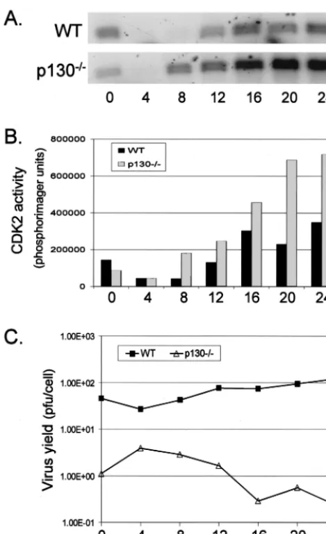
infected or infected with 50 PFU of HSV-1 per cell, harvested at 8 h p.i., and analyzed for CDK2 activity. A high MOI was used because we had previously determined that MOI 5 infec-tions of NIH 3T3 and 129/BL6 3T3 cells resulted in up to a 10-fold decrease in virus yield per cell compared to infections of Vero cells at the same MOI (T. I. McLean and S. L. Bachen-heimer, unpublished data), and we wanted to ensure maximally synchronous infection and efficient expression of IE and DE proteins. CDK2 complexes were immunoprecipitated from protein equivalent amounts of whole-cell lysates and the kinase activity of these complexes was determined in an in vitro kinase assay. As had previously been shown in various human cell lines (18, 71), HSV-1 infection of WT 3T3 cells resulted in an up to 60% reduction of CDK2 activity compared to mock-infection when activity was normalized to CDK2 protein levels (Fig. 1, lanes 1 and 2). p107⫺/⫺ and pRb⫺/⫺cell lines also
displayed a 60 to 80% decrease in CDK2 activity following HSV-1 infection (Fig. 1, lanes 3, 4, 9, and 10). Assays of p130⫺/⫺/p107⫺/⫺ lysates demonstrated a 30% decline in
CDK2 activity (Fig. 1, lanes 7 and 8). In contrast, infected p130⫺/⫺cells displayed no loss in CDK2 activity compared to
mock-infected samples (Fig. 1, lanes 5 and 6). These results suggested that HSV-1 inhibits cyclin kinase activities indepen-dently of pRb and p107. The failure to detect any effect of HSV-1 on CDK2 activity in the p130⫺/⫺cells suggests that
p130 is essential for HSV-1-dependent regulation of cyclin kinase activity. Alternatively, p130 may be important for virus-mediated effects on the infected cell upstream of CDK2 inhi-bition. While conducting these assays, we observed that in addition to not supporting a decrease in CDK2 activity, p130⫺/⫺cells did not display a cytopathic effect even 10 h after
HSV-1 infection (data not shown). This observation prompted
us to examine several additional aspects of the HSV-1 repli-cation cycle in the p130⫺/⫺cell line.
HSV-1 replication is severely impaired in p130ⴚ/ⴚ cells.
Due to the inability of HSV-1 to inhibit CDK2 activity or to induce cytopathic effect in cells lacking p130, we hypothesized that these cells would be impaired in their ability to support HSV-1 replication. We performed a one-step growth curve, comparing HSV-1 replication in p130⫺/⫺cells to that in WT
cells and other knockout cell lines. WT, pRb⫺/⫺, p107⫺/⫺,
p130⫺/⫺, and p130⫺/⫺/p107⫺/⫺cells were infected with 5 PFU
per cell and harvested at 8, 12, 16, or 24 h p.i. The amount of progeny virus produced by each culture was determined by standard plaque assay and expressed as the number of PFU per 105cells (Fig. 2). Although all of these mouse cell lines were
inefficient at supporting virus replication compared to human or other primate cell lines, it was readily apparent that the p130⫺/⫺cells were impaired to a greater extent than WT cells.
The virus yield from p130⫺/⫺cells was reduced 2 logs
com-pared to that from WT cells at 16 h p.i. The difference in yield narrowed to 1 log at 24 h p.i., as virus production in WT cells leveled off and production in p130⫺/⫺ cells continued to
in-crease. Virus production in p130⫺/⫺ cells, however, never
reached levels comparable to that in WT cells, even at 40 h p.i. (data not shown). Yield experiments performed at MOIs of 50 and 20 revealed that the 16-h HSV-1 yields were 1 and 2.5 logs lower, respectively, in p130⫺/⫺ cells than in WT cells. This
confirmed that the HSV-1 replication impairment in p130⫺/⫺
cells was not MOI dependent (data not shown; see Fig. 7B). Cells lacking pRb or p107 showed less significant differences in yield compared to WT cells. Virus yield produced by pRb⫺/⫺cells closely mirrored that produced by WT cells, while
virus yield produced by p107⫺/⫺cells exceeded that of WT
[image:3.612.313.551.77.228.2]cells at 12, 16, and 24 h p.i. Interestingly, cells lacking both FIG. 1. Regulation of CDK2 activity in HSV-1 infected 3T3 cell lines. WT and mutant 3T3 cell lines were mock infected (M) or in-fected with HSV-1 at 50 PFU/cell (I) and harvested after 8 h of infection. CDK2 was immunoprecipitated (IP) from protein equivalent amounts of whole-cell lysate. One half of the immunoprecipitate was used to determine the relative kinase activity of CDK2 using histone H1 substrate by in vitro kinase assay. The other half of the immuno-precipitate was used to detect CDK2 protein by Western blotting (WB). The bar graph represents the kinase activity of each infected sample relative to that of the corresponding mock-infected sample, normalized to the amount of CDK2 protein in the immunoprecipitate. Kinase activity was quantified by phosphorimaging. Protein levels were quantified by laser densitometry.
VOL. 75, 2001 p130 AND HSV-1 REPLICATION 7151
on November 9, 2019 by guest
http://jvi.asm.org/
p130 and p107 were not as impaired in supporting virus repli-cation as the p130 single-knockout cells, with HSV-1 yield being approximately 1 log lower than that of the WT at 16 h p.i. This result was consistent through three separate experiments (see Discussion).
WT and p130ⴚ/ⴚ cells are equally susceptible to HSV-1
infection. Due to the severe impairment in virus replication
and the failure to detect loss of CDK2 activity following HSV-1 infection in p130⫺/⫺cells, we were interested in whether the
absence of p130 affects the ability of the virus to efficiently bind and enter cells or whether p130 is important for a function downstream of entry. We used GFP expression as a marker for HSV-1 susceptibility by infecting cells withd107, a virus engi-neered to express GFP under the control of the human cyto-megalovirus (HCMV) IE promoter (64). We hypothesized that if HSV-1 is equally able to bind and enter both WT and p130⫺/⫺cells, GFP expression should be observed in an
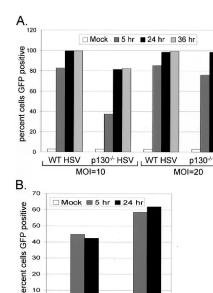
equiv-alent number of cells of both genotypes. To that end, subcon-fluent populations of WT and p130⫺/⫺cells were infected with d107 at an MOI of 10 or 20. At 5, 24, and 36 h after infection, cells were harvested by trypsinization and fixed, and the per-cent GFP-positive cells was determined by flow cytometry analysis. At 5 h p.i. the fraction of GFP-positive p130⫺/⫺cells
was smaller than the fraction of GFP-positive WT cells at both MOIs tested (Fig. 3A). However, at 24 and 36 h p.i., WT cell populations were 100% GFP-positive and p130⫺/⫺cell
popu-lations were 80% GFP positive when infected at an MOI of 10, and both WT and p130⫺/⫺cell populations were nearly 100%
GFP positive when infected at an MOI of 20. These data indicate that although p130⫺/⫺ cells are initially delayed in
expression of the GFPtransgene, the two cell types are equally susceptible to HSV-1 infection.
The delay in GFP expression in p130⫺/⫺cells at 5 h was
unexpected, as this expression was under the control of a strongly constitutive HCMV IE promoter. Because the DNA genome of HSV-1 requires transport into the nucleus before viral gene expression can occur, we determined whether a cytoplasmically replicating virus would show a similar impair-ment in GFP expression in p130⫺/⫺cells. Infection of p130⫺/⫺
cells with GFP-expressing Sindbis virus, a cytoplasmically rep-licating RNA virus, resulted in efficient GFP expression ex-ceeding that seen in WT cells (Fig. 3B). Thus, expression of GFP in the cytoplasm was not delayed in p130⫺/⫺cells
com-pared to WT cells. These data suggest that p130⫺/⫺cells may
impose a block to efficient nuclear DNA virus-mediated gene expression or at the very least to expression from the HCMV IE promoter.
Accumulation of viral proteins is delayed and reduced in
p130ⴚ/ⴚcells.The failure to detect significant progeny virus
[image:4.612.68.278.74.281.2]production in cells lacking p130, despite the fact that every cell was capable of receiving at least one infectious particle, sug-gested that the absence of p130 impaired the virus replication cycle at a point after entry and localization of viral DNA in the nucleus. To assess the replication cycle at the level of viral gene expression we analyzed the kinetics of infected-cell protein production by Western blot analyses. We examined proteins from the IE, DE, and L gene classes. Subconfluent monolayers of WT, p130⫺/⫺, and p130⫺/⫺/p107⫺/⫺cells were infected with
FIG. 2. One-step growth curve of HSV-1 on 3T3 cell lines. Sub-confluent monolayers of WT and mutant cell lines were infected with 5 PFU of HSV-1/cell. The virus titer of each culture harvested at 8, 12, 16, and 24 h p.i. was determined by standard plaque assay as described in Materials and Methods. The virus yield displayed is representative of three separate experiments and is expressed on a semilogarithmic scale.
FIG. 3. Susceptibility of WT and p130⫺/⫺cells to HSV-1 infection.
Subconfluent monolayers were infected with GFP-expressing viruses: HSVd107 at 10 or 20 PFU/cell (A) or Sindbis-GFP at 10 PFU/cell (B). Cells were harvested by trypsinization at 5, 24, or 36 h p.i. and fixed in 1% paraformaldehyde. The percent GFP-positive cells was determined by flow cytometry.
on November 9, 2019 by guest
http://jvi.asm.org/
[image:4.612.321.542.368.671.2]HSV-1 at an MOI of 5 and harvested at various intervals af-ter infection from 2 to 16 h (IE and DE proteins) or from 0 to 30 h (L proteins). Whole-cell lysates were prepared, separated by sodium dodecyl sulfate (SDS)-polyacrylamide gel electro-phoresis, transferred to polyvinylidene difluoride membranes, and then probed with antibodies to viral proteins.
The Western blots (Fig. 4) revealed that the kinetics of viral protein accumulation were similar between WT and p130⫺/⫺/
p107⫺/⫺cells for all three classes of viral proteins, although
the level of protein accumulation was slightly reduced in the p130⫺/⫺/p107⫺/⫺ cells. IE proteins ICP0, ICP4, and ICP27
were first detected between 2 and 3 h p.i., DE protein ICP8 was first detected at 3 h p.i., and L proteins gC and Us11 were first detected at 8 h p.i. gC accumulation was undetectable between 2 and 8 h p.i. (data not shown). In contrast, p130⫺/⫺ cells
showed a delay in viral protein accumulation ranging from 4 to 6 h. Specifically, ICP4, ICP27, and ICP8 were first detected no earlier than 4 to 5 h after these proteins first appeared in WT cells (Fig. 4A and B), and ICP0 was barely detectable in p130⫺/⫺cells even at 16 h p.i. (Fig. 4A). Interestingly, by 12 h
p.i., levels of ICP27 and ICP8 were comparable to levels de-tected in WT cells by 5 to 6 h p.i. Infections performed at 20 PFU per cell provided the same pattern of protein accumula-tion kinetics for all IE and DE proteins shown (data not shown).
In experiments using 5 PFU per cell, it appeared that L protein gC was not detectable in p130⫺/⫺ cells, so we
per-formed the experiments presented in Fig. 4C at 50 PFU per cell. Even at the higher MOI, gC was below the level of de-tection in the experiment shown. However, in other experi-ments and with longer exposures, we were able to detect gC at very low levels. In these experiments, L proteins gC and Us11 accumulated in p130⫺/⫺cells to levels that were less than 10%
of those found in WT cells, and their accumulation was delayed
about 8 h compared to that in WT cells (Fig. 4C). p130⫺/⫺
cells, therefore, can support a program of HSV-1 viral gene expression that lags behind that of a productive infection in WT cells. One exception is the inability to detect significant levels of ICP0, which may itself contribute to the delay in accumulation of other IE, DE, and L gene products in the absence of p130. The virus yield obtained with an ICP0-null virus (d99) on WT cells at 24 h p.i. was almost identical to that of WT KOS virus on the p130⫺/⫺cells (both reduced 2.5 logs
compared to WT KOS on WT cells; data not shown). Addi-tionally, thed99 virus yield was reduced only 0.5 log compared to the WT KOS virus on the p130⫺/⫺cells at 24 h p.i. (data
not shown). These data support the idea that the failure of p130⫺/⫺cells to support expression of significant levels of ICP0
protein may contribute to the overall replication deficiencies seen in this cell line.
p130ⴚ/ⴚcells are impaired in the formation of viral DNA
replication compartments and support reduced levels of viral
DNA replication. Since expression of true L genes depends
upon the onset of viral DNA replication (62), we hypothesized that the low levels of gC and Us11 seen in p130⫺/⫺cells were
due to an inhibition of viral DNA replication in these cells. We used two separate methods to compare viral DNA synthesis in WT and p130⫺/⫺cells. First, we looked for the formation of
[image:5.612.142.461.72.279.2]viral DNA replication compartments as visualized by immuno-fluorescence microscopy for ICP8. This virus-encoded DNA binding protein is absolutely required for complete prerepli-cative complex formation and DNA synthesis (3, 12, 15, 23, 39, 40, 55, 59). Prior to the onset of viral DNA synthesis, ICP8 accumulates at either a small number of ND10s or a larger number of sites corresponding to sites of cellular DNA syn-thesis (41, 74). The former are also sites where parental ge-nomes have been detected (28, 46). At early times and prior to the onset of viral DNA replication the ICP8 staining pattern FIG. 4. Kinetics of viral protein accumulation in 3T3 cell lines. WT, p130⫺/⫺/p107⫺/⫺, and p130⫺/⫺cells were infected with HSV-1 at 5 PFU/cell
(A and B) or 50 PFU/cell (C) and harvested at various times after infection. Cells were scraped into SDS sample buffer, and cell-equivalent amounts of lysate were separated by SDS-polyacrylamide gel electrophoresis. The resolved proteins were transferred to a polyvinylidene difluoride membrane, and viral proteins were visualized by Western blotting with antibodies directed to IE proteins ICP0, ICP4, and ICP27, DE protein ICP8, or true L proteins gC and Us11.
VOL. 75, 2001 p130 AND HSV-1 REPLICATION 7153
on November 9, 2019 by guest
http://jvi.asm.org/
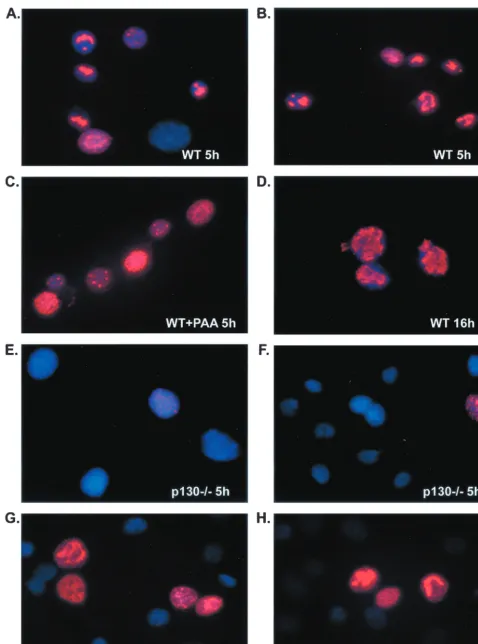
takes on a punctate appearance. As viral DNA synthesis pro-ceeds, the staining pattern evolves to a globular appearance as replication compartments begin to fill the nucleus (15, 48, 59). In WT cells infected with 20 PFU of HSV-1 per cell, a punctate or globular nuclear ICP8 staining pattern was observed in almost 100% of cells at 5 h p.i. (Fig. 5A and B). By 16 h p.i. the ICP8 staining pattern had transitioned to globular structures in most cells, indicating progression of viral DNA replication (Fig. 5D). In the presence of 400g of the viral DNA poly-merase inhibitor phosphonoacetic acid (PAA; Sigma) per ml, only punctate or diffuse nuclear ICP8 staining was observed (Fig. 5C). This concentration of PAA was sufficient to reduce viral DNA synthesis to undetectable levels in these cell lines (data not shown). As expected from the Western blot data for ICP8 protein (Fig. 4B), less than 1% of p130⫺/⫺cells displayed
ICP8 staining at 5 h p.i. (Fig. 5E and F), and this staining was punctate or globular and reminiscent of that seen in WT cells at 5 h. At 16 h p.i., however, approximately 10 to 20% of p130⫺/⫺ cells displayed punctate or globular ICP8 staining
(Fig. 5G and H). In a separate experiment we saw that the proportion of ICP8-positive p130⫺/⫺cells increased an
addi-tional 17% between 16 and 24 h p.i. (data not shown). The results of ICP8 immunofluorescence microscopy indi-cated that formation of viral DNA replication compartments was impaired in cells lacking p130, since few if any cells showed evidence of punctate or globular ICP8 staining patterns. In order to directly assess the level of viral DNA replication in p130⫺/⫺cells, we performed Southern blot analyses on DNA
extracted from HSV-1-infected cells. Subconfluent monolayers of WT, p107⫺/⫺, p130⫺/⫺, and p130⫺/⫺/p107⫺/⫺ cells were
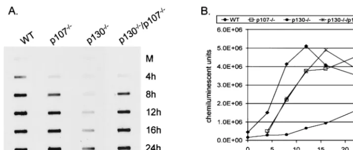
infected with HSV-1 at 20 PFU per cell. Cells were harvested at 4, 8, 12, 16, and 24 h p.i., total DNA was extracted, and samples were slot blotted to nylon membranes and hybridized with an HSV-1-specific DNA probe (Fig. 6A). The amount of viral DNA in each sample was quantified, and the data are presented as the number of BLU per sample plotted as a function of hours postinfection (Fig. 6B). The slot blot re-vealed impaired viral DNA synthesis in p130⫺/⫺ cells
com-pared to each of the other cell types (Fig. 6A). Again, the onset of viral DNA accumulation was delayed in p130⫺/⫺cells
com-pared to WT cells and was 12% of the WT value at 12 h p.i., 22% of the WT value at 16 h p.i., and 53% of the WT value at 24 h p.i. (Fig. 6B). Of interest was the observation that at 12 h p.i., when p130⫺/⫺cells expressed significant amounts of ICP4,
ICP27, and ICP8, DNA replication was only 12% of that of WT cells (Fig. 4B). These data corroborate the ICP8 immu-nofluorescence studies, indicating that viral DNA replication is significantly impaired and delayed in cells lacking p130 even in the presence of seemingly adequate amounts of viral IE and DE proteins. The detection of limited viral DNA synthesis in p130⫺/⫺cells by 16 h p.i. is consistent with the ability to detect
minimal amounts of gC and Us11 proteins at this time point (Fig. 4C). In the experiment presented, it appeared that the levels of viral DNA peaked in WT cells at 12 h p.i. and then declined. However, in two additional experiments we found that the level of viral DNA plateaued in WT cells beginning at 12 h p.i. (data not shown).
Efficiency of HSV-1 replication in p130ⴚ/ⴚcells is cell cycle
dependent. Southern blot analyses showed that overall viral
DNA synthesis was reduced in p130⫺/⫺cells compared to that
in WT cells. We found by immunofluorescence microscopy, however, that some cells displayed a pattern and kinetics of ICP8 staining similar to those of WT cells. In addition, by 16 and 24 h p.i., the fraction of p130⫺/⫺cells with punctate or
globular ICP8 staining had increased considerably. We hypoth-esized that at the time of infection of this asynchronous cell population, only cells at one or more discrete stages of the cell cycle were capable of or had the potential to support virus replication. Furthermore, as additional cells within the popu-lation cycled through these discrete stages, they gained the ability to support viral gene expression. In order to test the idea that HSV-1 replication in p130⫺/⫺cells is cell cycle dependent,
WT and p130⫺/⫺ cells were synchronized in G
0 by contact
inhibition, released by replating at lower density, and then infected at different times after release from G0. Cells were
infected at 4-h intervals from 0 to 24 h after replating and harvested 16 h after infection. Progeny virus production was determined by standard plaque assay and expressed as PFU per cell (Fig. 7C). Additionally, we harvested cells from two duplicate plates at each time of infection to assess the cell cycle status and level of CDK2 activity in each culture at the onset of infection. Cell cycle profiles were determined by measuring the DNA content of propidium iodide-stained cells by flow cytom-etry analyses. CDK2 activity was determined by in vitro kinase assay as described above. An average of the cell cycle profiles of synchronized WT and p130⫺/⫺ cells from three separate
experiments (Table 1), as well as the timing of CDK2 activa-tion (Fig. 7A and B), revealed that WT cells transiactiva-tioned from G1to S phase between 16 and 20 h after release, while p130⫺/⫺
cells transitioned from G1to S phase between 8 and 12 h after
release. Analyses of virus yield revealed that progeny virus production in WT cells did not vary with the phase of the cell cycle that cells were in at the time of infection or with the level of CDK2 activity (Fig. 7C). In contrast, we found that p130⫺/⫺
cells showed an inverse correlation between viral progeny pro-duction and CDK2 activity. In p130⫺/⫺cells, we observed the
highest levels of HSV-1 production in cultures infected 4 and 8 h postplating (early and mid-G1 phase), when CDK2 levels were low, and the lowest levels of virus production in cultures infected 16 to 24 h postplating (S to G2/M phase), when CDK2
levels were high (Fig. 7B and C). The increased virus yield observed as a function of cell cycle could reflect either in-creased efficiency of virus replication in a small number of cells or an increased number of cells able to support virus repli-cation. To differentiate between these possibilities, synchro-nized WT and p130⫺/⫺cell populations were infected at
dif-ferent times after replating and the pattern of ICP8 staining was determined by immunofluorescence microscopy at 5 or 16 h p.i. In one experiment, when p130⫺/⫺cells were infected
in G0or early G1(0 or 8 h after replating), 63 and 60% of cells
subsequently demonstrated globular ICP8 staining at 16 h p.i. (Table 2). Only 2% of cells demonstrated globular ICP8 stain-ing when p130⫺/⫺cells were infected during S phase (16 h after
replating) or G2/M phase (24 h after replating). The great
majority of WT cells demonstrated globular ICP8 staining at 5 h p.i. regardless of the cell cycle phase at the time of infection (Table 2). These data indicate that in p130⫺/⫺cells, but not
WT cells, HSV-1 replication is cell cycle dependent. This find-ing lends credence to the idea that the individual p130⫺/⫺cells
displaying punctate or globular ICP8 staining at earlier times
on November 9, 2019 by guest
http://jvi.asm.org/
FIG. 5. Formation of viral DNA replication compartments in WT and p130⫺/⫺cells. WT and p130⫺/⫺cells were seeded onto 12-mm glass cover
circles and later infected with 20 PFU of HSV-1/cell. Cells were fixed in 1% paraformaldehyde in PBS and permeabilized in 0.2% Triton X-100 in PBS. Viral replication compartments were visualized by detection of DE protein ICP8 (red) by immunofluorescence at 5 or 16 h p.i. (5h and 16h, respectively). Cellular DNA was detected by DAPI staining (blue).
7155
on November 9, 2019 by guest
http://jvi.asm.org/
than the rest of the culture were passing through a discrete window in early to mid-G1phase when infected.
DISCUSSION
In an attempt to understand the mechanism by which HSV-1 regulates CDK2 activity after infection, we have discovered that the pocket protein p130 is critical for HSV-1 replication. First, p130⫺/⫺ cells did not support a reduction in CDK2
activity after HSV-1 infection, as we had observed in other pocket protein-null cells and a variety of other human and primate cell lines. Second, infection of p130⫺/⫺cells resulted
in a temporal lag in viral IE and DE protein synthesis, accom-panied by a delay and decrease in viral DNA replication and L protein synthesis. The accumulation of IE proteins ICP4 and ICP27 was delayed 5 h in p130⫺/⫺cells, while accumulation of
ICP0 was barely at the level of detection at any time. This significant reduction in ICP0 production could account for the delay in IE, DE, and L protein synthesis, because ICP0 has a well-documented role in promoting viral gene transcription both in vitro and in vivo (5, 65). Additionally, both the reduc-tion in viral DNA replicareduc-tion (as much as 10-fold at 12 h p.i.) and the reduction in abundance of L proteins such as gC to levels less than 10% of that in WT cells help to explain the 2-log reduction in progeny virus formation in p130⫺/⫺cells,
despite equal susceptibility to virus binding and entry. These data suggest a delay in the onset of viral gene transcription after binding and entry, as well as additional impairment in viral DNA synthesis, leading to severe reduction in L protein synthesis. We also observed that although IE and DE proteins (with the exception of ICP0) eventually accumulated to almost WT levels in p130⫺/⫺cells, levels of viral DNA synthesis and L
protein accumulation did not reach WT levels even by 24 h p.i. These data suggest an uncoupling between IE and DE protein synthesis and DNA replication in p130⫺/⫺ cells. It appears,
thus, that the impairment in viral DNA replication, and not the delay in IE and DE protein accumulation, is rate limiting for progeny virus production in p130⫺/⫺cells.
3T3 cells lacking pocket protein pRb or p107 supported HSV-1 replication at levels comparable to that found in the
parental WT cells. Additionally, in the absence of either pocket protein, CDK2 activity declined after virus infection, suggesting that neither of these proteins plays a critical role in HSV-1-dependent cyclin kinase regulation. p107⫺/⫺cells, in
fact, supported HSV-1 progeny formation at levels slightly higher than those of the WT cells, leading us to propose that deletion of p107 may actually enhance HSV-1 replication. The current understanding of p107 function suggests that the role of p107 differs from those of pRb and p130 despite their similar structures and functional capabilities. pRb, p107, and p130 are all known to bind histone deacetylase, recruit tran-scriptional repression activities, and inhibit E2F-responsive gene expression (25). One notable difference, however, is that pRb-E2F and p130-E2F complexes are present primarily in G0
and early-G1cells, while p107-E2F complexes first appear in
late G1following cell cycle reentry from quiescence. In cycling
[image:8.612.125.482.70.222.2]cells the p107-E2F complex persists, suggesting a proliferation-related role for p107 rather than a role as a growth suppressor like pRb and p130 (53). Additionally, chromatin immunopre-cipitation has revealed that cell cycle-regulated gene promot-ers are occupied by different pocket protein-E2F complexes at various phases of the cell cycle (72, 75). These studies reveal that the regulation of genes by pocket proteins and E2F is complex and each pocket protein may play a unique and nec-essary role in cell cycle control. If, indeed, p107 is a prolifer-ation-promoting factor, then perhaps a virus that acts to halt cell proliferation would benefit from its absence. These ideas may help to explain why, in the p130/p107 double-knockout cell line, HSV-1 replication was only modestly reduced and kinetics of viral protein accumulation were comparable to those in WT cells. If the absence of growth suppressor p130 is inhibitory to HSV-1 infection and the absence of proliferating factor p107 is favorable for HSV-1 infection, then the elimi-nation of both proteins would tend to cancel out their individ-ual effects on HSV-1 replication. Further investigation into the mechanisms by which p130 interacts with the HSV-1 infection process will help us to clarify the results presented by the p130/ p107 double-knockout cell line. Additionally, though structur-ally and functionstructur-ally related, p107 and p130 have distinct cell cycle expression patterns and roles in cellular gene expression. FIG. 6. HSV-1 DNA replication in WT and p130⫺/⫺cells. (A) Subconfluent 100-mm monolayers of WT and mutant cells were infected with
HSV-1 at 20 PFU/cell and harvested at the indicated times. Total cellular and viral DNA was extracted as described in Materials and Methods. Equivalent amounts of DNA were slot blotted onto positively charged nylon membranes, and viral DNA was detected by hybridization with a DNA probe generated from the HSV-1BamHI-SP2 fragment. (B) Quantification of viral DNA was determined as BLU per sample and plotted as a function of hours postinfection.
on November 9, 2019 by guest
http://jvi.asm.org/
The present study reveals an additional functional dichotomy, since p107⫺/⫺ cells support virus replication as efficiently as
WT cells, while p130⫺/⫺cells do not. Current efforts directed
at determining whether impaired regulation of cyclin kinase or E2F can explain the virus replication phenotype of p130⫺/⫺
cells may shed additional light on the differing roles of p107 and p130.
The impaired infection observed in the p130⫺/⫺3T3 cell line
may be due to secondary mutations that the cells acquired during the process of immortalization rather than to the ab-sence of p130. We acknowledge the importance of verifying the direct role of p130 in efficient HSV-1 replication, and to that
end we have attempted to repair the p130 mutation by rein-troducing p130 via transient transfection or transient expres-sion from an adenovirus vector. To date, we have been un-successful in detecting ectopic p130 expression and thus in repairing the replication phenotype by either method. An al-ternative approach will be to establish repaired p130⫺/⫺cell
lines by stable transfection with a p130 expression vector. We do still feel that our data are significant for the following reason: infection of cells lacking the pocket proteins p107 and pRb, which are closely related to p130, resulted in a normal HSV-1 replication cycle like that observed in WT cells. The WT, p107⫺/⫺, pRb⫺/⫺, and p130⫺/⫺cell lines were all created
by the 3T3 method (10); however, more total passages were required to obtain an immortalized population of p130⫺/⫺
cells. The documented secondary mutations (i.e., p53 and p16/ p19ARF status) acquired are the same in WT, pRb⫺/⫺, and
p130⫺/⫺ cells (10), yet the HSV-1 replication phenotype is
different. However, due to the high number of passages re-quired to isolate the p130⫺/⫺cell line, it is probable that this
cell line acquired additional secondary mutations that have yet to be characterized. Thus, the creation of a repaired cell line would provide the most conclusive evidence that the observed HSV-1 replication phenotype in p130⫺/⫺cells is due directly to
the absence of p130.
[image:9.612.55.290.70.455.2]The question then remains as to the role of the pocket protein p130 in the HSV-1 replication cycle. The two known roles for p130 in cell growth control are the regulation of E2F transcriptional activities and the negative regulation of CDKs. FIG. 7. Cell cycle dependence of HSV-1 in p130⫺/⫺cells. WT and
p130⫺/⫺cells were synchronized in G
0by contact inhibition. Cells were
[image:9.612.311.552.91.227.2]released from contact inhibition by replating at a lower cell density and infected with HSV-1 at 5 PFU/cell at 4-h intervals from 0 to 24 h after release. The virus titer of each culture harvested at 16 h p.i. was determined by standard plaque assay as described in Materials and Methods. Duplicate plates were harvested at each time of infection and used to determine the cell cycle status by flow cytometry (Table 1) and to assess CDK2 activity by in vitro kinase assay. (A) CDK2 activity as measured by phosphorylation of histone H1 substrate; (B) kinase activity, expressed as phosphorimager units per reaction; (C) virus yield, expressed as PFU per cell on a semilogarithmic scale.
TABLE 1. Cell cycle profile of WT and p130⫺/⫺cells
synchronized by contact inhibition
Cell type Time (h) % of cells in:
G0/G1 S G2/M
WT 0 69 23 8
8 64 29 7
16 66 29 5
20 41 53 6
24 27 53 20
p130⫺/⫺ 0 82 13 5
8 88 7 5
16 21 77 2
20 11 82 7
24 50 25 25
TABLE 2. Cell cycle dependency of replication compartment formation in WT versus p130⫺/⫺cells
Infection time (h)a
% of cells showing ICP8 stainingb
Expt 1 Expt 2,
p130⫺/⫺
WT p130⫺/⫺
0 87.5c 63.1 66.4c
8 71.7 60.0 42.8
16 79.3 2.3 5.6
24 62.0 2.4 2.6
aTime after release from contact inhibition.
bDetermined by scoring 200 to 600 cells at 5 h p.i. for WT cells and 16 h p.i.
for p130⫺/⫺cells.
cDue to experimental variability, only 60 to 100 total cells were scored.
VOL. 75, 2001 p130 AND HSV-1 REPLICATION 7157
on November 9, 2019 by guest
http://jvi.asm.org/
[image:9.612.311.552.606.694.2]Both of these functions ultimately affect the availability, local-ization, and/or activation of transcription factor complexes di-rectly or indidi-rectly and thus regulate gene expression. The HSV-1 replication program in p130⫺/⫺cells displays both a lag
in initiation of viral protein production and a decrease in the amount of viral protein produced, especially ICP0. Northern blot analyses confirm that the delay in protein accumulation is also reflected at the transcriptional level (S. DeWire and S. L. Bachenheimer, unpublished data). It is plausible that regula-tion of E2F-dependent transcripregula-tion by p130 affects the switch from a cellular transcription program to a viral transcription program, perhaps by sequestering cellular transcription factors and redirecting RNA polymerase II to viral promoters. Alter-natively, p130-E2F complexes may help to regulate ICP0 ex-pression specifically, thus affecting viral gene transcription as a whole. The critical role for ICP0 in HSV-1 replication in the p130⫺/⫺cell line is supported by the observation that an
ICP0-null virus replicates to similar levels in WT cells as KOS virus replicates in p130⫺/⫺cells, and that ICP0-null virus does not
show much reduction in yield compared to KOS virus in p130⫺/⫺cells (data not shown).
A second possible role for p130 in HSV-1 replication is to reduce CDK activities in order to create a cellular environment more favorable for virus infection. We have shown previously that HSV-1 infection results in a decline in CDK2 activity in all human cell types tested (18). Here we show that infection causes a decrease in CDK2 activity in WT 3T3 cells but not in p130⫺/⫺cells. These observations support a model in which
p130-dependent inhibition of CDK2 activity contributes to ef-ficient HSV-1 infection, perhaps by eliminating competition between cellular and viral DNA synthesis machinery. CDK2 activity is normally regulated in a cell cycle-dependent manner, with low activity in early G1, increased activity at the G1/S
border, and decreased activity again in late S phase (reviewed in references 51, 57, and 69). It is tantalizing to consider that p130 is necessary for HSV-1 replication in a cell cycle-depen-dent manner as well. Individual cells in the p130⫺/⫺culture
were able to initiate HSV-1 DNA replication, as revealed by ICP8 immunofluorescence, at earlier times than the bulk pop-ulation. Also, infection of synchronized WT and p130⫺/⫺cells
showed that HSV-1 yield was highest when cells were infected during early G1 phase when CDK2 activity levels are lowest.
Immunofluorescence microscopy revealed that when infected in early G1phase, p130⫺/⫺cells displayed an increase in the
number of cells forming replication compartments, indicating that more cells were able to pass to late stages of HSV repli-cation. We suggest that in the absence of p130, individual cells within the asynchronous population pass through a discrete cell cycle window during which CDK2 activities are low and HSV-1 is able to initiate gene transcription. In a WT cell population, this cell cycle window would be less important because the virus is able to down regulate CDK2 activities in a p130-dependent manner, regardless of the cell cycle status of the cell at the time of infection. Interestingly, it was previously observed that an ICP0-null virus (7134) can be complemented by a cellular factor present during early G1phase and that this
mutant virus shows cell cycle dependency, much as the WT KOS does in the p130⫺/⫺cells (4). Taking into consideration
the cell cycle dependency of ICP0-null viruses and the obser-vation that a WT KOS infection in p130⫺/⫺cells resembles an
ICP0-null virus infection in WT cells, a functional interaction between ICP0 and p130 becomes plausible. It is possible that in WT cells ICP0 is able to eliminate the cell cycle dependency of HSV-1 infection, perhaps through reduction in CDK activities or regulation of E2F-dependent gene expression. Further in-vestigation is necessary in order to clarify these possible mod-els.
Previous work by Schang and colleagues indicated that roscovitine, a drug known to inhibit several cellular CDKs, including CDK2, CDK1 (CDC2), CDK5, and CDK7, blocks HSV-1 replication at several levels (30, 66–68). In their studies, roscovitine blocked HSV-1 IE and DE gene expression and viral DNA replication independently, and they hypothesized that one or more roscovitine-sensitive CDKs are necessary at multiple steps in HSV-1 replication. Specifically, they pro-posed that CDK2 may be required for HSV-1 DNA replica-tion. However, the lack of a CDK2-specific drug does not allow conclusive determination of the contribution of CDK2 alone to HSV-1 replication. Our results are in direct contrast to Schang and colleagues’ proposed role for CDK2 in HSV-1 replication. We show in p130⫺/⫺ cells an inverse relationship between
CDK2 activity and HSV-1 replication measured as either virus yield or viral DNA replication compartment formation. Our results suggest that HSV-1 DNA replication proceeds most efficiently when CDK2 activities are low, or at least during a time in the cell cycle when CDK2 is least active. It seems possible that the impairment in the HSV-1 replication program in the presence of roscovitine can be attributed to an alterna-tive target of this drug, such as CDK7, which is known to be important for phosphorylation of the RNA polymerase II C-terminal domain (13, 24).
Finally, we have considered a role for p130 in the localiza-tion of parental HSV-1 genomes to sites within the nucleus associated with ND10 (also called PML oncogenic domains). Viral DNA replication and much of late gene transcription occur in replication compartments that formed in proximity to ND10 (28, 33, 41, 46, 56, 60, 61, 74). It has also recently been shown that pocket proteins pRb, p107, and p130 localize to perinucleolar foci in G1and early S phase and that these sites
overlap with sites of early cellular DNA replication (31), al-though a direct role for p130 in DNA replication has not been shown. We consider a third model in which p130 is important for localization of viral genomes to sites within the nucleus where viral gene transcription will occur and in which, in the absence of p130, viral genomes are delayed in their localization to those sites. A different, but related, idea is that p130 is important for the dispersal of PML from ND10. Everett and others have shown that ICP0 is necessary for the dispersal of PML from ND10 and that this dispersal is important for effi-cient viral gene expression (19–21, 45). We observe that ICP0 protein accumulation is reduced in the absence of p130, and it follows that without ICP0, PML is not dispersed and the viral transcription program is temporally delayed. However, to prove a role for p130 in remodeling nuclear architecture we would need to visualize HSV-1 genome localization in WT and p130⫺/⫺nuclei by in situ hybridization or some other means.
Thus, at present we suggest three possible models by which the pocket protein p130 plays a critical role in HSV-1 replica-tion: (i) regulation of E2F transcription activities in order to favor viral gene expression, (ii) inhibition of CDK activities in
on November 9, 2019 by guest
http://jvi.asm.org/
order to create a cell state favoring viral DNA replication, and/or (iii) remodeling nuclear architecture in order to aid in localization of viral genomes to ND10. While further studies are necessary to obtain direct evidence that p130 interacts with HSV-1 in one or more of these proposed ways, it is clear that the presence of p130 in the cell is critical to timely and efficient HSV-1 replication. Dissection of its role in viral growth will direct us to a deeper understanding of virus host interactions.
ACKNOWLEDGMENTS
G.L.E. was supported by NIH grants NIGMS T32 GM-07092 and NIAID T32 AI 07419. These studies were supported by PHS Program Project Grant CA 19014 to S.L.B.
We extend special thanks to Marie Classon and Ed Harlow for providing the 3T3 cell lines for these studies. We thank Scott DeWire for performing Northern blot analyses, Suman Vidyarthi for techni-cal assistance, Robert Bagnell and Victoria Madden for assistance with immunofluorescence microscopy and image capturing, and Tim McLean for critical review of the manuscript.
REFERENCES
1.Advani, S. J., R. R. Weichselbaum, and B. Roizman.2000. E2F proteins are posttranslationally modified concomitantly with a reduction in nuclear
bind-ing activity in cells infected with herpes simplex virus 1. J. Virol.74:7842–
7850.
2.Brehm, A., E. A. Miska, D. J. McCance, J. L. Reid, A. J. Bannister, and T. Kouzarides.1998. Retinoblastoma protein recruits histone deacetylase to
repress transcription. Nature391:597–601.
3.Bush, M., D. R. Yager, M. Gao, K. Weisshart, A. I. Marcy, D. M. Coen, and D. M. Knipe.1991. Correct intranuclear localization of herpes simplex virus DNA polymerase requires the viral ICP8 DNA-binding protein. J. Virol.
65:1082–1089.
4.Cai, W., and P. A. Schaffer.1991. A cellular function can enhance gene expression and plating efficiency of a mutant defective in the gene for ICP0,
a transactivating protein of herpes simplex virus type 1. J. Virol.65:4078–
4090.
5.Cai, W., and P. A. Schaffer.1992. Herpes simplex virus type 1 ICP0 regulates expression of immediate-early, early, and late genes in productively infected
cells. J. Virol.66:2904–2915.
6.Canhoto, A. J., A. Chestukhin, L. Litovchick, and J. A. DeCaprio.2000. Phosphorylation of the retinoblastoma-related protein p130 in
growth-arrested cells. Oncogene19:5116–5122.
7.Castano, E., Y. Kleyner, and B. D. Dynlacht. 1998. Dual cyclin-binding domains are required for p107 to function as a kinase inhibitor. Mol. Cell.
Biol.18:5380–5391.
8.Cheng, L., F. Rossi, W. Fang, T. Mori, and D. Cobrinik.2000. Cdk2-depen-dent phosphorylation and functional inactivation of the pRB-related p130
protein in pRB(-), p16INK4A(⫹) tumor cells. J. Biol. Chem.275:30317–
30325.
9.Clarke, A. R., E. R. Maandag, M. van Roon, N. M. van der Lugt, M. van der Valk, M. L. Hooper, A. Berns, and H. te Riele.1992. Requirement for a
functional Rb-1 gene in murine development. Nature359:328–330.
10. Classon, M., S. Salama, C. Gorka, R. Mulloy, P. Braun, and E. Harlow.2000. Combinatorial roles for pRB, p107, and p130 in E2F-mediated cell cycle
control. Proc. Natl. Acad. Sci. USA97:10820–10825.
11. Cobrinik, D., M. H. Lee, G. Hannon, G. Mulligan, R. T. Bronson, N. Dyson, E. Harlow, D. Beach, R. A. Weinberg, and T. Jacks.1996. Shared role of the pRB-related p130 and p107 proteins in limb development. Genes Dev.
10:1633–1644.
12. Conley, A. J., D. M. Knipe, P. C. Jones, and B. Roizman.1981. Molecular genetics of herpes simplex virus. VII. Characterization of a temperature-sensitive mutant produced by in vitro mutagenesis and defective in DNA
synthesis and accumulation of gamma polypeptides. J. Virol.37:191–206.
13. Dahmus, M. E.1996. Reversible phosphorylation of the C-terminal domain
of RNA polymerase II. J. Biol. Chem.271:19009–19012.
14. Dannenberg, J.-H., A. van Rossum, L. Schuijff, and H. te Riele.2000. Ab-lation of the retinoblastoma gene family deregulates G1 control causing immortalization and increased cell turnover under growth-restricting
condi-tions. Genes Dev.14:3051–3064.
15. de Bruyn Kops, A., and D. M. Knipe.1988. Formation of DNA replication structures in herpes virus-infected cells requires a viral DNA binding protein.
Cell55:857–868.
16. De Luca, A., T. K. MacLachlan, L. Bagella, C. Dean, C. M. Howard, P. P. Claudio, A. Baldi, K. Khalili, and A. Giordano.1997. A unique domain of
pRb2/p130 acts as an inhibitor of cdk2 kinase activity. J. Biol. Chem.272:
20971–20974.
17. Dyson, N.1998. The regulation of E2F by pRB-family proteins. Genes Dev.
12:2245–2262.
18. Ehmann, G. L., T. I. McLean, and S. L. Bachenheimer.2000. Herpes simplex virus type 1 infection imposes a G(1)/S block in asynchronously growing cells
and prevents G(1) entry in quiescent cells. Virology267:335–349.
19. Everett, R. D.1999. A surprising role for the proteasome in the regulation of
herpesvirus infection. Trends Biochem. Sci.24:293–295.
20. Everett, R. D., and G. G. Maul.1994. HSV-1 IE protein Vmw110 causes
redistribution of PML. EMBO J.13:5062–5069.
21. Everett, R. D., M. Meredith, and A. Orr.1999. The ability of herpes simplex virus type 1 immediate-early protein Vmw110 to bind to a ubiquitin-specific protease contributes to its roles in the activation of gene expression and
stimulation of virus replication. J. Virol.73:417–426.
22. Ezhevsky, S. A., H. Nagahara, A. M. Vocero-Akbani, D. R. Gius, M. C. Wei, and S. F. Dowdy.1997. Hypo-phosphorylation of the retinoblastoma protein (pRb) by cyclin D:Cdk4/6 complexes results in active pRb. Proc. Natl. Acad.
Sci. USA94:10699–10704.
23. Goodrich, L. D., P. A. Schaffer, D. I. Dorsky, C. S. Crumpacker, and D. S. Parris.1990. Localization of the herpes simplex virus type 1 65-kilodalton DNA-binding protein and DNA polymerase in the presence and absence of
viral DNA synthesis. J. Virol.64:5738–5749.
24. Greenblatt, J.1997. RNA polymerase II holoenzyme and transcriptional
regulation. Curr. Opin. Cell Biol.9:310–319.
25. Harbour, J. W., and D. C. Dean.2000. The Rb/E2F pathway: expanding roles
and emerging paradigms. Genes Dev.14:2393–2409.
26. Harbour, J. W., R. X. Luo, A. Dei Santi, A. A. Postigo, and D. C. Dean.1999. Cdk phosphorylation triggers sequential intramolecular interactions that
progressively block Rb functions as cells move through G1. Cell98:859–869.
27. Hilton, M. J., D. Mounghane, T. McLean, N. V. Contractor, J. O’Neil, K. Carpenter, and S. L. Bachenheimer.1995. Induction by herpes simplex virus
of free and heteromeric forms of E2F transcription factor. Virology213:
624–638.
28. Ishov, A. M., and G. G. Maul.1996. The periphery of nuclear domain 10
(ND10) as site of DNA virus deposition. J. Cell Biol.134:815–826.
29. Jacks, T., A. Fazeli, E. M. Schmitt, R. T. Bronson, M. A. Goodell, and R. A. Weinberg.1992. Effects of an Rb mutation in the mouse. Nature359:295– 300.
30. Jordan, R., L. Schang, and P. A. Schaffer.1999. Transactivation of herpes simplex virus type 1 immediate-early gene expression by virion-associated factors is blocked by an inhibitor of cyclin-dependent protein kinases. J.
Vi-rol.73:8843–8847.
31. Kennedy, B. K., D. A. Barbie, M. Classon, N. Dyson, and E. Harlow.2000. Nuclear organization of DNA replication in primary mammalian cells.
Genes Dev.14:2855–2868.
32. Klimstra, W. B., K. D. Ryman, and R. E. Johnston.1998. Adaptation of Sindbis virus to BHK cells selects for use of heparan sulfate as an attachment
receptor. J. Virol.72:7357–7366.
33. Knipe, D. M., D. Senechek, S. A. Rice, and J. L. Smith.1987. Stages in the nuclear association of the herpes simplex virus transcriptional activator
pro-tein ICP4. J. Virol.61:276–284.
34. Knudsen, E. S., and J. Y. J. Wang.1997. Dual mechanisms for the inhibition of E2F binding to RB by cyclin-dependent kinase-mediated RB
phosphor-ylation. Mol. Cell. Biol.17:5771–5783.
35. Lacy, S., and P. Whyte.1997. Identification of a p130 domain mediating interactions with cyclin A/cdk 2 and cyclin E/cdk 2 complexes. Oncogene
14:2395–2406.
36. LeCouter, J. E., B. Kablar, P. F. Whyte, C. Ying, and M. A. Rudnicki.1998. Strain-dependent embryonic lethality in mice lacking the
retinoblastoma-related p130 gene. Development125:4669–4679.
37. Lee, E. Y., C. Y. Chang, N. Hu, Y. C. Wang, C. C. Lai, K. Herrup, W. H. Lee, and A. Bradley.1992. Mice deficient for Rb are nonviable and show defects
in neurogenesis and haematopoiesis. Nature359:288–294.
38. Lee, M. H., B. O. Williams, G. Mulligan, S. Mukai, R. T. Bronson, N. Dyson, E. Harlow, and T. Jacks.1996. Targeted disruption of p107: functional
overlap between p107 and Rb. Genes Dev.10:1621–1632.
39. Liptak, L. M., S. L. Uprichard, and D. M. Knipe.1996. Functional order of assembly of herpes simplex virus DNA replication proteins into
prereplica-tive site structures. J. Virol.70:1759–1767.
40. Lukonis, C. J., and S. K. Weller.1996. Characterization of nuclear structures in cells infected with herpes simplex virus type 1 in the absence of viral DNA
replication. J. Virol.70:1751–1758.
41. Lukonis, C. J., and S. K. Weller.1997. Formation of herpes simplex virus type 1 replication compartments by transfection: requirements and
localiza-tion to nuclear domain 10. J. Virol.71:2390–2399.
42. Lundberg, A. S., and R. A. Weinberg.1998. Functional inactivation of the retinoblastoma protein requires sequential modification by at least two
dis-tinct cyclin-cdk complexes. Mol. Cell. Biol.18:753–761.
43. Luo, R. X., A. A. Postigo, and D. C. Dean.1998. Rb interacts with histone
deacetylase to repress transcription. Cell92:463–473.
44. Magnaghi-Jaulin, L., R. Groisman, I. Naguibneva, P. Robin, S. Lorain, J. P. Le Villain, F. Troalen, D. Trouche, and A. Harel-Bellan.1998.
Retinoblas-VOL. 75, 2001 p130 AND HSV-1 REPLICATION 7159