Biological significance of local TGF-
β
activation in liver
diseases
Hiromitsu Hayashi
1and Takao Sakai
1,2,3*
1Department of Biomedical Engineering, Lerner Research Institute, Cleveland Clinic, Cleveland, OH, USA 2Orthopaedic and Rheumatologic Research Center, Cleveland Clinic, Cleveland, OH, USA
3
Department of Anatomical Pathology, Pathology and Laboratory Medicine Institute, Cleveland Clinic, Cleveland, OH, USA
Edited by:
Honglei Weng, University of Heidelberg, Germany
Reviewed by:
Koichi Matsuzaki, Kansai Medical University, Japan
Maria L. Martinez Chantar, CIC bioGUNE, Spain
*Correspondence: Takao Sakai , Department of Biomedical Engineering/ND20, Lerner Research Institute, Cleveland Clinic, 9500 Euclid Avenue, Cleveland, OH 44195, USA.
e-mail: sakait@ccf.org
The cytokine transforming growth factor-
β
(TGF-
β
) plays a pivotal role in a diverse range of
cellular responses, including cell proliferation, apoptosis, differentiation, migration,
adhe-sion, angiogenesis, stimulation of extracellular matrix (ECM) synthesis, and downregulation
of ECM degradation. TGF-
β
and its receptors are ubiquitously expressed by most cell types
and tissues
in vivo
. In intact adult tissues and organs,TGF-
β
is secreted in a biologically
inac-tive (latent) form associated in a non-covalent complex with the ECM. In response to injury,
local latentTGF-
β
complexes are converted into activeTGF-
β
according to a tissue- and injury
type-specific activation mechanism. Such a well and tightly orchestrated regulation inTGF-
β
activity enables an immediate, highly localized response to type-specific tissue injury. In
the pathological process of liver fibrosis, TGF-
β
plays as a master profibrogenic cytokine in
promoting activation and myofibroblastic differentiation of hepatic stellate cells, a central
event in liver fibrogenesis. Continuous and/or persistent TGF-β
signaling induces sustained
production of ECM components and of tissue inhibitor of metalloproteinase synthesis.
Therefore, the regulation of locally activated TGF-β
levels is increasingly recognized as a
therapeutic target for liver fibrogenesis. This review summarizes our present knowledge of
the activation mechanisms and bioavailability of latent TGF-
β
in biological and pathological
processes in the liver.
Keywords: TGF-β, TSP-1,β6 integrin, fibronectin, local bioavailability, liver disease
INTRODUCTION
Transforming growth factor-
β
(TGF-
β
) is a member of the
TGF-β
super-family, which also includes bone morphogenic
pro-teins, activins, inhibins, and other related factors (
Moustakas and
Heldin, 2009
). Mammals have three different forms of TGF-
β
(
β
1,
β
2, and
β
3). The ligands initiate their cellular effects using
high-affinity cell surface receptors (TGF-
β
type I and type II
recep-tors). The importance of each TGF-
β
isoforms in mammalian
biology is highlighted by their lethal phenotypes in TGF-
β
-null
mice. For instance, 50 percent of TGF-
β
1-null mouse fetuses die
around 10.5 days
post coitum
(dpc) due to abnormal
develop-ment of the yolk sac, characterized by defective vasculogenesis
and/or anemia during embryonic development (
Dickson et al.,
1995
). Even in newborn TGF-
β
1-null mice, severe multi-organ
autoimmunity and multi-focal inflammation develop after birth:
the mice die within 3 weeks due to widespread inflammatory
dis-ease (
Shull et al., 1992
;
Kulkarni et al., 1993
). TGF-
β
2-null mice
die in the perinatal period due to cyanotic heart disease and
pul-monary insufficiency (
Sanford et al., 1997
). TGF-
β
3-null mice die
Abbreviations:α-SMA,α-smooth muscle actin; ADAMTS1, a disintegrin and
metalloproteinase with thrombospondin motifs 1; CCl4, carbon tetrachloride;
DMN, dimethylnitrosamine; ECM, extracellular matrix; HSC, hepatic stellate cell; HUVECs, human umbilical vein endothelial cells; LAP, latency-associated pep-tide; LTBP, latent TGF-βbinding protein; MMP, matrix metalloproteinase; TGF-β, transforming growth factor-β; TSP-1, thrombospondin-1.
2007
;
van Bezooijen et al., 2007
;
Wakefield and Stuelten, 2007
).
Because TGF-
β
and its receptors widely express in all cell types, a
tight regulation at every step of its synthesis, activation, and the
downstream signaling must be required for keeping tissue
home-ostasis in normal liver. Latency is one mechanism by which to
control the activity of a cytokine; latency prevents the cytokine
from eliciting a response until conversion to the active form and
may also allow the cytokine to circulate and reach its target cell.
TGF-
β
is synthesized and secreted as a latent complex and is
con-verted from the latent form into the active one to bind to its
high-affinity TGF-
β
receptor. Recently, activation of latent TGF-
β
is being increasingly recognized as a critical step in the control of
TGF-
β
activity (
Koli et al., 2001
;
Annes et al., 2003
;
Hyytiainen
et al., 2004
;
Wipff and Hinz, 2008
). Indeed, enhanced expression
of TGF-
β
mRNA and protein levels does not often correlate with
active TGF-
β
level (
Theodorescu et al., 1991
). In addition, latent
TGF-
β
activation can be induced independently of transcription
(
Boulanger et al., 1995
). The aim of this review is to summarize the
present knowledge of TGF-
β
activation mechanism in the liver.
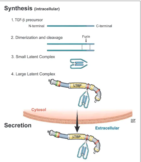
SYNTHESIS (PLEASE SEE FIGURE 1)
SMALL LATENT COMPLEX
The isoforms of TGF-
β
1, -2, and -3 are encoded as large precursor
proteins of 390–412 amino acids in size, and each isoform is the
product of a separate gene (
Derynck et al., 1985
;
Gentry and Nash,
1990
;
ten Dijke et al., 1990
;
Schlunegger and Grutter, 1992
). TGF-
β
proteins undergo several processing steps intracellularly prior to
their secretion. The most important step is the proteolytic
diges-tion of precursors by the endopeptidase furin, which cleaves the
TGF-
β
protein between amino acids 278 and 279 (
Dubois et al.,
1995
;
Blanchette et al., 1997
). The proteolysis yields two
prod-ucts that assemble into dimers. One is latency-associated peptide
(LAP), a 65- to 75-kDa dimer from the N-terminal region. The
other is mature TGF-
β
, a 25-kDa dimer from the C-terminal
por-tion (
Gentry and Nash, 1990
;
Blanchette et al., 1997
;
Munger et al.,
1997
). A common feature of TGF-
β
is that its N-terminal
por-tion (LAP) remains non-covalently associated with the rest of the
portion (termed small latent complex), despite the cleavage of the
precursor (
Derynck et al., 1985
;
Gentry and Nash, 1990
;
ten Dijke
et al., 1990
;
Schlunegger and Grutter, 1992
;
Dubois et al., 1995
;
Blanchette et al., 1997
;
Munger et al., 1997
). The presence of LAP
facilitates transit of TGF-
β
from the cell (
Lopez et al., 1992
).
Inter-estingly, cells transfected with mature sequences of TGF-
β
1 fail to
secrete proteins into medium, whereas cells transfected with LAP
and mature sequences of TGF-
β
1 can do this (
Gray and Mason,
1990
). Thus, LAP is required for the correct folding of the TGF-
β
homodimer and its secretion from cells. Furthermore, LAP shields
the receptor binding epitope of mature TGF-
β
, indicating that
LAP makes TGF-
β
biologically inactive and prevents interactions
of TGF-
β
with receptors.
LARGE LATENT COMPLEX
[image:2.595.305.551.62.344.2]Small latent complex is associated with a large protein termed
latent TGF-
β
binding protein (LTBP) via disulfide bonds.
Mam-malian cells express four different LTBP isoforms, of which three
(LTBP-1, -3, and -4) can associate with LAP. The trimolecular
complex of TGF-
β
, LAP, and LTBP is referred to as the large
FIGURE 1 | Illustration of the sequential steps in the synthesis of TGF-β
latent complex and secretion.(1) TGF-βis synthesized as a precursor protein; (2) Two TGF-βprecursor proteins dimerize through disulfide bridges; (3) TGF-β-dimer precursor is cleaved by furin to yield the small latent TGF-β complex, in which latency-associated peptide (LAP; light blue) and mature TGF-βpeptide (dark blue) are connected by non-covalent bonds; and (4) the large latent complex is formed by covalent linking between small latent complex and latent TGF-βbinding protein (LTBP), then secreted and incorporated into extracellular matrix.
TGF-
β
rapidly inside the cells, and this association is important
for the proper assembly and secretion of latent TGF-
β
complexes
(
Miyazono et al., 1991
).
ECM ANCHORING
After secretion, LTBP also plays a critical role in targeting small
latent TGF-
β
for deposition in the ECM (
Taipale et al., 1994
;
Nunes
et al., 1997
). LTBP belongs to the fibrillin family of ECM
pro-teins, and LTBP shares homology with fibrillins, which are major
constituents of connective tissue microfibrillar structure (
Kanzaki
et al., 1990
;
Munger et al., 1997
). The C-terminal region of LTBP-1
binds to the N-terminal region of fibrillin-1 (
Isogai et al., 2003
).
In addition, LTBP-1 is covalently cross-linked to ECM proteins
such as fibronectin (
Dallas et al., 2005
) via its N-terminal region
(
Taipale et al., 1994
;
Nunes et al., 1997
). Indeed, LTBP-1 colocalizes
with both fibrillin-1 and fibronectin
in vitro
by
immunostain-ing (
Taipale et al., 1996
;
Dallas et al., 2005
;
Massam-Wu et al.,
2010
), suggesting that fibrillin-1and fibronectin can associate with
LTBP-1. The treatment of fetal rat calvarial osteoblasts with a
70-kDa N-terminal fibronectin fragment, which inhibits fibronectin
assembly, impairs incorporation of LTBP-1 into the ECM in
cul-ture (
Dallas et al., 2005
). In addition, although fibronectin-null
embryonic fibroblasts secrete a large amount of LTBP-1 into
medium, they fail to incorporate LTBP-1 into the ECM (
Dal-las et al., 2005
). This covalent association between LTBP-1 and
the ECM depends on transglutaminase-mediated cross-linking,
because little LTBP-1 is recovered from matrix digests that have
been treated with transglutaminase inhibitors (
Nunes et al., 1997
).
Thus, the N-terminal and C-terminal binding sites of LTBP-1 are
central for anchoring latent TGF-
β
to fibrillin-1 and fibronectin
(
Hyytiainen et al., 1998
;
Unsold et al., 2001
), and this
anchor-ing influences the release of TGF-
β
from LAP, a process called
latent TGF-
β
activation (
Annes et al., 2004
). A number of genetic
studies suggest that the absence of or a mutation in LTBP-binding
ECMs such as fibrillin-1 and fibronectin results in increased
TGF-β
activity and Smad signaling, whereas the absence of or mutation
in latent TGF-
β
activators and LTBPs results in decreased
activ-ity (
Neptune et al., 2003
;
Koli et al., 2004
;
Mazzieri et al., 2005
;
Yoshinaga et al., 2008
;
Loeys et al., 2010
;
Table 1
). Efficient latent
TGF-
β
activation requires appropriate localization of latent
com-plexes in the ECM (
Annes et al., 2003
). Following the N-terminal
domain of LTBP, there is a protease-sensitive region, called the
hinge region. Large latent TGF-
β
is released from the ECM by
proteolytic cleavage at this region (
Taipale et al., 1994
). Thus, the
small latent complex, which consists of precursors of mature
TGF-β
and LAP, covalently interacts with LTBP and is secreted thereafter
as a large latent complex. The large latent complex is then
asso-ciated with the ECM such as fibrillin and fibronectin (
Figure 2
).
Extracellular TGF-
β
activity is regulated at the level of
bioavail-ability (release of latent TGF-
β
from ECM) and latency (release
of active TGF-
β
from the latent complex). Such a well and tightly
orchestrated regulation in latency of TGF-
β
enables the
imme-diate and highly localized response to type-specific tissue injury
without
de novo
synthesis. In this scenario, the ECM is potentially
an important regulator of latent TGF-
β
complexes. One of the
compelling themes in recent years is to clarify the crucial roles
of integrin
β
6, thrombospondin-1 (TSP-1), LTBPs, fibrillin-1, and
fibronectin in regulating local TGF-
β
activity and locally anchored
TGF-
β
availability in pathological process (
Hynes, 2009
).
MECHANISMS OF TGF-
β
ACTIVATION
[image:3.595.44.550.464.676.2]Local activation of latent (inactive) TGF-
β
complexes is a critical
event in regulating TGF-
β
function
in vivo
. In response to tissue
damage, TGF-
β
is released from LAP or latent TGF-
β
complex
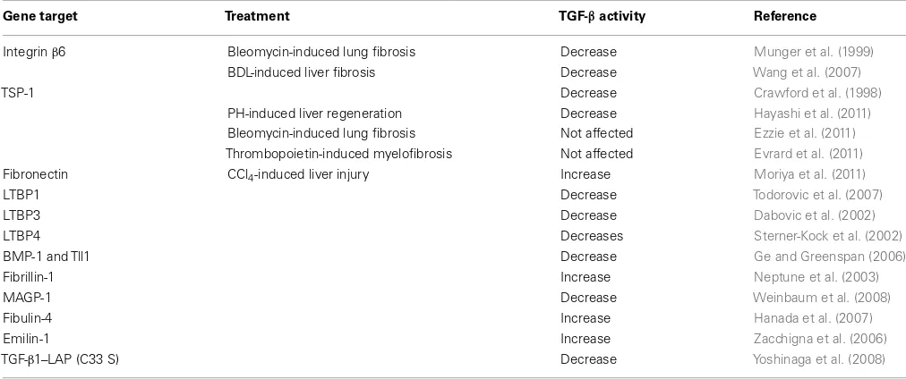
Table 1 | Altered TGF-βactivity in knockout or mutant mice.
Gene target Treatment TGF-βactivity Reference
Integrinβ6 Bleomycin-induced lung fibrosis Decrease Munger et al. (1999)
BDL-induced liver fibrosis Decrease Wang et al. (2007)
TSP-1 Decrease Crawford et al. (1998)
PH-induced liver regeneration Decrease Hayashi et al. (2011)
Bleomycin-induced lung fibrosis Not affected Ezzie et al. (2011)
Thrombopoietin-induced myelofibrosis Not affected Evrard et al. (2011)
Fibronectin CCl4-induced liver injury Increase Moriya et al. (2011)
LTBP1 Decrease Todorovic et al. (2007)
LTBP3 Decrease Dabovic et al. (2002)
LTBP4 Decreases Sterner-Kock et al. (2002)
BMP-1 and Tll1 Decrease Ge and Greenspan (2006)
Fibrillin-1 Increase Neptune et al. (2003)
MAGP-1 Decrease Weinbaum et al. (2008)
Fibulin-4 Increase Hanada et al. (2007)
Emilin-1 Increase Zacchigna et al. (2006)
TGF-β1–LAP (C33 S) Decrease Yoshinaga et al. (2008)
TGF-β, transforming growth factor-β; BDL, bile duct ligation; TSP-1, thrombospondin-1; PH, partial hepatectomy; LTBP, large latent TGF-β-binding protein; BMP-1, bone morphogenetic protein-1; TII1, tolloid-like 1; CCl4, Carbon tetrachloride; MAGP-1, microfibril-associated glycoprotein-1; LAP, latency-associated peptide; C33S, 33
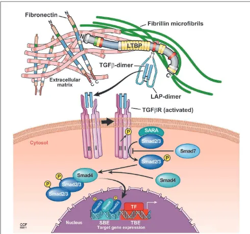
FIGURE 2 | Proposed model of TGF-βlatent complex and active TGF-β
signaling.The LTBP is covalently cross-linked to ECMs such as fibronectin and fibrillin via its N-terminal and C-terminal region, respectively. The small latent TGF-βcomplex is bound covalently to the third cysteine-rich domain of LTBP. In response to tissue injury, release of active TGF-βfrom latent complex and/or conformational change such as exposure of the TGF-β receptor binding site is induced. Binding of active TGF-βwith the TGF-β receptor type II leads to the phosphorylation and recruitment of TGF-β receptor type I into a heteromeric receptor complexes. The serine/threonine kinase activity of the activated complex phosphorylates Smad2 and Smad3 that both bind to Smad4 and translocate into the nucleus to enhance gene transcription by cooperating with DNA transcription factors.
undergoes conformational changes, and therefore, active
TGF-β
is exposed to the TGF-
β
receptor binding site (
Annes et al.,
2003
). Binding of active TGF-
β
to the TGF-
β
receptor type II
leads to the phosphorylation and recruitment of TGF-
β
receptor
type I into a heteromeric receptor complex. The serine/threonine
kinase activity of the activated complex phosphorylates Smad2 and
Smad3 that both bind to Smad4 and translocate into the nucleus
to enhance gene transcription by cooperating with DNA
tran-scription factors (
Feng and Derynck, 2005
;
Massague et al., 2005
;
Matsuzaki, 2012
;
Figure 2
). The activation mechanism to generate
active TGF-
β
from latent complexes has been extensively
stud-ied, and several modulators of TGF-
β
activity
in vivo
have been
proposed.
INTEGRIN-MEDIATED TGF-βACTIVATION
Recent studies show that integrins-related genetic models can
down-regulate TGF-
β
activation
in vivo
(
Munger et al., 1999
;
Yang
et al., 2007
). Integrins are heterodimeric cell adhesion molecules
and transmembrane receptors that link the ECM to the
cytoskele-ton, and have an important role in cell adhesion, cell proliferation,
differentiation, and cell migration (
Hynes, 1992, 2002
;
van der
Flier and Sonnenberg, 2001
). Integrins are composed of
α
- and
β
-subunits (18
α
- and 8
β
-subunits), both of which are glycoproteins
consisting of large extracellular domains and, in most cases, a short
cytoplasmic domain (
van der Flier and Sonnenberg, 2001
). Both
TGF-
β
1 and LAP have an RGD sequence, a binding motif in
lig-ands for
αv
integrins. Among the
αv
integrins, the integrins
αvβ1
,
αvβ3
,
αvβ5
,
αvβ6
,
αvβ8
,
α5β1
,
α8β1
, and
αIIbβ
3 are RGD
bind-ing integrins. Mice with the integrin-bindbind-ing RGD motif mutated
to RGD recapitulate all major phenotypes of TGF-
β
1-null mice,
including multi-organ inflammation and defects in vasculogenesis
(
Yang et al., 2007
). Two of these integrins (
αvβ6
and
αvβ8
) bind
and efficiently activate latent TGF-
β
1 and -
β
3 (
Munger et al., 1999
;
Annes et al., 2002
;
Mu et al., 2002
;
Araya et al., 2006
).
The integrin identified as a TGF-
β
activator
in vivo
is
αvβ6
,
which can directly activate latent TGF-
β
1 independently from
any proteolytic activity (
Munger et al., 1999
). The
αvβ6
integrin-mediated activation of latent TGF-
β
depends on a direct
inter-action between integrin
αvβ6
and the RGD amino acid sequence
present in LAP
β
1 and LAP
β
3. However, binding alone is not
suf-ficient to activate the latent complex (
Munger et al., 1999
). The
interaction of the
β6
cytoplasmic domain with the actin
cytoskele-ton is mandatory for this activation (
Munger et al., 1999
). LTBP-1
is identified as being a major regulatory factor in
αvβ6
-integrin-mediated TGF-
β
activation. This activation requires a covalent
interaction between LAP and the third cysteine-rich domain in
LTBP mediated by a hinge domain of LTBP-1 (
Annes et al., 2004
).
Since the hinge domain of LTBP-1 is not conserved among other
LTBP isoforms,
αvβ6
-mediated TGF-
β
activation via LTBP-1 is
isoform specific (
Annes et al., 2004
;
Wipff and Hinz, 2008
). In
response to tissue injury or tissue damage, integrin
αvβ6
induces
a conformational change of latent TGF-
β
via the interactions
between integrin
αvβ6
and actin cytoskeleton (
Munger et al., 1999
;
Wipff and Hinz, 2008
;
Shi et al., 2011
). Such a conformational
change makes it possible to cause mature TGF-
β
to interact with
the TGF-
β
type II receptor (
Munger et al., 1999
;
Wipff and Hinz,
2008
;
Shi et al., 2011
). There is no release of LAP or active TGF-
β
1
once latent TGF-
β
associates with integrin
αvβ6
. Although a
profi-brogenic drug bleomycin induces lung fibrosis in wild-type mice
by up-regulation of integrin
β
6 expression in alveolar epithelial
cells and activation of TGF-
β
, even high dose of bleomycin
admin-istration cannot cause fibrosis in integrin
β
6-null mice (
Munger
et al., 1999
).
TSP-1-MEDAITED TGF-βACTIVATION
the LAP and the LTBP (
Schultz-Cherry et al., 1994a
). Thus, TSP-1
binds small latent TGF-
β
and large latent TGF-
β
, and this
bind-ing interaction is sufficient to generate biologically active TGF-
β
.
The two sequences (GGWSHW and KRFK) located in the type
I repeat of TSP-1 comprise the region responsible for binding
and activating latent TGF-
β
, respectively (
Goundis and Reid, 1988
;
Schultz-Cherry et al., 1994b
). In LAP, a sequence LSKL near the
amino-terminus plays a pivotal role in the interaction and
TSP-1-mediated activation of latent TGF-
β
, because LSKL peptides
competitively inhibit TSP-1- or a KRFK-peptide-mediated latent
TGF-
β
activation (
Ribeiro et al., 1999
). Furthermore, mutations
of the LSKL sequence in LAP reduce the binding of LAP to the
mature TGF-
β
and induce the impaired ability of LAP to confer
latency to mature TGF-
β
(
Young and Murphy-Ullrich, 2004
). Such
a direct interaction between TSP-1 and LAP is supposed to induce
a conformational change of LAP in relation to mature TGF-
β
and
thereby presumably unmask the TGF-
β
receptor binding site that
can bind to its receptor (
Murphy-Ullrich and Poczatek, 2000
). To
further understand TSP-1-mediated TGF-
β
activation, the nature
of structural restraints imposed on LAP in the presence of bound
TSP-1 would be useful. TSP-1-null mice have an inflammatory
phenotype similar to that of TGF-
β
1-null mice in several organs,
including pancreas and lung, whereas the inflammatory changes
observed in TSP-1-null mice are not as severe as those in TGF-
β
1-null mice (
Crawford et al., 1998
). Treatment of TSP-1-null mice
with the TSP-1 derived peptide KRFK, which activates TGF-
β
1,
rescues the abnormal phenotypes in pancreas and lung (
Craw-ford et al., 1998
). In contrast, the treatment of wild-type mice
with the peptide LSKL, which blocks TGF-
β
activation, results in
the abnormalities in pancreas and lung, and these phenotypes are
very similar to those of TSP-1-null mice and TGF-
β
1-null mice
(
Crawford et al., 1998
).
PROTEASE-MEDIATED TGF-βACTIVATION
A number of proteases, including plasmin, matrix
metallopro-teinase (MMP)-2/9, and a disintegrin and metalloprometallopro-teinase with
TSP motifs 1 (ADAMTS1), have been identified as latent
TGF-β
activators
in vitro
(
Lyons et al., 1988
;
Sato and Rifkin, 1989
;
Yu and Stamenkovic, 2000
;
Bourd-Boittin et al., 2011
). Plasmin
and MMP-2/9 belong to the serine protease and
metallopro-teinase families, respectively. The prometallopro-teinase-sensitive hinge region
in LTBP is suggested to be a potential target for the release of
a still-latent remnant of the large latent complex (
Taipale et al.,
1994
). Using a co-culture system of endothelial cells and
peri-cytes or smooth muscle cells, plasmin has been identified as one
of the latent TGF-
β
activators
in vitro
: the active TGF-
β
forma-tion is blocked by plasmin inhibitors (
Sato and Rifkin, 1989
)
or prolonged by neutralizing antibody to plasminogen
activa-tor inhibiactiva-tor-1 (
Sato et al., 1990
). The treatments of
fibroblast-or Chinese hamster ovary cell-conditional medium with
plas-min can generate the active form of TGF-
β
(
Lyons et al., 1988,
1990
). Thus, the proteinase-mediated TGF-
β
activation system
appears to be important in activation of latent TGF-
β
in
sev-eral models
in vitro
. However, there is still no definite evidence of
proteinase-dependent TGF-
β
activation
in vivo
. Indeed,
plasmino-gen (plasmin pro-enzyme)-null mice show normal embryonic
development, survive to adulthood, are fertile, and display none of
the pathological features shown in TGF-
β
-null mice (
Bugge et al.,
1995
).
TGF-
β
ACTIVATION MECHANISM AND AVAILABILITY OF
LATENT TGF-
β
COMPLEX IN LIVER DISEASES
Transforming growth factor-
β
signal induces the transition of
quiescent hepatic stellate cells (HSCs) into contractile fibrogenic
myofibroblasts, which is the key event in the pathobiology of
liver fibrosis (
Bissell et al., 2001
;
Dooley et al., 2003
;
Gressner
and Weiskirchen, 2006
). Myofibroblasts play a central role in the
production of ECM components such as fibronectin, collagen
type I and III. Continuous and/or upregulated TGF-
β
-signaling,
such as in chronic liver injury, induces sustained activation of
myofibroblasts and results in liver fibrosis (
Bissell et al., 2001
;
Tahashi et al., 2002
;
Gressner and Weiskirchen, 2006
). Active
TGF-
β
-signaling acts on hepatocytes as an anti-proliferative factor
in culture, and administration of TGF-
β
inhibits the hepatocyte
proliferation during liver regeneration after partial hepatectomy
(
Russell et al., 1988
). Genetic ablation of TGF-
β
-signals using TGF
type II receptor-knockout mice actually accelerates hepatocyte
proliferation after partial hepatectomy (
Oe et al., 2004
). Thus,
locally activated TGF-
β
-signaling is involved in the repair
follow-ing liver injury, and the therapeutic strategies targetfollow-ing local TGF-
β
activation in fibrogenesis are now under intense investigation
(
Table 2
).
β6 INTEGRIN-MEDIATED LOCAL TGF-βBIOAVAILABILITY AND LIVER DISEASES
Expression of
αvβ6
-integrin is virtually absent except in
cholangio-cytes in normal liver, but its expression in cholangiocholangio-cytes is highly
upregulated in thioacetamide- and bile duct ligation-induced
fibrotic liver (
Wang et al., 2007
;
Popov et al., 2008
). Deficiency in
integrin
β6
attenuates liver fibrosis after bile duct ligation, which is
accompanied by a decrease in active TGF-
β
-signaling (
Wang et al.,
2007
). In human fibrogenic liver diseases (e.g., primary biliary
cir-rhosis, primary sclerosing cholangitis, alcoholic liver disease, and
hepatitis B or C),
α
vβ
6-integrin
mRNA levels are upregulated
com-pared to levels in healthy livers (
Popov et al., 2008
). Furthermore,
α
vβ
6-integrin
mRNA levels correlate with the progression of
fibro-sis in patients with chronic hepatitis C (
Popov et al., 2008
). These
findings suggest that
αvβ6
-integrin may be involved not only in
classic biliary type fibrosis but also in a variety of liver fibrogenic
diseases with different etiologies (
Popov et al., 2008
).
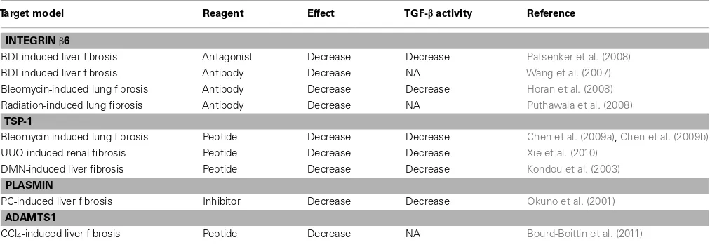
Table 2 | Therapeutic target for TGF-βactivation mechanismin vivo.
Target model Reagent Effect TGF-βactivity Reference
INTEGRINβ6
BDL-induced liver fibrosis Antagonist Decrease Decrease Patsenker et al. (2008)
BDL-induced liver fibrosis Antibody Decrease NA Wang et al. (2007)
Bleomycin-induced lung fibrosis Antibody Decrease Decrease Horan et al. (2008)
Radiation-induced lung fibrosis Antibody Decrease NA Puthawala et al. (2008)
TSP-1
Bleomycin-induced lung fibrosis Peptide Decrease Decrease Chen et al. (2009a),Chen et al. (2009b)
UUO-induced renal fibrosis Peptide Decrease Decrease Xie et al. (2010)
DMN-induced liver fibrosis Peptide Decrease Decrease Kondou et al. (2003)
PLASMIN
PC-induced liver fibrosis Inhibitor Decrease Decrease Okuno et al. (2001)
ADAMTS1
CCl4-induced liver fibrosis Peptide Decrease NA Bourd-Boittin et al. (2011)
TGF-β, transforming growth factor-β; PC, porcine serum; BDL, bile duct ligation; NA, not available; TSP-1, thrombospondin-1; UUO, Unilateral ureteral obstruction; DMN, dimethylnitrosamine; CCl4, carbon tetrachloride.
induces phosphorylation of Smad2. Furthermore, pretreatment
of primary hepatocytes with the TSP-1-inhibitory peptide LSKL
significantly suppresses conditioned media-induced
phosphory-lation of Smad2, whereas pretreatment of primary hepatocytes
with the control peptide SLLK shows no effects. Thus, these
findings indicate that TSP-1 plays a negative role in liver
regen-eration through local TGF-
β
1 activation (
Hayashi et al., 2011
).
Furthermore, treatment with the TSP-1 inhibitory peptide LSKL
reduces local TGF-
β
activity and suppresses the progression of liver
fibrosis in a dimethylnitrosamine (DMN)-treated rat liver fibrosis
model, suggesting that TSP-1-mediated local TGF-
β
activation is
associated with liver fibrogenesis (
Kondou et al., 2003
).
FIBRONECTIN-MEDIATED LOCAL TGF-βBIOAVAILABILITY AND LIVER DISEASES
Fibronectin is a major ECM component and exists in soluble
(plasma fibronectin) and insoluble form (cellular fibronectin) as a
part of the ECM (
Hynes, 1986
;
Mosher, 1989
). Fibronectin
partici-pates in the incorporation of LTBP-1 into the ECM
in vitro
(
Dallas
et al., 2005
). Fibronectin-null cells show poor activity vis-à-vis
LTBP-1 incorporation into the ECM and exhibit deficient
activa-tion of latent TGF-
β
1 even after transfection by the integrin
αvβ6
,
suggesting that fibronectin is necessary to the ECM scaffold for
LTBP-1 deposition (
Fontana et al., 2005
). To investigate the
inter-dependence of fibronectin and TGF-
β
on the fibrogenic response
to adult tissue damage, we recently established a null condition for
both fibronectin isoforms (plasma and cellular types) from adult
mouse liver. Since it has been proposed that collagen organization
and assembly depend on the fibronectin matrix in culture (
Sottile
and Hocking, 2002
;
Velling et al., 2002
;
Sottile et al., 2007
), we have
explored whether fibronectin would be a suitable molecular target
for preventing the extensive collagen deposits and scar formation
that could lead to liver fibrosis. We have demonstrated that the
lack of fibronectin does not actually interfere with reconstruction
of collagen fibril organization in response to carbon
tetrachloride-induced liver injury. Fibronectin deficiency results in elevated local
TGF-
β
bioavailability post injury, and it is mediated largely by
β
6
integrin. Furthermore, we have identified TGF-
β
-signaling and
type V collagen as essential elements for collagen fibrillogenesis
in adult tissue remodeling (
Moriya et al., 2011
). Our findings
imply that fibronectin regulates the balance of active and
inac-tive (latent) TGF-
β
, which in turn modulates ECM production
and remodeling and consequently maintains adult liver
home-ostasis. Indeed, mouse models of TGF-
β
1 overexpression show
dominant phenotypes such as advanced liver fibrosis (
Sanderson
et al., 1995
;
Ueberham et al., 2003
). These observations
sup-port the hypothesis that LTBP and its binding molecules such as
fibronectin determine the spatial localization of LTBP in tissues,
thereby regulating the extent of local TGF-
β
bioavailability. Since
locally activated TGF-
β
induces the production of type V collagen
in response to liver damage, it remains to be elucidated whether
type V collagen-nucleated collagen fibrillogenesis contributes to
adult chronic fibrotic diseases.
PROTEINASE-MEDIATED LOCAL TGF-βBIOAVAILABILITY AND LIVER DISEASES
porcine-serum-induced liver fibrosis in rats (
Okuno et al.,
2001
).
TRANSGLUTAMINASE-MEDIATED LOCAL TGF-βBIOAVAILABILITY AND LIVER DISEASES
Covalent cross-linking between proteins is catalyzed by
transg-lutaminases. This is an important process for tissue remodeling,
as it generates extra rigidity and a resistance against proteolytic
degradation. The family of transglutaminases (EC2.3.2.13)
con-sists of eight family members (transglutaminase 1–7 and plasma
coagulation factor XIII (
Beninati and Piacentini, 2004
). The
covalent association between LTBP-1 and the ECM depends on
transglutaminase-mediated cross-linking, because little LTBP-1
is recovered from matrix digests prepared from cultures treated
with transglutaminase inhibitors (
Nunes et al., 1997
).
Further-more, transglutaminase inhibitors prevent TGF-
β
1 activation in a
co-culture of bovine aortic endothelial cells and bovine smooth
muscle cells (
Kojima et al., 1993
). Thus, transglutaminase is
important for the LTBP anchoring to ECM, and transglutaminase
inhibitors affect TGF-
β
1 activity
in vitro
. Transglutaminase 2 is
involved in several human diseases, in wound healing and fibrosis,
and represents promising pharmacological targets (
Verderio et al.,
2004
;
Caccamo et al., 2010
). Indeed, transglutaminase 2 deficiency
reduces levels of active TGF-
β
1 and attenuates the interstitial renal
fibrosis induced by unilateral ureteral obstruction in mice (
Shweke
et al., 2008
).
Elevated transglutaminase 2 expression and activity are
observed in human fibrotic (
Grenard et al., 2001
) and mouse
CCl
4-treated livers (
Nardacci et al., 2003
;
Popov et al., 2011
).
Transglutaminase 2-null mice fail to clear hepatic necrotic tissues
and to rearrange the hepatic lobular architecture with a
progres-sive accumulation of ECM components and inflammatory cells
in CCl
4-induced chronic liver injury, indicating that
transglu-taminase 2 plays a protective role in tissue stability and repair
after liver injury (
Nardacci et al., 2003
;
Popov et al., 2011
). Very
recently,
Popov et al. (2011)
assessed whether transglutaminase 2
was implicated in irreversible collagen stabilization in liver fibrosis.
Transglutaminase 2 activity is upregulated during hepatic
fibroge-nesis or stabilization of collagen matrix. However, unexpectedly,
transglutaminase 2 deficiency does not promote regression of liver
fibrosis (
Popov et al., 2011
). Thus, there exists transglutaminase
2-independent irreversible collagen cross-linking in the progression
of liver fibrosis (
Popov et al., 2011
).
Although most antifibrotic strategies are directed against HSC
/myofibroblast proliferation and profibrogenic activation, few
studies have targeted ECM stabilization. Considering the evidence
that the instability of ECM bounded latent TGF-
β
complex results
in altered TGF-
β
availability (
Maeda et al., 2011
), the targeting
for ECM-mediated TGF-
β
bioavailability would be an alternative
therapeutic approach for preventing progression of liver fibrosis.
PERSPECTIVE
Transforming growth factor-
β
is synthesized as a small latent
com-plex (mature TGF-
β
and LAP) and thereafter covalently interacts
with LTBPs as the large latent complex. The large latent complex
is associated with ECM components, fibrillin, and fibronectin.
Such a well and tightly orchestrated regulation in latency of
TGF-
β
enables an immediate and highly localized response to
type-specific tissue injury without
de novo
synthesis.
Both
β
6 integrin- and TSP-1-mediated local TGF-
β
activation
are involved in the pathogenesis of liver diseases. Interestingly, in
other organs such as lung, the lack of TSP-1 does not affect
TGF-β
bioavailability and does not protect from bleomycin-induced
pulmonary fibrosis (
Ezzie et al., 2011
). In the
thrombopoietin-induced bone marrow myelofibrosis model, TSP-1 deficiency
does not attenuate local TGF-
β
bioavailability and myelofibrosis
(
Evrard et al., 2011
). Thus, mechanisms of local TGF-
β
activation
and their biological significance in the pathological process are
likely to be tissue specific.
Based on the TGF-
β
signaling pathway, four major strategies
allowing the modulation of TGF-
β
availability have emerged. The
first strategy is to block the production of TGF-
β
using anti-sense
oligonucleotide mRNA; a second is to use monoclonal antibodies
to block specific TGF-
β
isoforms; a third is to use the
intracellu-lar signal inhibitors that block the TGF-
β
receptor activation and
downstream signaling; and a fourth, which constitutes the main
focus of this review, is to block the local activation of latent
TGF-β
. Since TGF-
β
has so many important physiological functions, as
evidenced by TGF-
β
-null mice, a long-term global inhibition of
TGF-
β
activity might potentially lead to undesirable side effects,
such as aberrant immune activation, impaired wound healing,
and malignant cell transformation (
Kulkarni et al., 1993
;
Mallat
et al., 2001
;
Bhowmick et al., 2004
). Careful targeting of the TGF-
β
pathway to minimize systemic effects is clearly a highly
desir-able goal. Anti-TGF-
β
strategies to selectively block latent TGF-
β
activation at local sites where excess TGF-
β
activation occurs are
attractive and logical. The benefits of therapeutic strategies
target-ing local TGF-
β
activation are under intense investigation. Future
studies will yield valuable data about these strategies for liver
diseases.
ACKNOWLEDGMENTS
The authors are grateful to David R. Schumick, Center for Medical
Art and Photography, Cleveland Clinic, for his excellent artwork.
We also thank Christine Kassuba for editorial assistance. We wish
to acknowledge many outstanding contributions of investigators
in the field whose work could not be cited because of space
constraints. This work was supported by grants from the U.S.
National Institutes of Health (R01 DK074538 to Takao Sakai),
and the Byotai Taisha Research Foundation and Uehara Memorial
Foundation, Japan (to Hiromitsu Hayashi).
REFERENCES
Adams, J. C. (2001). Thrombospondins: multifunctional regulators of cell interactions. Annu. Rev. Cell Dev. Biol.17, 25–51.
Annes, J. P., Chen, Y., Munger, J. S., and Rifkin, D. B. (2004). Integrin
alphaVbeta6-mediated activation of latent TGF-beta requires the latent TGF-beta binding protein-1.J. Cell Biol.165, 723–734.
Annes, J. P., Munger, J. S., and Rifkin, D. B. (2003). Mak-ing sense of latent TGFbeta
activation. J. Cell. Sci. 116(Pt 2), 217–224.
Annes, J. P., Rifkin, D. B., and Munger, J. S. (2002). The inte-grin alphaVbeta6 binds and activates latent TGFbeta3. FEBS Lett. 511, 65–68.
Araya, J., Cambier, S., Morris, A., Finkbeiner, W., and Nishimura, S. L. (2006). Integrin-mediated trans-forming growth factor-beta activa-tion regulates homeostasis of the pulmonary epithelial-mesenchymal trophic unit. Am. J. Pathol. 169, 405–415.
Beninati, S., and Piacentini, M. (2004). The transglutaminase family: an overview: minireview article.Amino Acids26, 367–372.
Bhowmick, N. A., Chytil, A., Plieth, D., Gorska, A. E., Dumont, N., Shappell, S., Washington, M. K., Neilson, E. G., and Moses, H. L. (2004). TGF-beta signaling in fibroblasts modulates the oncogenic potential of adjacent epithelia.Science303, 848–851. Bissell, D. M., Roulot, D., and George,
J. (2001). Transforming growth fac-tor beta and the liver.Hepatology34, 859–867.
Blanchette, F., Day, R., Dong, W., Laprise, M. H., and Dubois, C. M. (1997). TGFbeta1 regulates gene expression of its own converting enzyme furin. J. Clin. Invest. 99, 1974–1983.
Blobe, G. C., Schiemann, W. P., and Lodish, H. F. (2000). Role of transforming growth factor beta in human disease.N. Engl. J. Med.342, 1350–1358.
Bornstein, P. (2009). Thrombospondins function as regulators of angio-genesis.J. Cell Commun. Signal.3, 189–200.
Boulanger, J., Reyes-Moreno, C., and Koutsilieris, M. (1995). Mediation of glucocorticoid receptor function by the activation of latent transform-ing growth factor beta 1 in MG-63 human osteosarcoma cells.Int. J. Cancer61, 692–697.
Bourd-Boittin, K., Bonnier, D., Leyme, A., Mari, B., Tuffery, P., Samson, M., Ezan, F., Baffet, G., and Theret, N. (2011). Protease profiling of liver fibrosis reveals the adam metal-lopeptidase with thrombospondin type 1 motif, 1 as a central acti-vator of TGF-beta.Hepatology 54, 2173–2184.
Bugge, T. H., Flick, M. J., Daugherty, C. C., and Degen, J. L. (1995). Plas-minogen deficiency causes severe thrombosis but is compatible with development and reproduction.
Genes Dev.9, 794–807.
Caccamo, D., Curro, M., and Ientile, R. (2010). Potential of transglutami-nase 2 as a therapeutic target.Expert Opin. Ther. Targets14, 989–1003. Chen, Y., Wang, X., Weng, D., Tao, S.,
Lv, L., and Chen, J. (2009a). A TSP-1 functional fragment inhibits activa-tion of latent transforming growth
factor-beta1 derived from rat alve-olar macrophage after bleomycin treatment.Exp. Toxicol. Pathol.61, 67–73.
Chen, Y., Wang, X., Weng, D., Tian, L., Lv, L., Tao, S., and Chen, J. (2009b). A TSP-1 synthetic peptide inhibits bleomycin-induced lung fibrosis in mice.Exp. Toxicol. Pathol.61, 59–65. Crawford, S. E., Stellmach, V., Murphy-Ullrich, J. E., Ribeiro, S. M., Lawler, J., Hynes, R. O., Boivin, G. P., and Bouck, N. (1998). Thrombospondin-1 is a major acti-vator of TGF-beta1 in vivo.Cell93, 1159–1170.
Dabovic, B., Chen, Y., Colarossi, C., Obata, H., Zambuto, L., Perle, M. A., and Rifkin, D. B. (2002). Bone abnormalities in latent TGF-[beta] binding protein (Ltbp)-3-null mice indicate a role for Ltbp-3 in mod-ulating TGF-[beta] bioavailability.J. Cell Biol.156, 227–232.
Dallas, S. L., Park-Snyder, S., Miya-zono, K., Twardzik, D., Mundy, G. R., and Bonewald, L. F. (1994). Characterization and autoregulation of latent transforming growth fac-tor beta (TGF beta) complexes in osteoblast-like cell lines. Production of a latent complex lacking the latent TGF beta-binding protein.J. Biol. Chem.269, 6815–6821.
Dallas, S. L., Sivakumar, P., Jones, C. J., Chen, Q., Peters, D. M., Mosher, D. F., Humphries, M. J., and Kielty, C. M. (2005). Fibronectin regulates latent transforming growth factor-beta (TGF factor-beta) by controlling matrix assembly of latent TGF beta-binding protein-1.J. Biol. Chem.280, 18871–18880.
Derynck, R., Jarrett, J. A., Chen, E. Y., Eaton, D. H., Bell, J. R., Assoian, R. K., Roberts, A. B., Sporn, M. B., and Goeddel, D. V. (1985). Human trans-forming growth factor-beta comple-mentary DNA sequence and expres-sion in normal and transformed cells.Nature316, 701–705. Dickson, M. C., Martin, J. S., Cousins,
F. M., Kulkarni, A. B., Karlsson, S., and Akhurst, R. J. (1995). Defective haematopoiesis and vasculogenesis in transforming growth factor-beta 1 knock out mice.Development121, 1845–1854.
Dooley, S., Hamzavi, J., Breitkopf, K., Wiercinska, E., Said, H. M., Loren-zen, J., Ten Dijke, P., and Gressner, A. M. (2003). Smad7 prevents activa-tion of hepatic stellate cells and liver fibrosis in rats.Gastroenterology125, 178–191.
Dubois, C. M., Laprise, M. H., Blanchette, F., Gentry, L. E., and Leduc, R. (1995). Processing of
transforming growth factor beta 1 precursor by human furin convertase. J. Biol. Chem. 270, 10618–10624.
Evrard, S., Bluteau, O., Tulliez, M., Rameau, P., Gonin, P., Zetter-berg, E., Palmblad, J., Bonnefoy, A., Villeval, J. L., Vainchenker, W., Giraudier, S., and Wagner-Ballon, O. (2011). Thrombospondin-1 is not the major activator of TGF-beta1 in thrombopoietin-induced myelofibrosis.Blood117, 246–249. Ezzie, M. E., Piper, M. G., Montague,
C., Newland, C. A., Opalek, J. M., Baran, C., Ali, N., Brigstock, D., Lawler, J., and Marsh, C. B. (2011). Thrombospondin-1-deficient mice are not protected from bleomycin-induced pulmonary fibrosis.Am. J. Respir. Cell Mol. Biol.44, 556–561. Feng, X. H., and Derynck, R. (2005).
Specificity and versatility in tgf-beta signaling through Smads.Annu. Rev. Cell Dev. Biol.21, 659–693. Fontana, L., Chen, Y., Prijatelj, P., Sakai,
T., Fassler, R., Sakai, L. Y., and Rifkin, D. B. (2005). Fibronectin is required for integrin alphavbeta6-mediated activation of latent TGF-beta com-plexes containing LTBP-1.FASEB J.
19, 1798–1808.
Galbreath, E., Kim, S. J., Park, K., Bren-ner, M., and Messing, A. (1995). Overexpression of TGF-beta 1 in the central nervous system of trans-genic mice results in hydrocephalus.
J. Neuropathol. Exp. Neurol. 54, 339–349.
Ge, G., and Greenspan, D. S. (2006). BMP1 controls TGFbeta1 activa-tion via cleavage of latent TGFbeta-binding protein. J. Cell Biol. 175, 111–120.
Gentry, L. E., and Nash, B. W. (1990). The pro domain of pre-pro-transforming growth factor beta 1 when independently expressed is a functional binding protein for the mature growth factor.Biochemistry
29, 6851–6857.
Goundis, D., and Reid, K. B. (1988). Properdin, the terminal complement components, thrombospondin and the circumsporozoite protein of malaria parasites contain similar sequence motifs.Nature335, 82–85. Gray, A. M., and Mason, A. J. (1990). Requirement for activin A and trans-forming growth factor – beta 1 pro-regions in homodimer assembly. Sci-ence247, 1328–1330.
Grenard, P., Bresson-Hadni, S., El Alaoui, S., Chevallier, M., Vuit-ton, D. A., and Ricard-Blum, S. (2001). Transglutaminase-mediated cross-linking is involved in the sta-bilization of extracellular matrix in
human liver fibrosis.J. Hepatol.35, 367–375.
Gressner, A. M., and Weiskirchen, R. (2006). Modern pathogenetic con-cepts of liver fibrosis suggest stellate cells and TGF-beta as major players and therapeutic targets.J. Cell. Mol. Med.10, 76–99.
Hanada, K., Vermeij, M., Garinis, G. A., de Waard, M. C., Kunen, M. G., Myers, L., Maas, A., Duncker, D. J., Meijers, C., Dietz, H. C., Kanaar, R., and Essers, J. (2007). Perturbations of vascular homeostasis and aortic valve abnormalities in fibulin-4 defi-cient mice.Circ. Res.100, 738–746. Hayashi, H., Sakai, K., Baba, H., and
Sakai, T. (2011). Thrombospondin-1 is a novel negative regulator of liver regeneration after par-tial hepatectomy via TGF-beta1 activation in mice. Hepatology. doi:10.1002/hep.24800. [Epub ahead of print].
Horan, G. S., Wood, S., Ona, V., Li, D. J., Lukashev, M. E., Weinreb, P. H., Simon, K. J., Hahm, K., Allaire, N. E., Rinaldi, N. J., Goyal, J., Feghali-Bostwick, C. A., Matteson, E. L., O’Hara, C., Lafyatis, R., Davis, G. S., Huang, X., Sheppard, D., and Vio-lette, S. M. (2008). Partial inhibition of integrin alpha(v)beta6 prevents pulmonary fibrosis without exacer-bating inflammation.Am. J. Respir. Crit. Care Med.177, 56–65. Hynes, R. O. (1986). Fibronectins.Sci.
Am.254, 42–51.
Hynes, R. O. (1992). Integrins: versatil-ity, modulation, and signaling in cell adhesion.Cell69, 11–25. Hynes, R. O. (2002). Integrins:
bidirec-tional, allosteric signaling machines.
Cell110, 673–687.
Hynes, R. O. (2009). The extracellular matrix: not just pretty fibrils.Science
326 1216–1219.
Hyytiainen, M., Penttinen, C., and Keski-Oja, J. (2004). Latent TGF-beta binding proteins: extracellular matrix association and roles in TGF-beta activation.Crit. Rev. Clin. Lab. Sci.41, 233–264.
Hyytiainen, M., Taipale, J., Heldin, C. H., and Keski-Oja, J. (1998). Recombi-nant latent transforming growth fac-tor beta-binding protein 2 assembles to fibroblast extracellular matrix and is susceptible to proteolytic process-ing and release.J. Biol. Chem.273, 20669–20676.
a microfibril-associated protein. J. Biol. Chem.278, 2750–2757. Kanzaki, T., Olofsson, A., Moren, A.,
Wernstedt, C., Hellman, U., Miya-zono, K., Claesson-Welsh, L., and Heldin, C. H. (1990). TGF-beta 1 binding protein: a component of the large latent complex of TGF-beta 1 with multiple repeat sequences.Cell
61, 1051–1061.
Kojima, S., Nara, K., and Rifkin, D. B. (1993). Requirement for transglut-aminase in the activation of latent transforming growth factor-beta in bovine endothelial cells.J. Cell Biol.
121, 439–448.
Koli, K., Saharinen, J., Hyytiainen, M., Penttinen, C., and Keski-Oja, J. (2001). Latency, activation, and binding proteins of TGF-beta.
Microsc. Res. Tech.52, 354–362. Koli, K., Wempe, F., Sterner-Kock, A.,
Kantola, A., Komor, M., Hofmann, W. K., von Melchner, H., and Keski-Oja, J. (2004). Disruption of LTBP-4 function reduces TGF-beta activa-tion and enhances BMP-4 signaling in the lung.J. Cell Biol.167, 123–133. Kondou, H., Mushiake, S., Etani, Y., Miyoshi, Y., Michigami, T., and Ozono, K. (2003). A blocking pep-tide for transforming growth factor-beta1 activation prevents hepatic fibrosis in vivo. J. Hepatol. 39, 742–748.
Kulkarni, A. B., Huh, C. G., Becker, D., Geiser, A., Lyght, M., Flanders, K. C., Roberts, A. B., Sporn, M. B., Ward, J. M., and Karlsson, S. (1993). Trans-forming growth factor beta 1 null mutation in mice causes excessive inflammatory response and early death.Proc. Natl. Acad. Sci. U.S.A.
90, 770–774.
Kyriakides, T. R., and Maclauchlan, S. (2009). The role of throm-bospondins in wound healing, ischemia, and the foreign body reac-tion. J. Cell Commun. Signal. 3, 215–225.
Loeys, B. L., Gerber, E. E., Riegert-Johnson, D., Iqbal, S., Whiteman, P., McConnell, V., Chillakuri, C. R., Macaya, D., Coucke, P. J., De Paepe, A., Judge, D. P., Wigley, F., Davis, E. C., Mardon, H. J., Handford, P., Keene, D. R., Sakai, L. Y., and Dietz, H. C. (2010). Mutations in fibrillin-1 cause congenital scleroderma: stiff skin syndrome.Sci. Transl. Med.2, 23ra20.
Lopez, A. R., Cook, J., Deininger, P. L., and Derynck, R. (1992). Domi-nant negative mutants of transform-ing growth factor-beta 1 inhibit the secretion of different transforming growth factor-beta isoforms. Mol. Cell. Biol.12, 1674–1679.
Lyons, R. M., Gentry, L. E., Purchio, A. F., and Moses, H. L. (1990). Mechanism of activation of latent recombinant transforming growth factor beta 1 by plasmin.J. Cell Biol.110, 1361–1367. Lyons, R. M., Keski-Oja, J., and Moses, H. L. (1988). Proteolytic activation of latent transforming growth factor-beta from fibroblast-conditioned medium.J. Cell Biol.
106, 1659–1665.
Maeda, T., Sakabe, T., Sunaga, A., Sakai, K., Rivera, A. L., Keene, D. R., Sasaki, T., Stavnezer, E., Iannotti, J., Schweitzer, R., Ilic, D., Baskaran, H., and Sakai, T. (2011). Conversion of mechanical force into TGF-beta-mediated biochemical signals.Curr. Biol.21, 933–941.
Mallat, Z., Gojova, A., Marchiol-Fournigault, C., Esposito, B., Kamate, C., Merval, R., Fradelizi, D., and Tedgui, A. (2001). Inhibition of transforming growth factor-beta signaling accelerates atherosclerosis and induces an unstable plaque phenotype in mice.Circ. Res. 89, 930–934.
Massague, J., Seoane, J., and Wotton, D. (2005). Smad transcription factors.
Genes Dev.19, 2783–2810. Massam-Wu, T., Chiu, M.,
Choud-hury, R., Chaudhry, S. S., Bald-win, A. K., McGovern, A., Baldock, C., Shuttleworth, C. A., and Kielty, C. M. (2010). Assembly of fib-rillin microfibrils governs extracel-lular deposition of latent TGF beta.
J. Cell Sci.123(Pt 17), 3006–3018. Matsuzaki, K. (2012). Smad
phospho-isoform signals in acute and chronic liver injury: similarities and differ-ences between epithelial and mes-enchymal cells.Cell Tissue Res.347, 225–243.
Mazzieri, R., Jurukovski, V., Obata, H., Sung, J., Platt, A., Annes, E., Karaman-Jurukovska, N., Gleizes, P. E., and Rifkin, D. B. (2005). Expres-sion of truncated latent beta-binding protein modulates TGF-beta signaling.J. Cell Sci.118(Pt 10), 2177–2187.
Miyazono, K., Olofsson, A., Colosetti, P., and Heldin, C. H. (1991). A role of the latent TGF-beta 1-binding pro-tein in the assembly and secretion of TGF-beta 1.EMBO J.10, 1091–1101. Miyazono, K., Thyberg, J., and Heldin, C. H. (1992). Retention of the trans-forming growth factor-beta 1 pre-cursor in the Golgi complex in a latent endoglycosidase H-sensitive form.J. Biol. Chem.267, 5668–5675. Moriya, K., Bae, E., Honda, K., Sakai, K., Sakaguchi, T., Tsujimoto, I., Kamisoyama, H., Keene, D. R., Sasaki, T., and Sakai, T. (2011).
A fibronectin-independent mecha-nism of collagen fibrillogenesis in adult liver remodeling. Gastroen-terology140, 1653–1663.
Mosher, D. F. (1989).Fibronectin. San Diego: Academic Press.
Mosher, D. F. (1990). Physiology of thrombospondin.Annu. Rev. Med.
41, 85–97.
Moustakas,A., and Heldin, C. H. (2009). The regulation of TGFbeta sig-nal transduction.Development136, 3699–3714.
Mu, D., Cambier, S., Fjellbirkeland, L., Baron, J. L., Munger, J. S., Kawakatsu, H., Sheppard, D., Broaddus, V. C., and Nishimura, S. L. (2002). The integrin alpha(v)beta8 medi-ates epithelial homeostasis through MT1-MMP-dependent activation of TGF-beta1.J. Cell Biol.157, 493–507. Munger, J. S., Harpel, J. G., Gleizes, P. E., Mazzieri, R., Nunes, I., and Rifkin, D. B. (1997). Latent transforming growth factor-beta: structural fea-tures and mechanisms of activation.
Kidney Int.51, 1376–1382. Munger, J. S., Huang, X., Kawakatsu, H.,
Griffiths, M. J., Dalton, S. L., Wu, J., Pittet, J. F., Kaminski, N., Garat, C., Matthay, M. A., Rifkin, D. B., and Sheppard, D. (1999). The integrin alpha v beta 6 binds and activates latent TGF beta 1: a mechanism for regulating pulmonary inflammation and fibrosis.Cell96, 319–328. Murphy-Ullrich, J. E., and Poczatek, M.
(2000). Activation of latent TGF-beta by thrombospondin-1: mech-anisms and physiology. Cytokine Growth Factor Rev.11, 59–69. Nardacci, R., O. Lo Iacono, Ciccosanti,
F., Falasca, L., Addesso, M., Amen-dola, A., Antonucci, G., Craxi, A., Fimia, G. M., Iadevaia,V., Melino, G., Ruco, L., Tocci, G., Ippolito, G., and Piacentini, M. (2003). Transglutam-inase type II plays a protective role in hepatic injury.Am. J. Pathol.162, 1293–1303.
Neptune, E. R., Frischmeyer, P. A., Ark-ing, D. E., Myers, L., Bunton, T. E., Gayraud, B., Ramirez, F., Sakai, L. Y., and Dietz, H. C. (2003). Dysregu-lation of TGF-beta activation con-tributes to pathogenesis in Marfan syndrome.Nat. Genet.33, 407–411. Nunes, I., Gleizes, P. E., Metz, C. N., and Rifkin, D. B. (1997). Latent trans-forming growth factor-beta bind-ing protein domains involved in activation and transglutaminase-dependent cross-linking of latent transforming growth factor-beta.J. Cell Biol.136, 1151–1163. Oe, S., Lemmer, E. R., Conner, E. A.,
Factor, V. M., Leveen, P., Larsson, J., Karlsson, S., and Thorgeirsson, S.
S. (2004). Intact signaling by trans-forming growth factor beta is not required for termination of liver regeneration in mice.Hepatology40, 1098–1105.
Okuno, M., Akita, K., Moriwaki, H., Kawada, N., Ikeda, K., Kaneda, K., Suzuki, Y., and Kojima, S. (2001). Prevention of rat hepatic fibrosis by the protease inhibitor, camo-stat mesilate, via reduced generation of active TGF-beta.Gastroenterology
120, 1784–1800.
Olofsson, A., Miyazono, K., Kanzaki, T., Colosetti, P., Engstrom, U., and Heldin, C. H. (1992). Transform-ing growth factor-beta 1, -beta 2, and -beta 3 secreted by a human glioblastoma cell line. Identification of small and different forms of large latent complexes.J. Biol. Chem.267, 19482–19488.
Patsenker, E., Popov, Y., Stickel, F., Jon-czyk, A., Goodman, S. L., and Schup-pan, D. (2008). Inhibition of inte-grin alphavbeta6 on cholangiocytes blocks transforming growth factor-beta activation and retards biliary fibrosis progression. Gastroenterol-ogy135, 660–670.
Popov, Y., Patsenker, E., Stickel, F., Zaks, J., Bhaskar, K. R., Niedobitek, G., Kolb, A., Friess, H., and Schuppan, D. (2008). Integrin alphavbeta6 is a marker of the progression of biliary and portal liver fibrosis and a novel target for antifibrotic therapies. J. Hepatol.48, 453–464.
Popov, Y., Sverdlov, D. Y., Sharma, A. K., Bhaskar, K. R., Li, S., Freitag, T. L., Lee, J., Dieterich, W., Melino, G., and Schuppan, D. (2011). Tis-sue transglutaminase does not affect fibrotic matrix stability or regression of liver fibrosis in mice. Gastroen-terology140, 1642–1652.
Proetzel, G., Pawlowski, S. A., Wiles, M. V., Yin, M., Boivin, G. P., Howles, P. N., Ding, J., Ferguson, M. W., and Doetschman, T. (1995). Transform-ing growth factor-beta 3 is required for secondary palate fusion. Nat. Genet.11, 409–414.
thrombospondin-1 interacts with the latency-associated peptide to regulate activation of latent transforming growth factor-beta.J. Biol. Chem.274, 13586–13593. Russell, W. E., Coffey, R. J. Jr.,
Ouel-lette, A. J., and Moses, H. L. (1988). Type beta transforming growth fac-tor reversibly inhibits the early pro-liferative response to partial hepate-ctomy in the rat.Proc. Natl. Acad. Sci. U.S.A.85, 5126–5130.
Saharinen, J., Taipale, J., and Keski-Oja, J. (1996). Association of the small latent transforming growth factor-beta with an eight cysteine repeat of its binding protein LTBP-1.EMBO J.
15, 245–253.
Sanderson, N., Factor, V., Nagy, P., Kopp, J., Kondaiah, P., Wakefield, L., Roberts, A. B., Sporn, M. B., and Thorgeirsson, S. S. (1995). Hepatic expression of mature transform-ing growth factor beta 1 in trans-genic mice results in multiple tissue lesions.Proc. Natl. Acad. Sci. U.S.A.
92, 2572–2576.
Sanford, L. P., Ormsby, I., Gittenberger-de Groot, A. C., Sariola, H., Fried-man, R., Boivin, G. P., Cardell, E. L., and Doetschman, T. (1997). TGF-beta2 knockout mice have multi-ple developmental defects that are non-overlapping with other TGF-beta knockout phenotypes. Develop-ment124, 2659–2670.
Sato, Y., and Rifkin, D. B. (1989). Inhibi-tion of endothelial cell movement by pericytes and smooth muscle cells: activation of a latent transforming growth factor-beta 1-like molecule by plasmin during co-culture.J. Cell Biol.109, 309–315.
Sato, Y., Tsuboi, R., Lyons, R., Moses, H., and Rifkin, D. B. (1990). Charac-terization of the activation of latent TGF-beta by co-cultures of endothe-lial cells and pericytes or smooth muscle cells: a self-regulating system.
J. Cell Biol.111, 757–763. Schlunegger, M. P., and Grutter, M. G.
(1992). An unusual feature revealed by the crystal structure at 2.2 A resolution of human transforming growth factor-beta 2. Nature 358, 430–434.
Schultz-Cherry, S., and Murphy-Ullrich, J. E. (1993). Throm-bospondin causes activation of latent transforming growth factor-beta secreted by endothelial cells by a novel mechanism.J. Cell Biol.122, 923–932.
Schultz-Cherry, S., Ribeiro, S., Gen-try, L., and Murphy-Ullrich, J. E. (1994a). Thrombospondin binds and activates the small and large forms of latent transforming
growth factor-beta in a chemically defined system.J. Biol. Chem.269, 26775–26782.
Schultz-Cherry, S., Lawler, J., and Murphy-Ullrich, J. E. (1994b). The type 1 repeats of thrombospondin 1 activate latent transforming growth factor-beta. J. Biol. Chem. 269, 26783–26788.
Sellheyer, K., Bickenbach, J. R., Roth-nagel, J. A., Bundman, D., Long-ley, M. A., Krieg, T., Roche, N. S., Roberts, A. B., and Roop, D. R. (1993). Inhibition of skin develop-ment by overexpression of trans-forming growth factor beta 1 in the epidermis of transgenic mice.
Proc. Natl. Acad. Sci. U.S.A. 90, 5237–5241.
Shi, M., Zhu, J., Wang, R., Chen, X., Mi, L., Walz, T., and Springer, T. A. (2011). Latent TGF-beta struc-ture and activation. Nature 474, 343–349.
Shull, M. M., Ormsby, I., Kier, A. B., Pawlowski, S., Diebold, R. J., Yin, M., Allen, R., Sidman, C., Proet-zel, G., Calvin, D., Annunziata, N., and Doetschman, T. (1992). Tar-geted disruption of the mouse trans-forming growth factor-beta 1 gene results in multifocal inflammatory disease.Nature359, 693–699. Shweke, N., Boulos, N., Jouanneau,
C., Vandermeersch, S., Melino, G., Dussaule, J. C., Chatziantoniou, C., Ronco, P., and Boffa, J. J. (2008). Tissue transglutaminase contributes to interstitial renal fibrosis by favor-ing accumulation of fibrillar col-lagen through TGF-beta activation and cell infiltration.Am. J. Pathol.
173, 631–642.
Siegel, P. M., and Massague, J. (2003). Cytostatic and apoptotic actions of TGF-beta in homeostasis and cancer.
Nat. Rev. Cancer3, 807–821. Sottile, J., and Hocking, D. C. (2002).
Fibronectin polymerization regu-lates the composition and stability of extracellular matrix fibrils and cell-matrix adhesions.Mol. Biol. Cell13, 3546–3559.
Sottile, J., Shi, F., Rublyevska, I., Chi-ang, H. Y., Lust, J., and Chandler, J. (2007). Fibronectin-dependent col-lagen I deposition modulates the cell response to fibronectin.Am. J. Phys-iol. Cell PhysPhys-iol.293, C1934–C1946. Sterner-Kock, A., Thorey, I. S., Koli,
K., Wempe, F., Otte, J., Bangsow, T., Kuhlmeier, K., Kirchner, T., Jin, S., Keski-Oja, J., and von Melch-ner, H. (2002). Disruption of the gene encoding the latent transform-ing growth factor-beta bindtransform-ing pro-tein 4 (LTBP-4) causes abnormal lung development, cardiomyopathy,
and colorectal cancer.Genes Dev.16, 2264–2273.
Tahashi, Y., Matsuzaki, K., Date, M., Yoshida, K., Furukawa, F., Sugano, Y., Matsushita, M., Himeno, Y., Ina-gaki, Y., and Inoue, K. (2002). Dif-ferential regulation of TGF-beta sig-nal in hepatic stellate cells between acute and chronic rat liver injury.
Hepatology35, 49–61.
Taipale, J., Miyazono, K., Heldin, C. H., and Keski-Oja, J. (1994). Latent transforming growth factor-beta 1 associates to fibroblast extracellular matrix via latent TGF-beta binding protein.J. Cell Biol.124, 171–181. Taipale, J., Saharinen, J., Hedman, K.,
and Keski-Oja, J. (1996). Latent transforming growth factor-beta 1 and its binding protein are com-ponents of extracellular matrix microfibrils.J. Histochem. Cytochem.
44, 875–889.
ten Dijke, P., and Arthur, H. M. (2007). Extracellular control of TGFbeta sig-nalling in vascular development and disease.Nat. Rev. Mol. Cell Biol.8, 857–869.
ten Dijke, P., Iwata, K. K., Thorikay, M., Schwedes, J., Stewart, A., and Pieler, C. (1990). Molecular characteriza-tion of transforming growth factor type beta 3.Ann. N. Y. Acad. Sci.593, 26–42.
Theodorescu, D., Bergsma, D., Man, M. S., Elshourbagy, N., Sheehan, C., Rie-man, D., and Kerbel, R. S. (1991). Cloning and overexpression of TGF-beta 1 cDNA in a mammary ade-nocarcinoma: in vitro and in vivo effects.Growth Factors5, 305–316. Todorovic, V., Frendewey, D., Gutstein,
D. E., Chen, Y., Freyer, L., Finnegan, E., Liu, F., Murphy, A., Valenzuela, D., Yancopoulos, G., and Rifkin, D. B. (2007). Long form of latent TGF-beta binding protein 1 (Ltbp1L) is essential for cardiac outflow tract septation and remodeling. Develop-ment134, 3723–3732.
Ueberham, E., Low, R., Ueberham, U., Schonig, K., Bujard, H., and Gebhardt, R. (2003). Conditional tetracycline-regulated expression of TGF-beta1 in liver of transgenic mice leads to reversible intermediary fibrosis.Hepatology37, 1067–1078. Unsold, C., Hyytiainen, M.,
Bruckner-Tuderman, L., and Keski-Oja, J. (2001). Latent TGF-beta bind-ing protein LTBP-1 contains three potential extracellular matrix inter-acting domains.J. Cell Sci.114(Pt 1), 187–197.
van Bezooijen, R. L., Deruiter, M. C., Vilain, N., Monteiro, R. M.,Visser,A., van der Wee-Pals, L., van Munsteren, C. J., Hogendoorn, P. C., Aguet,
M., Mummery, C. L., Papapoulos, S. E., Ten Dijke, P., and Lowik, C. W. (2007). SOST expression is restricted to the great arteries during embryonic and neonatal cardiovas-cular development.Dev. Dyn.236, 606–612.
van der Flier, A., and Sonnenberg, A. (2001). Function and interactions of integrins. Cell Tissue Res. 305, 285–298.
Velling, T., Risteli, J., Wennerberg, K., Mosher, D. F., and Johansson, S. (2002). Polymerization of type I and III collagens is dependent on fibronectin and enhanced by inte-grins alpha 11beta 1 and alpha 2beta 1.J. Biol. Chem.277, 37377–37381. Verderio, E. A., Johnson, T., and Griffin,
M. (2004). Tissue transglutaminase in normal and abnormal wound healing: review article.Amino Acids
26, 387–404.
Wakefield, L. M., and Stuelten, C. (2007). Keeping order in the neigh-borhood: new roles for TGFbeta in maintaining epithelial homeostasis.
Cancer Cell12, 293–295.
Wang, B., Dolinski, B. M., Kikuchi, N., Leone, D. R., Peters, M. G., Weinreb, P. H., Violette, S. M., and Bissell, D. M. (2007). Role of alphavbeta6 inte-grin in acute biliary fibrosis. Hepa-tology46, 1404–1412.
Weinbaum, J. S., Broekelmann, T. J., Pierce, R. A., Werneck, C. C., Segade, F., Craft, C. S., Knutsen, R. H., and Mecham, R. P. (2008). Deficiency in microfibril-associated glycoprotein-1 leads to complex phenotypes in multiple organ systems. J. Biol. Chem.283, 25533–25543. Wipff, P. J., and Hinz, B. (2008).
Inte-grins and the activation of latent transforming growth factor beta1 – an intimate relationship.Eur. J. Cell Biol.87, 601–615.
Xie, X. S., Li, F. Y., Liu, H. C., Deng, H. C., Li, Z., and Fan, J. M. (2010). LSKL, a peptide antagonist of thrombospondin-1, attenuates renal interstitial fibro-sis in rats with unilateral ureteral obstruction. Arch. Pharm. Res.33, 275–284.
Yang, Z., Mu, Z., Dabovic, B., Jurukovski, V., Yu, D., Sung, J., Xiong, X., and Munger, J. S. (2007). Absence of integrin-mediated TGFbeta1 activa-tion in vivo recapitulates the pheno-type of TGFbeta1-null mice.J. Cell Biol.176, 787–793.
(TGF)-beta1 association with latent TGF-beta binding protein yields inflammation and tumors.
Proc. Natl. Acad. Sci. U.S.A. 105, 18758–18763.
Young, G. D., and Murphy-Ullrich, J. E. (2004). Molecular inter-actions that confer latency to transforming growth factor-beta. J. Biol. Chem. 279, 38032–38039.
Yu, Q., and Stamenkovic, I. (2000). Cell surface-localized matrix metalloproteinase-9 proteolyt-ically activates TGF-beta and promotes tumor invasion and
angiogenesis. Genes Dev. 14, 163–176.
Zacchigna, L., Vecchione, C., Notte, A., Cordenonsi, M., Dupont, S., Maretto, S., Cifelli, G., Ferrari, A., Maffei, A., Fabbro, C., Braghetta, P., Marino, G., Selvetella, G., Aretini, A., Colonnese, C., Bettarini, U., Russo, G., Soligo, S., Adorno, M., Bonaldo, P., Volpin, D., Piccolo, S., Lembo, G., and Bressan, G. M. (2006). Emilin1 links TGF-beta maturation to blood pressure homeostasis. Cell 124, 929–942.
Zhou, L., Dey, C. R., Wert, S. E., and Whitsett, J. A. (1996). Arrested
lung morphogenesis in transgenic mice bearing an SP-C-TGF-beta 1 chimeric gene. Dev. Biol. 175, 227–238.
Conflict of Interest Statement: The authors declare that the research was conducted in the absence of any com-mercial or financial relationships that could be construed as a potential con-flict of interest.
Received: 14 December 2011; paper pend-ing published: 06 January 2012; accepted: 17 January 2012; published online: 06 February 2012.
Citation: Hayashi H and Sakai T (2012) Biological significance of local TGF-β activation in liver diseases. Front. Physio.
3:12. doi: 10.3389/fphys.2012.00012 This article was submitted to Frontiers in Gastrointestinal Sciences, a specialty of Frontiers in Physiology.