0022-538X/11/$12.00 doi:10.1128/JVI.00767-11
Copyright © 2011, American Society for Microbiology. All Rights Reserved.
Multiple CCR5 Conformations on the Cell Surface Are Used
Differentially by Human Immunodeficiency Viruses
Resistant or Sensitive to CCR5 Inhibitors
䌤
†
Reem Berro,
1Per Johan Klasse,
1Danny Lascano,
1Ayanna Flegler,
3Kirsten A. Nagashima,
4Rogier W. Sanders,
1,5Thomas P. Sakmar,
2Thomas J. Hope,
3and John P. Moore
1*
Department of Microbiology and Immunology, Weill Medical College of Cornell University, New York, New York1; Laboratory of
Molecular Biology and Biochemistry, Rockefeller University, New York, New York2; Department of Cell and Molecular Biology,
Northwestern University, Chicago, Illinois3; Progenics Pharmaceuticals, Inc., Tarrytown, New York4; and Department of
Medical Microbiology, Academic Medical Center, University of Amsterdam, Amsterdam, The Netherlands5
Received 16 April 2011/Accepted 25 May 2011
Resistance to small-molecule CCR5 inhibitors arises when HIV-1 variants acquire the ability to use inhib-itor-bound CCR5 while still recognizing free CCR5. Two isolates, CC101.19 and D1/85.16, became resistant via four substitutions in the gp120 V3 region and three in the gp41 fusion peptide (FP), respectively. The binding characteristics of a panel of monoclonal antibodies (MAbs) imply that several antigenic forms of CCR5 are expressed at different levels on the surfaces of U87-CD4-CCR5 cells and primary CD4ⴙT cells, in a cell-type-dependent manner. CCR5 binding and HIV-1 infection inhibition experiments suggest that the two CCR5 inhibitor-resistant viruses altered their interactions with CCR5 in different ways. As a result, both mutants became generally more sensitive to inhibition by CCR5 MAbs, and the FP mutant is specifically sensitive to a MAb that stains discrete cell surface clusters of CCR5 that may correspond to lipid rafts. We conclude that some MAbs detect different antigenic forms of CCR5 and that inhibitor-sensitive and -resistant viruses can use these CCR5 forms differently for entry in the presence or absence of CCR5 inhibitors.
The small-molecule CCR5 inhibitors maraviroc (MVC) and vicriviroc (VVC) are, or have been, used to treat human im-munodeficiency virus type 1 (HIV-1) infection. They bind in the transmembrane helices and stabilize CCR5 in a conforma-tion the viral Env complex cannot use efficiently (14, 26, 47). Resistant viruses usually gain the ability to enter cells via in-hibitor-bound CCR5 while retaining the use of free CCR5 (46, 57). Virus-CCR5 binding involves interactions between the Tyr-sulfated N terminus (NT) and the second extracellular loop (ECL2) of the coreceptor and the 4-stranded bridging sheet and V3 region of the gp120 glycoprotein, respectively (20, 21). In the most common genetic route to resistance, multiple sequence changes in V3 make the virus more depen-dent on the CCR5 NT (4, 7, 27, 37–39, 55). A much rarer pathway involves changes in the fusion peptide (FP) of the gp41 protein, but the resistance mechanism is unknown (3). These pathways were followed when resistant isolates CC101.19 and D1/85.16 were derived from CC1/85 under se-lection by two similar inhibitors, AD101 and VVC, in periph-eral blood mononuclear cells (PBMCs); the most critical re-sistance-associated substitutions in the escape mutant viruses were four in V3 and three in the FP (27, 33). In this study, we used infectious Env chimeric clones, Res-4V3 derived from CC101.19 and Res-3FP from D1/85.16, together with the pa-rental clones Par-4V3 and Par-3FP, derived from CC1/85,
which were chosen based on sequence similarities with Res-4V3 and Res-3FP (7).
The HIV-1 coreceptors CCR5 and CXCR4 exist in hetero-geneous forms (6, 29), influenced by factors such as posttrans-lational modifications, coupling to G proteins, and the lipid environment (5, 8, 15, 34, 35). CCR5 monoclonal antibodies (MAbs) can vary considerably in how they stain different cell types in a way that is not always explained by CCR5 expression levels (18, 29, 40). It is possible that some of the MAb staining differences reflect the presence of CCR5 antigenic variants created by structural variations or posttranslational modifica-tions. Of note, among the various MAbs that bind to CCR5, only a few can inhibit HIV-1 infection, irrespective of how well they stain the same cells (23, 24, 28, 29, 40).
In this study, we quantified the binding properties of 10 CCR5 MAbs to various epitopes and assessed whether paren-tal and inhibitor-resistant clones representative of the V3 and FP resistance pathways use distinct CCR5 variants for entry. Different antigenic forms of CCR5 were seen on the surfaces of U87-CD4-CCR5 cells and primary CD4⫹T cells. The only three MAbs able to inhibit replication of both VVC-sensitive and -resistant viruses in one or both cell types recognized epitopes in the NT (PA11), NT-ECL2 (PA14), and ECL2 (2D7). There was no strict correlation between the antiviral activity of a MAb and either its affinity or the amount of CCR5 it detected. Overall, the two inhibitor-resistant viruses were more sensitive than the parental clones to PA14 and 2D7 in both cell types. We also observed selective inhibition of certain viruses by some MAbs; for example, the NT MAb CTC5 pref-erentially inhibited Res-4V3 in primary cells, while the ECL2 MAb 45531 inhibited Res-3FP only in U87-CD4-CCR5 cells. Cell surface staining, cholesterol depletion, and microscopy * Corresponding author. Mailing address: 1300 York Avenue,
W-805, New York, NY 10021. Phone: (212) 4462. Fax: (212) 746-8340. E-mail: [email protected].
† Supplemental material for this article may be found at http://jvi .asm.org/.
䌤Published ahead of print on 15 June 2011.
8227
on November 7, 2019 by guest
http://jvi.asm.org/
studies together yield evidence suggesting that MAb 45531 binds to an antigenic form of CCR5 located in distinct clusters that might represent cholesterol-rich membrane domains or “lipid rafts” and that can be preferentially used by Res-3FP in the presence of VVC.
Overall, we conclude that the target cell type has an influ-ence on how VVC-resistant viruses interact with CCR5, due at least in part to variation in the nature and quantity of the CCR5 antigenic variants present on different cells.
MATERIALS AND METHODS
Antibodies.Mouse anti-CCR5 MAbs PA8, PA10, PA11, and PA14 (Progenics Pharmaceuticals Inc., Tarrytown, NY) have been described previously (40). Mouse anti-CCR5 MAbs CTC5, 45502, 45523, 45531, and 45549 were purchased from R&D Systems (Minneapolis, MN) (29). Mouse anti-CCR5 MAbs 2D7 and fluorescein isothiocyanate (FITC)-conjugated goat mouse secondary anti-body were from BD Biosciences (San Jose, CA).
Cells and cell culture.U87-CD4 and U87-CD4-CCR5 cells, contributed by Hong Kui Deng and Dan Littman, were obtained from the NIH AIDS Research and Reference Reagent Program. 293T cells were from the American Type Culture Collection. HOS-CCR5-GFP cells were obtained from Tom Hope. The lines were maintained in Dulbecco’s modified Eagle medium plus 10% fetal bovine serum
(FBS), 100 U/ml penicillin, 100g/ml streptomycin, andL-glutamine.
PBMCs were purified and stimulated as previously described (27). Blood from
leukopacks were depleted of CD8⫹cells using RosetteSep (StemCell
Technol-ogies) and purified on a Ficoll gradient. Cells from each donor were split into two cultures, one stimulated for 3 days with surface-immobilized anti-CD3 MAb
(clone OKT3) and the other with 5g/ml phytohemagglutinin (Sigma) in the
presence of interleukin 2 (IL-2) (ARRRP; donated by Hoffmann-La Roche) in
both cases. CD4⫹T cells were purified from PBMCs using a Dynal CD4
positive-selection kit (Invitrogen) after 3 days of stimulation.
Flow cytometry.CD4⫹T cells or EDTA-detached U87-CD4-CCR5 cells were transferred into flow tubes, pelleted, and washed once with fluorescence-acti-vated cell sorter (FACS) buffer (phosphate-buffered saline [PBS] plus 10% FBS).
CD4⫹T cells from a CCR5-⌬32 homozygous (CCR5⫺) donor or U87-CD4 cells
were used as a negative control. For MAb titration experiments, cells (3⫻105
) were incubated for 1 h with an unconjugated CCR5 MAb at 4°C and washed before addition of a FITC-conjugated secondary MAb for 30 min at 4°C. Flow cytometry was performed using an LSRII digital cytometer (BD Bioscience).
Binding parameters were determined by fitting the function for degree of
binding:y⫽Bmax⫻{[(x/Kd)L]/[1⫹(x/Kd)L]}, wherexis the concentration of
MAb,Bmaxis maximal binding (expressed as the geometric mean fluorescence
intensity [GMFI]),Kdis the dissociation constant (50% effective concentration
[EC50] or half-maximal binding) andLis the Hill coefficient or slope
(con-strained to 1) by nonlinear regression (Prism; Graphpad).
For competition experiments, cells were incubated with the indicated concen-trations of unlabeled MAb for 1 h at 4°C, followed by the addition of FITC-labeled MAb for a further 1 h at the same temperature.
Visualization of CCR5 MAb binding by fluorescence microscopy. U87-CD4-CCR5 or HOS-U87-CD4-CCR5-GFP cells were plated on 12-mm glass coverslips and allowed to adhere overnight. Cells were blocked and stained in blocking solution containing 10% normal goat serum (Vector Laboratories; S-1000) in Dulbecco’s modified Eagle’s medium (DMEM). Incubations were performed at 4°C to prevent internalization of MAb CCR5 complex. Unlabeled primary MAbs were added for 1 h, followed by addition of a labeled secondary Ab against IgG1 or IgG2b for 30 min. When double staining was performed, the first primary Ab was added, followed by the corresponding labeled secondary Ab. The second primary Ab was added next, followed by the corresponding secondary Ab labeled with a different fluorophore. Washes were done prior to the addition of each antibody. Following CCR5 labeling with MAbs, nuclei were stained with Hoechst and coverslips were mounted onto glass slides with Gel Mount (Biomedia). All antibodies were titrated prior to imaging. The final concentrations were chosen to minimize the nonspecific signal obtained on the CCR5-negative U87-CD4 control cells, as a background subtraction procedure could not be used in the microscopy studies. Images were collected in a z series using an Olympus IX 71 microscope. Following image acquisition, out-of-focus light was removed using softWoRx Applied Precision deconvolution software. Volume projections were generated using the softWoRx program.
Cholesterol depletion.For cholesterol depletion, cells were treated with 10
mM hydroxypropyl--cyclodextrin (BCD) (Sigma, St. Louis, MO) for 30 min at
37°C in serum-free medium and then washed twice with HEPES buffer before further processing.
Infection inhibition assay.The pNL4-3/envplasmids were constructed as de-scribed previously (3, 27). Infectious clonal virus stocks were prepared by
tran-sient transfection of 293T cells with pNL4-3/envplasmids using Lipofectamine
2000 (Invitrogen, Carlsbad, CA), as described previously (27). All stocks of
infectious viruses were passed through a 0.45-m filter and stored in aliquots at
⫺80°C. The 50% tissue culture infectious doses (TCID50) for PBMCs were
determined by standard methods (22). The sensitivity of the viral clones to CCR5
MAbs was assessed as described previously (27, 45). Briefly, 5⫻103
U87-CD4-CCR5 cells or 2⫻105
CD4⫹T cells were seeded per well of a 96-well plate. The
CD4⫹T cells, from a single donor, consisted of equal numbers from each of the
two stimulation conditions outlined above. MAbs were diluted in culture me-dium to twice the final concentration and added to the cells for 1 h at 37°C.
Infection was initiated by adding 100 TCID50of a clone. Production of HIV-1
p24 antigen after 7 days was quantified by enzyme-linked immunosorbent assay (ELISA) (53). Replication inhibition in the presence of MAbs was calculated as
follows: 100⫻[1⫺(p24MAb/p24control)], the control being infection without
MAb. All titration curves generated using Prism (Graphpad Software, San
Di-ego, CA) were used to derive maximal percent inhibition (MPI) and EC50s.
For infection with pseudotyped viruses, 1⫻104
U87-CD4-CCR5 or CD4⫹T
cells were preincubated with increasing concentrations of CCR5 MAbs or VVC before addition of supernatants containing pseudotyped viruses (as previously described [7]). Briefly, Freshly harvested Env pseudoviruses were preincubated with magnetic beads (ViroMag R/L; Oz Biosciences, Marseille, France) for 15 min, added to the cells at a volume of 100 ml, and placed on a Super Magnetic Plate (Oz Biosciences) for 10 min, as recommended by the manufacturer. The cultures were then maintained for 72 h at 37°C. The cell supernatants were then removed, the collected cells were lysed, and the lysate was mixed with Bright-Glo Luciferase Substrate (Promega Inc.). After 20 min, the plates were analyzed in a Victor3 1420 plate-reading luminometer (Perkin Elmer, Wellesly, MA). There was no measurable luminescence from uninfected cells. Of note, similar results were obtained when we infected cells by spinoculation for 2 h at 2,000 rpm and 22°C (data not shown).
Correlation analyses.Spearman nonparametric correlation analyses were
per-formed using the following parameters: two-tailedPvalue and 95% confidence
interval. Spearman correlation coefficients were calculated to determine whether
correlations existed in each cell type between the GMFI and EC50s obtained
from Table 1 and for the GMFI and EC50s obtained using the two cell types.P
values ofⱕ0.05 were considered statistically significant.
RESULTS
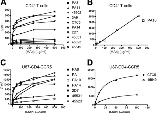
Different antigenic forms of CCR5 are present on primary CD4ⴙ T cells. We stained purified, activated CD4⫹ T cells from a single individual (donor A) with 10 different CCR5 MAbs for 1 h at 4°C. A FITC-conjugated secondary antibody was then added at a concentration (150g/ml) that had pre-viously been determined to be saturating for all the isotypes represented by the MAbs in the test panel. Each CCR5 MAb was titrated to determine maximal binding levels and half-maximal binding (EC50) concentrations (Fig. 1 and Table 1). GMFI values were corrected for nonspecific MAb binding by subtracting signals derived using CD4⫹T cells from a
CCR5-⌬32 homozygous individual (see Fig. S1 in the supplemental material).
With two exceptions (45549 and PA10), the MAb titration curves tended to plateau, which is indicative of saturation bind-ing. MAb 45549 did not bind detectably to cells from this donor (Fig. 1A). The PA10 curve trended continuously upward without reaching a plateau, and its slope was lower than for the other MAbs (Fig. 1B). The maximal corrected GMFI values derived from the flow cytometry histograms (see Fig. S1 in the supplemental material) ranged from 0 to 2,600 (Fig. 1 and Table 1). The GMFI rank order was PA10⬎PA14⬎45531⬎ PA11 ⬎ 2D7 ⬎ CTC5 ⬎ 45523 ⬎ PA8 ⫽ 45502 ⬎ 45549 (45549 did not bind detectably). We note that 2D7, thede facto
on November 7, 2019 by guest
http://jvi.asm.org/
standard reagent for CCR5 detection and quantification, had only the 5th highest GMFI value. The rank order varied in repeat experiments using cells from three more donors (B, C, and D), but PA10 always ranked highest, and PA8, 45502, and 45549 were consistently the least reactive (see Table S1 in the supplemental material).
The EC50s (which approximate dissociation constants [25])
ranged from 1.5 to 31g/ml (10 to 207 nM) (Table 1). Exclud-ing the nonreactive MAb 45549, the affinity (reciprocal EC50) rank order for the CCR5 forms each MAb recognized was 2D7⬎CTC5⬎45531⬎ PA14⬇PA11⬎PA8⬎ 45523⬎ 45502⬎PA10. The rank orders based on maximal binding and affinity values were not concordant (Spearman correlation co-efficient [r] ⫽ ⫺0.07; P ⫽ 0.8). For example, 2D7 has the TABLE 1. Affinities and maximal binding of CCR5 MAbs to CD4⫹T cells and U87-CD4-CCR5 cells
MAb Isotype Epitopea
CD4⫹T cellsb
U87-CD4-CCR5 cellsb
EC50
(g/ml)
Maximal GMFI
EC50
(g/ml)
Maximal GMFI
PA8 IgG1 NT 22⫾9.9 44⫾4.0 12⫾6.7 400⫾37
PA11 IgG1 NT 9.8⫾6.9 410⫾47 34⫾11 1,500⫾177
45502 IgG2B NT 85⫾19 36⫾6.8 NA 0c
CTC5 IgG1 NT 3.6⫾2.6 210⫾20 19⫾2.7 2,000⫾80
PA10 IgG1 NT-ECL2d ⬎200 ⬎2,600 215⫾138 380⫾106
PA14 IgG1 NT-ECL2 9.1⫾4.1 500⫾36 10⫾3.6 1,300⫾78
2D7 IgG2A ECL2 1.5⫾0.76 340⫾22 1.2⫾0.26 980⫾26
45531 IgG2B ECL2 6.3⫾1.0 490⫾12 16⫾6.2 1,400⫾98
45523 IgG2B MD 31⫾6.8 75⫾3.6 139⫾47 1,600⫾209
45549 IgG2B MD NA 0c 40⫾8.6 800⫾67
aAs defined in references 29 and 40. MD, multidomain.
bEC
50and GMFI values (⫾standard errors of the mean关SEM兴) are based on the fitted curves generated from the data points shown in Fig. 1. EC50is the MAb
concentration giving half-maximal binding. Maximal GMFI is the fitted plateau value. Therefore, it might not always be the same as the highest value reached in Fig.
1. The data were derived from one experiment of two performed for each cell type. The purified CD4⫹T cells expressed high levels of CD4 (GMFI⫽5,000). NA,
not applicable.
cNo binding detectable.
dThe PA10 epitope was originally reported to involve both the NT and ECL2 (40), but the MAb was later shown to bind to a tyrosine-sulfated NT peptide (12).
[image:3.585.45.542.81.216.2]Whether ECL2 residues are also involved is not clear.
FIG. 1. Binding of CCR5 MAbs to CD4⫹ T cells and U87-CD4-CCR5 cells. Cells were incubated with anti-CCR5 MAbs and a FITC-conjugated secondary Ab. Shown is binding of CCR5 MAbs to CD4⫹T cells from a single donor (A and B) and U87-CD4-CCR5 cells (C and D) (one representative experiment). Flow cytometry data are expressed as background-corrected GMFI values. Corrected GMFI values were obtained after subtraction of GMFI values derived using CD4⫹T cells from a CCR5-⌬32 homozygous (CCR5⫺) donor (A and B) or from CCR5-negative U87-CD4 cells (C and D). (B) PA10 binding to CD4⫹T cells is shown on a different GMFI scale. (D) GMFI values for CTC5 and 45549 staining of U87-CD4-CCR5 cells are not plotted at⬎100g/ml because of high background binding. For visual clarity, data points were connected by straight lines as opposed to the fitted curves; however, the related data in Table 1 were based on the plateau values obtained by the nonlinear-regression curve fitting. TheR2values were⬎0.81 for all reactive MAbs except PA10 (Fig. 1B), for which saturation binding was never reached.
Hence, theR2value, an indicator of how well the function fits the data, could not be determined for this MAb.
on November 7, 2019 by guest
http://jvi.asm.org/
[image:3.585.134.448.414.640.2]highest affinity but an intermediate maximum binding level, while PA10 has the highest maximum binding level, but its affinity is among the lowest. The lack of correlation shows that the maximum GMFI values do not simply reflect the strength of binding, which was a possibility, since flow cytometry is not an equilibrium binding system and weakly bound MAbs can dissociate during washing. Hence, the striking differences in the extents to which different MAbs bind most likely indicate that antigenically distinct subpopulations of CCR5 are present on CD4⫹ T cells. In other words, some MAbs (e.g., PA10, PA14, and 45531) recognize a much greater proportion of the total CCR5 molecules than others (e.g., PA8, 45502, and 45549). The unusual binding curve for PA10 could be ex-plained by its binding heterogeneously to several different forms of CCR5 at a wide range of affinities. However, it is also possible that PA10, when present at high concentrations, cross-reacts with a similar epitope on molecules other than CCR5 on the surfaces of CD4⫹T cells that are absent from U87-CD4-CCR5 cells.
Different antigenic forms of CCR5 are also present on U87-CD4-CCR5 cells.To see whether CCR5 also exists in antigeni-cally distinct conformations on a cell line, U87-CD4-CCR5 cells were stained at 4°C with the same 10 MAbs (Fig. 1 and Table 1). Background GMFI values obtained using U87-CD4 cells were subtracted to derive titration curves (Fig. 1C and D). MAb 45502 did not stain U87-CD4-CCR5 cells at levels above background (see Fig. S2 in the supplemental material).
Among the reactive MAbs, the maximal GMFI rank order was CTC5 ⬎ 45523 ⬎ PA11 ⬎ 45531 ⬎ PA14 ⬎ 2D7 ⬎
45549⬎PA8⬎PA10, and for affinities it was 2D7⬎PA14⬎ PA8⬎45531⬎CTC5⬎PA11⬎45549⬎45523⬎PA10. As with CD4⫹T cells, the plateau binding levels and the affinities were markedly discordant (r ⫽0.0;P⫽1.0). Hence, the pla-teau value again reflects the number of CCR5 molecules rec-ognized by each MAb, not just the strength of binding.
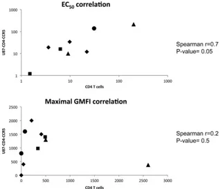
The binding studies show that various antigenic forms of CCR5 are present in different amounts on the two cell types. For example, MAbs 45549 and 45523 either did not bind de-tectably to CD4⫹T cells or bound to only a low level, but their extrapolated GMFI plateaus on the U87-CD4-CCR5 line (GMFI ⫽ 800 ⫾ 67 and 1,600 ⫾ 209) indicate they bind significantly to these cells. Conversely, PA10 had the lowest GMFI of the MAbs binding detectably to U87-CD4-CCR5 cells, but it was the most reactive with CD4⫹T cells. Of the eight MAbs reactive with both cell types, all except PA10 bound to a higher level to U87-CD4-CCR5 cells than to CD4⫹ T cells, but their maximal GMFI values did not correlate be-tween the two cell types (r ⫽ ⫺0.3;P⫽ 0.4). The maximal GMFI values for the whole set of 10 MAbs also did not cor-relate between the two cell types (r⫽0.2;P⫽0.5). In contrast, there was a strong correlation between the EC50s, and hence the affinity estimates (r⫽0.7;P⫽0.05) (Fig. 2).
Some CCR5 MAbs can inhibit VVC-sensitive and/or -resis-tant viruses.To assess whether VVC-sensitive and -resistant viruses have preferences among the different CCR5 antigenic variants revealed by the MAb staining patterns, we tested the infection-inhibitory activities of the various MAbs in a multi-cycle replication assay using infectious clonal viruses. The tar-FIG. 2. Correlations between CCR5 MAb affinity and maximal binding measured using CD4⫹T cells and U87-CD4-CCR5 cells. The affinity of each MAb for CCR5, expressed as the EC50, and its reactivity with CD4⫹T cells, expressed as the maximal GMFI value, were plotted as a
function of the same parameters derived from U87-CD4-CCR5 cells. The epitope clusters are as follows: NT, diamonds; NT-ECL2, triangles; ECL2, squares; multidomain (MD), circles.
on November 7, 2019 by guest
http://jvi.asm.org/
[image:4.585.132.454.69.343.2]get cells were incubated with the MAbs for 1 h at 37°C prior to virus addition, and MPIs and/or 50% inhibitory concentrations (IC50s) were quantified whenever this could be done with
suf-ficient precision. Only three MAbs, PA11, PA14, and 2D7, were broadly inhibitory in that they were active against VVC-sensitive and -resistant viruses in one or both cell types, albeit not universally (Table 2). Two MAbs, CTC5 and 45531, selec-tively inhibited infection by only one of the resistant viruses in a cell-specific manner, as further discussed below (Table 2 and Fig. 2 and 3). The remaining MAbs had no significant inhibi-tory effect against any of the viruses.
MAbs 2D7 and PA14 were potent inhibitors of the two VVC-resistant infectious clones in both CD4⫹ T cells and U87-CD4-CCR5 cells (IC50s, 0.062 to 3.4 g/ml). However, they were much less active, or completely inactive (IC50s
be-tween⬃20- and⬎600-fold higher), against the parental clones, particularly in U87-CD4-CCR5 cells (Table 2). Of note, a similar inhibition pattern was seen when Env-pseudotyped vi-ruses were used to infect U87-CD4-CCR5 cells in the presence of 2D7 and PA14, although the MAbs were more potent under these conditions and gave complete inhibition (see Table S2 in the supplemental material). MAbs 2D7 and PA14 recognize an ECL2 and a composite NT-ECL2 epitope, respectively (Table 1). As they both target regions of CCR5 important for gp120 binding, and they have high affinities, their inhibition of HIV-1 infection is understandable. The fact that the two VVC-resis-tant viruses were clearly more sensitive than the parental vi-ruses to PA14 and 2D7 suggests that they interact with CCR5 in a quantitatively or qualitatively different manner.
[image:5.585.42.541.81.221.2]Of the four MAbs to NT epitopes, only PA11 had antiviral activity against the four test viruses (Table 2). PA11 inhibited infection of U87-CD4-CCR5 cells by Res-3FP completely (MPI ⫽ 100%) and Par-3FP substantially (MPI ⫽ 72% ⫾ 1.0%), with similar IC50s. However, PA11 was only moderately active against Par-4V3 and Res-4V3 infection (MPI⫽42%⫾ 18% and 52%⫾ 20%, respectively), and only at the highest concentration (Table 2). A different pattern was seen with CD4⫹T cells, where PA11 inhibited Res-4V3 infection with an IC50⬃20-fold lower than that of Par-4V3 (Table 2). The other
TABLE 2. Inhibition of HIV-1 replication in CD4⫹T cells and U87-CD4-CCR5 cells by CCR5 MAbs
Cell type Clone
Value for epitope (MAb)a
NT (PA11) NT-ECL2 (PA14) ECL2 (2D7)
IC50⫾SEM
(g/ml) MPI (%)
IC50⫾SEM
(g/ml) MPI (%)
IC50⫾SEM
(g/ml) MPI (%)
CD4⫹T cells Par-4V3 14⫾6.0 91⫾9.0 30⫾1.8 100 4.6⫾2.0 100
Res-4V3 0.80⫾0.20 95⫾5.3 0.29⫾0.22 100 0.71⫾0.37 100
Par-3FP ⬎200 45⫾15 3.1⫾0.42 100 2.0⫾1.3 100
Res-3FP 4.6⫾3.8 85⫾15 0.34⫾0.17 100 0.27⫾0.15 100
U87-CD4-CCR5 Par-4V3 ⬎200 42⫾18 46⫾7.8 100 ⬎80 11⫾3
Res-4V3 200 52⫾20 0.32⫾0.00073 100 3.4⫾1.0 100
Par-3FP 2.1⫾0.72 72⫾1.0 5.1⫾1.5 100 ⬎80 3.2⫾2.6
Res-3FP 8.8⫾6.8 100 0.062⫾0.014 100 0.13⫾0.053 100
aMPI values (means⫾SEM) represent the percent inhibition of HIV-1 replication at the highest MAb concentration that could be used without any toxic effects
(200g/ml for PA11 and PA14; 80g/ml for 2D7). The sodium azide preservative present in stock solutions of 2D7, but not the other test MAbs, had toxic effects
at the highest concentrations tested (⬎80g/ml), as assessed by trypan blue exclusion. Hence, inhibition by 2D7 was assessed in a lower concentration range than the
others to avoid confounding influences. IC50is the MAb concentration that reduced p24 production by 50% compared to the control (no MAb). This reduction is not
always half the MPI value, so the IC50, defined in this way, can only be determined when the MPI is⬎50%. For U87-CD4-CCR5 cells, the values are from 3 independent
[image:5.585.316.522.317.590.2]experiments, for CD4⫹T cells they are from 3 experiments using different donors each time.
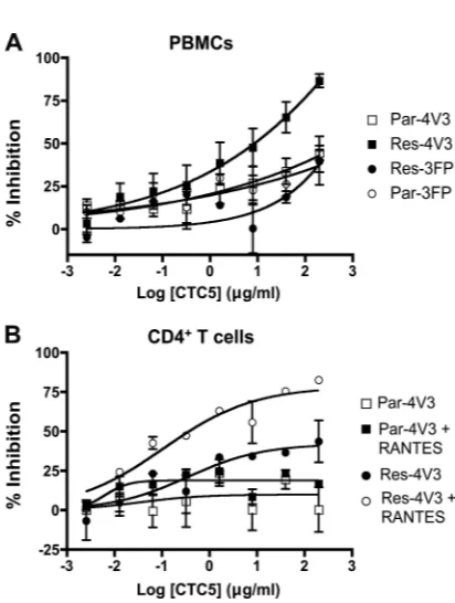
FIG. 3. Effects of MAb CTC5 on HIV-1 replication in primary cells or U87-CD4-CCR5 cells. (A) PBMCs were incubated with increasing concentrations of CTC5 before infection with chimeric molecular clones containingenvgenes derived from Par-4V3, Res-4V3, Par-3FP, or Res-3FP. (B) CD4⫹T cells were incubated with increasing concen-trations of CTC5 in the presence or absence of RANTES (8 nM) before infection with chimeric molecular clones containingenvgenes derived from Par-4V3 or Res-4V3, as indicated. The RANTES con-centration used approximates the IC50for inhibition of both viruses.
The data points in panels A and B represent the percent inhibition of HIV-1 replication (measured by p24 ELISA after 7 days) relative to that in the absence of MAb (⫽100%) and with or without RANTES, as indicated. The data represent the means⫾standard errors of the mean (SEM) of values derived from three independent experiments using three different PBMC and CD4⫹T cell donors.
on November 7, 2019 by guest
http://jvi.asm.org/
pair of viruses was also differentially sensitive to PA11 in CD4⫹ T cells, in that Res-3FP was 40-fold more potently inhibited than Par-3FP, and to a greater extent (Table 2). The most striking observation made with PA11 involved Res-4V3, which was highly sensitive in CD4⫹T cells but only minimally inhib-ited in U87-CD4-CCR5 cells (Table 2).
When Env-pseudotyped viruses were used instead of fully infectious viruses to infect U87-CD4-CCR5 cells in the pres-ence of CCR5 MAbs, the data pattern was slightly different. Thus, although Res-4V3 and Par-4V3 behaved comparably in the two assays, PA11 inhibited Res-3FP to a more substantial extent (MPI⫽72%⫾1.8%) than Par-3FP (MPIⱕ35%) in the Env pseudotype assay (compare Table S2 in the supple-mental material to Table 2).
As noted above, PA14 and 2D7 were consistently more potent inhibitors of the VVC-resistant viruses in both cell types. In contrast, the infection inhibition patterns seen with PA11 differed between U87-CD4-CCR5 cells and CD4⫹ T cells, which is indicative of a cell-type-dependent influence on how viruses use the CCR5 antigenic forms recognized by this MAb. We have found that PA11 preferentially binds to a Tyr-sulfated version of a CCR5-NT peptide (see Fig. S3 in the supplemental material) (12). It is therefore possible that its cell surface reactivity is dependent on the extent to which different CCR5 variants are Tyr sulfated in the two cell types. Unlike the MAb binding experiments described above, the infection inhi-bition assays were performed at 37°C instead of 4°C. To con-trol for potential differences in the binding properties of the various MAbs at the two temperatures, we compared the cell surface staining at a saturating concentration of every test MAb to the cell surface by FACS at 37°C and 4°C. In all cases, the staining levels were similar at the two temperatures, except with PA10 on U87-CD4-CCR5 and CD4⫹T cells and PA11 on U87-CD4-CCR5 cells (but not CD4⫹T cells). In those cases, staining was substantially lower at 37°C than at 4°C (data not shown). PA11 may, therefore, induce CCR5 internalization in a cell-type-dependent manner, which might be an influence on how PA11 inhibits HIV-1 infection in different cell types.
Res-4V3 has an altered interaction with the CCR5 NT.
PA11, which is directed to an epitope in the CCR5 NT, blocked at least a proportion (⬎40%) of all four viruses on both cell types. CTC5, however, also binds to an NT epitope, yet it does not normally inhibit HIV-1 infection (28, 40). These two MAbs might bind to nonoverlapping epitopes within the same CCR5 protein or to nonoverlapping antigenic forms of CCR5, as further discussed below (see Fig. S3 and S4B in the supplemental material). In a PBMC infection assay, we found that CTC5 inhibited infection by Res-4V3 (MPI⫽80%) more strongly than the other three test viruses (MPI⬇40%) (Fig. 3A). This outcome suggests that Res-4V3 is more sensitive than the other viruses to occlusion of its binding site by CTC5; an implication is that Res-4V3 has acquired a new contact site in the distal part of the CCR5 NT. Previously, we showed that entry of Res-4V3 was sensitive to single amino acid substitu-tions in the CCR5 NT and concluded that the resistance-associated V3 sequence changes had rendered the virus more reliant on the NT than the other test viruses (7). This enhanced interaction between Res-4V3 gp120 and the CCR5 NT be-comes particularly relevant when the gp120-ECL2 interaction is weakened by the presence of CCR5 inhibitors.
In CD4⫹T cells, CTC5 partially inhibited Res-4V3 (MPI⫽ 45%), but it had a more substantial effect when the cells were pretreated with RANTES (MPI⫽80%) (Fig. 3B). In contrast, CTC5 did not inhibit any of the other viruses whether RANTES was present or not (Fig. 3B and data not shown). Of note, purified CD4⫹T cells express CCR5 at higher levels than the PBMCs from which they were derived, possibly because CC-chemokine levels are substantially (at least 10-fold) lower than in PBMC cultures (data not shown). We suggest that RANTES potentiates the inhibitory effect of CTC5 indirectly by reducing the number of CCR5 molecules available for HIV-1 entry and MAb binding. If so, this factor would allow the antiviral activity of a weak inhibitor, such as CTC5, to become more apparent, particularly with Res-4V3. In contrast to CD4⫹T cells, CTC5 did not inhibit Res-4V3 infection of U87-CD4-CCR5 cells (data not shown). The higher overall CCR5 level and the absence of CC-chemokines could, alone or together, explain why CTC5 is inactive against Res-4V3 infection of U87-CD4-CCR5 cells while being active in PBMCs or when CD4⫹T cells are supplemented with RANTES.
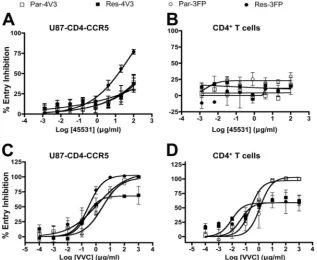
Different sensitivities of Res-3FP to MAb 45531 and VVC in the two cell types.MAb 45531 shares many features with the more broadly inhibitory MAbs 2D7 and PA14, including high levels of surface binding by FACS, a high affinity for CCR5, and an epitope involving ECL2. The 2D7 and PA14 epitopes include residues located in the N-terminal segment of ECL2 (residues K171/E172 and R168/Y176, respectively), while that for 45531 involve residues in the C-terminal region (residues Y184/F189) (40). Unlike 2D7 and PA14, however, 45531 has a limited ability to inhibit HIV-1 infection that could only be detected in an Env pseudotype virus infection assay using U87-CD4-CCR5 cells. In that system, Res-3FP was the virus most sensitive to inhibition by 45531, with an MPI of 80% versus
⬃40% for the other three (Fig. 4A; see Table S2 in the sup-plemental material). Of note, 45531 did not inhibit Res-3FP infection of PBMCs or CD4⫹ T cells (Fig. 4B and data not shown). Cumulatively, these results again indicate that a VVC-resistant virus can have an altered interaction with CCR5 and that the target cell type influences the nature of this interac-tion.
It seems unlikely that the lack of effect of 45531 on Res-3FP infection of primary cells is attributable to cell-type-dependent affinity differences; if anything, 45531 has a lower affinity for CCR5 in U87-CD4-CCR5 cells (Table 1). Alternative expla-nations for the cell type dependency are that Res-3FP uses different CCR5 variant forms on the two cell types or that the forms of CCR5 it can use are available in different amounts. Res-3FP emerged under the selection pressure of VVC in a PBMC culture (33). Hence, whatever the mechanism by which FP changes confer resistance, it seems likely they must render Res-3FP better able to use the VVC-CCR5 complex in a mem-brane environment that is present on primary CD4⫹T cells. We therefore investigated whether there are differences in how Res-3FP uses the VVC-CCR5 complex in the two cell systems. Of note, Res-FP was resistant to VVC in CD4⫹T cells (MPI⫽ 42%) but was completely inhibited in U87-CD4-CCR5 cells (Fig. 4C and D). For comparison, both parental Env-pseu-dotyped viruses were completely inhibited by VVC in both cell types, while Res-4V3 was consistently VVC resistant (Fig. 4C and D). Thus, Res-3FP can clearly use the inhibitor-bound
on November 7, 2019 by guest
http://jvi.asm.org/
form of CCR5 efficiently to enter CD4⫹T cells but not U87-CD4-CCR5 cells, which is consistent with our earlier conclu-sion (2, 3).
VVC binding induces conformational changes in CCR5 that create alternative binding sites for resistant, but not parental, viruses and for some, but not all, MAbs. For example, we found that when U87-CD4-CCR5 or CD4⫹T cells were incu-bated with a saturating VVC concentration for 30 min at 37°C, the subsequent binding of 45531 was decreased by⬎60% com-pared to when VVC was absent. In contrast, 2D7 or PA14 binding was reduced by⬍30% under the same conditions (see Fig. S4A in the supplemental material). These results indicate that PA14 and 2D7, but not 45531, recognize a VVC-stabilized conformation of CCR5. Thus, not only can the exposure of certain epitopes differ among CCR5 conformational variants, their recognition by MAbs and viruses also varies. To further address these points, we characterized the CCR5 variants that are seen by MAbs 2D7, PA14, and 45531 by determining whether they stain distinct or overlapping CCR5 conforma-tional subsets and assessing where these subsets are localized on the cell surface.
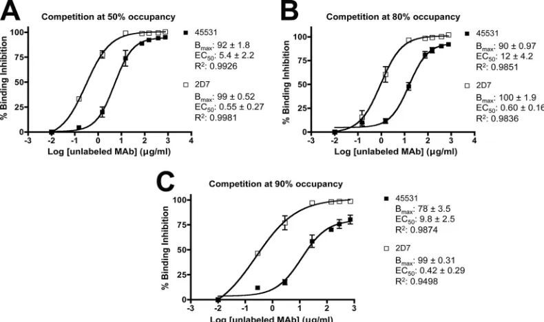
MAbs 2D7 and 45531 recognize overlapping CCR5 sub-populations.We used a competition binding assay to evaluate the extent of overlap in the binding of FITC-labeled 2D7 and unlabeled 45531 or, for comparison, unlabeled 2D7 to U87-CD4-CCR5 cells. These two MAbs were chosen because, while both have ECL2 epitopes that may be overlapping, they differ in their abilities to inhibit infection of the cells. We first incu-bated the cells with increasing concentrations of FITC-labeled
2D7 for 1 h at 4°C. The resulting EC50of 2.0⫾0.21g/ml was
similar to that for unconjugated 2D7, as detected using a la-beled secondary antibody (data not shown and Table 1). Since the EC50approximates theKd, we estimated that when labeled 2D7 was used at concentrations equivalent to 1⫻, 5⫻, and 10⫻ the EC50, it occupies 50%, 80%, and 90% of the available CCR5 molecules, respectively.
To assess and quantify how the two MAbs competed for binding to CCR5, we incubated U87-CD4-CCR5 cells for 1 h at 4°C with a range of concentrations of unlabeled 2D7 or 45531 prior to the addition of FITC-2D7. Both unlabeled MAbs inhibited FITC-2D7 binding, but with subtly different characteristics (Fig. 5). The EC50s for inhibition of FITC-2D7
binding by unlabeled 2D7 and unlabeled 45531 varied only minimally with the amount of labeled 2D7 used, ranging from 0.42 to 0.60g/ml and 5.4 to 12g/ml, respectively (Fig. 5). The 10- to 20-fold-lower value for 2D7 is consistent with its higher affinity for CCR5 (Table 1). As expected, the binding of FITC-2D7, at all three concentrations tested (CCR5 occu-pancy levels of 50%, 80%, or 90%), was completely inhibited (Bmax ⫽ 99%) by the prior addition of sufficient unlabeled
[image:7.585.133.450.70.330.2]2D7. In contrast, 45531 inhibited the binding of labeled 2D7 only incompletely, as indicated by the appearance of a plateau in the binding curve below 100% (Fig. 5). The resultingBmax values were estimated to be 92%, 90%, and 78% at 50%, 80%, and 90% occupancy, respectively, of CCR5 by FITC-2D7. Hence, depending on the amount of FITC-2D7 used, between 8 and 22% of the CCR5 molecules that bind 2D7 do so in the presence of a saturating concentration of 45531. Thus, there FIG. 4. Effects of MAb 45531 and VVC on HIV-1 entry in primary cells or U87-CD4-CCR5 cells. U87-CD4-CCR5 cells or CD4⫹T cells were incubated with a range of concentrations of 45531 (A and B) or VVC (C and D) before infection with the Env-pseudotyped virus Par-4V3, Res-4V3, Par-3FP, or Res-3FP. The data points represent percent HIV-1 entry (measured by luciferase expression after 3 days) relative to that in the absence of MAb or VVC, as appropriate (⫽100%). The data represent the means⫾SEM of values derived from three independent experiments.
on November 7, 2019 by guest
http://jvi.asm.org/
may be a small residual CCR5 subpopulation that binds 2D7 but is not recognized by 45531.
MAb 45531 stains distinct clusters on the cell surface, and its binding is dependent on cholesterol levels.To further assess the presence and properties of different CCR5 antigenic forms, we used fluorescence deconvolution microscopy to study the surface distribution of CCR5 molecules on live U87-CD4-CCR5 and HOS-U87-CD4-CCR5-GFP cells. Of note, the native and GFP fusion versions of CCR5 are overexpressed in the stably transfected U87-CD4-CCR5 and HOS-CCR5-GFP cells, re-spectively. The use of the GFP fusion protein allows the total CCR5 population in HOS-CCR5-GFP cells to be readily de-tected via the tag, which is directly fused to the CCR5 C terminus. The two test MAbs, PA14 and 45531, recognize different epitopes that include ECL2 residues and have com-parable affinities for CCR5 (Table 1). Saturating concentra-tions of PA14 and 45531 were added to the target cells at 4°C to prevent CCR5 internalization and recycling. The distribu-tion patterns for PA14- and 45531-stained CCR5 were clearly different. PA14 staining was homogeneously distributed over most of the U87-CD4-CCR5 cell surface and was clearly visible on protruding membrane structures that are likely to be mi-crovilli or membrane ruffles (Fig. 6A) (50). In similar exper-iments, the 2D7 staining pattern was similar to that for PA14, indicating that the two MAbs might bind to com-pletely overlapping CCR5 subsets (data not shown). In con-trast, 45531-stained CCR5 molecules were located in dis-tinct clusters on the cell surface and were absent from the
membrane protrusions (Fig. 6C). Under the conditions used, PA14 and 45531 stained only the cell surface; z sec-tions from the middle of the cells showed no internal stain-ing by either MAb (Fig. 6B and D).
A similar distribution was observed using HOS-CCR5-GFP cells. The PA14-stained CCR5 molecules were homogeneously distributed and completely overlapped with the CCR5-GFP staining pattern, both on the cell surface in general and on the membrane protrusions (Fig. 6E to H). In contrast, the 45531 label overlapped with the CCR5-GFP signal only in distinct clusters on the cell surface and was absent from the membrane protrusions (Fig. 6I to L). The CCR5-GFP tag was also de-tected in the perinuclear regions of HOS-CCR5-GFP cells as a result of CCR5 internalization and recycling (Figure 6F and J), but PA14 and 45531 were not detected at these sites because the antibody-staining procedure was carried out at a temper-ature nonpermissive for endocytosis (Fig. 6H and L).
[image:8.585.93.488.71.305.2]When U87-CD4-CCR5 cells were costained first with 45531 and then with PA14, the results were consistent with the pat-tern described above. Thus, the PA14 stain was again homo-geneously distributed over most of the cell surface, while the 45531 label tended to cluster in distinct areas (Fig. 6M to O). There was very little overlap in the distributions of the two MAbs within the predominantly 45531-stained clusters. How-ever, z sections from the middle of the cells did reveal some areas of overlap between PA14 and 45531 outside the 45531-stained clusters (Fig. 6P to R). Of note, the MAb concentra-tions of 4 g/ml used for microscopy (Fig. 6) were 125- to FIG. 5. Binding competition between CCR5 MAbs. In the competition binding assay, U87-CD4-CCR5 cells were incubated for 1 h at 4°C with a range of concentrations of unlabeled MAb 2D7 or 45531 before addition of FITC-conjugated 2D7 for a further 1 h, without washing, at concentrations corresponding to the EC50(A), 5 times the EC50(B), and 10 times the EC50(C) (i.e., 2, 10, or 20g/ml, calculated to yield 50%,
80%, or 90% occupancy, respectively). The cells were then washed before flow cytometry analysis. The percent inhibition of FITC-2D7 binding was calculated using the following formula: inhibition (%)⫽100⫺[(GMFIx⫺GMFIbackground)/(GMFI0⫺GMFIbackground)]⫻100, where GMFIx
is the binding of FITC-2D7 in the presence of the various concentrations of unlabeled MAb (atxg/ml) and GMFI0is the binding of FITC-labeled
2D7 in the presence of an unlabeled isotype control MAb. Background binding was measured as GMFIbackground, as determined using FITC-2D7
and CCR5-negative U87-CD4-CCR5 cells. TheBmax, EC50, andR
2values were derived from the resulting sigmoidal curves. The data represent
the averages of 3 independent experiments.
on November 7, 2019 by guest
http://jvi.asm.org/
FIG. 6. Visualizing MAb binding to CCR5 by fluorescence microscopy. Live U87-CD4-CCR5 cells were incubated for 1 h at 4°C with MAb PA14 (A and B) or MAb 45531 (C and D) at 4g/ml and then with Rhodamine Red X-labeled anti-mouse IgG1 (A and B) or IgG2b (C and D). The images are shown as volume projections (A and C) or as one middle plane from the same z series. Live HOS-CCR5-GFP cells were incubated with PA14 (E to H) or 45531 (I to L) under conditions similar to those described above. Staining with PA14 and 45531 is displayed in red (E and I) and CCR5-GFP in green (F and J). In panels G and K, the yellow stain indicates areas where the MAbs and CCR5-GFP overlap. Nuclear staining (Hoechst) is in blue. The images in panels E to G and I to K are volume projections, and the images in panels H and L are middle-plane sections of the same z series. (M to R) Live U87-CD4-CCR5 cells were costained with 45531 and PA14. MAb 45531 staining is displayed in green (M and P) and PA14 in red (N and Q), and double staining was overlaid (O and R). Panels M to O represent volume projections, and P to R are middle-plane sections of the same z series. The scale bars correspond to 15m.
8235
on November 7, 2019 by guest
200-fold lower than those shown to give maximal staining in the flow cytometry studies (Fig. 1) or used to compete out 2D7 binding (Fig. 5). Hence the intensities of the signals cannot be directly compared between the two experimental systems; the microscopy data are more qualitative than quantitative.
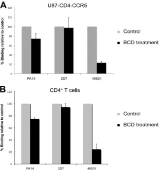
The clustered pattern of 45531 staining suggests that CCR5 molecules stained by the MAb might be localized in high-cholesterol membrane domains or “lipid rafts.” To test this hypothesis, we measured the surface binding of 45531 to cells treated with a BCD derivative. BCDs are cholesterol-depleting agents that have been shown to disrupt lipid rafts in primary cells and cell lines (36). The binding of 45531 to the surfaces of BCD-treated U87-CD4-CCR5 cells or CD4⫹T cells was sub-stantially decreased (by 77% [P⫽0.0002] and by 76% [P ⫽ 0.0132], respectively) compared to control cells. In contrast, PA14 binding was only modestly reduced by BCD treatment (by 26% [P⫽0.07] and by 25% [P⫽0.003], respectively) and 2D7 binding not at all, i.e., by⬍6% (Fig. 7A and B).
Overall, the staining experiments clearly demonstrate the existence of different subpopulations of CCR5 on the cell sur-face that can be present in distinct locations and that poten-tially involve different membrane microdomains or other dis-crete substructures. That PA14, but not 45531, stains a CCR5
population(s) present in microvilli is of particular note given that these protruding membrane structures are believed to be sites of CD4 and CCR5 coclustering where HIV-1 entry occurs (16, 48, 50, 51). In addition, the clustering of 45531 staining and its decreased binding after cholesterol depletion suggest that this MAb recognizes a form of CCR5 that is located in cholesterol-rich membrane domains or lipid rafts.
DISCUSSION
[image:10.585.135.454.68.403.2]We investigated how parental and VVC-resistant HIV-1 variants enter primary CD4⫹T cells and U87-CD4-CCR5 cells via naturally expressed and stably transfected CCR5, respec-tively. We used activated CD4⫹ T cells because, unlike un-stimulated cells, they are highly permissive to HIV-1 replica-tion. We first used a panel of 10 MAbs to different CCR5 epitopes and four viruses that recognize CCR5 in different ways and then conducted additional analytical studies with a subset of the more diagnostically useful MAbs. Our principal conclusion is that CCR5 is present on the cell surface in mul-tiple antigenic forms that VVC-sensitive and -resistant clones use for entry with a range of efficiencies. The compositions of these CCR5 subpopulations differ between primary CD4⫹T FIG. 7. Effects of cholesterol depletion on the cell surface binding of CCR5 MAbs. U87-CD4-CCR5 (A) or CD4⫹T cells (B) were incubated with or without 10 mM BCD for 30 min at 37°C in serum-free medium, washed, and then incubated with the indicated CCR5 MAbs for 1 h at 4°C. CCR5 binding was measured by flow cytometry after addition of a FITC-labeled secondary Ab and expressed as GMFI. The binding of the various MAbs to the BCD-treated cells was expressed as a percentage of binding to control cells (no BCD). The data represent the mean values from three independent experiments, with error bars representing the SEM.
on November 7, 2019 by guest
http://jvi.asm.org/
cells and cell lines (U87-CD4-CCR5 and HOS-CCR5-GFP) and, presumably, also vary on other cell types. We discuss here what these subpopulations might be, where they may be lo-cated, and how they may be used differently by VVC-sensitive and -resistant viruses.
In the flow cytometry studies, the plateau level of binding did not reflect the affinity of a MAb for CCR5. For example, PA10 has a low affinity for CD4⫹T cells but a high maximal binding level; conversely CTC5 binds with high affinity but to only a low level. In general, the CCR5 affinities measured on CD4⫹T cells and U87-CD4-CCR5 cells correlated well, but the maximal binding levels did not. These findings are com-patible with a model in which some MAbs preferentially detect distinct CCR5 subpopulations that have the same antigenic determinants on the two cell types but different absolute and relative abundances. Genetic and related host factors seem, however, to have a limited influence on the prevalences of the various antigenic forms of CCR5, as only moderate differences in MAb staining levels were found among four CD4⫹T cell donors (see Table S1 in the supplemental material). MAb 2D7 is commonly used for CCR5 detection and quantification, but it had only intermediate reactivity with CCR5 on both cell types. Using 2D7 might underestimate the total CCR5 cell surface expression level under some conditions. For example, some apparently CCR5-negative cells, including primary lym-phocytes, do express sufficient CCR5 to be targets for HIV-1 infection (11, 13, 41, 43, 44).
PA11, PA14, and 2D7 were active against more than one VVC-sensitive and -resistant virus in one or both cell types, while 45531 and CTC5 were more selectively active, each in-hibiting only one VVC-resistant virus strongly in a cell-type-specific manner. Overall, although affinity is likely to be im-portant for the antiviral activity of a CCR5 MAb, it was not the only determinant, and the extent of cell surface binding did not predict whether a MAb was inhibitory. For example, CTC5 and 45531 have higher or comparable affinities for epitopes con-tiguous to those for PA11 and 2D7, respectively, but they clearly have a narrower ability to inhibit infection. Both the VVC-resistant clones were more sensitive than the parental viruses to PA11, PA14, and 2D7 in CD4⫹T cells and to PA14 and 2D7 in U87-CD4-CCR5 cells. Overall, the emergence of mutant viruses that are panresistant to small-molecule CCR5 inhibitors but hypersensitive to CCR5 MAbs against various epitopes is compatible with the escape phenotype involving a reduction in the efficiency of the Env-CCR5 interaction. To compensate, there may be a requirement for a greater number of MAb-unencumbered CCR5 molecules for triggering the fusion reaction. This scenario would be consistent with the lower MAb inhibitory concentrations, and therefore lower minimal MAb-CCR5 occupancies, that we have observed with VVC-resistant viruses compared to their parents (Table 2; see Table S2 in the supplemental material). Additional studies will be required to test this hypothesis.
As noted, some MAbs inhibited infection in a cell-type-dependent manner. For example, Res-4V3 was highly sensitive to PA11 in CD4⫹T cells but only minimally inhibited in U87-CD4-CCR5 cells. As sulfated tyrosines at residues 10 and 14 are critical both for the binding of some NT MAbs (e.g., PA10 and PA11, but not PA8) and for HIV-1 entry, it is possible that cell-type-dependent variation in the expression of partially and
fully sulfated forms of CCR5 might influence PA10 or PA11 staining (12, 15). If, for example, CCR5 overexpression in U87-CD4-CCR5 cells saturates the cellular sulfotransferases, there may be proportionately more partially sulfated or non-sulfated CCR5 variants on the surfaces of these cells than on CD4⫹T cells (15). Another consideration is that, according to our preliminary observations, PA11 can induce CCR5 inter-nalization into U87-CD4-CCR5 cells, but not CD4⫹ T cells (whereas PA10 downmodulates CCR5 in both), thereby reduc-ing the number of coreceptors available for HIV-1 entry.
Res-4V3 was previously shown to be more dependent than its parent on specific residues in the CCR5 NT (7). We now show that it is the virus most sensitive to CTC5, which blocks an NT epitope. Other CCR5 inhibitor-resistant viruses are also known to become highly sensitive to CTC5, which may be a corollary of a weakened interaction between gp120 and ECL2 (28, 38). PA11 also recognizes an NT epitope, and while it inhibited all four of the test viruses to different extents in CD4⫹ T cells, it was most active against Res-4V3. Using a competition binding assay, we found that preincubation of U87-CD4-CCR5 or CD4⫹T cells with a saturating concentra-tion of PA11 had no effect on CTC5 binding, implying that the two MAbs bind either nonoverlapping epitopes within the same CCR5 protein or nonoverlapping antigenic forms of CCR5 (see Fig. S4B in the supplemental material). CTC5 binding to the NT seems to be less dependent than PA11 on tyrosine sulfation but more dependent on CCR5 having a na-tive conformation. Thus, PA11 binds to a linear peptide span-ning amino acids 1 to 22 of the CCR5 NT, and more strongly if the peptide is sulfated on residues Tyr-10 and -14 (12) (see Fig. S3 in the supplemental material). In contrast, CTC5 does not bind the NT peptides but is highly dependent on residue Asp-2 in the context of native CCR5 (see Fig. S3 in the sup-plemental material). Whether PA11 and CTC5 recognize the same or different CCR5 antigenic variants is now under inves-tigation.
We used microscopy to identify where CCR5 conforma-tional variants were located on the cell surface. PA14-reactive CCR5 molecules were homogeneously distributed over most of the cell surface, while 45531 staining was concentrated in dis-tinct clusters that were not generally costained by PA14. In addition, alone among the test MAbs, 45531 binding was sig-nificantly decreased by cholesterol depletion. Cumulatively, these observations suggest that 45531 recognizes CCR5 vari-ants that are concentrated in cholesterol-rich membrane sub-domains or lipid rafts. Palmitoylation of the C-terminal tail reportedly influences whether CCR5 is located within lipid rafts, which are considered to be sites where signaling compo-nents are also enriched (10, 42). The interactions of CCR5 intracellular domains with various signaling or endocytic adap-tor proteins, in a cell-type-dependent manner, could affect the conformation of external domains and hence their recognition by MAbs and HIV-1.
G-protein-coupled-receptors, including CCR5, can acquire multiple conformations as they transit from the signaling-ac-tive, agonist-bound conformation to the more stable, antago-nist-bound, and signaling-inactive conformation. Between these two ends of the spectrum lie multiple intermediate con-formations corresponding to various levels of activation and stability (58, 59). Most of the structural information on CCR5
on November 7, 2019 by guest
http://jvi.asm.org/
is based on the rhodopsin crystal structure. Rhodopsin transi-tions from the inactive to the active state as a result of a tilt in transmembrane helix 6 that leads to a rearrangement of the overall structure (1, 49, 59). Whether MAb 45531 binds to a signaling-active conformation of CCR5 remains to be deter-mined, although there is some indirect support for this sce-nario. The binding of small-molecule inhibitors interferes with the binding of only a few CCR5 MAbs, a prominent example being 45531 (19, 30, 31, 52, 54). We found that the CCR5 binding of 45531, but not other MAbs, such as 2D7 and PA14, was significantly reduced when VVC was added (see Fig. S4A in the supplemental material). VVC and other small-molecule inhibitors all bind within the hydrophobic cavity formed by the transmembrane domains of CCR5, but in subtly different ways (17, 30, 31). Moreover, all these inhibitors stabilize distinct conformations of CCR5 with different abilities to activate G proteins. In other words, some inhibitors are more effective than others at stabilizing the G-protein-uncoupled, inactive states of CCR5 (17, 26, 30, 31). Nonetheless, irrespective of precisely how the various inhibitors bind to CCR5, they all seem to stabilize the receptor in a conformation that is not efficiently recognized by 45531 but is still visible to other MAbs, such as 2D7, PA14, and CTC5 (19, 30, 31, 54). Whether 45531 binds to signaling-active CCR5 molecules is now being directly studied using pharmacological agents that block vari-ous signaling pathways downstream of CCR5, as well as a panel of CCR5 mutants with altered signaling properties.
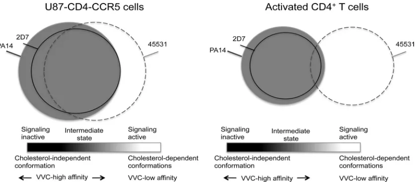
We propose the existence of a CCR5 conformational gradi-ent in which inactive conformations bind PA14, signaling-ac-tive conformations bind 45531, and intermediate conforma-tions bind both MAbs, as well as 2D7, to different extents (Fig.
8). U87-CD4-CCR5 cells overexpress transfected CCR5, which may be in excess over G proteins, but they do not produce chemokine ligands for CCR5 (9). A considerably larger pool of CCR5 uncoupled from G proteins, and with a higher affinity for small-molecule inhibitors (17), may therefore exist in U87-CD4-CCR5 cells than in activated CD4⫹T cells (Fig. 8). The extent to which CCR5 is incorporated into lipid rafts is likely to correlate with the degree of signaling activity; both processes would conceivably be less common in U87-CD4-CCR5 cells than in CD4⫹T cells.
We postulate that PA14 binds the greatest proportion of CCR5 molecules that are permissive for HIV-1 entry (Fig. 8). This scenario agrees with the capacity, noted earlier, of PA14 to block entry of all four viruses, although it does so with distinctly varying potencies. MAb 45531, which mostly binds to cell surface clusters that are not stained by PA14, and which only partly colocalizes with PA14, is only a weak inhibitor of HIV-1 infection; its activity is strongest against Res-3FP but is detectable only with Env pseudovirus on U87-CD4-CCR5 cells. The increased sensitivity of Res-3FP to 45531 in these cells does not indicate that the virus has switched to using CCR5 in the lipid rafts, because it is even more potently in-hibited by PA14 than the parental virus. Moreover, unlike Par-3FP, Res-3FP is effectively blocked by 2D7 in U87-CD4-CCR5 cells. Both mutants, but particularly Res-3FP, seem to have a lower inhibitory threshold of MAb occupancy on the subset of CCR5 molecules that are relevant to entry. Even if 45531 binds the same CCR5 subset as 2D7, its weaker inhibi-tion can be explained by the different locainhibi-tion of its epitope. For 45531, and also the more potent MAbs, inhibiting infec-FIG. 8. Model summarizing the functional and structural properties of some CCR5 variants and their usage for HIV-1 entry. Three CCR5 subsets recognized by MAbs PA14 (gray), 2D7 (solid black lines), and 45531 (dashed lines) are represented by a Venn diagram. The diameter of each subset is proportional to the relative amounts of each variant as indicated by the maximal GMFI values in Table 1. Differences in binding plateaus between the cell types are represented by the sizes of the circles (but are not to scale): Maximum GMFI values were higher on U87-CD4-CCR5 cells than on CD4⫹T cells. The positions of the circles on the gradient depict the relative amounts of the different forms of CCR5, but the shades of the circles do not necessarily coincide with the gradient shades. The gradient represents the various activation states of CCR5 from the inactive to the active conformations as illustrated for GPCRs in general in Fig. 5 of Yao et al. (58). The degree of overlap between the subsets and their functional properties were partly predicted from the various data sets in this study. There is less overlap between the active 45531-bound CCR5 conformations and the inactive PA14- and 2D7-bound conformations in CD4⫹T cells than in U87-CD4-CCR5 cells. This depiction reflects the greater proportion of CCR5 molecules that are involved in chemokine-mediated signaling, and hence coupled to G proteins, in activated CD4⫹T cells. Of note, CCR5 molecules that are permissive to viral entry in the absence of VVC largely coincide with the PA14-bound subset shaded in gray.
on November 7, 2019 by guest
http://jvi.asm.org/
[image:12.585.84.502.68.251.2]tion of U87-CD4-CCR5 cells requires MAb binding to sub-stantial numbers of largely signaling-inactive CCR5 molecules. In contrast, we predict that fewer CCR5 molecules are com-pletely uncoupled from G proteins, or are signaling inactive, in CD4⫹T cells, as depicted by sliding the circles to the right in Fig. 8. In the intermediate zone, the overlap between PA14 and 2D7 would therefore be greater, which could explain why, unlike on U87-CD4-CCR5 cells, 2D7 is as potent and effective an inhibitor as PA14. The failure of 45531 to inhibit even Res-3FP Env pseudovirus infection of CD4⫹T cells is depicted by a smaller overlap in binding between 45531 and 2D7 or PA14 than occurs on U87-CD4-CCR5 cells; the ratio between signaling-active and -inactive or intermediate conformations is modeled to be higher in these cells (Fig. 8).
Overall, a model of intersecting, antigenically heterogeneous forms of CCR5 can account for the differential inhibitory ef-fects of the MAbs on HIV-1 infection in the absence of VVC (Fig. 8). In this scenario, a pool of CCR5 molecules with a high affinity for VVC, which we propose is better recognized by PA14 than the other MAbs, may be preferentially used for entry by the different viruses. The VVC-resistant viruses ap-pear to require a greater density of such receptors in the absence of VVC (3). We suggest those receptors are mostly located outside cholesterol-rich subregions of the cell mem-brane. CCR5 forms located within the latter regions may, how-ever, be highly relevant to how resistant viruses enter in the presence of VVC. We have modeled the different VVC inhi-bition patterns observed in primary cells and cell lines (3). Consistent with our current results, a virus similar to Res-3FP was strongly resistant to VVC in PBMCs but significantly more sensitive in the TZM-bl line. Moreover, the VVC inhibition pattern was not only cell type dependent, but also PBMC donor dependent (3). The model predicted two forms of CCR5, one with low affinity for VVC and the other with high affinity (3). When the proportions of the two CCR5 popula-tions and their affinities for VVC were varied, the model cre-ated infection inhibition curves that fit data sets from both cell types. We proposed that VVC-sensitive viruses can enter via one or both of the free forms of CCR5 to various extents but can use neither of the VVC-bound forms. In contrast, Res-3FP could only use the VVC complex of the low-VVC-affinity ver-sion of CCR5 while losing the capacity to use the unbound low-affinity form and retaining or increasing its ability to use the unbound high-affinity form. The VVC inhibition pattern in cell lines is consistent with the high-VVC-affinity form of CCR5 predominating over the low-VVC-affinity form (3). It may be relevant that TAK-779 and, to a lesser extent, MVC was recently shown to have a higher affinity for G-protein-uncoupled CCR5 molecules than for G-protein-coupled forms (17).
A synthesis of our previous and new models is that Res-3FP may be able to use the low-VVC-affinity form of CCR5 (when occupied by VVC), which we argue corresponds to a subset of 45531-stained CCR5 molecules (when not occupied by VVC). These coreceptors may be predominantly localized in choles-terol-rich membrane microdomains (Fig. 8) (3). However, we do not know the identities or lipid characteristics of the clus-ters on the surfaces of U87-CD4-CCR5 cells that are stained by 45531, nor do we know the sublocalizations of CCR5 mol-ecules recognized by this MAb on CD4⫹T cells. The isolate
from which Res-3FP was cloned emerged under the selection pressure of VVC in PBMCs (33). We propose that in doing so it became adapted to using the VVC complex of the low-VVC-affinity form of CCR5 in lipid rafts, a CCR5 subpopulation that is proportionately more abundant in activated CD4⫹T cells than in U87-CD4-CCR5 cells (Fig. 8). Cholesterol is not re-quired for wild-type HIV-1 Env-induced fusion if the receptor density is high (56). Instead, the presence of cholesterol pro-motes fusion and entry by increasing the mobility of the core-ceptor and thereby allowing local enrichment at the sites where fusion occurs (32, 50). The cholesterol requirements for the resistant virus in the presence of VVC could be different from what applies to wild-type viruses. We do not yet know whether the FP substitutions directly affect the lipid interactions of gp41 or how the fusion process is triggered by different forms of CCR5 (3). One possibility is that the FP sequence changes permit fusion to be triggered by VVC-CCR5 complexes in a cholesterol-rich lipid environment. We are now investigating this hypothesis.
ACKNOWLEDGMENTS
This work was supported by NIH grant R01 AI41420 to J.P.M. and by a Mathilde Krim amfAR fellowship to R.B.
We thank Thomas Ketas and Mira Patel for technical support, Katie Matthews for helpful discussions, and Julie Strizki for supplying CCR5 inhibitors.
REFERENCES
1.Altenbach, C., A. K. Kusnetzow, O. P. Ernst, K. P. Hofmann, and W. L. Hubbell.2008. High-resolution distance mapping in rhodopsin reveals the pattern of helix movement due to activation. Proc. Natl. Acad. Sci. U. S. A.
105:7439–7444.
2.Anastassopoulou, C. G., et al.2011. Resistance of a human immunodefi-ciency virus type 1 isolate to a small molecule CCR5 inhibitor can involve
sequence changes in both gp120 and gp41. Virology413:47–59.
3.Anastassopoulou, C. G., T. J. Ketas, P. J. Klasse, and J. P. Moore.2009. Resistance to CCR5 inhibitors caused by sequence changes in the fusion
peptide of HIV-1 gp41. Proc. Natl. Acad. Sci. U. S. A.106:5318–5323.
4.Baba, M., H. Miyake, X. Wang, M. Okamoto, and K. Takashima.2007. Isolation and characterization of human immunodeficiency virus type 1 resistant to the small-molecule CCR5 antagonist TAK-652. Antimicrob.
Agents Chemother.51:707–715.
5.Bannert, N., et al.2001. Sialylated O-glycans and sulfated tyrosines in the NH2-terminal domain of CC chemokine receptor 5 contribute to high
affin-ity binding of chemokines. J. Exp. Med.194:1661–1673.
6.Baribaud, F., et al.2001. Antigenically distinct conformations of CXCR4.
J. Virol.75:8957–8967.
7.Berro, R., R. W. Sanders, M. Lu, P. J. Klasse, and J. P. Moore.2009. Two HIV-1 variants resistant to small molecule CCR5 inhibitors differ in how
they use CCR5 for entry. PLoS Pathog.5:e1000548.
8.Chabot, D. J., H. Chen, D. S. Dimitrov, and C. C. Broder.2000. N-linked glycosylation of CXCR4 masks coreceptor function for CCR5-dependent
human immunodeficiency virus type 1 isolates. J. Virol.74:4404–4413.
9.Chabre, M., P. Deterre, and B. Antonny.2009. The apparent cooperativity of some GPCRs does not necessarily imply dimerization. Trends Pharmacol.
Sci.30:182–187.
10.Chan, W. E., H. H. Lin, and S. S. Chen.2005. Wild-type-like viral replication potential of human immunodeficiency virus type 1 envelope mutants lacking
palmitoylation signals. J. Virol.79:8374–8387.
11.Chanel, C., et al.2002. Low levels of co-receptor CCR5 are sufficient to
permit HIV envelope-mediated fusion with resting CD4 T cells. AIDS16:
2337–2340.
12.Cormier, E. G., D. N. Tran, L. Yukhayeva, W. C. Olson, and T. Dragic.2001. Mapping the determinants of the CCR5 amino-terminal sulfopeptide inter-action with soluble human immunodeficiency virus type 1 gp120-CD4
com-plexes. J. Virol.75:5541–5549.
13.Dejucq, N., G. Simmons, and P. R. Clapham.1999. Expanded tropism of
primary human immunodeficiency virus type 1 R5 strains to CD4(⫹) T-cell
lines determined by the capacity to exploit low concentrations of CCR5.
J. Virol.73:7842–7847.
14.Dragic, T., et al.2000. A binding pocket for a small molecule inhibitor of HIV-1 entry within the transmembrane helices of CCR5. Proc. Natl. Acad.
Sci. U. S. A.97:5639–5644.