1
“
STRAIN USG ELASTOGRAPHY WITH GRAY
SCALE AND COLOUR DOPPLER IMAGING OF
THYROID NODULES –EFFICACY COMPARED
WITH FNAC
”Dissertation submitted to
THE TAMIL NADU DR.M.G.R.MEDICAL UNIVERISTY
CHENNAI
In Partial Fulfillment of the Regulations for the Award of the degree
M.D. DEGREE EXAMNATION
BRANCH VIII RADIODIAGNOSIS
MADRAS MEDICAL COLLEGE
&
RAJIV GANDHI GOVERNMENT GENERAL HOSPITAL,
CHENNAI -600 003.
THE TAMIL NADU DR.M.G.R.MEDICAL UNIVERISTY
CHENNAI -600 003. TAMIL NADU
2
BONAFIDE CERTIFICATE
Certified that this dissertation is the bonafide work of
DR.A.KARPAGAVALLI
on
“STRAIN USG ELASTOGRAPHY
WITH GRAY SCALE AND COLOUR DOPPLER IMAGING
OF THYROID NODULES – EFFICACY COMPARED WITH
FNAC
” during her MD(RADIODIAGNOSIS) in Barnard Institute of Radiology in the academic year of 2015-2018 at the Madras MedicalCollege and Rajiv Gandhi Government General Hospital,Chennai
-600003.
PROF.DR.N .KAILASANATHAN, MD, DMRD
FORMER DIRECTOR AND PROFESSOR, Department of Radiodiagnosis,
Barnard institute of Radiology,
Rajiv Gandhi Government General Hospital, Madras Medical College,
Chennai -600003.
PROF.DR.RAVI MD, DMRD
Director,
Barnard Institute of Radiology, Madras Medical College & Rajiv Gandhi Government General Hospital,
Chennai – 600 003.
PROF.DR.R.NARAYANABABU, MD., D.Ch.,
Dean
Madras Medical College &
3
DECLARATION
I, certainly declare that this dissertation titled “
STRAIN USG
ELASTOGRAPHY WITH GRAY SCALE AND COLOUR
DOPPLER
IMAGING
OF
THYROID
NODULES
–
EFFICACY COMPARED WITH FNAC
” represent a genuine work of mine. The contribution of any supervisors to the research areconsistant with normal supervisory practice, and are acknowledged.
I, also affirm that this bonafide work or part of this work was not
submitted by me or any others for any award, degree or diploma to any
other university board, neither in India or abroad. This is submitted to
The Tamil Nadu Dr.MGR Medical University, Chennai in partial
fulfilment of the rules and regulation for the award of Master of
Radiodiagnosis Branch VIII.
DR.KARPAGAVALLI .A
Date :
4
ACKNOWLEDGEMENT
I would like to express my deep sense of gratitude to
PROF.DR.R.NARAYANA BABU, MD., D.Ch., Dean, Madras Medical
College & Rajiv Gandhi Government General Hospital Chennai, for
giving me permission to conduct the study in this institution.
With extreme gratefulness, I express my indebtedness to
PROF.DR.RAVI, MD.,DMRD, Director, Barnard Institute of
radiology, MMC & RGGGH, for allowing me to undertake this study on
“STRAIN USG ELASTOGRAPHY WITH GRAY SCALE AND
COLOUR DOPPLER IMAGING OF THYROID NODULES –
EFFICACY COMPARED WITH FNAC”
I would like to express my deep gratitude and respect to my guide
FORMER PROF.DR.N .KAILASANATHAN, MD, D.M.R.D whose
advice and insight was invaluable to me. This work would not have been
possible without his guidance, support and encouragement.
I was able to carry out my study to my fullest satisfaction, thanks
to guidance, encouragement, motivation and constant supervision
extended to me, by my beloved Head of the Department PROF.DR.
5
My sincere thanks to Prof.Dr.S.Kalpana,MD.,DNB for her
practical comments and guidance especiall y at the inception of the study
and I also wish to thank Prof.Dr.S.Babu Peter,MD.,DNB for his
valuable support through out the study.
I am also extremely indepted to Prof.Dr.D.Ramesh MD for his
valuable suggestions.
I am extremely thankful to my Associate Professor
Dr.GangaDevi MD.,DMRD.,FRCR for her invaluable contribution
,personal attention, constructive cricticism she has made during my
study.
I am bound by ties of gratitude to my respected Associate
Professors, Dr.Manimekala MD.,DNB and Dr.KasiVisalakshi,MD and
Assistant professors Dr.K.Geetha MD, Dr.J.Chezhian MD,
Dr.G.Geetha MD , Dr.S.Iyengaran, Dr.Mohideen Ashraf MD,
Dr.Saranya DMRD , Dr.Balan DMRD, DR.Dheeba MD, Dr.Karthick
MD in general, for placing and guiding me on the right track from the
very beginning of my career in Radiodiagnosis till this day.
I am fortunate to have my postgraduate colleagues
Dr.Elavazhagan, Dr.Kanmani kiruba, Dr.Sakthivel raja and
Dr.Swarnalakshmi for their valuable suggestions, relentless help for
6
for their unseen contributions. My lovable thanks to my family for their
moral support.
I would be failing in my duty if I don’t place on record my sincere
thanks to those patients who inspite of their sufferings extended their
fullest co-operation.
7
INDEX
SL.NO CONTENTS PAGE
1 INTRODUCTION 1
2 ANATOMY AND EMBRYOLOGY 3
3 APPROACH TO THYROID LESIONS 13
4 DIFFUSE THYROID LESIONS 14
5 NODULAR THYROID LESIONS 19
6 EVALUATION OF THYROID NODULES
BY USG 35
7 THYROID SONOELASTOGRAPHY 48
8 AIMS AND OBJECTIVES 55
9 MATERIALS AND METHODS 56
10 REPRESENTATIVE CASES 59
11 STATISTICAL ANALYSIS 64
12 RESULTS &DISCUSSION 78
13 CONCLUSION 84
14 BIBLOGRAPHY
16 ANNXEURE
PROFORMA, ABBREVIATION, MASTER CHART
ETHICAL COMMITTEE CERTIFICATE PLAGIARISM CERTIFICATE
PLAGIARISM CERTIFICATE
This is to certify that this dissertation work titled “STRAIN USG
ELASTOGRAPHY WITH GRAY SCALE AND COLOUR DOPPLER IMAGING
OF THYROID NODULES –EFFICACY COMPARED WITH FNAC” of the
candidate Dr.A.KARPAGAVALLI with Registration Number 201518002 for the
award of M.D RADIODIAGNOSIS. I personally verified the urkund.com website for
plagiarism check. I found that the uploaded file containing from introduction to
conclusion pages shows a result of 1% plagiarism in this dissertation.
1
INTRODUCTION
Thyroid nodules are very commonly observed on thyroid
ultrasonography . Conventional US has been widely used to
determine which nodules should be biopsied.
Although conventional US can provide meaningful
information in thyroid nodule diagnosis, there has been considerable
variation in diagnostic performances.
According to the AmericanThyroidAssociation guidelines, no
single US feature or combination of features is adequately sensitive or
specific to identify all malignant nodules [9]. For this reason, fine
needle aspiration cytology (FNAC) is required for the nodules with
suspicious ultrasound signs . However, FNAC has inherent limitations
due to indeterminate and nondiagnostic results
On physical examination, a hard or firm nature is associated
with thyroid malignancy. But, palpation is very subjective and
limited in patients with multinodular goiter or small deep -seated
nodules. As a consequence, a significant number of patients eve ntually
receive unnecessary thyroid surgery.
Therefore, improvement and refinement of noninvasive
2
elastography (USE) has recently been introduced in the clinical workup
of thyroid nodules.
USE is a US-based technique to assess the biomechanical
properties of tissue in the clinical setting . Meanwhile, US -based
elastography can provide an objective evaluation of tissue stiffnes.
There are two kinds of elastography (strain and s hear wave
elastography) that are currently used in clinical practice. Among
these types of USE, strain USE was the first to be introduced into
commercially available systems.
Although many reports have compared conventional US with
elastography, in clinical practice, the final decision or diagnosis is
usually based on a combination of conventional US and
3
EMBRYOLOGY
Thyroid is a butterfly-shaped gland situiated in the neck deep to
the strap muscle. It develops as an evagination of the primitive foregut
at the level of foramen cecum in the base of tongue.
Endodermal cells at the floor of the primitive foregut start to
thicken and form the median primordium(bud) of the thyroid, which
descends anterior to the structures forming the laryngx. An
epithelium-lined thyroglossal duct connects the foramen cecum and the median
primordium .
The cells from the median primordium become thyroid hormone
producing follicular cells. The paired lateral primordium(bud)s develop
from the fourth pharyngeal pouches and fuse the median primordium ,
forming ultimobranchial bodies. The neuroectodermal lateral
primordium (ultimobranchial bodies) provide the parafollicular C cells
producing calcitonin.
ANATOMY:
1,2The bilateral thyroid lobe is connected with each other by the
midline isthmus.
A pyramidal lobe can arise from the isthmus in 50% of patients.
4
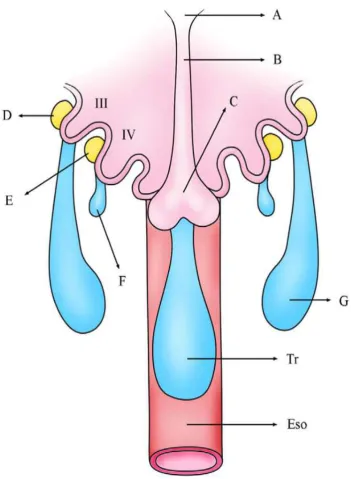
FIG 1. Early development of thyroid gland.The epithelium lined
median primordium (c) fuse with the neuroectodermal lateral
primordium(ultimobranchhial bodies, F), formig the thyroid gland.The
ultimobranchial bodies provide the parafolllicular c cells producing
[image:12.595.118.470.65.545.2]5
Notes: III, third branchial pouch; four branchia l pouch;A, foramen
caecum; B thyroglossal duct; C ,median thyroid;D,inferior parathyroid;
E, superior parathyroid ;F,ultimobranchial body; G,thymus; Tr,trachea;
Eso,esophagus
RELATIONS:
The thyroid gland is anterior to the prevertebral and paraspinal
musculature and deep to the sternothyroid and sternohyoid muscles.
ARTERIAL SUPPLY:
The thyroid gland has a rich blood supply, derived from the
superior, inferior, and the small inferior ima arteries that often directly
originate from the aortic arch.
VENOUS DRAINAGE:
Venous drainage is via multiple surface veins draining into the
superior, middle, and inferior thyroid veins.
PHYSIOLOGY:
Thyroid gland produces 2 types of thyroid hormones: thyroxine
(T4) and triiodothyronine (T3), which are iodinated derivat ives of
tyrosine. The primary internal regulation of thyroid activity is through
the anterior pituitary gland secreting thyroid-stimulating hormone by
way of thyrotropin-releasing hormone released by the hypothalamus.
Most circulating thyroid hormones are bound to plasma proteins; the
6
Fig -2: Thyroid gland is highly vascular . It is supplied by superior and inferior thyroid arteries .It is drained via superior ,middle and
inferior thyroid veins .
[image:14.595.176.430.443.711.2]7
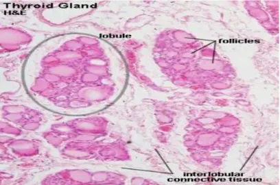
[image:15.595.95.504.107.377.2]HISTOLOGY
FIG.7.Lobule of thyroid
DISTINGUISING FEATURES OF THYROID
Follicle is a structural unit. Follicle contains follicular epithelial cells and parafollicular cells.
Follicle shows follicular cavity in whic h contains gel-like colloid.
Inter-follicular spaces are filled by reticular connective tissue and
8
FIG.8: follicles and Colloid of thyroid gland
COLLOID
Cavity of thyroid follicle is filled with semi –fluid or gel like
substance, called thyroid colloid
the endocrine secretion of epithelia cells and composed of
nucleoproteins, thyroglobulin and proteolytic enzymes.
Among the endocrine glands, thyroid is unique because it
utiloizes an inorganic element iodine for the synthesis of its
9
FIG. 9: Follicular cells of thyroid gland
FOLLICULAR CELLS
These are cuboidal epithelial cells with their basal ends resting on
basement membrane.
These cells show the changes in shape depending on state of
gland.
When the gland is inactive, cells exhibit squamous structure and
columnar when hyperactive.
The follicular cells show central or basal round nucleus with one
or more eccentric nuclei.
10
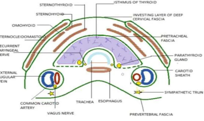
CROSS SECTIONAL ANATOMY
3 [image:18.595.93.505.315.559.2]
FIG 10, Transverse sonogram made with 7.5-MHz linear array transducer Tr, tracheal air shadow; C, common carotid artery; J,
jugular vein.
FIG .7, Relations of thyroid gland
IMAGING MODALITIES
Imaging modalities for the thyroid gland include cross -sectional
imaging techniques, such as ultrasound (US), computed tomography
(CT), magnetic resonance imaging (MRI), or positron emission
11
Ultrasound is the most frequently performed examination for
thyroid lesions . US plays a pivotal role in the diagnosis and
management of benign and malignant thyroid nodules.
US allows characterization of thyroid nodules, detection of cervical
lymphadenopathy, and guidance for FNA as well as percutaneous intervention,
such as ethanol, radio frequency, or laser ablation.
American Thyroid Association recommended in the revised
guidelines on 2009 that thyroid US should be performed in patients with
palpable or incidental thyroid nodules on CT, MRI, or
18fluorodeoxyglucose (FDG)-PET scan.
THYROID USG TECHNIQUE :
Thyroid US should be performed with high-frequency linear
probes ranging from 7 to 12 MHz.
Patients are in supine position with hyperextension of the neck.
Scanning field includes the central neck from the lower border of the
mandible to the sternal notch and the bilateral cervical nodal chains and
the supraclavicular area, to detect accompanying nodal metastasis from
thyroid cancer.
The thyroid gland should be evaluated in both transverse and
longitudinal scans for each individual lobe and isthmic lob e separately.
12
microcalcifications, and echogenicity in combination, are used to access
the malignant potential of each lesion. When a thyroid nodule is
detected by US, the size in its maximum dimensi on should be recorded
for the management and follow-up reference. Although malignancy risk
becomes 4 times higher in the nodules equal to or 4 cm than that of
smaller ones, size is not an independent predictor for malignancy.
Smaller nodules could have as much risk of malignancy as larger
nodules.
COMPUTED TOMOGRAPHY/ MAGNETIC RESONANCE
IMAGING:
CT and MR imaging provide important information about the
extension of thyroid disease into adjacent structures, including the
trachea and larynx, esophagus, and great vessels.
CT or MRI has a complimentary role with US in preoperative
staging and postoperative surveillance of thyroid cancer, and evaluation
of mediastinal goiter or ectopic thyroid. CT is also helpful in the
evaluation of nodal status of thyroid cancer, especially in
US-inaccessible area (retropharyngeal space or mediastinum). MRI is
preferred to CT in the evaluation of the extent of thyroid neoplasm due
to superior soft-tissue contrast. MR does not require iodinated contrast
agent, which is advantageous for patients planned for radioactive iodine
13
APPROACH TO THYROID LESIONS:
4Thyroid lesions are classified into diffuse and nodular
thyroid lesions.
Several thyroid diseases are characterized by diffuse rather than
focal involvement.
This usually results in generalized enlargement of the gland
(goiter) and no palpable nodules. Specific conditions that produce such
diffuse enlargement include chronic autoimmune lymphocytic
thyroiditis (Hashimoto’s thyroiditis), colloid or adenomatous goiter, and
Graves’ disease.
These conditions are usually diagnosed on the basis of clinical
and laboratory findings and occasionally by FNA biopsy. Sonography
is seldom indicated. However, high-resolution sonography can be
helpful when the underlying diffuse disease causes asymmetrical
thyroid enlargement, which suggests a mass in the larger lobe. The
sonographic finding of generalized parenchymal abnormality may alert
the clinician to consider diffuse thyroid disease as the underlying c ause.
FNA, with sonographic guidance if necessary, can be performed if a
14
DIFFUSE THYROID LESIONS – SONOGRAPHIC
EVALUATION
Recognition of diffuse thyroid enlargement on sonography can
often be facilitated by noting the thickness of the isthmus, normally a
thin bridge of tissue measuring only a few millimeters in AP dimension.
With diffuse thyroid enlargement, the isthmus may be up to 1 cm or
more in thickness.
DIFFUSE THYROID DISEASES:
39,40 Acute suppurative thyroiditisSubacute granulomatous thyroiditis
Hashimoto’s thyroiditis (chronic lymphocytic thyroiditis)
Adenomatous or colloid goiter
Painless (silent) thyroiditis
Acute suppurative thyroiditis is a rare inflammatory disease
usually caused by bacterial infection and affecting c hildren. Sonography
can be useful in select patients to detect the development of a frank
thyroid abscess. The infection usually begins in the perithyroidal soft
tissues. On ultrasound images, an abscess is seen as a poorly defined,
hypoechoic heterogeneousmass with internal debris, with or without
15
Subacute granulomatous thyroiditis or de Quervain’s
disease,40,41 is a spontaneously remitting inflammatory disease probably
caused by viral infection. Sonographically, the gland may appear
enlarged and hypoechoic, with normal or decreased vascularity caused
by diffuse edema of the gland, or the process may appear as focal
hypoechoic regions. Although usually not necessary, sonography can be
usto assess evolution of de Quervain’s disease after medical therapy.
Fig 11. Focal areas of subacute thyroiditis. Longitudinal power
Doppler image of the thyroid gland shows two poorly defined
hypoechoic areas(arrow) caused by subacute thyroiditis .
The most common type of thyroiditis is chronic autoimmune
lymphocytic thyroiditis, or Hashimoto’s thyroiditis. It typically
occurs as a painless, diffuse enlargement of the thyroid gland in a young
16
typical sonographic appearance of Hashimoto’s thyroiditis is diffuse,
coarsened, parenchymal echotexture, generally more hypoechoic than a
normal thyroid.
In most cases the gland is enlarged. Multiple, discrete hypoechoic
micronodules from 1 to 6 mm in diameter are strongly suggestive of
chronic thyroiditis; this appearance has been called micronodulation43 .
Micronodulation is a highly sensitive sign of chronic thyroiditis .
micronodules represent lobules of thyroid parenchyma that have been
infiltrated by lymphocytes and plasma cells.
Fig 12. Hashimoto’s thyroiditis: coarse septations.
These lobules are surrounded by multiple linear echogenic
fibrous septations . These fibrotic septations may give the parenchyma a
“pseudolobulated” appearance. Both benign and malignant thyroid
nodules may coexist with chronic lymphocytic thyroiditis, and FNA is
17
autoimmune disorders, there is an increased risk of malignancy, with a
B-cell malignant lymphoma most often arising within the gland.
Although the appearance of diffuse parenchymal inhomogeneity
and micronodularity is typical of Hashimoto’s thyroiditis, other diffuse
thyroid diseases, most frequently multinodular or adenomatous goiter,
may have a similar sonographic appearance.44 Most patients with
adenomatous goiter have multiple discrete nodules separated by
otherwise normal-appearing thyroid parenchyma. others have
enlargement with rounding of the poles of the gland, diffus e
parenchymal inhomogeneity, and no recognizable normal tissue.
Adenomatous goiter affects women three times more often than men.
Graves’ disease45
is a common diffuse abnormality of the
thyroid gland and is usually biochemically characterized by
hyperfunction (thyrotoxicosis). The echotexture may be more
inhomogeneous than in diffuse goiter, mainly because of numerous
large, intraparenchymal vessels. Further, especially in young patients,
the parenchyma may be diffusely hypoechoic because of the extensive
lymphocytic infiltration or the predominantly cellular content of the
parenchyma, which becomes almost devoid of colloid substance. Color
Doppler sonography often demonstrates a hypervascular pattern
referred to as the thyroid inferno. Spectral Doppler will often
demonstrate peak systolic velocities exceeding 70 cm/sec, which is the
18
flow velocities in the superior and inferior thyroid arteries after medical
treatment has been reported.
The rarest type of inflammatory thyroid disease is invasive
fibrous thyroiditis, also called Riedel’s struma. This disease
primarily affects women and often progresses to complete destruction
of the gland.
Some cases may be associated with mediastinal or retroperitoneal
fibrosis or sclerosing cholangitis. The primary reason for sonography is
to check for extrathyroid extension of the inflammatory process, with
encasement of the adjacent vessels . Such information can be particularly
useful in surgical planning. Open biopsy is generally required to
distinguish this condition from anaplastic thyroid carcinoma. The
sonographic findings in these two diseases may be identical.
Fig 13.Transverse color Doppler image of the left lobe show s increased vascularity, indicating an acute stage of the Graves’
19
NODULAR THYROID LESIONS :
Many thyroid diseases can present clinically with one or more
thyroid nodules. Such nodules represent common and controversial
clinical problems. women affected more frequently than men. Exposure
to ionizing radiation increases the incidence of benign and malignant
nodules, with 20% to 30% of a radiationexposed population having
palpable thyroid disease. Although nodular thyroid disease is relatively
common, thyroid cancer is rare and accounts for less than 1% of all
malignant neoplasms. The majority of thyroid nodules are benign. The
clinical challenge is to distinguish the few clinically significant
malignant nodules from the many benign nodules and thus identify
patients who need surgical excision. This task is complicated because
nodular disease of the thyroid gland often is clinically occult (<10-15
mm), although it can be readily detected by high-resolution sonography.
HYPERPLASIA AND GOITER:
Approximately 80% of nodular thyroid disease is caused by
hyperplasia5,6 of the gland and occurs in up to 5% of any population .
Its etiology includes iodine deficiency (endemic), disorders of
hormonogenesis (hereditary familial forms), and poor utilization of
iodine as a result of medication. When hyperplasia leads to an overall
increase in size or volume of the gland, the te rm goiter is used. The
peak age of patients with goiter is 35 to 50 years, and women are
20
Sonographically, most hyperplastic or adenomatous nodules are
isoechoic compared to normal thyroid tissue , but may bec ome
hyperechoic because of the numerous interfaces between cells and
colloid substance8 . Less frequently, a hypoechoic spongelike or
honeycomb pattern is seen . When the nodule is isoechoic or
hyperechoic, a thin peripheral hypoechoic halo is typically seen, most
likely caused by perinodular blood vessels and mild edema or
compression of the adjacent normal parenchyma. Perinodular blood
vessels are typically detected by color Doppler sonogram9,10.
Histologically, the initial stage is cellular hyperplasia of the
thyroid acini, followed by micronodule and macronodule formation,
often indistinguishable from normal thyroid parenchyma, even at
histology. Hyperplastic nodules often undergo liquefactive degeneration
with the accumulation of blood, serous fluid, and colloid substance11.
21
Pathologically, they are often referred to as hyperplastic,
adenomatous, or colloid nodules. Many (if not all) cystic thyroid
lesions are hyperplastic nodules that have undergone extensive
liquefactive degeneration. Pathologically, true epithelial -lined cysts of
the thyroid gland are rare.
In the course of this cystic degenerative process, calcification,
which is often coarse and perinodular, may occur. Hyperplastic nodule
function may have decreased, may have remained normal, or may have
increased (toxic nodules).
Hyperfunctioning (autonomous) nodules often exhibit an
abundant perinodular and intranodular vascularity; however, because
of the hypervascular pattern shown in most solid thyroid nodules on
high-sensitivity Doppler systems, this feature does not allow detection
of hyperfunctioning nodules within multinodular goiters with
sonography.9,10
The degenerative changes of goitrous n odules correspond to their
sonographic appearances . Purely anechoic areas are caused by serous
or colloid fluid. Echogenic fluid or moving fluid-fluid levels
correspond to hemorrhage.11 Bright echogenic foci with comet-tail
artifacts are likely caused by microcrystals or aggregates of colloid
substance, which may also move slowly, like snowflakes, within the
22
Thin, intracystic septations probably correspond to attenuated
strands of thyroid tissue and appear completely avascular on color
Doppler ultrasound. These degenerative processes may also lead to the
formation of calcifications, which may be either thin, peripheral
shells (“eggshell”) or coarse, highly reflective foci with associated
acoustic shadows, scattered throughout the gland.
ADENOMA
1Adenomas represent only 5% to 10% of all nodular disease of the
thyroid and are seven times more common in women than me5 . Most
result in no thyroid dysfunction.
Most adenomas are solitary, but may also develop as part of a
multinodular process. The benign follicular adenoma is a true thyroid
neoplasm, characterized by compression of adjacent tissues and fibrous
encapsulation.
Various subtypes of follicular adenoma include the fetal
adenoma, Hürthle cell adenoma, and embryonal adenoma, each
distinguished according to the type of cell proliferation. The cytologic
features of follicular adenomas are generally indistinguishable from
those of follicular carcinoma.
Vascular and capsular invasion are the hallmarks of follicular
carcinoma, identified by histologic rather than cytologic analysis.
23
Sonographically, adenomas are usually solid masses that may be
hyperechoic, isoechoic, or hypoechoic . They often have a thick, smoot h
peripheral hypoechoic halo resulting from the fibrous capsule and blood
vessels, which can be readily seen by color Doppler imaging. Often,
vessels pass from the periphery to the central regions of the nodule,
sometimes creating a “spoke and wheel” appearance. This vascular
pattern is usually seen in both hyperfunctioning and poorly functioning
adenomas and thus does not allow the detection of hyperfunctioning
[image:31.595.128.469.351.555.2]lesions.
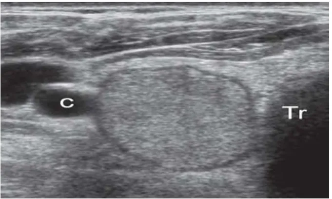
Fig 15. homogeneous, hypoechoic, round to oval masses with a surrounding thin halo, the capsule of the adenoma; Tr, tracheal air
shadow; C, carotid artery.
CARCINOMA:
Most primary thyroid cancers are of epithelial origin and are
24
of mesenchymal origin are exceedingly rare, as are metastases to the
thyroid.
Most thyroid cancers are well differentiated, and papillary
carcinoma (including so-called mixed papillary and follicular
carcinoma) accounts for 75% to 90% of all cases. In contrast, medullary,
follicular, and anaplastic carcinomas (combined) represent only 10% to
25% of all thyroid carcinomas.
PAPILLARY CARCINOMA OF THYROID:
Although it can occur in patients of any age, prevalence of
papillary thyroid carcinoma peaks in both the third and t he seventh
decade of life.12 Women are affected more often than men. On
microscopic examination, the tumor is multicentric within the thyroid
gland in at least 20% of cases14. Round, laminated calcifications
(psammoma bodies) in the cytoplasm of papillary cancer cells are seen
in approximately 35% of patients. The major route of spread of
papillary carcinoma is through the lymphatics to nearby cervical lymph
nodes.
In fact, a patient with papillary thyroid cancer may present with
enlarged cervical nodes and a palpably normal thyroid gland.17
Interestingly, the presence of nodal metastasis in the neck generally
does not appear to worsen the prognosis for this malignancy. Distant
25
and lung . After 20 years, the cumulative mortality from papillary
thyroid cancer is typically only 4% to 8%.
Sonographic characteristics of papillary carcinoma are relatively distinctive, as follows ;
Hypoechogenicity (90% of cases), resulti ng from closely packed
cell content, with minimal colloidsubstance.
Microcalcifications, appearing as tiny, punctuate hyperechoic
foci, either with or without acoustic shadows . In rare, but
usually aggressive cases of papillary carcinomas of childh ood,
microcalcifications may be the only sonographic sign of the
neoplasm, even without evidence of a nodular lesion. .
Hypervascularity (90% of cases), with disorganized vascularity,
mostly in well-encapsulated forms.
Cervical lymph node metastases, which may contain tiny,
punctate echogenic foci caused by microcalcifications . These are
mainly located in the caudal half of the deep jugular chain.
Occasionally, metastatic nodes may be cystic as a result of
extensive degeneration . Cystic nodal metastases show a thickened outer
wall, internal nodularity, and septations in most cases, although they
may appear purely cystic in younger patients. Cystic lymph node
metastases in the neck occur almost exclusively in association with
26
carcinomas. The overwhelming majority of papillary carcinomas
appear as a predominantly solid mass.
Invasion of adjacent muscles is infrequently visualized by
ultrasound but indicates that the mass is malignant. . A follicular
variant accounts for 10% of cases of papillary carcinoma and appears
similar to a follicular neoplasm on gross pathologic inspection and
ultrasound.
Papillary microcarcinoma is a rare, nonencapsulated sclerosing
tumor measuring 1 cm or less in diameter . Most patient (80%) present
with enlarged cervical nodes and a palpably normal thyroid gland.17
Papillary microcarcinoma can be imaged by high -frequency ultrasound.
27
FOLLICULAR CARCINOMA :
16Follicular carcinoma is the second subtype of well -differentiated
thyroid cancer. It accounts for 5% to 15% of all cases of thyroid
cancer, affecting women more often than men. The two variants of
follicular carcinoma differ greatly in histology and clinical course. The
minimally invasive follicular carcinomas are encapsulated, and only the
histologic demonstration of focal invasion of capsular blood vessels of
the fibrous capsule itself permits differentiation from follicular
[image:35.595.131.464.346.586.2]adenoma.
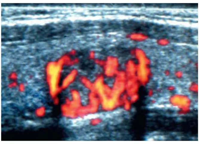
Fig 17. Power Doppler image shows that nodule is hypervascular and has flow in the center and at the periphery.
The widely invasive follicular carcinomas are not well
encapsulated, and invasion of the vessels and the adjacent thyroid is
more easily demonstrated. Both variants of follicular carcinoma tend to
28
metastases to bone, lung, brain, andliver are more likely than metastases
to cervical lymphnodes.
No unique sonographic features allow differentiation of follicular
carcinoma from adenoma, which is not surprising, given the cytologic
and histologic similarities ofthese two tumors . Similarly, fine -needle
aspiration is not reliable in differentiating benign from malignant
follicular neoplasms because the pathologic diagnosis is not based on
cellular appearance but rather on capsular and vascular invasion.
Therefore, most follicular nodules must be surgically removed for
accurate pathologic diagnosis. Features that suggest follicular
carcinoma are rarely seen but include irregular tumor margins, a thick
irregular halo, and a tortuous or chaotic arrangement of internal blood
vessels on color Doppler imaging.
FOLLICULAR THYROID CARCINOMA
SONOGRAPHIC FEATURES:
Irregular tumor margins
Thick, irregular halo
29
Fig 18 A, Left lobe, and18 B, right lobe, of the thyroid show round, homogeneous hypoechoic masses that appear identical except for size differences on transverse images; Tr, tracheal air shadow. The smaller
30
Fig 19. Microscopic appearance of the capsule shows invasion of the follicular cells into the capsule (arrows). This is one of the
microscopic features that allows a pathologic diagnosis of
malignancy but is not visible by ultrasound .
MEDULLARY CARCINOMA:
Medullary carcinoma accounts for about 5% of all malignant
thyroid diseases. It is derived from the parafollicular cells, or C cells,
and typically secretes the hormone calcitonin, which can be a useful
serum marker. This cancer is frequently familial(20%) and is an
essential component of the multiple endocrine neoplasia (MEN) type
II syndromes. The disease is multicentric and/or bilateral in about 90%
of the familial cases . There is a high incidence of metastatic
involvement of lymph nodes. The prognosis for patients with
medullary cancer is somewhat worse than for follicular cancer.
The sonographic appearance of medullary carcinoma is usually
similar to that of papillary carcinoma and is seen most often as a
hypoechoic solid mass. Calcifications are often seen (histologically
31
coarse than the calcifications of typical papillary carcinoma.
Calcifications can be seen not only in the primary tumor but also in
lymph node metastases and even in hepatic metastases.
Fig 20.Transverse dual image in patient with multiple endocrine neoplasia type II (MEN II) shows bilateral hypoechoic masses (arrows) that contain areas of coarse calcification; C, carotid arteries;
Tr, trachea; E, esophagus.
ANAPLASTIC THYROID CARCINOMA.
15Anaplastic thyroid carcinoma is typically a disease of elderly
persons; it represent one of the most lethal of solid tumors. Although it
accounts for less than 2% of all thyroid cancers, it carries the worst
prognosis, with a 5-year mortality rate of more than 95%. The tumor
typically presents as a rapidly enlarging mass extending beyond the
gland and invading adjacent structures. It is often inoperable at
presentation. Anaplastic carcinomas may often be associated with
papillary or follicular carcinomas, presumably representing a
32
lymphatics but instead are prone to aggressive local invasion of muscles
and vessels.
Sonographically, anaplastic thyroid carcinomas are usually
hypoechoic and often encase or invade bloodvessels and neck mu scles .
Often these tumors cannot be adequately examined by ultrasound
because of their large size. Instead, computed tomography (CT) or
magnetic resonance imaging (MRI) of the neck usually demonstrates
the extent of disease more accurately.
ANAPLASTIC THYROID CARCINOMA:
SONOGRAPHIC FEATURES
Large, hypoechoic mass
Encase or invade blood vessels
Invade neck muscles
LYMPHOMA
18Lymphoma accounts for approximately 4% of all thyroid
malignancies. It is mostly of the non-Hodgkin’s type andusually affects
older women. The typical clinical sign is
a rapidly growing mass that may cause symptoms of obstruction
such as dyspnea and dysphagia. In 70% to 80% of patients, lymphoma
arises from a preexisting chronic lymphocytic thyroiditis (Hashimoto’s
thyroiditis) with subclinical or overt hypothyroidism. The prognosis is
33
survival ranges from almost 90% in early-stage cases to less than 5% in
advanced, disseminated disease.
Sonographically, lymphoma of the thyroid appears as an
extremely hypoechoic and lobulated mass. Large areas of cystic necrosis
may occur, as well as encasement of adjacent neck vessels. On color
Doppler imaging, both nodular and diffuse thyroid lymphomas may
appear mostly hypovascular or may show blood, vessels with chaotic
distribution and AV shunts. The adjacent thyroid parenchyma may be
heterogeneous as a result of associated chronic thyroiditis.
Fig21. Transverse image of left lobe of the thyroid shows diffuse mass enlarging the lobe and extending into the soft tissues (arrows) surrounding the common carotid artery (c); Tr, tracheal air shadow.
THYROID METASTASES:
Metastases to the thyroid are infrequent, occurring late in the
34
less frequently a lymphatic route. Metastases usually are from
melanoma (39%), breast (21%), and renal cell (10%) carcinoma.
Metastases may appear as solitary, well-circumscribed nodules
or as diffuse involvement of the gland. On sonography, thyroid tumors
are solid, homogeneously hypoechoic masses, without calcifications.
[image:42.595.131.464.285.511.2]
35
NODULAR THYROID DISEASE:
SONOGRAPHIC EVALUATION
Determine location of palpable neck mass (e.g., thyroid or
extrathyroid).
Characterize benign versus malignant nodule features.
Detect occult nodule in patient with history of head and
neck irradiation or MEN II syndrome.
Determine extent of known thyroid malignancy.
Detect residual, recurrent, or metastatic carcinoma.
Guide fine-needle aspiration of thyroid nodule or cervical
lymph nodes.
The fundamental anatomic features of a thyroid n odule on high-resolution sonography are as follows:
Internal consistency (solid, mixed solid and cystic, or purely
cystic)
Echogenicity relative to adjacent thyroid parenchyma
Margin
Shape
Presence and pattern of calcification
36
Presence and distribution of blood flow signals
INTERNAL CONTENTS :
Usually, approximately 70% of thyroid nodules are solid,
whereas the remaining 30% exhibit various amounts of cystic
change. A nodule that has a significant cystic component is
usually a benign adenomatous (colloid) nodule that has undergone
degeneration or hemorrhage.
All cystic thyroid lesions seen with high-resolution ultrasound
demonstrate some wall irregularity and internal solid elements or
debris caused by nodule degeneration.
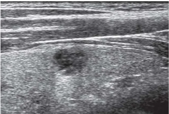
[image:44.595.119.476.404.646.2].
Fig 23. honeycomb-like or cystic changes, with nodules showing larger cystic spaces indicate a very high probability of a benign
37
Fig 24.Typical appearance of colloid cysts. Nodules that are mostly cystic are considered benign. These nodules have tiny echogenic foci that are thought to be microcrystals. A few of these foci are associated
with comet-tail artifacts .
Comet-tail artifacts are frequently encountered in cystic
thyroid nodules and are likely related to the presence of
microcrystals . These comet-tail arti- facts can be located in the
cyst walls and internal septations or in the cyst fluid .
When a more densely echogenic fluid is gravitationally
layered in the posterior portion of a cystic cavity, the likelihood
of hemorrhagic debris is very high. Frequently, patients with
hemorrhagic debris present clinically with a rapidly growing, often
tender neck mass. The spongiform appearance of thyroid nodules,
related to the presence of tiny colloid changes, is an extremely
uncommon finding in malignant nodules, particularly when it is
38
isoechogenicity. This pattern is highly predictive of a benign
nodule . Papillary carcinomas may rarely exhibit varying amounts
of cystic change and appear almost indistinguishable from benign
cystic nodules. In cystic papillary carcinomas, the frequent
sonographic detection of a solid elements or projections (≥1 cm
with blood flow signals and/or microcalcifications) into the lumen
can lead to suspicion of malignancy . Cervical metastatic lymph
nodes from either a solid or a cystic primary papillary cancer may
also demonstrate a cystic pattern; this is likely pathognomonic of
malignant adenopathy.
SHAPE:
A taller-than-wide shape, in which the AP diameter is equal
or less than its transverse diameter on a transverse or longitudinal
plane, is specific for differentiating malignant nodules from benign
nodules, because malignant neoplasms (taller than wide) grow
across normal tissue planes, whereas benign nodules grow parallel
39
Fig 25. Hyperplastic nodule. Oval homogeneous nodule – follicular adenoma.
ECHOGENICITY :
Thyroid cancers are usually hypoechoic relative to the
adjacent normal thyroid parenchyma . Many benign thyroid
nodules are also hypoechoic. Marked hypoechogenicity is highly
specific for diagnosing malignant nodules, whereas the
hypoechogenicity often found in benign lesions is usua lly less
marked. A predominantly hyperechoic nodule, although relatively
uncommon, is more likely to be benign. The isoechoic nodule,
visible because of a peripheral sonolucent rim that separates it
from the adjacent normal parenchyma, has an intermediate to low
risk of malignancy. Isoechogenicity has low sensitivity but high
specificity and positive predictive value for the diagnosis of
40
Fig 26. Solitary hyperechoic nodule, which was ben ign on fine-needle aspiration biopsy
HALO :
A peripheral sonolucent halo that completely or incompletely
surrounds a hypoechogenicity often found in benign lesions is
usually less marked. A predominantly hyperechoic nodule, although
relatively uncommon, is more likely to be benign.
41
The isoechoic nodule, visible because of a peripheral
sonolucent rim that separates it from the adjacent normal
parenchyma, has an intermediate to low risk of malignancy.
Isoechogenicity has low sensitivity but high specificity and positive
predictive value for the diagnosis of benign nodules.
Histologically, it is thought to represent the capsule of the
nodule, but hyperplastic nodules that have no capsule often have
this sonographic feature. The hypothesis that it represents
compressed normal thyroid parenchyma seems accept able, especially
for rapidly growing thyroid cancers, which often have thick,
irregular, and incomplete halos that are hypovascular or avascular
on color Doppler scans.
Color and power Doppler imaging demonstrates that the
thin, complete peripheral halo, which is strongly suggestive of
benign nodules, represents blood vessels coursing around the
42
Fig 28 .Isoechoic nodule with surrounding thin rim of halo( arro ws)
MARGIN:
Benign thyroid nodules tend to have sharp, well -defined
margins, whereas malignant lesions tend to have irregular,
spiculated, or poorly defined margins. For any given nodule,
however, the appearance of the outer mar gin cannot reliably
predict the histologic features because many exceptions to these
general trends have been identified, even if the association of
spiculated margins with malignant nodules has recently been
demonstrated as highly specific.
CALCIFICATION:
Calcification can be detected in about 10% to 15% of all
thyroid nodules, but the location and pattern of the calcification
have a more predictive value in distinguishing benign from
43
rarely present, has traditionally been considered a characteristic of
a benign nodule . Thickened and interrupted peripheral
calcifications, particularly if associated with h ypoechoic halo, have
very high sensitivity for the diagnosis of malignant nature.
Scattered echogenic foci of calcification with or without associated
acoustic shadows are more common. When these calcifications are
large and coarse (usually related to fibrosis and degeneration), the
nodule is more likely to be a benign nodule, with long disease
duration. When the calcifications are fine and punctate, however,
malignancy is more likely. Pathologically, these fine calcifications
may be caused by psammoma bodies, typically seen in papillary
cancers . Medullary thyroid carcinomas often exhibit bright echogenic
foci either within the primary tumor or within metastatically
involved cervical lymph nodes.The larger echogenic foci are
usually associated with acousticshadowing. Pathologically, these
densities are caused by reactive fibrosis and calcification around
amyloid deposits, which are characteristic of medullary carcinoma.
In the appropriate clinical setting (e.g., MEN II syndrome,
increased serum calcitonin level), the finding of echogenic foci
within a hypoechoic thyroid nodule or a cervical node can be
highly suggestive of medullary carcinoma. There is a strong
association between sonographically detected thyroid calcifications
44
a solitary thyroid nodule. The presence of calcification s within a
solitary nodule increases the incidence of malignancy. Various
sonographic features seen in thyroid nodules, microcalcifications
show the highest accuracy (76%), specificity (93%), and positive
predictive value (70%) for malignancy as a single sign. However,
sensitivity is low (36%) and insufficient to be reliable for
detection of malignancy.
DOPPLER FLOW PATTERN.
Most hyperplastic nodules are hypovascular lesions and are
less vascular than normal thyroid parenchyma.
On the contrary, most well-differentiated thyroid carcinomas
are generally hypervascular, with irregular tortuous vessels and AV
shunting .
Poorly differentiated and anaplastic carcinomas are often
hypovascular because of the extensive necrosis associated with
their rapid growth.
Quantitative analysis of flow velocities is not accurate in
differentiating benign from malignant nodules, so the only Doppler
45
The two main categories of vessel distribution are nodules
with peripheral vascularity and nodules with internal vascularity
(with or without a peripheral component).
80% to 95% of hyperplastic, goitrous, and adenomatous
nodules display peripheral vascularity.
70% to 90% of thyroid malignancies display internal
vascularity, with or without a peripheral component.
Gray-scale and color Doppler ultrasound findings bec ome
highly predictive for malignancy only when multiple signs are
simultaneously present in a nodule.
The combination of absent halo sign plus microcalcifications
plus intranodular flow pattern achieved a 97.2% specificity for th e
diagnosis of thyroid malignancy.
In a recent report the presence of at least one malignant
sonographic finding (tallerthan-wide shape, spiculated margin,
marked hypoechogenicity, microcalcification and macrocalcification)
had sensitivity of 83.3%, specificity of 74.0%, and diagnostic
accuracy of 78.0%.
The presence of other findings (e.g., rim calcification)
showed no statistical significance in the differentiation of a
46
Following table shows reliability of sonographic features in
differentiation of benign from malignant thyroid nodules .
FNAC :
It is recognized that FNAC is the most effective method for
diagnosing malignancy in a thyroid nodule. FNA C has had a
substantial impact on the management of thyroid nodules because
47
diagnostic technique. It is safe, inexpensive, and results in better
selection of patients for surgery.
Fine-needle thyroid aspirates are often classified
cytopathologically into the following four categories:
1) Negative (no malignant cells)
2) Positive for malignancy
3) Suggestive of malignancy
4) Nondiagnostic
If a nodule is classified in either of the first two
categories, the results are highly sensitive and specific.The major
limitation of the technique is the lack of specificity in the third
group, whose results are suggestive of malignancy. In these cases,
surgical excision is required for diagnosis. In addition, up to 20%
of aspirates may be nondiagnostic, approximately half of which
result from inadequate cell sampling of cystic lesions.
In these cases, repeat FNAC under sonographic guidance can
be performed for selective sampling of the solid elements of the
mass. In the literature, FNAC of thyroid nodules has a sensitivity
48
THYROID SONOELASTOGRAPHY :
A new sonographic technique called sonoelastography (or
elastosonography) has been applied to the study of thyroid nodules,
following the results achieved for breast nodules.
Sonoelastography provides information on tissue elasticity, based
on the pathologic processes such as cancer alter the physical
characteristics of the involved tissue.
Sonoelastographic measurements are performed during the
ultrasound examination, using the same ultrasound machine and the
same transducer . The ultrasound elastogram is displayed over the
typical B-mode gray-scale ultrasound scan in a color scale and
classified by using the elasticity score33,34. .To minimize interobserver
and intraobserver variability, the freehand compression applied on the
neck region is standardized by real-time measurement displayed to
maintain an intermediate level optimal for elastographic evaluation.
USE is used to assess the biomechanical properties of tissue in the
clinical setting. Among different types of USE, strain USE was the first
to be introduced into commercially available systems. It is based upon
the principle that, under compression, the softer parts of tissues deform
easier than the harder parts 47. The concept of USE was firstly conceived
and realized in 1991 by Ophir et al46. and gradually developed into a
robust US examination method. It has recently gained great interest and
49
applications, including the thyroid nodules 48. As shown by a number of
studies, USE of thyroid nodules seems promising in differentiating
benign from malignant nodules 49,50,51. The American Thyroid
Association guidelines in 2009 stated that USE is an emerging and
promising technique that requires additional vali dation with prospective
studies .
PHYSICAL PRINCIPLES AND TECHNIQUE OF STRAIN
USE :
A deformation force is applied to tissue resulting in changes in
dimensions and shape, which are then used to calculate the stiffness of
the tissue. This is the underlying physical mechanism on which all forms
of current commercially available USE methods are based. However, the
alternative technologies differ according to the method used to deform
tissue and the way they display deformation, leading to 3 main types of
USE: strain USE, acoustic radiation force impulse (ARFI), and shear
wave USE.
Strain USE detects the local deformation (strain) under slight
pressure and displays it as a relative value in comparison to the strain
values of the different tissues within the region of interest. Strain USE is
also named real-time ultrasound elastography (RTE), or strain
elastography (SE), or free-hand elastography and is the most widely
available type of USE. The pressure is performed either by the hand
50
pulsation). This results in the elastographic image, also known as
elastogram, which is represented as a color coded image superimposed
on the B-mode image and displayed next to it on the screen. The quality
of the operator’s freehand pressure is visualized on the screen as a sine
-wave , allowing the operator to assess the validity of the compression
cycles in real-time. For computing strain images without noise, the light
and cyclic probe pressure has to be harmonic wi th a near constant rate of
displacement . In general, a rectangular, or elliptic, or rounded region of
interest (ROI) is used, large enough to include the entire nodule as well
as a large portion of the surrounding thyroid and perithyroid tissue. This
technique allows a qualitative and a semiquantitative assessment of
nodule elasticity. The qualitative assessment (elastogram) represents a
mapping of the amount of tissue strain at each location . Color coding
depends on the system and usually blue represents hard, stiff tissue
(with lowest elastic strain or no strain), red represents soft tissue (with
greatest elastic strain), and green or orange represents intermediate level
51
Fig 29.Benign thyroid nodule that appeared soft at SE, with score 2.
Fig 30.Malignant thyroid nodule that appeared hard at SE, with score 4.
QUALITATIVE USE SCORING SYSTEMS OF THYROID
NODULES:
Strain elastograms of nodules are qualitatively evaluated with a
stepwise scoring system, according to the prevalent color in th e nodule.
The two principal scoring systems are those classified by Asteria et
al.52and Rago et al.53. The first one, based on the breast strain USE scale
52
with scores 1 and 2 are considered benign (Figure 1) and those with scores 3 and 4 are classified as suspicious for malignancy52.
A modified Asteria scale was used by Rubaltelli et al. both for
thyroid nodules 60 and neck lymph nodes 61. It consists of a five-step
system that divides Asteria score 3 into patterns 3A and 3B, with a scale
description as follows.
Pattern 1: the entire nodule section is diffusely elastic.
Pattern 2: the formation appears to be largely elastic with the
inconstant appearance of anelastic areas during the real -time
examination.
Pattern 3: constant presence of large anelastic areas is seen at the
periphery
(Pattern 3A) or center (Pattern 3B) of the formation.
Pattern 4: uniformly displayed anelasticity throughout the whole
nodule.
Lesions that present Pattern 1 or 2 are classified as probably
benign, while Patterns 3 and 4 are indicative of probable
53
DIAGNOSTIC PERFORMANCE OF USE :
For the differentiation of malignant and benign thyroid nodules, a
number of literature reports show encouraging results for SE. The
diagnostic performances of the main studies are presented in Table 1.
Diagnostic performance in malignancy detection, of SE with color
coded scale for elasticity evaluation, in selected studied .
Study Number of nodules Sensitivity % Specificity % Reference standard
Rago et al., 2007 [29]
92 97 100 Surgery
Asteria et al., 2008 [28]
86 94 81 FNAB or surgery
Tranquart et al., 2008 [60]
108 100 93 FNAB
Hong et al., 2009 [23]
145 88 90 Surgery
Rubaltelli et al., 2009 [32]
51 82 86 FNAB or surgery
Lippolis et al., 2011 [49]
102 89 6 Presurgery of indeterminate cytology (follicular)
Moon et al., 2012 [44]
703 65 58 FNAB or surgery
Azizi et al., 2013 [31]
912 80 70 FNAB or surgery
Ko et al., 2014 [61]
367 89 81 FNAB or surgery
Mehrotra et al., 2013 [62]
54
FUTURE PROSPECTS
SE is expected to technically evolve in the upcoming years.
Volumetric 3D elasticity images with 3D probes are currently being
developed and resulting in high-resolution 3D strain-volumetric images
. Initial data in thyroid , breast and testis show that in vivo 3D strain
imaging is feasible and may have the potential to reduce noise and helps
to differentiate cystic and solid lesions . More work has to be done on
standardization of the technique. Dedicated thyroid USE protocols are
55
AIMS AND OBJECTIVES
AIM
To compare sonography combined with USE findings of thyroid
lesion with the cytological results of fine-needle aspiration cytology and
determine the accuracy of ultrasound combined USE findings in the
diagnosis of thyroid lesions.
OBJECTIVE
Ultrasonographic and USE evaluation of thyroid lesions as
benign ,intermediate or malignant. To compare the accuracy of
Ultrasonographic findings with fine needle aspiration cytology
(FNAC) in the diagnosis, sensitivity, specificity ,PPV,NPV and overall
56
MATERIALS AND METHODS
This is a prospective study with 100 patients. A written
“informed consent” was taken from patients before performing
Ultrasound , USE and US- FNAC. Ultrasound guided fine needle
aspiration cytology will be collected from patients presented for a
thyroid lesion after detailed sonographic evaluation .
EVALUATION OF PATIENTS
Detailed sonographic evaluation of thyroid lesion noted under the
following headings
Location
Internal content -solid, cystic, mixed
Homogenous or heterogenous Parenchymal interface
Echogenicity
Posterior Sound TransmissionEnhancement, shadow, no change
Lateral edge shadowing present or absent.
Cystic lesions with septations.
Result of the examination will be interpreted on basis of these
findings and diagnosis will be proposed after considering history and
57
METHOD OF US- FNAC
FNAC will be performed by using 10 ml disposable syringe with
24-gauge needle by using a perpendicular puncture and without l ocal
anesthesia. After a sample was obtained, the specimen was mounted
immediately onto a glass slide. Specimens are fixed with 95% ethanol
and will be sent for pathological evaluation.
SUBJECT SELECTION`
Inclusion Criteria1) Physical examination suggestive of palpable thyroid swelling in
lower neck in midline or oneither side.
2) Signs and symptoms suggestive of thyroid Disorder(hypo or
hyperthyroidism)
Exclusion Criteria
1) Patient not willing for study
2) Patient already diagnosed and treated for thyroid disorder
3) Pregnancy
4) FNAC showing inadequate aspirated material
ASSESSMENTS OF PARAMETERS:
Ultrasound Findings Echogenecity
58 Calcifications
Shape
Consistency -Cystic, solid or mixed
Impression
Benign
Intermediate
Malignant
USE
Benign
Malgnant
FNAC
Benign
Suspicious for malignancy